differential diagnosis of wide qrs complex tachycardia gholamreza davoodi m.d tehran heart center...
TRANSCRIPT
Differential Differential Diagnosis of Diagnosis of Wide QRS Wide QRS Complex Complex
TachycardiaTachycardiaGholamreza Davoodi M.DGholamreza Davoodi M.D
Tehran Heart CenterTehran Heart Center
TUMSTUMS
Importance of diagnosis Importance of diagnosis of WCTof WCT
-Correct diagnosis is important both for acute -Correct diagnosis is important both for acute management and also subsequent management and also subsequent management.management.
-If we inject verapamil to a patient with VT -If we inject verapamil to a patient with VT and low EF , prolonged hypotension and and low EF , prolonged hypotension and hemodynamic deterioration happens.hemodynamic deterioration happens.
-Non of the criteria is perfect but they can be -Non of the criteria is perfect but they can be helpful.helpful.
-The clinical situation of the patient with WCT -The clinical situation of the patient with WCT usually don’t allow leisurely analysis of usually don’t allow leisurely analysis of ECG ,so the criteria must be not only accurate ECG ,so the criteria must be not only accurate but easily applied and easily remembered.but easily applied and easily remembered.
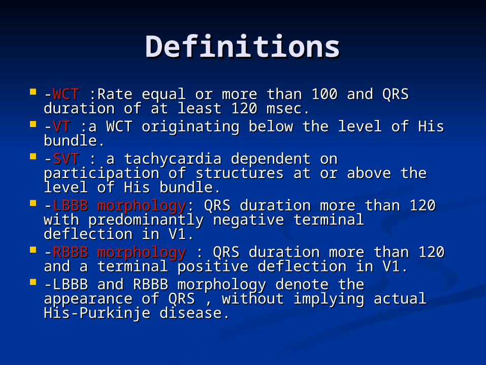
DefinitionsDefinitions --WCTWCT :Rate equal or more than 100 and QRS :Rate equal or more than 100 and QRS
duration of at least 120 msec.duration of at least 120 msec. --VTVT :a WCT originating below the level of His :a WCT originating below the level of His
bundle.bundle. --SVTSVT : a tachycardia dependent on participation : a tachycardia dependent on participation
of structures at or above the level of His bundle.of structures at or above the level of His bundle. --LBBB morphologyLBBB morphology: QRS duration more than 120 : QRS duration more than 120
with predominantly negative terminal deflection with predominantly negative terminal deflection in V1.in V1.
--RBBB morphologyRBBB morphology : QRS duration more than : QRS duration more than 120 and a terminal positive deflection in V1.120 and a terminal positive deflection in V1.
-LBBB and RBBB morphology denote the -LBBB and RBBB morphology denote the appearance of QRS , without implying actual His-appearance of QRS , without implying actual His-Purkinje disease.Purkinje disease.
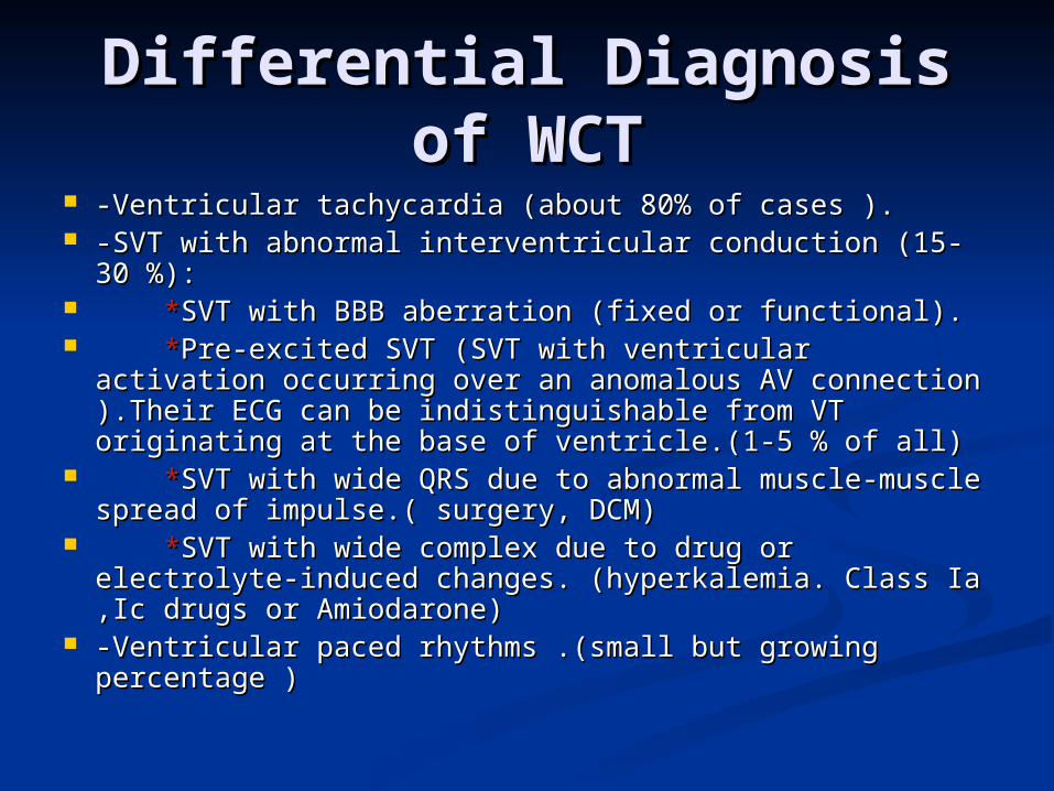
Differential Diagnosis of Differential Diagnosis of WCTWCT
-Ventricular tachycardia (about 80% of cases ).-Ventricular tachycardia (about 80% of cases ). -SVT with abnormal interventricular conduction (15-30 %):-SVT with abnormal interventricular conduction (15-30 %): **SVT with BBB aberration (fixed or functional).SVT with BBB aberration (fixed or functional). **Pre-excited SVT (SVT with ventricular activation Pre-excited SVT (SVT with ventricular activation
occurring over an anomalous AV connection ).Their ECG occurring over an anomalous AV connection ).Their ECG can be indistinguishable from VT originating at the base of can be indistinguishable from VT originating at the base of ventricle.(1-5 % of all)ventricle.(1-5 % of all)
**SVT with wide QRS due to abnormal muscle-muscle SVT with wide QRS due to abnormal muscle-muscle spread of impulse.( surgery, DCM)spread of impulse.( surgery, DCM)
**SVT with wide complex due to drug or electrolyte-SVT with wide complex due to drug or electrolyte-induced changes. (hyperkalemia. Class Ia ,Ic drugs or induced changes. (hyperkalemia. Class Ia ,Ic drugs or Amiodarone)Amiodarone)
-Ventricular paced rhythms .(small but growing -Ventricular paced rhythms .(small but growing percentage )percentage )
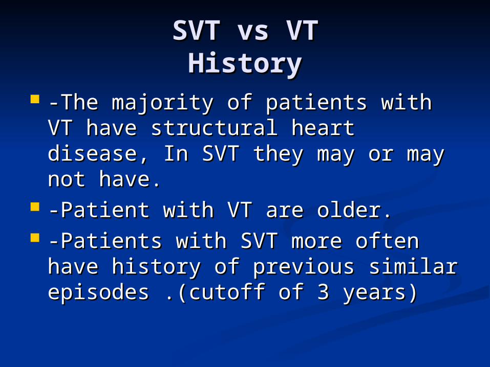
SVT vs VTSVT vs VTHistoryHistory
-The majority of patients with VT -The majority of patients with VT have structural heart disease, In SVT have structural heart disease, In SVT they may or may not have.they may or may not have.
-Patient with VT are older.-Patient with VT are older. -Patients with SVT more often have -Patients with SVT more often have
history of previous similar episodes .history of previous similar episodes .(cutoff of 3 years)(cutoff of 3 years)
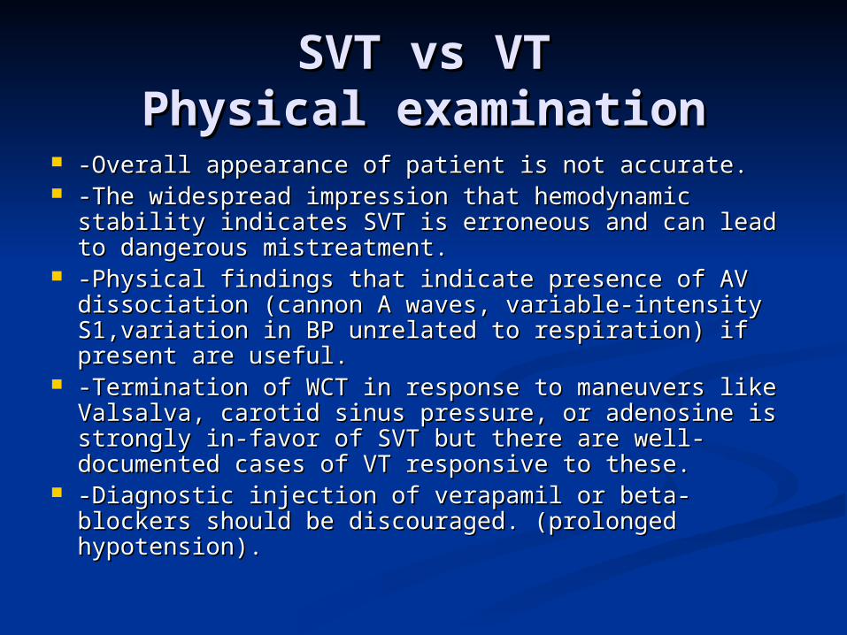
SVT vs VTSVT vs VTPhysical examinationPhysical examination
-Overall appearance of patient is not accurate.-Overall appearance of patient is not accurate. -The widespread impression that hemodynamic -The widespread impression that hemodynamic
stability indicates SVT is erroneous and can lead to stability indicates SVT is erroneous and can lead to dangerous mistreatment.dangerous mistreatment.
-Physical findings that indicate presence of AV -Physical findings that indicate presence of AV dissociation (cannon A waves, variable-intensity dissociation (cannon A waves, variable-intensity S1,variation in BP unrelated to respiration) if present S1,variation in BP unrelated to respiration) if present are useful.are useful.
-Termination of WCT in response to maneuvers like -Termination of WCT in response to maneuvers like Valsalva, carotid sinus pressure, or adenosine is Valsalva, carotid sinus pressure, or adenosine is strongly in-favor of SVT but there are well-strongly in-favor of SVT but there are well-documented cases of VT responsive to these.documented cases of VT responsive to these.
-Diagnostic injection of verapamil or beta-blockers -Diagnostic injection of verapamil or beta-blockers should be discouraged. (prolonged hypotension).should be discouraged. (prolonged hypotension).
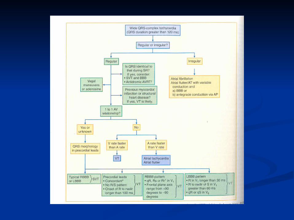
SVT vs VTSVT vs VTECG criteriaECG criteria
-A fundamentally simple approach: If WCT -A fundamentally simple approach: If WCT is due to SVT with aberration, the QRS is due to SVT with aberration, the QRS must be compatible with some form of BBB must be compatible with some form of BBB or FB.or FB.
-QRS duration:70% of VTs have QRS -QRS duration:70% of VTs have QRS duration >140, but no SVT has it. VT is duration >140, but no SVT has it. VT is probable when QRS> 140 with RBBB and probable when QRS> 140 with RBBB and >160 with LBBB pattern.Anti arrhythmic >160 with LBBB pattern.Anti arrhythmic drugs may prolong QRS. Some patients drugs may prolong QRS. Some patients with VT may have QRS of 120-140 specially with VT may have QRS of 120-140 specially in those without structural heart disease.in those without structural heart disease.
SVT vs VTSVT vs VTECG criteria contd,ECG criteria contd,
--QRS axisQRS axis:: *The more leftward the axis , the more *The more leftward the axis , the more
probable VT.probable VT. * The quadrant between -90 and 180 * The quadrant between -90 and 180
(northwest )can’t be achieved with any (northwest )can’t be achieved with any combination of FB or BBB.combination of FB or BBB.
*Some has suggested that if the axis in *Some has suggested that if the axis in sinus rhythm is more than 40 degrees sinus rhythm is more than 40 degrees different with WCT the diagnosis is VT , but different with WCT the diagnosis is VT , but it is not widely accepted.it is not widely accepted.
SVT vs VTSVT vs VTECG criteria contdECG criteria contd
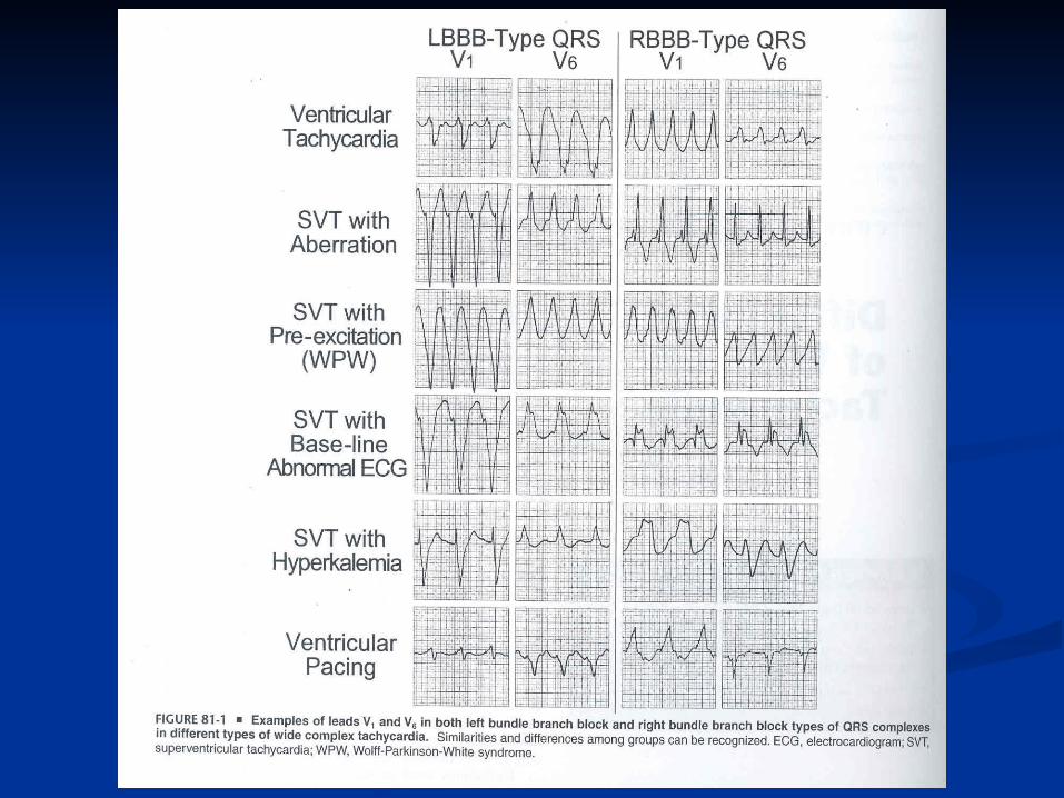
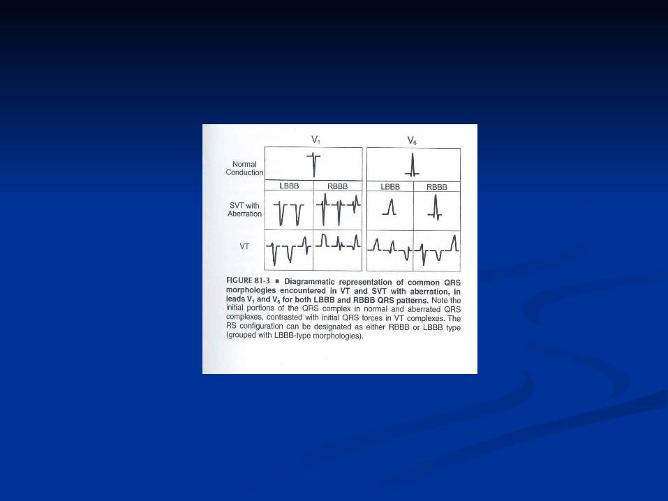
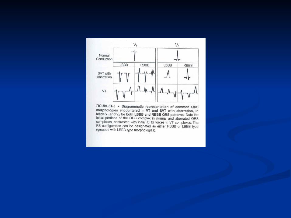
-Specific morphologies ;-Specific morphologies ; If with RBBB patternIf with RBBB pattern:: *In *In V1V1:During aberration there is no :During aberration there is no
change in initial portion of QRS so we may change in initial portion of QRS so we may see see rSr’ ,rR’ , rsr’ or rSR’.rSr’ ,rR’ , rsr’ or rSR’. But a But a monophasic R wave , or a broad (> 30 monophasic R wave , or a broad (> 30 msec) R with any following terminal QRS msec) R with any following terminal QRS forces or qR are highly suggestive of VT.forces or qR are highly suggestive of VT.
* In * In V6V6 :During aberration :During aberration qRs , Rs ,or RSqRs , Rs ,or RS (with R/S ratio >1) are seen but in VT we (with R/S ratio >1) are seen but in VT we may see may see rS,Qrs , QS orrS,Qrs , QS or QRQR or monophasic R or monophasic R wave. If RS pattern is present R/S must be wave. If RS pattern is present R/S must be less than 1.less than 1.
SVT vs VTSVT vs VTECG criteria contdECG criteria contd
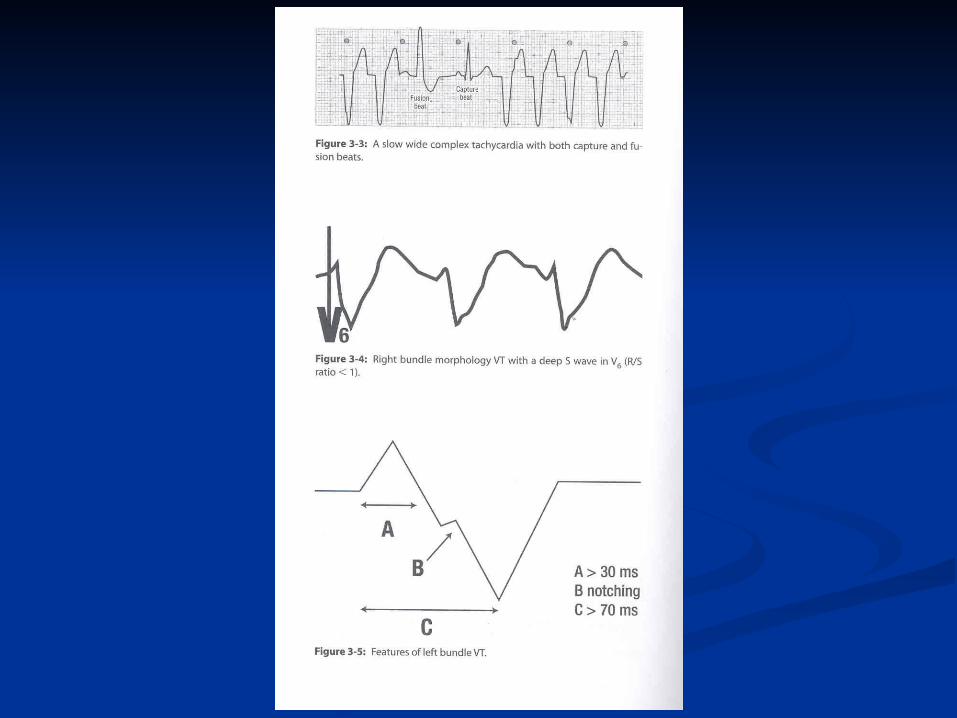
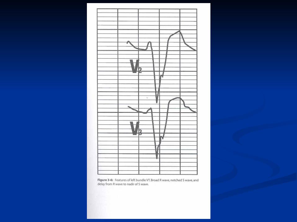
LBBB patternLBBB pattern:: * In * In V1V1 :Either rS or QS with rapid initial forces :Either rS or QS with rapid initial forces
(narrow R with rapid smooth descent to S )is seen in (narrow R with rapid smooth descent to S )is seen in LBBB type aberration. Any other pattern such as LBBB type aberration. Any other pattern such as broad R/deep S or QS with slow descent to S wave broad R/deep S or QS with slow descent to S wave nadir will imply VT.nadir will imply VT.
If the initial R is wider than 30 msec it suggest VT , If the initial R is wider than 30 msec it suggest VT , the wider the R , the greater the likelihood of VT.the wider the R , the greater the likelihood of VT.
Notching in the down-stroke of S or interval from the Notching in the down-stroke of S or interval from the onset of QRS to the S wave nadir greater than 60 onset of QRS to the S wave nadir greater than 60 msec strongly suggest VT.msec strongly suggest VT.
*In *In V6V6 :In aberrancy there is no initial Q wave and :In aberrancy there is no initial Q wave and we see RR’, or monophasic R. During VT common we see RR’, or monophasic R. During VT common patterns are patterns are QR ,QSQR ,QS ,QrS ,or Rr’,QrS ,or Rr’ although patterns although patterns compatible with SVT may also be seen.compatible with SVT may also be seen.
SVT vs VTSVT vs VTECG criteria contdECG criteria contd
Combination of LBBB and RAD is almost Combination of LBBB and RAD is almost always due to VT .always due to VT .
RBBB with a normal axis is very uncommon in RBBB with a normal axis is very uncommon in VT.VT.
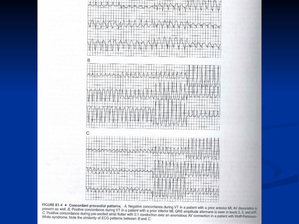
Concordant pattern in precordial leads is Concordant pattern in precordial leads is uncommon in SVT ,with the exception of pre-uncommon in SVT ,with the exception of pre-excited tachycardia .The specificity of excited tachycardia .The specificity of concordant pattern for VT is >90% but concordant pattern for VT is >90% but sensitivity is low(20%).sensitivity is low(20%).
Negative concordance in limb leads is another Negative concordance in limb leads is another way of describing NW axis and suggests VT.way of describing NW axis and suggests VT.
SVT vs VTSVT vs VTECG criteria contdECG criteria contd
Q waves during WCT show old MI and Q waves during WCT show old MI and are in favor of VT. Generally patients are in favor of VT. Generally patients with old Q waves maintain it during with old Q waves maintain it during WCT .WCT .
Some patients with DCM may have Q Some patients with DCM may have Q during VT while they don’t show it in SR.during VT while they don’t show it in SR.
Pseudo Q waves may be seen in some Pseudo Q waves may be seen in some SVTs with aberrancy. (AVNRT or pre-SVTs with aberrancy. (AVNRT or pre-excited with a posterior AV connection ).excited with a posterior AV connection ).
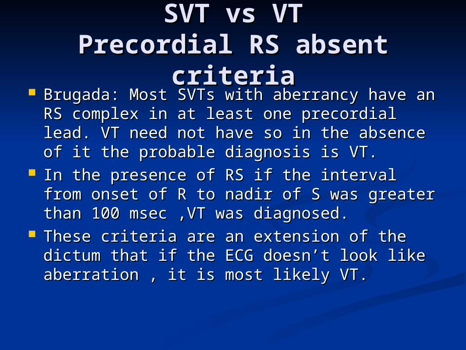
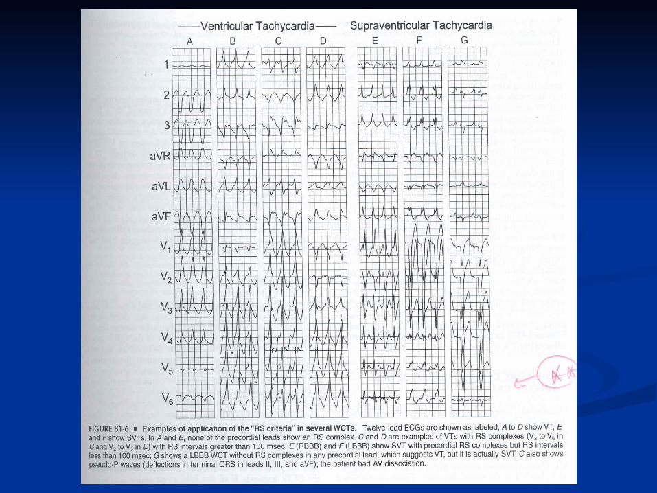
SVT vs VTSVT vs VTPrecordial RS absent Precordial RS absent
criteriacriteria Brugada: Most SVTs with aberrancy have Brugada: Most SVTs with aberrancy have
an RS complex in at least one precordial an RS complex in at least one precordial lead. VT need not have so in the absence lead. VT need not have so in the absence of it the probable diagnosis is VT.of it the probable diagnosis is VT.
In the presence of RS if the interval from In the presence of RS if the interval from onset of R to nadir of S was greater than onset of R to nadir of S was greater than 100 msec ,VT was diagnosed. 100 msec ,VT was diagnosed.
These criteria are an extension of the These criteria are an extension of the dictum that if the ECG doesn’t look like dictum that if the ECG doesn’t look like aberration , it is most likely VT.aberration , it is most likely VT.
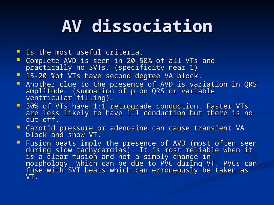
AV dissociationAV dissociation Is the most useful criteria.Is the most useful criteria. Complete AVD is seen in 20-50% of all VTs and practically Complete AVD is seen in 20-50% of all VTs and practically
no SVTs. (specificity near 1)no SVTs. (specificity near 1) 15-20 %of VTs have second degree VA block.15-20 %of VTs have second degree VA block. Another clue to the presence of AVD is variation in QRS Another clue to the presence of AVD is variation in QRS
amplitude. (summation of p on QRS or variable ventricular amplitude. (summation of p on QRS or variable ventricular filling).filling).
30% of VTs have 1:1 retrograde conduction. Faster VTs 30% of VTs have 1:1 retrograde conduction. Faster VTs are less likely to have 1:1 conduction but there is no cut-are less likely to have 1:1 conduction but there is no cut-off.off.
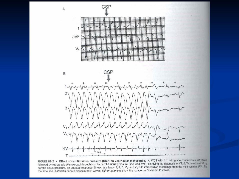
Carotid pressure or adenosine can cause transient VA Carotid pressure or adenosine can cause transient VA block and show VT.block and show VT.
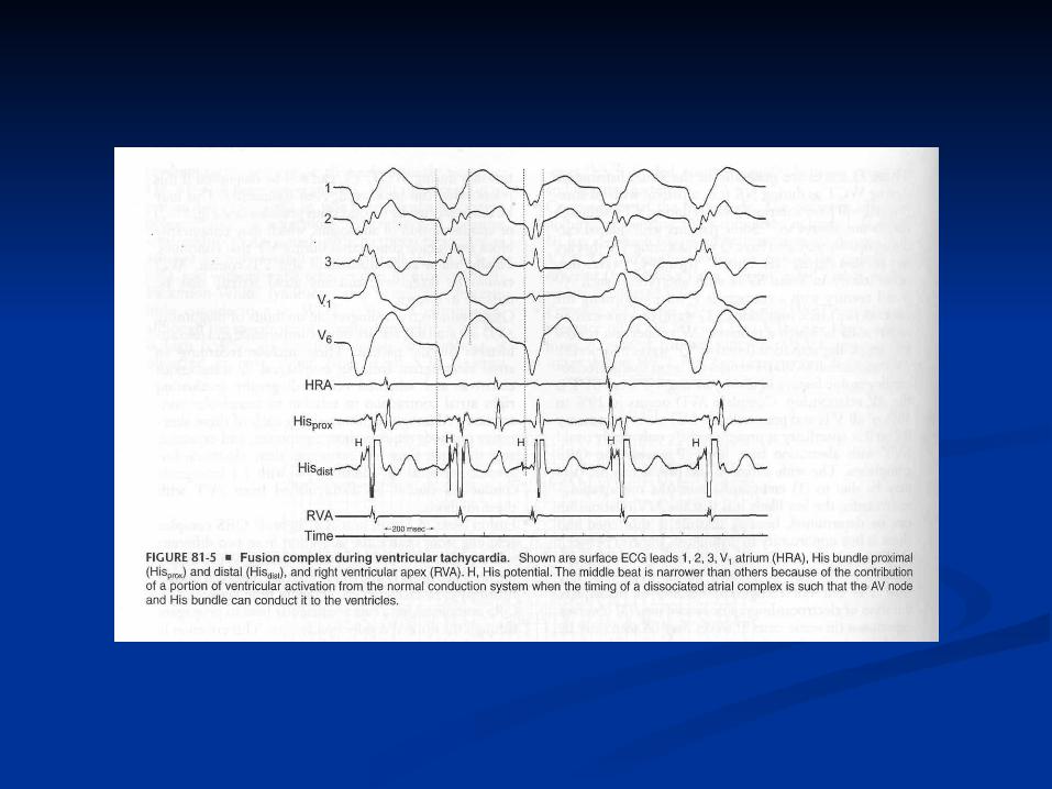
Fusion beats imply the presence of AVD (most often seen Fusion beats imply the presence of AVD (most often seen during slow tachycardias). It is most reliable when it is a during slow tachycardias). It is most reliable when it is a clear fusion and not a simply change in morphology. Which clear fusion and not a simply change in morphology. Which can be due to PVC during VT. PVCs can fuse with SVT can be due to PVC during VT. PVCs can fuse with SVT beats which can erroneously be taken as VT.beats which can erroneously be taken as VT.
SVT vs VTSVT vs VT
WCT with QRS narrower than NSR is WCT with QRS narrower than NSR is strongly in favor of VT.strongly in favor of VT.
Contralateral BBB in NSR and WCT strongly Contralateral BBB in NSR and WCT strongly suggests VT. Because if RBBB is present in suggests VT. Because if RBBB is present in SR and LBBB develops with SVT, then CHB SR and LBBB develops with SVT, then CHB must happen. (except in rare cases).must happen. (except in rare cases).
QRS alternans is not an important QRS alternans is not an important diagnostic factor.diagnostic factor.
Presence of multiple WCT configurations is Presence of multiple WCT configurations is in favor of VT.in favor of VT.
Special cases of WCTSpecial cases of WCT Pre-excited SVTs (ventricular activation Pre-excited SVTs (ventricular activation
entirely or primarily over an anomalous AV entirely or primarily over an anomalous AV connection) has an ECG pattern consistent with connection) has an ECG pattern consistent with VT.VT.
In BB reentry the ECG criteria suggest SVT, In BB reentry the ECG criteria suggest SVT, but it is VT.(AVD is frequent in these patients but it is VT.(AVD is frequent in these patients and helps ).and helps ).
Both SVT and VT are usually regular, marked Both SVT and VT are usually regular, marked irregularity is in favor of AF but remember that irregularity is in favor of AF but remember that VTs can be irregular particularly in the first 30 VTs can be irregular particularly in the first 30 seconds of the episode. VTs with CL seconds of the episode. VTs with CL irregularity are usually seen in patients taking irregularity are usually seen in patients taking anti -arrhythmic drugs.anti -arrhythmic drugs.
Narrow QRS complex VTNarrow QRS complex VT
VT can have QRS duration less than VT can have QRS duration less than the cutoff of 140 msec. Possible the cutoff of 140 msec. Possible explanations are:explanations are:
1-Septal origin of VT.1-Septal origin of VT. 2- Early penetration in the His- 2- Early penetration in the His-
Purkinje system.Purkinje system.