diagnosis of noncultivatable gastroenteritis viruses, …cmr.asm.org/content/14/1/15.full.pdf ·...
TRANSCRIPT
CLINICAL MICROBIOLOGY REVIEWS,0893-8512/01/$04.0010 DOI: 10.1128/CMR.14.1.15–37.2001
Jan. 2001, p. 15–37 Vol. 14, No. 1
Copyright © 2001, American Society for Microbiology. All Rights Reserved.
Diagnosis of Noncultivatable Gastroenteritis Viruses,the Human Caliciviruses
ROBERT L. ATMAR AND MARY K. ESTES*
Departments of Medicine and Molecular Virology & Microbiology,Baylor College of Medicine, Houston, Texas 77030
INTRODUCTION .........................................................................................................................................................15Importance and Impact of Gastroenteritis ...........................................................................................................15Viral Causes of Gastroenteritis ..............................................................................................................................16
HISTORY OF HUMAN CALICIVIRUSES ...............................................................................................................16Recognition ................................................................................................................................................................16Classification .............................................................................................................................................................17
Morphology ............................................................................................................................................................17Genomic organization and classification...........................................................................................................17Cross-protection and serologic studies ................................................................................................................5
FIRST-GENERATION DIAGNOSTIC TESTS USING HUMAN REAGENTS ...................................................20Electron Microscopy.................................................................................................................................................20
Direct electron microscopy ..................................................................................................................................20IEM.........................................................................................................................................................................20
Immune Adherence Hemagglutination Assay .......................................................................................................21Radioimmunoassay ...................................................................................................................................................21Enzyme Immunoassay ..............................................................................................................................................21Western Blot Assay...................................................................................................................................................21
DEVELOPMENT AND APPLICATION OF NEWER DIAGNOSTIC TESTS.....................................................22Antigen Detection......................................................................................................................................................22
EIA with hyperimmune animal sera ..................................................................................................................22EIA with MAbs......................................................................................................................................................22
Antibody Detection....................................................................................................................................................23EIA with VLPs.........................................................................................................................................................9Detection of infection caused by heterotypic HuCVs.......................................................................................24
Nucleic Acid Detection .............................................................................................................................................24Hybridization assays.............................................................................................................................................25RT-PCR ..................................................................................................................................................................25
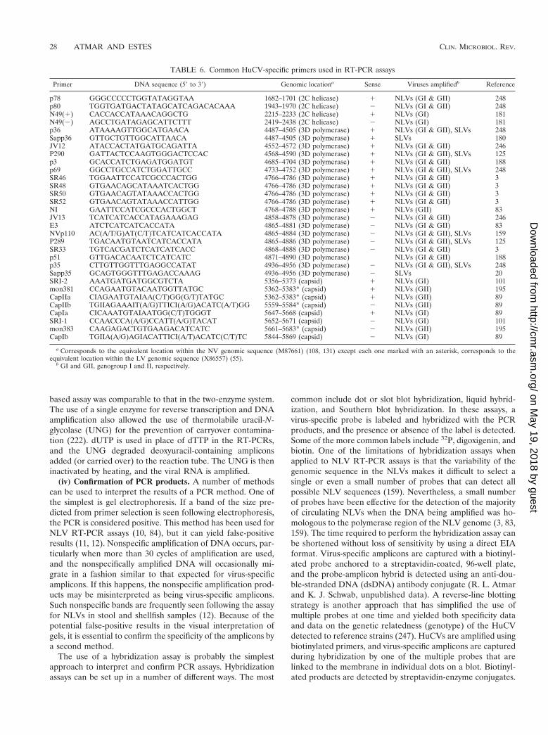
(i) Extraction methods .....................................................................................................................................25(ii) Primer selection..........................................................................................................................................26(iii) Other PCR conditions ..............................................................................................................................27(iv) Confirmation of PCR products................................................................................................................28(v) Internal standard........................................................................................................................................29(vi) Application to clinical and environmental specimens..........................................................................29
Use of Newer Diagnostic Tests ...............................................................................................................................30Future Directions......................................................................................................................................................31
CONCLUSIONS ...........................................................................................................................................................31ACKNOWLEDGMENTS .............................................................................................................................................32REFERENCES ..............................................................................................................................................................32
INTRODUCTION
Importance and Impact of Gastroenteritis
Acute gastroenteritis is one of the most common diseases ofhumans. In the United States, it is second only to acute viralrespiratory disease as a cause of acute illness (61). Worldwide,more than 700 million cases of acute diarrheal disease areestimated to occur annually just in children under the age of 5
years (230). Gastroenteritis most commonly is manifested clin-ically as mild diarrhea, but more severe disease, ranging fromupper gastrointestinal symptoms (nausea and vomiting) to pro-fuse diarrhea leading to dehydration and death, may occur.The annual mortality associated with gastroenteritis has beenestimated to be 3.5 to 5 million, with the majority of deathsoccurring in developing countries (21, 97, 250).
Acute gastroenteritis is caused by a number of differentagents, including bacteria, viruses, and parasites. Until re-cently, many cases were attributed to viruses because of thefailure to identify a bacterial or parasitic pathogen. AlthoughReimann et al. (214) and Gordon et al. (77) suggested morethan 50 years ago that viruses are a cause of diarrhea after
* Corresponding author. Mailing address: Department of MolecularVirology & Microbiology, Baylor College of Medicine, 1 Baylor Plaza,Houston, TX 77030. Phone: (713) 798-3585. Fax: (713) 798-3586. E-mail: [email protected].
15
on May 19, 2018 by guest
http://cmr.asm
.org/D
ownloaded from
on M
ay 19, 2018 by guesthttp://cm
r.asm.org/
Dow
nloaded from
on May 19, 2018 by guest
http://cmr.asm
.org/D
ownloaded from
inducing illness in volunteers with stool filtrates free of bacte-ria, it was only in 1972 that a virus (Norwalk virus) was defin-itively identified as a cause of acute gastroenteritis (139). Sincethat time, the number of viral agents associated with acutegastroenteritis has increased progressively.
Viral Causes of Gastroenteritis
Many different viruses have been found in the stools ofpersons with gastroenteritis. However, a causative role for eachof these viruses has not been established. Criteria to define avirus as an etiologic agent of gastroenteritis include (i) identi-fication of the virus more frequently in subjects with diarrheathan in controls, (ii) demonstration of an immune response tothe specific agent, and (iii) demonstration that the beginningand end of the illness correspond to the onset and terminationof virus shedding, respectively (148). Table 1 lists those virusesestablished as etiologic agents of gastroenteritis and other vi-ruses found in stools that have not yet fulfilled the aforemen-tioned criteria (9). Coronaviruses (34, 62), picobirnavirusesand picotrirnaviruses (35, 96, 174), pestiviruses (13, 257), andtoroviruses (17, 18, 251) are candidate diarrheal agents be-cause they are associated with diarrheal illness in animals andhave been found in the stools of humans with gastroenteritis.However, these viruses have not fulfilled the criteria needed toestablish them as diarrheal agents in humans (134, 148) andwill not be considered further in this review. Non-group Fadenoviruses and several enteroviruses (e.g., Coxsackie A andB viruses) are found in the stools of non-ill individuals withfrequencies similar to those seen in ill individuals (148).
Of the viruses that have been shown definitively to be causesof acute gastroenteritis, only the human caliciviruses cannot begrown in cell culture. The study of rotaviruses, enteric adeno-viruses, and astroviruses has been facilitated greatly by theability to propagate these viruses in cell culture. The ability tocultivate these viruses has allowed the production of reagentsfor use in diagnostic studies, a better understanding of factorscorrelated with immunity to infection, and the elucidation ofeach virus’s life cycle. Although human caliciviruses have de-fied numerous attempts to propagate them in cell culture todate, recent developments in their study by using molecular
biology techniques have increased our understanding of thisgroup of viruses. This article will review the new diagnosticassays that have resulted from these advances.
HISTORY OF HUMAN CALICIVIRUSES
Recognition
In 1968, an outbreak of acute gastroenteritis (termed wintervomiting disease) occurred among students and teachers in aschool in Norwalk, Ohio (1). The primary attack rate was 50%,with a secondary attack rate of 32%. Illness was characterizedby nausea and vomiting in .90% and diarrhea in 38% of af-fected individuals, and the duration of illness was usually 12 to24 h. Subsequently, organism-free filtrates of stools collectedfrom affected individuals induced similar illness in human vol-unteers, and different treatments of the inoculum suggestedthat the causative agent was a small (,36 nm), ether-resistant(nonenveloped), relatively heat-stable virus (24, 57, 58). At-tempts to propagate the agent in cell culture and organ culturewere unsuccessful (24, 57).
In 1972, Kapikian et al. (139) used immune electron micros-copy (IEM) to identify 27-nm viral particles, the Norwalkagent, in a fecal filtrate used to induce illness in human volun-teers. Virus particles were precipitated in an antigen-antibodyreaction using convalescent-phase serum from a volunteer whobecame ill following inoculation with the fecal filtrate. Anti-gen-antibody complexes were then visualized with an electronmicroscope. The assay was modified to quantify the amount ofantibody in serum and demonstrated that significantly moreantibody was present in convalescent-phase serum than inacute-phase serum. Because of these data, Norwalk virus (NV)was proposed to be the etiologic agent of the Norwalk, Ohio,outbreak of gastroenteritis (30). Later studies demonstratedthat other small, round-structured viruses (SRSVs) morpho-logically similar to NV were associated with outbreaks of gas-troenteritis (7, 59, 238), but NV remained the prototype ofthese fecal viruses.
Morphologically typical caliciviruses were first recognized instool samples by Madeley and Cosgrove in 1976 (176). Theseinvestigators found calicivirus particles in the fecal specimens
TABLE 1. Causal relationship to diarrhea of enteric virusesa.
Group Viruses Cultivation reportedb
(reference)
Causal relationship demonstrated Rotaviruses Yes (66)Human caliciviruses NoAstroviruses Yes (155, 156)Enteric (group F) adenoviruses Yes (147)
Candidate agents (etiologic relationship not yet determined) Coronaviruses Yes (215)Echovirus type 22 Yes (148)Picobirnaviruses, picotrirnaviruses No (35, 96, 174)Pestiviruses No (13, 257)Toroviruses No (17, 18, 251)
Other agentsc (causal relationship not demonstrated) Non-group F adenoviruses Yes (148)Coxsackie A and B viruses Yes (148)Echoviruses Yes (148)
a Viruses are listed by relative clinical significance. Data are modified from R. L. Atmar and M. K. Estes, Clin. Microbiol. Newsl: 19:177–182, 1997 (9), with permissionfrom Elsevier Science.
b Virus cultivated from human enteric sample.c Present in stools of non-ill individuals with frequency similar to that seen in ill subjects.
16 ATMAR AND ESTES CLIN. MICROBIOL. REV.
on May 19, 2018 by guest
http://cmr.asm
.org/D
ownloaded from
of 10 children, but some of the children were asymptomatic, sothat no conclusions as to the pathogenicity of the virus could bemade. Later that year, Flewett and Davies (68) identified cali-civirus particles in the small bowel from a fatal case of gastro-enteritis, but because adenovirus particles also were present inlarge numbers, the significance of the calicivirus particles couldnot be determined. However, in the next several years, calici-viruses were definitively associated with several outbreaks ofgastroenteritis (37, 38, 44, 45, 185) and became recognized asanother virus group associated with gastroenteritis (33, 175).
Based on properties of NV and its appearance by electronmicroscopy, NV and other small round viruses were thought tobe parvovirus-like (57, 137). However, in 1981 Greenberg et al.(93) published data suggesting that NV has a single structuralprotein with an estimated molecular mass of 59 kDa and pro-posed that Norwalk virus might be a calicivirus. Nine yearslater, Jiang et al. (216) provided molecular evidence that NV isa calicivirus by demonstrating that the viral genome consists ofpositive-sense, single-stranded, polyadenylated RNA. Subse-quent elucidation of the complete sequence of NV and relatedSRSVs confirmed the genetic relatedness of these viruses toother caliciviruses (55, 108, 131, 153, 154, 225).
Classification
The classification of viruses responsible for acute gastroen-teritis was first based on morphology (Table 2). For example,NV was the prototype of a group of agents initially calledSRSVs. Recently, rapid advances in molecular biology allowedthese viruses to be classified based on their genome character-istics, and most of the previously named SRSVs were shown tobelong to the Caliciviridae.
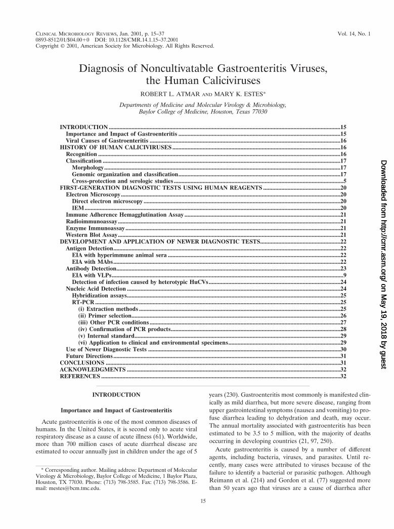
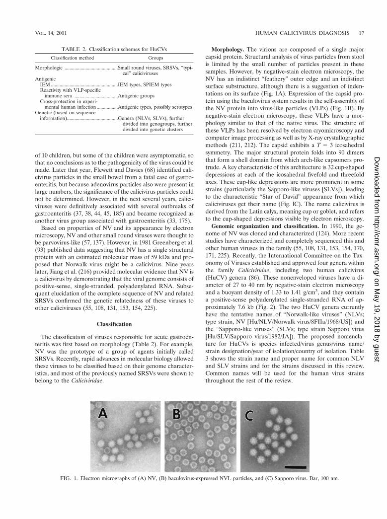
Morphology. The virions are composed of a single majorcapsid protein. Structural analysis of virus particles from stoolis limited by the small number of particles present in thesesamples. However, by negative-stain electron microscopy, theNV has an indistinct “feathery” outer edge and an indistinctsurface substructure, although there is a suggestion of inden-tations on its surface (Fig. 1A). Expression of the capsid pro-tein using the baculovirus system results in the self-assembly ofthe NV protein into virus-like particles (VLPs) (Fig. 1B). Bynegative-stain electron microscopy, these VLPs have a mor-phology similar to that of the native virus. The structure ofthese VLPs has been resolved by electron cryomicroscopy andcomputer image processing as well as by X-ray crystallographicmethods (211, 212). The capsid exhibits a T 5 3 icosahedralsymmetry. The major structural protein folds into 90 dimersthat form a shell domain from which arch-like capsomers pro-trude. A key characteristic of this architecture is 32 cup-shapeddepressions at each of the icosahedral fivefold and threefoldaxes. These cup-like depressions are more prominent in somestrains (particularly the Sapporo-like viruses [SLVs]), leadingto the characteristic “Star of David” appearance from whichcaliciviruses get their name (Fig. lC). The name calicivirus isderived from the Latin calyx, meaning cup or goblet, and refersto the cup-shaped depressions visible by electron microscopy.
Genomic organization and classification. In 1990, the ge-nome of NV was cloned and characterized (124). More recentstudies have characterized and completely sequenced this andother human viruses in the family (55, 108, 131, 153, 154, 170,171, 225). Recently, the International Committee on the Tax-onomy of Viruses established and approved four genera withinthe family Caliciviridae, including two human calicivirus(HuCV) genera (86). These nonenveloped viruses have a di-ameter of 27 to 40 nm by negative-stain electron microscopyand a buoyant density of 1.33 to 1.41 g/cm3, and they containa positive-sense polyadenylated single-stranded RNA of ap-proximately 7.6 kb (Fig. 2). The two HuCV genera currentlyhave the tentative names of “Norwalk-like viruses” (NLVs;type strain, NV [Hu/NLV/Norwalk virus/8FIIa/1968/US]) andthe “Sapporo-like viruses” (SLVs; type strain Sapporo virus[Hu/SLV/Sapporo virus/1982/JA]). The proposed nomencla-ture for HuCVs is species infected/virus genus/virus name/strain designation/year of isolation/country of isolation. Table3 shows the strain name and proper name for common NLVand SLV strains and for the strains discussed in this review.Common names will be used for the human virus strainsthroughout the rest of the review.
TABLE 2. Classification schemes for HuCVs
Classification method Groups
Morphologic ...........................................Small round viruses, SRSVs, “typi-cal” caliciviruses
AntigenicIEM ......................................................IEM types, SPIEM typesReactivity with VLP-specific
immune sera ...................................Antigenic groupsCross-protection in experi-
mental human infection .................Antigenic types, possibly serotypesGenetic (based on sequence
information).........................................Genera (NLVs, SLVs), furtherdivided into genogroups, furtherdivided into genetic clusters
FIG. 1. Electron micrographs of (A) NV, (B) baculovirus-expressed NVL particles, and (C) Sapporo virus. Bar, 100 nm.
VOL. 14, 2001 HUMAN CALICIVIRUS DIAGNOSIS 17
on May 19, 2018 by guest
http://cmr.asm
.org/D
ownloaded from
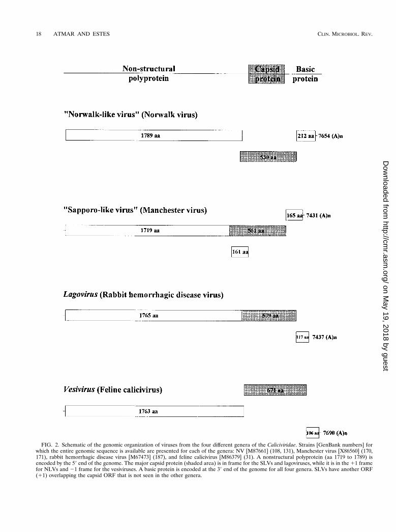
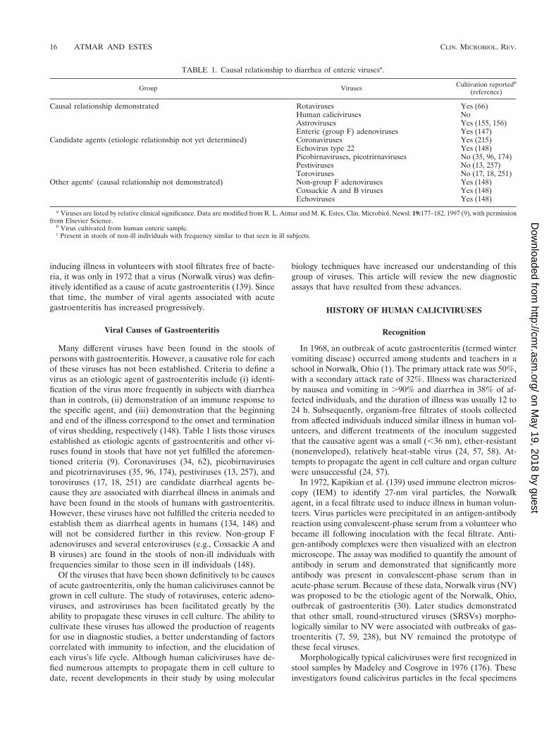
FIG. 2. Schematic of the genomic organization of viruses from the four different genera of the Caliciviridae. Strains [GenBank numbers] forwhich the entire genomic sequence is available are presented for each of the genera: NV [M87661] (108, 131), Manchester virus [X86560] (170,171), rabbit hemorrhagic disease virus [M67473] (187), and feline calicivirus [M86379] (31). A nonstructural polyprotein (aa 1719 to 1789) isencoded by the 59 end of the genome. The major capsid protein (shaded area) is in frame for the SLVs and lagoviruses, while it is in the 11 framefor NLVs and 21 frame for the vesiviruses. A basic protein is encoded at the 39 end of the genome for all four genera. SLVs have another ORF(11) overlapping the capsid ORF that is not seen in the other genera.
18 ATMAR AND ESTES CLIN. MICROBIOL. REV.
on May 19, 2018 by guest
http://cmr.asm
.org/D
ownloaded from
Calicivirus strains that infect animals (bovine and swine) andhave characteristics that place them in the NLV and SLVgenera have also been described (51, 100, 172, 233). In con-trast, the other two genera within the family Caliciviridae (Lago-virus [type strain, rabbit hemorrhagic disease virus, Ra/LV/RHDV/V351/1987/CK] and Vesivirus [type strain, swine vesic-ular exanthema virus, Sw/VV/VESV/A48/1948/US]) are cur-rently recognized to contain strains that naturally only infectanimals (and not humans). There is a single case report ofinfection of a person who was isolating a vesivirus in the lab-oratory (229).
The genome of NLVs is organized in three major openreading frames (ORFs). For NV, the first ORF at the 59 endencodes a large polyprotein of 1738 amino acids (aa) with apredicted molecular weight of 193.5 (193.5K). This polyproteincontains short motifs of similarity with the 2C (helicase), 3C(cysteine protease), and 3D (RNA-dependent RNA polymer-ase) proteins of picornaviruses. Thus, the 59 end of the genomeof the NLVs codes for a precursor of the nonstructural pro-teins. ORF2 encodes a 530-aa (56.6K) protein, the capsid pro-tein. The ORF2 protein expressed in insect cells self-assemblesinto VLPs as explained below (Fig. 1B). ORF3 at the 39 end ofthe genome is predicted to code for a small protein of 212 aa(22.5K) with a very basic charge (isoelectric point of 10.99).The ORF3 protein does not have sequence similarity with anyother proteins in the GenBank database, and its function re-mains unknown. Recent studies indicate that the ORF3 pro-tein is a minor structural protein, based on its being found inVLPs expressed from cDNA constructs that contain bothORF2 and ORF3 and in virus particles purified from stool(75).
The genome of SLVs is organized slightly differently. ForManchester virus, the first ORF codes for the nonstructuralproteins as well as the capsid protein, which is found in-frameat the end of the nonstructural proteins (170, 171). This ge-nome organization is similar to that found in the animal cali-civirus rabbit hemorrhagic disease virus belonging to the genusLagovirus (Fig. 2) (187). ORF2 encodes a predicted small,highly basic protein of unknown function, similar to ORF3 for
NV. Manchester virus contains a third ORF within the capsidprotein that could encode another small basic protein. Thesignificance of this ORF is unclear, as this ORF is not seen inany of the other calicivirus genomes sequenced thus far, andthe small protein it potentially encodes shows no sequencehomology to other viral proteins in the database (171).
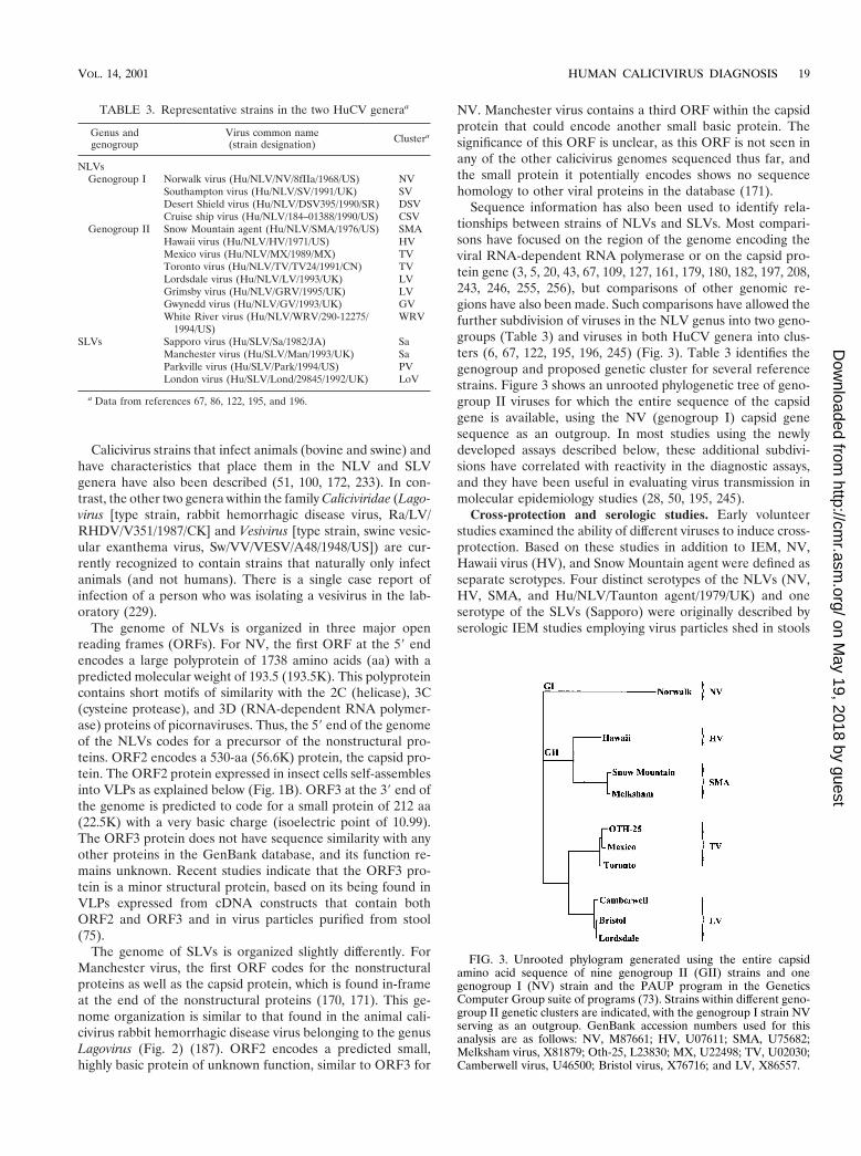
Sequence information has also been used to identify rela-tionships between strains of NLVs and SLVs. Most compari-sons have focused on the region of the genome encoding theviral RNA-dependent RNA polymerase or on the capsid pro-tein gene (3, 5, 20, 43, 67, 109, 127, 161, 179, 180, 182, 197, 208,243, 246, 255, 256), but comparisons of other genomic re-gions have also been made. Such comparisons have allowed thefurther subdivision of viruses in the NLV genus into two geno-groups (Table 3) and viruses in both HuCV genera into clus-ters (6, 67, 122, 195, 196, 245) (Fig. 3). Table 3 identifies thegenogroup and proposed genetic cluster for several referencestrains. Figure 3 shows an unrooted phylogenetic tree of geno-group II viruses for which the entire sequence of the capsidgene is available, using the NV (genogroup I) capsid genesequence as an outgroup. In most studies using the newlydeveloped assays described below, these additional subdivi-sions have correlated with reactivity in the diagnostic assays,and they have been useful in evaluating virus transmission inmolecular epidemiology studies (28, 50, 195, 245).
Cross-protection and serologic studies. Early volunteerstudies examined the ability of different viruses to induce cross-protection. Based on these studies in addition to IEM, NV,Hawaii virus (HV), and Snow Mountain agent were defined asseparate serotypes. Four distinct serotypes of the NLVs (NV,HV, SMA, and Hu/NLV/Taunton agent/1979/UK) and oneserotype of the SLVs (Sapporo) were originally described byserologic IEM studies employing virus particles shed in stools
TABLE 3. Representative strains in the two HuCV generaa
Genus andgenogroup
Virus common name(strain designation) Clustera
NLVsGenogroup I Norwalk virus (Hu/NLV/NV/8fIIa/1968/US) NV
Southampton virus (Hu/NLV/SV/1991/UK) SVDesert Shield virus (Hu/NLV/DSV395/1990/SR) DSVCruise ship virus (Hu/NLV/184–01388/1990/US) CSV
Genogroup II Snow Mountain agent (Hu/NLV/SMA/1976/US) SMAHawaii virus (Hu/NLV/HV/1971/US) HVMexico virus (Hu/NLV/MX/1989/MX) TVToronto virus (Hu/NLV/TV/TV24/1991/CN) TVLordsdale virus (Hu/NLV/LV/1993/UK) LVGrimsby virus (Hu/NLV/GRV/1995/UK) LVGwynedd virus (Hu/NLV/GV/1993/UK) GVWhite River virus (Hu/NLV/WRV/290-12275/
1994/US)WRV
SLVs Sapporo virus (Hu/SLV/Sa/1982/JA) SaManchester virus (Hu/SLV/Man/1993/UK) SaParkville virus (Hu/SLV/Park/1994/US) PVLondon virus (Hu/SLV/Lond/29845/1992/UK) LoV
a Data from references 67, 86, 122, 195, and 196.
FIG. 3. Unrooted phylogram generated using the entire capsidamino acid sequence of nine genogroup II (GII) strains and onegenogroup I (NV) strain and the PAUP program in the GeneticsComputer Group suite of programs (73). Strains within different geno-group II genetic clusters are indicated, with the genogroup I strain NVserving as an outgroup. GenBank accession numbers used for thisanalysis are as follows: NV, M87661; HV, U07611; SMA, U75682;Melksham virus, X81879; Oth-25, L23830; MX, U22498; TV, U02030;Camberwell virus, U46500; Bristol virus, X76716; and LV, X86557.
VOL. 14, 2001 HUMAN CALICIVIRUS DIAGNOSIS 19
on May 19, 2018 by guest
http://cmr.asm
.org/D
ownloaded from
as the antigen and paired sera from infected individuals as thesource of antibody (133). Additional antigenic groups wereproposed subsequently (165, 202). These serotype designationsassumed that antibody reactivity by IEM reflects the reactivityof neutralizing antibodies. This may not be the case, and cleardefinition of serotypes remains difficult to achieve due to thelack of a cultivation system. More recently, the antigenic rela-tionships between a subset of these viruses have been exam-ined by enzyme-linked immunosorbentassay (ELISA) usinghyperimmune antisera raised against VLPs. By this method,Norwalk, Mexico, and Grimsby viruses are antigenically dis-tinct (102), as are NV and Desert Shield virus (161), NV andHV (87), and NV and Sapporo viruses (198).
FIRST-GENERATION DIAGNOSTIC TESTSUSING HUMAN REAGENTS
During the 1970s and 1980s, tests for the diagnosis of NVinfections were designed using reagents from previously in-fected humans. The number of virus particles in the stools ofinfected subjects is sufficiently small that hyperimmune seracould not be produced in animals. (An exception was the useof partially purified Sapporo virus to produce hyperimmunesera in guinea pigs [192].) The inability to propagate NV andrelated viruses in cell culture also prevented the production ofanimal hyperimmune sera. Thus, stools of acutely infectedindividuals served as a source of virus antigen, and convales-cent-phase sera from infected individuals were used as hyper-immune sera. These restrictions limited the general availabilityof these diagnostic tools to only a few research laboratories(40). However, a number of tests were developed with thesereagents and used to begin to define the epidemiology of NVand other human caliciviruses.
Electron Microscopy
Direct electron microscopy. Detection of enteric viruses instool specimens using direct electron microscopy requires virusconcentrations of at least 106 per ml of stool (56). In manylaboratories, stool is mixed with phosphate-buffered saline ortissue culture medium to form a 10 to 20% suspension beforebeing clarified by low-speed centrifugation. Subsequent con-centration of virus may be achieved by ultracentrifugation orprecipitation with ammonium sulfate. Virus particles are neg-atively stained using one of a number of available electron-dense stains, including phosphotungstic acid, uranyl acetate,and ammonium molybdate (56). Viral particles must be distin-guished from nonviral material, and this can be particularlydifficult for the NLVs that do not have typical calicivirus mor-phology (56, 140). The small numbers of viral particles presentin fecal samples make direct electron microscopy, even afterconcentration, relatively insensitive. Nevertheless, direct elec-tron microscopy is used to screen fecal specimens for entericviruses in the public health laboratories of many countries, andit is clear that talented electron microscopists who receive fecalsamples collected early in the course of an infection can oftendetect viruses (52, 179, 246, 256). However, this method re-quires highly skilled microscopists and expensive equipment,making it not feasible for large epidemiological or clinicalstudies.
IEM . The use of immune serum to aid in virus identificationwas first described for tobacco mosaic virus in 1941 (2). Thismethod had been used for only a few human viruses (22, 49,135) before Kapikian et al. (139) adapted it for the detection ofNV in stool filtrates. Since then, it has been used to identifymany other SRSVs in stool samples (27, 59, 141, 238, 242). Aclarified stool suspension is incubated with a reference serumor saline (as a control) for 1 h at 37°C, and immune complexesare pelleted by ultracentrifugation (140). The pellet is sus-pended in a few drops of distilled water and negatively stainedas for direct electron microscopy. The saline control is neces-sary to demonstrate the specificity of the immune serum be-cause virus clumping may occur in the absence of antibody (56,194). A positive sample has aggregates of viral particles whichare absent in the saline control sample. Although IEM was firstused to detect NV, it is positive on stool samples from onlyapproximately half of volunteers who become ill following ar-tificial challenge with NV (237). When IEM has been appliedto outbreaks of gastroenteritis associated serologically with NVinfection, it has been positive in only about 20% of the out-breaks, and just over one third of stool samples from affectedindividuals in these outbreaks are IEM positive (141). The lackof detection probably reflects the very low concentration ofvirus in many stool samples and the lack of collection of thefirst early diarrheal stool samples.
Modifications to the IEM method have been made to im-prove the ease with which viral particles are detected and tosimplify the performance of the test. Solid-phase IEM (SPIEM)has been used to capture viral particles directly onto the grid(56, 164, 168). Virus-specific immunoglobulin or broad-spec-trum immunoglobulin (gamma globulin) is used to coat thegrids, and virus particles from a fecal suspension are thencaptured by interaction with antiviral antibodies. Protein A hasbeen used to capture the antibody onto the grid before expo-sure to fecal suspensions; this may increase the exposure ofantigen-binding sites by capturing the antibodies through theirFc receptor (56, 149, 165). SPIEM has been used as a tool for“serotyping” HuCVs (165, 166). Another modification of IEMhas been the use of colloidal gold-protein A conjugates to la-bel in suspension clumps of virus and antibody; this modifica-tion allows specific antigen-antibody interactions to be distin-guished from nonspecific clumping (149).
IEM has also been used to detect seroresponses to viralantigen (37, 59, 139, 202). In this assay, the source of viralantigen is a stool filtrate in which viral particles are easilydetected by electron microscopy or a stool from which virushas been partially purified. Antigen is mixed with a 1:5 dilutionof the serum to be tested, and the mixture is examined. Theamount of antibody is determined by the appearance of theviral particles and is rated 0 to 41, with 0 being no viralaggregates noted and 41 being nonglistening, heavily coatedviral aggregates (139). This assay is type specific and has beenused to evaluate the association of HuCVs with outbreaks ofgastroenteritis (37, 56, 59, 202). While IEM is an importantcomponent of the diagnostic armamentarium for the noncul-tivatable caliciviruses, like direct electron microscopy, the ap-plication of IEM is limited and not readily applied to largeepidemiological studies.
20 ATMAR AND ESTES CLIN. MICROBIOL. REV.
on May 19, 2018 by guest
http://cmr.asm
.org/D
ownloaded from
Immune Adherence Hemagglutination Assay
IEM was found to be a specific and reproducible method forantibody determination, but it is laborious, cumbersome, andtime-consuming to perform. The immune adherence hemag-glutmation assay (IAHA) was developed to allow the evalua-tion of Norwalk antibody levels in greater numbers of sera sothat epidemiological studies of seroprevalence could be per-formed (138). Viral particles are purified from stool and usedas antigen, and antigen-antibody-complement interactions aredetected in a microtiter plate format by agglutination of sen-sitive human O erythrocytes. Kapikian et al. used the IAHA todemonstrate seroprevalence rates ranging from ,20% in chil-dren to approximately 50% in adults in the fifth and sixthdecades of life (91, 138). Although the assay has the advantageof requiring less antigen for its performance than complementfixation antibody assays, it was soon replaced with anotherassay, the blocking radioimmunoassay (RIA), which uses evenless antigen and is more sensitive (91, 94, 136). IAHA alsocould not be adapted to detect viral antigen in stool specimens.
Radioimmunoassay
RIA was developed as an alternative to IEM for the detec-tion of NV antigen in stool (91, 94). The assay is used in amicrotiter format, and it detects both particulate and solubleantigen (133). Preinfection and convalescent sera from a vol-unteer experimentally infected with NV and known to have ahigh-titered antibody response (by IEM and IAHA) are usedto capture virus antigen in duplicate wells. Immunoglobulin G(IgG) purified from a convalescent serum of a NV-infectedchimpanzee or human volunteer and radiolabeled with 125I isused as a detector system. The use of preinfection (negative)and convalescent (positive) serum differentially captures NVantigen and is indicated by greater radioactivity (counts perminute) in the convalescent than in the preinfection sample.Positive-negative (P/N) ratios greater than or equal to 2 indi-cate the presence of virus antigen in the sample (94). RIAs forthe SMA and the morphologically typical HuCV were devel-oped later (60, 191). In the latter assay, pre- and postvaccina-tion sera from guinea pigs hyperimmunized with purified virusare used in place of human-derived reagents (191). For opti-mal specificity, positive samples must be confirmed using ablocking assay. Convalescent serum from a calicivirus-infectedindividual is incubated with captured virus antigen prior toaddition of the radiolabeled IgG; a reduction in bound radio-activity of 50% or more indicates specific capture of virusantigen in samples with a P/N ratio of 2 or more (191). TheRIAs using human-derived reagents do not require a blockingassay for confirmation of positive samples. All of these assaysare specific (i.e., do not detect unrelated HuCVs or otherenteric viruses) and 10- to 100-fold more sensitive than IEM(60, 94, 191). However, RIAs are negative when applied tostool samples from as many as one third of symptomatic vol-unteers experimentally infected with NV (231).
RIAs have been modified to detect virus-specific antibodiesusing a blocking format similar to that described above. Aconvalescent-phase serum from an HuCV-infected subject isused to capture partially purified virus antigen. Serial twofolddilutions of the serum to be tested for antibody determinationare added next, and after an overnight incubation, 125I-labeled,
virus-specific IgG is added. If virus-specific antibodies arepresent in the serum being tested, binding of the radiolabeledIgG is blocked. A reduction in bound radioactivity of 50% orgreater is used to define the presence of virus-specific antibod-ies, and the reciprocal of the last dilution at which 50% orgreater blocking occurs is the titer of virus-specific antibody(94). RIA blocking assays have been developed for the detec-tion of antibody to NLVs (NV and SMA and SLVs (60, 91, 94,191). The RIA blocking assay for NV-specific antibody is 10 to.200 times more sensitive than the IAHA (91, 94).
RIA antigen and antibody detection assays were used tofurther characterize infection and illness in experimentally in-duced human infection (23, 48, 231), to perform seropreva-lence studies in different populations (47), and to investigateoutbreaks of gastroenteritis (14, 15, 46, 76, 92, 95, 98, 99,141–144, 151, 169, 235, 253). The application of these assayshelped identify NV and related viruses as a common cause ofnonbacterial gastroenteritis outbreaks. For example, a reviewof 74 outbreaks of gastroenteritis investigated by the Centersfor Disease Control between 1976 and 1980 showed that 42%of the outbreaks were associated with NLVs and an additional23% were possibly associated with NLVs (141).
Enzyme Immunoassay
As the technology to perform immunoassays using noniso-topic reporters developed, enzyme immunoassays (EIAs) uti-lizing the same principles as the RIAs described above weredeveloped for the detection of NV infection. Partially purifiedIgG is labeled with biotin or horseradish peroxidase in place of125I. The antigen detection EIAs detect NV in stool sampleswith a frequency similar to that seen with RIAs (72, 115, 178).Blocking EIAs for the detection of serum antibody are approx-imately twofold more sensitive than the comparable blockingRIA, although both assays detect fourfold or greater rises inserum antibody with similar frequency. An advantage of EIAsover RIAs is the increased stability of the reagents used toperform the tests. 125I-labeled anti-NV IgG has a much shortershelf life (several days to 2 weeks) than does anti-NV IgGlabeled with biotin (3 months or more at 220°C) or horserad-ish peroxidase (6 months at 4°C) (72, 115). Other advantagesof the EIA include the elimination of the use of radioisotopesand the decreased time needed to perform the blocking EIAcompared to the blocking RIA (3 days and 6 days, respectively)(72).
EIAs for the detection of SMA and HV were developedlater (178, 241), as were EIAs for the detection of IgM and IgAserum antibody responses (64, 65). The EIAs were applied ina fashion similar to RIAs, being used on specimens from ex-perimental human infection studies (71, 177) and from out-break investigations (16, 29, 114, 117, 249).
Western Blot Assay
Another approach used for the evaluation of antibody re-sponses has been the Western blot assay. For this assay, viruswas partially purified from stool for use as an antigen (112).Multiple bands appeared on blots used to assay human serumspecimens, but when acute- and convalescent-phase sera weretested, increasing reactivity with a protein of approximately 63kDa was identified. The specificity of this reactivity was con-
VOL. 14, 2001 HUMAN CALICIVIRUS DIAGNOSIS 21
on May 19, 2018 by guest
http://cmr.asm
.org/D
ownloaded from
firmed using virus purified by isopycnic cesium chloride densitygradient centrifugation (112). In some assays convalescent seraalso showed increasing reactivity with a second band with anapproximate molecular size of 33 kDa (201), which may rep-resent the soluble protein identified following proteolyticcleavage of the capsid protein (111). When applied to clinicalspecimens from outbreaks of gastroenteritis, the Western blotassay gave results comparable to those obtained using IEM,although antibody detected by Western blot persisted for alonger period of time than did that detected by IEM (112, 145).The Western blot assay may detect a larger number of epitopesthan IEM because it is able to detect antibody to relatedviruses that is not recognized by IEM (112). While this assayhas theoretical advantages for the confirmation of HuCV in-fections, it has not been used by many laboratories, probablybecause of high background reactivities and lack of a repro-ducible source of viral antigen.
DEVELOPMENT AND APPLICATIONOF NEWER DIAGNOSTIC TESTS
The successful cloning of NV led to the development of newreagents and methods for the diagnosis of infections caused byHuCVs. When the NV capsid protein was expressed in a bac-ulovirus expression system, VLPs were generated (130). TheseVLPs were subsequently shown to be morphologically andantigenically similar to native virus particles (88). The VLPswere used to immunize different animal species to producepolyclonal and monoclonal immune sera that could then beused to establish EIA-based diagnostic assays. Virus sequencewas used to design primer pairs for the detection of HuCVsusing reverse transcription (RT)-PCR. The development andapplication of these newer assays are described below.
Antigen Detection
EIA with hyperimmune animal sera. The production of NVVLPs provided sufficient quantities of viral capsid antigen toallow the generation of hyperimmune sera in mice, guinea pigs,and rabbits (130). Hyperimmune sera from these animalshave NV-specific antibody titers of 1:256,000 to .1:1,000,000.Subsequently, VLPs have been produced for other HuCVs,including Mexico virus (MX), SMA, HV, Desert Shield virus,Toronto virus (TV), Grimsby virus (GRV), Sapporo virus,Southampton virus, and Lordsdale virus (LV) (55, 87, 102, 109,126, 160, 161, 198). Polyclonal hyperimmune animal sera pro-duced by immunization of different animal species with VLPshave been used to develop antigen detection EIAs for use inclinical specimens (102, 121, 128). These immune sera havebeen quite specific, detecting homologous recombinant VLPsin an EIA format but not reacting with heterologous VLPs.
The antigen detection EIA utilizing polyclonal animal hy-perimmune sera is set up in a sandwich format (79, 121).Rabbit hyperimmune serum is used for capture of viral anti-gen, and guinea pig hyperimmune serum is used to detect thecaptured antigen. The presence or absence of guinea pig serumis determined using a goat anti-guinea pig serum conjugated tohorseradish peroxidase. EIAs using hyperimmune sera gener-ated from recombinant NV or recombinant MX (rMX) VLPsdetect similar amounts of antigen, identifying an estimated 105
to 106 VLP particles per well (79, 121). These antigen detec-
tion assays were found to detect native virus in stool sampleswith a sensitivity comparable to that of RT-PCR. The NVantigen detection assay was found to be more sensitive thanRIA (79, 199). Of 50 EIA-positive stool samples obtained fromexperimental human infection studies, only 24 samples werepositive by RIA; the 26 RIA-negative samples were shown tocontain virus by RT-PCR. In contrast to the results of IEMstudies in which viral shedding could not be documented 100 hfollowing experimental human challenge, the antigen detectionEIA identified NV in stool for up to 13 days (203, 237). Thus,the antigen detection EIA using hyperimmune sera raisedagainst VLPs is more sensitive than earlier assays that relied onhuman reagents (88). The specificity of the assays for HuCVshas been shown by the lack of reactivity with other entericviruses, including rotaviruses, adenoviruses, astroviruses, hep-atitis A virus, and enteroviruses (79, 121, 199).
A limitation of these antigen detection assays was recog-nized when the assays were applied to clinical samples con-taining other HuCVs. For example, the antigen detection assaythat utilizes hyperimmune sera raised to rNV VLPs only de-tects a subset of genogroup I NLVs and does not detect geno-group II NLVs (128, 161). Only the most closely related virusesin genogroup I ($90% aa identity in the polymerase region)were detected in this assay. Similarly, the antigen detectionassay that utilizes hyperimmune sera raised to rMX VLPs ismost efficient at detecting genogroup II NLVs that are themost closely related to MX and does not detect genogroup Iviruses and (104, 121). For example, an assay using hyperim-mune sera raised to rMX VLPs does not detect the genogroupII NLV GRV in stool samples, and conversely, an assay usinghyperimmune serum raised against rGRV VLPs does not de-tect MX in stool (102). These assays also do not detect SLVs.Thus, the lack of an EIA that is broadly reactive with a rangeof HuCVs has limited the utility of these assays. When appliedto specimens in epidemiological studies (Table 4), positivestool samples have been identified infrequently with these as-says in most studies (43, 118, 127, 193, 199, 213, 228, 254). Ina few reports of outbreaks, antigen detection EIAs have beenpositive in more than 20% of samples tested (146, 167). Whenmultiple assays have been used, results have been better, but itremains unclear how many individual assays will be needed toensure the detection of most human caliciviruses. No EIAsusing polyclonal sera are available yet commercially.
EIA with MAbs. Monoclonal antibodies (MAbs) have beenprepared using native NV, native SMA, and rNV VLPs (110,113, 239). Similar to what was seen with polyclonal sera, theseMAbs are often type specific, recognizing the capsid protein ofthe immunizing virus but not that of other NLVs. The MAbshave been evaluated in limited studies for the detection ofvirus in stool samples. One assay format used a pool of twoMAbs for antigen capture and also antigen detection. Thedetector antibodies were conjugated to horseradish peroxi-dase. This assay was reported to have a twofold greater sensi-tivity (as measured by the amount of virus detected) thanassays using polyclonal sera, detected NV in 15 of 15 stoolsamples from subjects infected with NV, and failed to detectHV in any of nine stool samples from infected subjects (113).
In a separate study, a panel of 10 different MAbs were usedfor antigen capture, and antigen detection was performed us-ing guinea pig anti-NV polyclonal antisera and horseradish
22 ATMAR AND ESTES CLIN. MICROBIOL. REV.
on May 19, 2018 by guest
http://cmr.asm
.org/D
ownloaded from
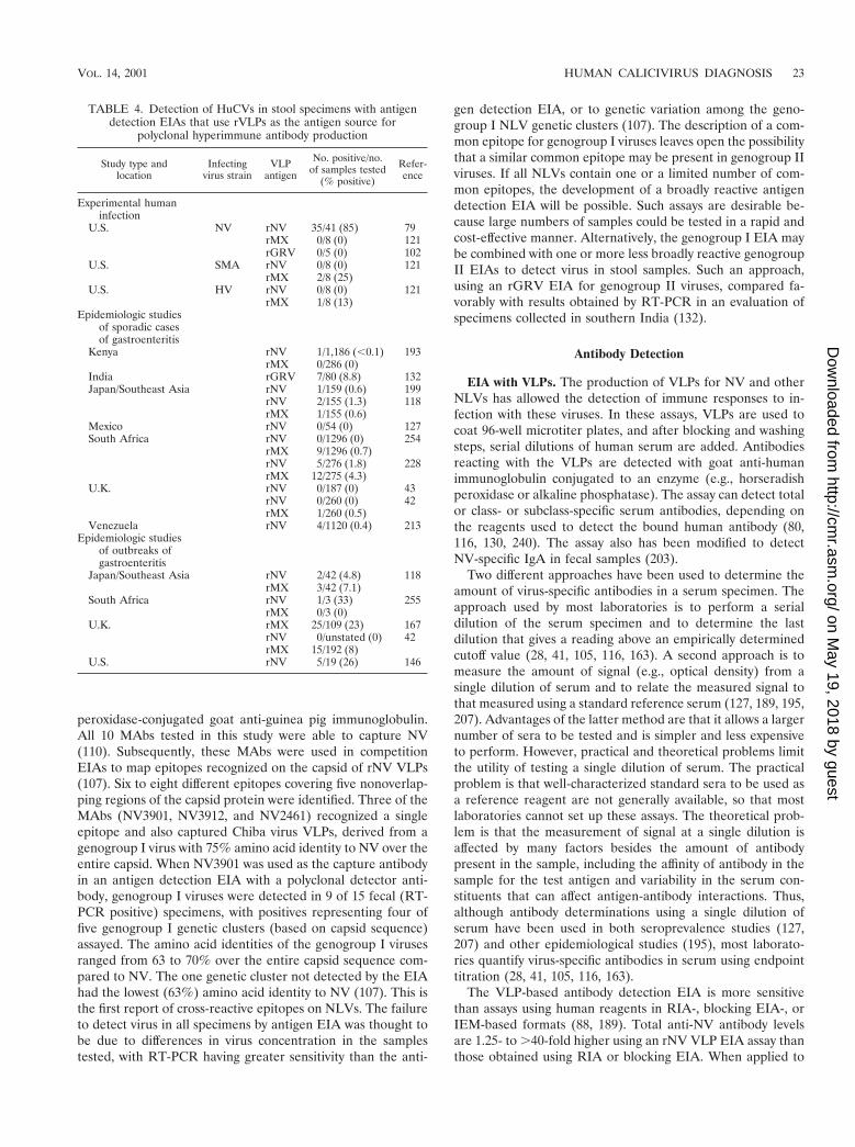
peroxidase-conjugated goat anti-guinea pig immunoglobulin.All 10 MAbs tested in this study were able to capture NV(110). Subsequently, these MAbs were used in competitionEIAs to map epitopes recognized on the capsid of rNV VLPs(107). Six to eight different epitopes covering five nonoverlap-ping regions of the capsid protein were identified. Three of theMAbs (NV3901, NV3912, and NV2461) recognized a singleepitope and also captured Chiba virus VLPs, derived from agenogroup I virus with 75% amino acid identity to NV over theentire capsid. When NV3901 was used as the capture antibodyin an antigen detection EIA with a polyclonal detector anti-body, genogroup I viruses were detected in 9 of 15 fecal (RT-PCR positive) specimens, with positives representing four offive genogroup I genetic clusters (based on capsid sequence)assayed. The amino acid identities of the genogroup I virusesranged from 63 to 70% over the entire capsid sequence com-pared to NV. The one genetic cluster not detected by the EIAhad the lowest (63%) amino acid identity to NV (107). This isthe first report of cross-reactive epitopes on NLVs. The failureto detect virus in all specimens by antigen EIA was thought tobe due to differences in virus concentration in the samplestested, with RT-PCR having greater sensitivity than the anti-
gen detection EIA, or to genetic variation among the geno-group I NLV genetic clusters (107). The description of a com-mon epitope for genogroup I viruses leaves open the possibilitythat a similar common epitope may be present in genogroup IIviruses. If all NLVs contain one or a limited number of com-mon epitopes, the development of a broadly reactive antigendetection EIA will be possible. Such assays are desirable be-cause large numbers of samples could be tested in a rapid andcost-effective manner. Alternatively, the genogroup I EIA maybe combined with one or more less broadly reactive genogroupII EIAs to detect virus in stool samples. Such an approach,using an rGRV EIA for genogroup II viruses, compared fa-vorably with results obtained by RT-PCR in an evaluation ofspecimens collected in southern India (132).
Antibody Detection
EIA with VLPs. The production of VLPs for NV and otherNLVs has allowed the detection of immune responses to in-fection with these viruses. In these assays, VLPs are used tocoat 96-well microtiter plates, and after blocking and washingsteps, serial dilutions of human serum are added. Antibodiesreacting with the VLPs are detected with goat anti-humanimmunoglobulin conjugated to an enzyme (e.g., horseradishperoxidase or alkaline phosphatase). The assay can detect totalor class- or subclass-specific serum antibodies, depending onthe reagents used to detect the bound human antibody (80,116, 130, 240). The assay also has been modified to detectNV-specific IgA in fecal samples (203).
Two different approaches have been used to determine theamount of virus-specific antibodies in a serum specimen. Theapproach used by most laboratories is to perform a serialdilution of the serum specimen and to determine the lastdilution that gives a reading above an empirically determinedcutoff value (28, 41, 105, 116, 163). A second approach is tomeasure the amount of signal (e.g., optical density) from asingle dilution of serum and to relate the measured signal tothat measured using a standard reference serum (127, 189, 195,207). Advantages of the latter method are that it allows a largernumber of sera to be tested and is simpler and less expensiveto perform. However, practical and theoretical problems limitthe utility of testing a single dilution of serum. The practicalproblem is that well-characterized standard sera to be used asa reference reagent are not generally available, so that mostlaboratories cannot set up these assays. The theoretical prob-lem is that the measurement of signal at a single dilution isaffected by many factors besides the amount of antibodypresent in the sample, including the affinity of antibody in thesample for the test antigen and variability in the serum con-stituents that can affect antigen-antibody interactions. Thus,although antibody determinations using a single dilution ofserum have been used in both seroprevalence studies (127,207) and other epidemiological studies (195), most laborato-ries quantify virus-specific antibodies in serum using endpointtitration (28, 41, 105, 116, 163).
The VLP-based antibody detection EIA is more sensitivethan assays using human reagents in RIA-, blocking EIA-, orIEM-based formats (88, 189). Total anti-NV antibody levelsare 1.25- to .40-fold higher using an rNV VLP EIA assay thanthose obtained using RIA or blocking EIA. When applied to
TABLE 4. Detection of HuCVs in stool specimens with antigendetection EIAs that use rVLPs as the antigen source for
polyclonal hyperimmune antibody production
Study type andlocation
Infectingvirus strain
VLPantigen
No. positive/no.of samples tested
(% positive)
Refer-ence
Experimental humaninfection
U.S. NV rNV 35/41 (85) 79rMX 0/8 (0) 121rGRV 0/5 (0) 102
U.S. SMA rNV 0/8 (0) 121rMX 2/8 (25)
U.S. HV rNV 0/8 (0) 121rMX 1/8 (13)
Epidemiologic studiesof sporadic casesof gastroenteritis
Kenya rNV 1/1,186 (,0.1) 193rMX 0/286 (0)
India rGRV 7/80 (8.8) 132Japan/Southeast Asia rNV 1/159 (0.6) 199
rNV 2/155 (1.3) 118rMX 1/155 (0.6)
Mexico rNV 0/54 (0) 127South Africa rNV 0/1296 (0) 254
rMX 9/1296 (0.7)rNV 5/276 (1.8) 228rMX 12/275 (4.3)
U.K. rNV 0/187 (0) 43rNV 0/260 (0) 42rMX 1/260 (0.5)
Venezuela rNV 4/1120 (0.4) 213Epidemiologic studies
of outbreaks ofgastroenteritis
Japan/Southeast Asia rNV 2/42 (4.8) 118rMX 3/42 (7.1)
South Africa rNV 1/3 (33) 255rMX 0/3 (0)
U.K. rMX 25/109 (23) 167rNV 0/unstated (0) 42rMX 15/192 (8)
U.S. rNV 5/19 (26) 146
VOL. 14, 2001 HUMAN CALICIVIRUS DIAGNOSIS 23
on May 19, 2018 by guest
http://cmr.asm
.org/D
ownloaded from
sera from experimental human or chimpanzee infection andfrom outbreaks of gastroenteritis, the rNV VLP EIA detectsfourfold or greater increases in serum antibody levels morefrequently than IEM and at least as frequently as RIA andblocking EIA (88, 189). The antibody rNV VLP EIA identifiedinfection following experimental human challenge better thanthe antigen detection EIA, detecting 40 of 41 (98%) infectionscompared to 36 of 41 (88%) infections (79). Assays utilizingother VLPs, including rMX, rTV, rHV, rSouthampton virus,and rLV, have also been developed (127, 195, 206).
The antibody detection EIA has been used to characterizeIgG, IgM, and IgA serologic responses following experimentalhuman infection with NV (80). Eight of 13 infected subjectshad fourfold or greater increases in virus-specific antibodylevels between 8 and 11 days following infection; ill subjects(eight of nine) were more likely to have these early responses,while antibody rises were seen in asymptomatic subjects onlyafter 15 days. All infected subjects (n 5 14) developed virus-specific IgM serum antibody. Virus-specific IgM serum anti-body was present as early as 9 days following infection, but itdid not develop in some subjects until 2 weeks after infection.IgM serum antibody could still be detected 3 months later insome subjects. Fourfold or greater increases in virus-specificIgA serum antibody were detected in all nine symptomaticinfections but in only two of five asymptomatic infections.Virus-specific geometric mean serum antibody levels of in-fected and uninfected subjects were similar 3 months afterchallenge (80). The kinetics of the IgM and IgA responses aresimilar to those seen in earlier studies using human reagents(64, 65). The presence of neither virus-specific serum IgG,IgM, or IgA nor of fecal IgA is associated with protection frominfection (80, 203, 240). Low serum antibody levels appear tobe associated with a decreased likelihood of infection followingexperimental human challenge and natural exposure (79, 226).
Virus-specific IgM serum antibody has also been detectedusing an IgM capture assay. In this assay, goat anti-human IgMis used to coat microtiter plates, and dilutions of the test serumare then applied. VLPs are added in the next step, and VLPsbound by virus-specific IgM are detected using hyperimmuneanti-NV rabbit serum (240). An alternative method uses avirus-specific MAb for VLP detection (28). Antibody levels arefour- to eightfold higher using the IgM capture assay comparedto the assay in which IgM bound to VLPs is detected (240). Intwo different studies following experimental human infection,IgM capture assays detected IgM responses in 15 of 15 and14 of 15 subjects, with infection documented by fourfold orgreater IgG responses (28, 240).
Detection of infection caused by heterotypic HuCVs. Theserologic responses measured using VLP-based antibody de-tection EIAs have been characterized using sera collected dur-ing studies of experimental human infection and during eval-uations of gastroenteritis outbreaks. Heterologous rNV IgGresponses occur following experimental human infection withHV or SMA although they are present at a lower frequencyand magnitude than is seen following infection with NV (240)or when rMX (SMA-like) VLPs are used in the assay (206).Heterologous rHV IgG responses also can be demonstratedfollowing NV infection (41). The results are similar to thoseobtained using human reagents in blocking EIAs, althoughheterologous seroresponses as measured by the older and
newer assays occurred in different subjects (177, 240). Heter-ologous seroresponses appear to be limited to subjects whoalso have an IgG seroresponse to homologous viral antigenand who are ill. Heterologous IgM and IgA responses occurinfrequently (240).
Similar results have been obtained when these assays havebeen applied to sera collected during outbreak investigations(28, 105, 195, 206). IgG responses occur with a higher fre-quency and magnitude when the assay utilizes VLPs that aremore closely related to the outbreak strain (105, 195). Noel etal. (195) found homologous IgG seroresponses to rNV VLPswhen the infecting NLV was a genogroup I NLV with as muchas 38.5% amino acid divergence from NV in the capsid region.In contrast, homologous seroresponses were seen for geno-group II NLVs only when the amino acid divergence from thetest antigen was less than 6.5%. In general, the likelihood ofdetecting an IgG seroresponse for genogroup II NLVs wasgreatest when the test antigen was derived from a strain closelyrelated to the infecting virus, less when the test antigen wasderived from an unrelated genogroup II NLV, and least whenthe test antigen was rNV (genogroup I). Hale et al. (105)examined several outbreaks caused by genogroup II NLVs andfound that rMX IgM responses occurred in 14 of 19 (74%)subjects with an IgG seroresponse. IgM responses occurredmore frequently when the outbreak virus was MX-like (9 of 10)than when it was an unrelated genogroup II virus (4 of 9). Fourof these subjects also had an rNV IgG seroresponse, but noneof them had a rNV IgM seroresponse. Brinker et al. (28)obtained similar results applying the rNV and rMX IgM EIAsto genogroup I and genogroup II outbreaks. Twenty-four of 25subjects infected with genogroup I viruses had rNV IgM re-sponses, while only 3 of these subjects had rMX IgM respons-es; 28 of 47 subjects infected with genogroup II viruses hadrMX IgM responses while none of them had rNV IgM re-sponses. Taken together, these results indicate that IgM re-sponses may be able to provide data on the genogroup of viruscausing an infection.
The antibody detection EIAs have been used in seropreva-lence surveys and in longitudinal studies of antibody acquisi-tion (41, 54, 70, 82, 116, 118, 127, 163, 190, 193, 199, 204, 206,207, 213, 227, 228, 236). These studies confirmed and extendedthe results obtained using human reagents showing that NLVscause infection worldwide and seroprevalence increases withage. New findings include serologic evidence of NLV infectionoccurring in young children in developed countries that wasnot recognized in early studies (163) and of transplacentaltransfer of NLV-specific antibodies from mother to child (41,118, 199, 206, 228). In addition, seroprevalence rates variedbetween regions within a country, between countries, and bythe NLV VLP antigen used in the assay.
Nucleic Acid Detection
Nucleic acid detection assays are the third group of newassays that have been developed in the last decade since thecloning of the NV genome. Knowledge of the sequence of theNV genome led to the design of primers from the polymeraseregion that were able to amplify fragments of other NLVs andSLVs, and this led to sequencing of the complete genomes ofmany HuCVs. Although there have been a few reports of the
24 ATMAR AND ESTES CLIN. MICROBIOL. REV.
on May 19, 2018 by guest
http://cmr.asm
.org/D
ownloaded from
use of hybridization assays, the primary nucleic acid detectionassay that is used is RT-PCR. RT-PCR is currently being usedworldwide because of the lack of a commercially available,broadly reactive EIA. Both nucleic acid detection diagnosticapproaches will be discussed below.
Hybridization assays. Only a few hybridization assays havebeen described for the detection of HuCVs. This is most likelydue to the availability of the more sensitive RT-PCR assays atthe time that cDNAs for HuCVs first became available. Jianget al. (129) described a hybridization assay using 32P-labeledcDNAs covering the same region of the genome as was ampli-fied in RT-PCR assays. Stool suspensions (10 to 50%) wereextracted with trichlorotrifluoroethane, and the viral nucleicacids were partially purified from the aqueous phase by diges-tion with proteinase K, extractions in phenol-chloroform andchloroform, and precipitation in ethanol. The hybridizationassay detected NV with a sensitivity similar to that of RIA, butdetected 100-fold less viral RNA and detected virus in stoolsamples 27% less frequently than an RT-PCR assay applied tothe same samples. Thus, the hybridization assay was unable todetect NV when low titers of virus were present in a stoolsample (129).
A hybridization assay using a digoxigenin-labeled cDNAprobe derived from the polymerase region of Sapporo virus hasalso been described (150). Viral nucleic acids were partiallypurified as described above, and approximately 105 viral par-ticles could be detected per dot. This level of detection is lowerthan the 1 3 104 to 2 3 104 particles detected by RIA and EIAsspecific for Sapporo virus. The interpretation of test results wasalso hampered by the colorimetric detection system. Somestool samples gave false-positive results due to the color of thestool or to nonspecific binding of the probe to substances in thestool. The problem of false-positive results was addressed byinclusion of digoxigenin-labeled vector (pBR322) DNA in theassay as a negative control. The signal intensities obtained withvirus-specific and control probes were compared to referencedots containing 10-fold serial dilutions of Sapporo virus cDNA,and positive samples were those in which the reaction with thevirus-specific probe was stronger than with the control probe.
The Sapporo virus-specific dot blot assay was used to eval-uate 100 stool samples in which HuCVs were detected byelectron microscopy. Eight of 10 samples that tested positiveusing the SLV/Sapporo/82 EIA were positive by the dot blotassay, and an additional 13 samples were EIA negative and dotblot hybridization positive. Seventy-seven samples were nega-tive by both assays. The investigators speculated that the im-proved sensitivity of the dot blot hybridization assay comparedto the EIA may have been due to the greater conservation ofsequence among SLVs in the polymerase region (targeted inthe dot blot assay) than in the capsid region (targeted in theEIA assay) or to the presence of substances in stool samplesthat had a greater inhibitory effect on EIA than on dot blot hy-bridization assays. A potential advantage of the dot blot hybrid-ization assay over RT-PCR assays is the lower cost of the assayand decreased risk of cross-contamination. Nevertheless, RT-PCR assays have been the major nucleic acid detection assayused for the diagnosis of HuCV infections.
RT-PCR. The first RT-PCR assays were described within 2years of the initial report of the successful cloning of the NVgenome (53, 129). Since then a number of different RT-PCR
assay formats have been developed, and these assays havebecome one of the principal means for the diagnosis of HuCVinfections. A number of factors can affect the sensitivity andspecificity of RT-PCR assays, including the sample being as-sayed, the method used for purification of viral nucleic acids,the primers used in amplification, and the method used forinterpretation of test results. Approaches to addressing each ofthese factors are discussed below.
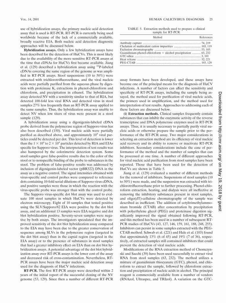
(i) Extraction methods. Clinical samples frequently containsubstances that can inhibit the enzymatic activity of the reversetranscriptase and DNA polymerase enzymes used in RT-PCRassays. Thus, it is usually necessary to partially purify viral nu-cleic acids or otherwise prepare the sample prior to the per-formance of the RT-PCR assay. Two major considerations inselecting an extraction method are its efficiency of viral nucleicacid recovery and its ability to remove or inactivate RT-PCRinhibitors. Secondary considerations include the ease of per-formance of the method and the number of samples that canbe processed at one time. A number of different approachesfor viral nucleic acid purification from stool samples have beenreported. Those that have been used for the detection ofHuCVs are shown in Table 5.
Jiang et al. (129) evaluated a number of different methodsfor the removal of inhibitors. Suspensions of stool samples (10to 50%) were made, and the suspension was extracted with tri-chlorotrifluoroethane prior to further processing. Phenol-chlo-roform extraction, heating, and dialysis were all ineffective atremoving the inhibitory substances present in stool samples,and oligo(dT)-cellulose chromatography of the sample wasdescribed as inefficient. The addition of cetyltrimethylammo-nium bromide (CTAB) after concentration by precipitationwith polyethylene glycol (PEG) and proteinase digestion sig-nificantly improved the signal obtained following RT-PCR,and this method has been used in a number of subsequent RT-PCR studies of HuCVs (43, 127, 146, 159, 161, 188, 248, 255).Inhibitors can persist in some samples extracted with the PEG-CTAB method. Schwab et al. (221) and Hale et al. (103) foundthat approximately 13% (6 of 45) and 19% (7 of 36), respec-tively, of extracted samples still contained inhibitors that couldprevent the detection of viral nucleic acids.
Modifications of the RNA extraction method of Chomczyn-ski and Sacchi (39) have been used successfully to extract viralRNA from stool samples (63, 232). The method utilizes amixture of guanidinium thiocyanate (GTC), phenol, and chlo-roform to extract the sample, followed by chloroform extrac-tion and precipitation of nucleic acids in alcohol. The principalreagent is commercially available from a number of vendors(RNAzol, Ultraspec, and TRIzol). A variation on the GTC-
TABLE 5. Extraction methods used to prepare a clinicalsample for RT-PCR
Method References
Antibody capture ................................................................................ 74, 219Chelation of multivalent cation impurities ..................................... 103, 119Exclusion chromatography ................................................................ 53, 103Guanidinium-phenol-chloroform 1 alcohol precipitation ............ 63, 232GTC-silica ........................................................................................... 3, 101Heat release ........................................................................................ 32, 221PEG-CTAB ......................................................................................... 103, 129
VOL. 14, 2001 HUMAN CALICIVIRUS DIAGNOSIS 25
on May 19, 2018 by guest
http://cmr.asm
.org/D
ownloaded from
based extraction procedure was described by Boom et al. (26),in which a GTC-containing buffer is used to release viral RNAfrom the viral capsid. The viral RNA is adsorbed onto size-fractionated silica particles and washed in successive steps witha second GTC-containing buffer, 70% ethanol, and acetone.The viral RNA is then eluted from the silica particles withwater. Other variations have also been used successfully (3,101). The GTC-silica method has been reported to be quitesuccessful in removing inhibitors of PCR in two comparativestudies, performing better than the PEG-CTAB method in thedetection of NLVs (103) and being approximately equivalentto PEG-CTAB for the detection of hepatitis A virus in stoolsamples (8).
Exclusion chromatography using spin columns containingSephadex G200 was one of the original methods used to ex-tract virus from stool samples (53). Although the method issensitive, it is inconsistent at removing inhibitors from clinicalsamples (103). Similarly, a method based on the chelation ofmultivalent cation impurities has been used successfully fordetection of NLVs but is unreliable at removing inhibitors ofRT-PCR (8, 103). Unexpectedly, NLVs can be detected byRT-PCR in stool samples after simple heating of the sample to95 to 99°C for 5 min (32, 221). The heat may inactivate someinhibitors and is thought to denature the viral capsid, allowingthe release of viral RNA. Prior to the heat release procedure,10 to 20% suspensions of the fecal samples are clarified andextracted with trichlorotrifluoroethane or pelleted through a45% (wt/vol) sucrose cushion. The trichlorotrifluoroethane-treated samples must be diluted 100-fold, as amplification isinhibited in the majority of specimens at a lower dilution (221).
Antibody capture has been used for virus purification priorto amplification (74, 219). Virus-specific antibodies are boundin a 96-well plate or to paramagnetic beads before exposure toa virus-containing sample. After an incubation period to allowantigen capture, the wells or beads are washed repeatedly toremove inhibitors and other substances. Viral genomic RNA isthen released from its capsid by heating. This method hasworked well for hepatitis A virus, but its use in the detection ofHuCVs has been limited. The principal reason this method hasnot been explored further is the lack of high-titered antiserathat react with a broad range of NLVs. Nevertheless, Schwabet al. (219) were able to use human immunoglobulin prepara-tions as a source of antibody to detect NLVs from water sam-ples in which viruses had been concentrated by filtration andPEG precipitation. Polyclonal hyperimmune animal sera havealso been coupled to paramagnetic beads and used to purifyNV from fecal specimens prior to RT-PCR amplification (74).This strategy was found to yield a greater number of positiveRT-PCR results from stools collected during experimental hu-man infection studies than other processing protocols. Asbroadly reactive antisera become available, further studies ofthis extraction method are likely to be performed.
(ii) Primer selection. The sensitivity and specificity of RT-PCR assays depend in large part on primer selection. Severalfactors affect the ability of a primer pair to detect a given NLVor SLV strain, including primer sequence, the amount of viruspresent in the sample to be assayed, and the temperature usedfor primer annealing during the PCR amplification process.The genetic diversity of NLVs and SLVs has made it difficult toselect a single primer set with adequate sensitivity and speci-
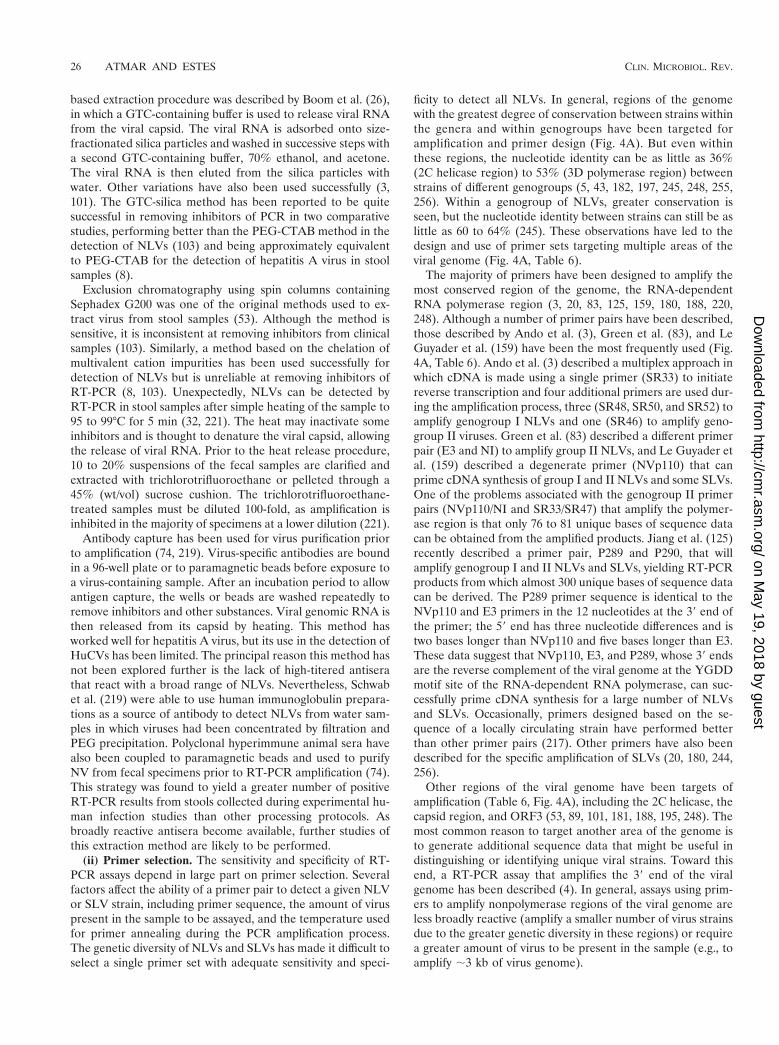
ficity to detect all NLVs. In general, regions of the genomewith the greatest degree of conservation between strains withinthe genera and within genogroups have been targeted foramplification and primer design (Fig. 4A). But even withinthese regions, the nucleotide identity can be as little as 36%(2C helicase region) to 53% (3D polymerase region) betweenstrains of different genogroups (5, 43, 182, 197, 245, 248, 255,256). Within a genogroup of NLVs, greater conservation isseen, but the nucleotide identity between strains can still be aslittle as 60 to 64% (245). These observations have led to thedesign and use of primer sets targeting multiple areas of theviral genome (Fig. 4A, Table 6).
The majority of primers have been designed to amplify themost conserved region of the genome, the RNA-dependentRNA polymerase region (3, 20, 83, 125, 159, 180, 188, 220,248). Although a number of primer pairs have been described,those described by Ando et al. (3), Green et al. (83), and LeGuyader et al. (159) have been the most frequently used (Fig.4A, Table 6). Ando et al. (3) described a multiplex approach inwhich cDNA is made using a single primer (SR33) to initiatereverse transcription and four additional primers are used dur-ing the amplification process, three (SR48, SR50, and SR52) toamplify genogroup I NLVs and one (SR46) to amplify geno-group II viruses. Green et al. (83) described a different primerpair (E3 and NI) to amplify group II NLVs, and Le Guyader etal. (159) described a degenerate primer (NVp110) that canprime cDNA synthesis of group I and II NLVs and some SLVs.One of the problems associated with the genogroup II primerpairs (NVp110/NI and SR33/SR47) that amplify the polymer-ase region is that only 76 to 81 unique bases of sequence datacan be obtained from the amplified products. Jiang et al. (125)recently described a primer pair, P289 and P290, that willamplify genogroup I and II NLVs and SLVs, yielding RT-PCRproducts from which almost 300 unique bases of sequence datacan be derived. The P289 primer sequence is identical to theNVp110 and E3 primers in the 12 nucleotides at the 39 end ofthe primer; the 59 end has three nucleotide differences and istwo bases longer than NVp110 and five bases longer than E3.These data suggest that NVp110, E3, and P289, whose 39 endsare the reverse complement of the viral genome at the YGDDmotif site of the RNA-dependent RNA polymerase, can suc-cessfully prime cDNA synthesis for a large number of NLVsand SLVs. Occasionally, primers designed based on the se-quence of a locally circulating strain have performed betterthan other primer pairs (217). Other primers have also beendescribed for the specific amplification of SLVs (20, 180, 244,256).
Other regions of the viral genome have been targets ofamplification (Table 6, Fig. 4A), including the 2C helicase, thecapsid region, and ORF3 (53, 89, 101, 181, 188, 195, 248). Themost common reason to target another area of the genome isto generate additional sequence data that might be useful indistinguishing or identifying unique viral strains. Toward thisend, a RT-PCR assay that amplifies the 39 end of the viralgenome has been described (4). In general, assays using prim-ers to amplify nonpolymerase regions of the viral genome areless broadly reactive (amplify a smaller number of virus strainsdue to the greater genetic diversity in these regions) or requirea greater amount of virus to be present in the sample (e.g., toamplify ;3 kb of virus genome).
26 ATMAR AND ESTES CLIN. MICROBIOL. REV.
on May 19, 2018 by guest
http://cmr.asm
.org/D
ownloaded from

Very few studies have been reported that describe the quan-tity of virus genome that must be present in order for a primerpair to detect the virus (159). Instead, most studies only reportwhether a primer pair can detect a virus strain without regardto the amount of virus present in the sample. However, suc-cessful detection of virus may be enhanced when larger quan-tities of virus are present. For example, Le Guyader et al. (159)noted 10- to 1,000-fold differences in the quantities of severalNLV strains that could be detected by two primer pairs,NVp110/p36 and NVp110/p69. In other words, when a largequantity of a virus strain is present, both primer pairs cansuccessfully detect it, but when small quantities of the strainare present, only one primer pair allows successful virus de-tection. The reason for the differences in virus detection isprimer homology, with primers having lower homology requir-ing the presence of larger quantities of virus for successfulamplification to occur. Another approach used to improve thechances of successful amplification has been to lower theprimer annealing temperature to as low as 37°C (248). Thisstrategy has the disadvantage of increasing the likelihood thatnonspecific amplicons will be generated and may increase theamount of virus that must be present in the sample for suc-cessful virus detection.
The use of nested or seminested PCR is another methodthat has been used to increase the likelihood of detectingNLVs (84, 200, 234). This approach utilizes two rounds of PCRamplification, with one (seminested) or both (nested) primersused in the second round of amplification targeting a region ofthe genome inside that targeted by the primers used in theinitial amplification. This strategy has been reported to be 10 to1,000 times more sensitive than single-round RT-PCR (84) andhas also been used to detect the presence of multiple viralstrains within a single sample (200, 234). However, a majordisadvantage of this approach is the increased and very realpossibility that carryover contamination may occur betweensamples.
(iii) Other PCR conditions. The conditions (e.g., magnesiumconcentration and primer annealing temperature) of the RT-PCR assay are dictated, in part, by the primers used. The useof different DNA polymerases during the PCR amplificationprocess has also been reported. Ando et al. (4) used a combi-nation of Taq DNA polymerase and Pwo DNA polymerase forthe amplification of a large (3 kb) fragment of the viral ge-nome. Schwab et al. (222) utilized Tth polymerase in place ofavian myeloblastosis virus reverse transcriptase and Taq poly-merase and found that detection of NV in the Tth polymerase-
FIG. 4. (A) Schematic representation of the calicivirus genome and the regions amplified by common primer pairs. Modified from reference220 with permission from Technomic Publishing Co, Inc., copyright 2000. See Table 6 for further details about primer sequences. hel, helicase; pol,polymerase. (B) Schematic representation of the NLV genome from which an internal standard control for genogroup I NLVs was made. Therelative locations of selected primers and probes are noted for virus and internal standard (Int. Std.) RNA. The internal standard control RNAyields amplicons that are 123 bp shorter (347 bp) than those from NV genomic RNA (470 bp). A portion of the genomic sequence targeted byvirus-specific probes (e.g., SR65 and p116) is not present in the internal standard control, allowing differentiation of virus-specific and internalstandard amplicons by nucleic acid hybridization (221).
VOL. 14, 2001 HUMAN CALICIVIRUS DIAGNOSIS 27
on May 19, 2018 by guest
http://cmr.asm
.org/D
ownloaded from
based assay was comparable to that in the two-enzyme system.The use of a single enzyme for reverse transcription and DNAamplification also allowed the use of thermolabile uracil-N-glycolase (UNG) for the prevention of carryover contamina-tion (222). dUTP is used in place of dTTP in the RT-PCRs,and the UNG degraded deoxyuracil-containing ampliconsadded (or carried over) to the reaction tube. The UNG is theninactivated by heating, and the viral RNA is amplified.
(iv) Confirmation of PCR products. A number of methodscan be used to interpret the results of a PCR method. One ofthe simplest is gel electrophoresis. If a band of the size pre-dicted from primer selection is seen following electrophoresis,the PCR is considered positive. This method has been used forNLV RT-PCR assays (10, 84), but it can yield false-positiveresults (11, 12). Nonspecific amplification of DNA occurs, par-ticularly when more than 30 cycles of amplification are used,and the nonspecifically amplified DNA will occasionally mi-grate in a fashion similar to that expected for virus-specificamplicons. If this happens, the nonspecific amplification prod-ucts may be misinterpreted as being virus-specific amplicons.Such nonspecific bands are frequently seen following the assayfor NLVs in stool and shellfish samples (12). Because of thepotential false-positive results in the visual interpretation ofgels, it is essential to confirm the specificity of the amplicons bya second method.
The use of a hybridization assay is probably the simplestapproach to interpret and confirm PCR assays. Hybridizationassays can be set up in a number of different ways. The most
common include dot or slot blot hybridization, liquid hybrid-ization, and Southern blot hybridization. In these assays, avirus-specific probe is labeled and hybridized with the PCRproducts, and the presence or absence of the label is detected.Some of the more common labels include 32P, digoxigenin, andbiotin. One of the limitations of hybridization assays whenapplied to NLV RT-PCR assays is that the variability of thegenomic sequence in the NLVs makes it difficult to select asingle or even a small number of probes that can detect allpossible NLV sequences (159). Nevertheless, a small numberof probes have been effective for the detection of the majorityof circulating NLVs when the DNA being amplified was ho-mologous to the polymerase region of the NLV genome (3, 83,159). The time required to perform the hybridization assay canbe shortened without loss of sensitivity by using a direct EIAformat. Virus-specific amplicons are captured with a biotinyl-ated probe anchored to a streptavidin-coated, 96-well plate,and the probe-amplicon hybrid is detected using an anti-dou-ble-stranded DNA (dsDNA) antibody conjugate (R. L. Atmarand K. J. Schwab, unpublished data). A reverse-line blottingstrategy is another approach that has simplified the use ofmultiple probes at one time and yielded both specificity dataand data on the genetic relatedness (genotype) of the HuCVdetected to reference strains (247). HuCVs are amplified usingbiotinylated primers, and virus-specific amplicons are capturedduring hybridization by one of the multiple probes that arelinked to the membrane in individual dots on a blot. Biotinyl-ated products are detected by streptavidin-enzyme conjugates.
TABLE 6. Common HuCV-specific primers used in RT-PCR assays
Primer DNA sequence (59 to 39) Genomic locationa Sense Viruses amplifiedb Reference
p78 GGGCCCCCTGGTATAGGTAA 1682–1701 (2C helicase) 1 NLVs (GI & GII) 248p80 TGGTGATGACTATAGCATCAGACACAAA 1943–1970 (2C helicase) 2 NLVs (GI & GII) 248N49(1) CACCACCATAAACAGGCTG 2215–2233 (2C helicase) 1 NLVs (GI) 181N49(2) AGCCTGATAGAGCATTCTTT 2419–2438 (2C helicase) 2 NLVs (GI) 181p36 ATAAAAGTTGGCATGAACA 4487–4505 (3D polymerase) 1 NLVs (GI & GII), SLVs 248Sapp36 GTTGCTGTTGGCATTAACA 4487–4505 (3D polymerase) 1 SLVs 180JV12 ATACCACTATGATGCAGATTA 4552–4572 (3D polymerase) 1 NLVs (GI & GII) 246P290 GATTACTCCAAGTGGGACTCCAC 4568–4590 (3D polymerase) 1 NLVs (GI & GII), SLVs 125p3 GCACCATCTGAGATGGATGT 4685–4704 (3D polymerase) 1 NLVs (GI & GII) 188p69 GGCCTGCCATCTGGATTGCC 4733–4752 (3D polymerase) 1 NLVs (GI & GII), SLVs 248SR46 TGGAATTCCATCGCCCACTGG 4766–4786 (3D polymerase) 1 NLVs (GI & GII) 3SR48 GTGAACAGCATAAATCACTGG 4766–4786 (3D polymerase) 1 NLVs (GI & GII) 3SR50 GTGAACAGTATAAACCACTGG 4766–4786 (3D polymerase) 1 NLVs (GI & GII) 3SR52 GTGAACAGTATAAACCATTGG 4766–4786 (3D polymerase) 1 NLVs (GI & GII) 3NI GAATTCCATCGCCCACTGGCT 4768–4788 (3D polymerase) 1 NLVs (GII) 83JV13 TCATCATCACCATAGAAAGAG 4858–4878 (3D polymerase) 2 NLVs (GI & GII) 246E3 ATCTCATCATCACCATA 4865–4881 (3D polymerase) 2 NLVs (GI & GII) 83NVp110 AC(A/T/G)AT(C/T)TCATCATCACCATA 4865–4884 (3D polymerase) 2 NLVs (GI & GII), SLVs 159P289 TGACAATGTAATCATCACCATA 4865–4886 (3D polymerase) 2 NLVs (GI & GII), SLVs 125SR33 TGTCACGATCTCATCATCACC 4868–4888 (3D polymerase) 2 NLVs (GI & GII) 3p51 GTTGACACAATCTCATCATC 4871–4890 (3D polymerase) NLVs (GI & GII) 188p35 CTTGTTGGTTTGAGGCCATAT 4936–4956 (3D polymerase) 2 NLVs (GI & GII), SLVs 248Sapp35 GCAGTGGGTTTGAGACCAAAG 4936–4956 (3D polymerase) 2 SLVs 20SRI-2 AAATGATGATGGCGTCTA 5356–5373 (capsid) 1 NLVs (GI) 101mon381 CCAGAATGTACAATGGTTATGC 5362–5383* (capsid) 1 NLVs (GII) 195CapIIa CIAGAATGTAIAA(C/T)GG(G/T)TATGC 5362–5383* (capsid) 1 NLVs (GII) 89CapIIb TGIIAGAAAIT(A/G)TTICI(A/G)ACATC(A/T)GG 5559–5584* (capsid) 2 NLVs (GII) 89CapIa CICAAATGTAIAATGG(C/T)TGGGT 5647–5668 (capsid) 1 NLVs (GI) 89SRI-1 CCAACCCA(A/G)CCATT(A/G)TACAT 5652–5671 (capsid) 2 NLVs (GI) 101mon383 CAAGAGACTGTGAAGACATCATC 5661–5683* (capsid) 2 NLVs (GII) 195CapIb TGIIA(A/G)AGIACATTICI(A/T)ACATC(C/T)TC 5844–5869 (capsid) 2 NLVs (GI) 89
a Corresponds to the equivalent location within the NV genomic sequence (M87661) (108, 131) except each one marked with an asterisk, corresponds to theequivalent location within the LV genomic sequence (X86557) (55).
b GI and GII, genogroup I and II, respectively.
28 ATMAR AND ESTES CLIN. MICROBIOL. REV.
on May 19, 2018 by guest
http://cmr.asm
.org/D
ownloaded from
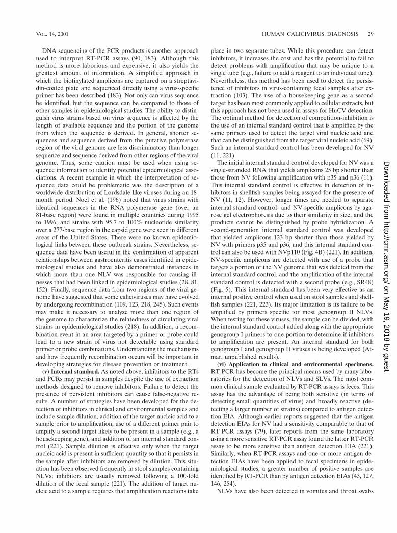
DNA sequencing of the PCR products is another approachused to interpret RT-PCR assays (90, 183). Although thismethod is more laborious and expensive, it also yields thegreatest amount of information. A simplified approach inwhich the biotinylated amplicons are captured on a streptavi-din-coated plate and sequenced directly using a virus-specificprimer has been described (183). Not only can virus sequencebe identified, but the sequence can be compared to those ofother samples in epidemiological studies. The ability to distin-guish virus strains based on virus sequence is affected by thelength of available sequence and the portion of the genomefrom which the sequence is derived. In general, shorter se-quences and sequence derived from the putative polymeraseregion of the viral genome are less discriminatory than longersequence and sequence derived from other regions of the viralgenome. Thus, some caution must be used when using se-quence information to identify potential epidemiological asso-ciations. A recent example in which the interpretation of se-quence data could be problematic was the description of aworldwide distribution of Lordsdale-like viruses during an 18-month period. Noel et al. (196) noted that virus strains withidentical sequences in the RNA polymerase gene (over an81-base region) were found in multiple countries during 1995to 1996, and strains with 95.7 to 100% nucleotide similarityover a 277-base region in the capsid gene were seen in differentareas of the United States. There were no known epidemio-logical links between these outbreak strains. Nevertheless, se-quence data have been useful in the confirmation of apparentrelationships between gastroenteritis cases identified in epide-miological studies and have also demonstrated instances inwhich more than one NLV was responsible for causing ill-nesses that had been linked in epidemiological studies (28, 81,152). Finally, sequence data from two regions of the viral ge-nome have suggested that some caliciviruses may have evolvedby undergoing recombination (109, 123, 218, 245). Such eventsmay make it necessary to analyze more than one region ofthe genome to characterize the relatedness of circulating viralstrains in epidemiological studies (218). In addition, a recom-bination event in an area targeted by a primer or probe couldlead to a new strain of virus not detectable using standardprimer or probe combinations. Understanding the mechanismsand how frequently recombination occurs will be important indeveloping strategies for disease prevention or treatment.
(v) Internal standard. As noted above, inhibitors to the RTsand PCRs may persist in samples despite the use of extractionmethods designed to remove inhibitors. Failure to detect thepresence of persistent inhibitors can cause false-negative re-sults. A number of strategies have been developed for the de-tection of inhibitors in clinical and environmental samples andinclude sample dilution, addition of the target nucleic acid to asample prior to amplification, use of a different primer pair toamplify a second target likely to be present in a sample (e.g., ahousekeeping gene), and addition of an internal standard con-trol (221). Sample dilution is effective only when the targetnucleic acid is present in sufficient quantity so that it persists inthe sample after inhibitors are removed by dilution. This situ-ation has been observed frequently in stool samples containingNLVs; inhibitors are usually removed following a 100-folddilution of the fecal sample (221). The addition of target nu-cleic acid to a sample requires that amplification reactions take
place in two separate tubes. While this procedure can detectinhibitors, it increases the cost and has the potential to fail todetect problems with amplification that may be unique to asingle tube (e.g., failure to add a reagent to an individual tube).Nevertheless, this method has been used to detect the persis-tence of inhibitors in virus-containing fecal samples after ex-traction (103). The use of a housekeeping gene as a secondtarget has been most commonly applied to cellular extracts, butthis approach has not been used in assays for HuCV detection.The optimal method for detection of competition-inhibition isthe use of an internal standard control that is amplified by thesame primers used to detect the target viral nucleic acid andthat can be distinguished from the target viral nucleic acid (69).Such an internal standard control has been developed for NV(11, 221).
The initial internal standard control developed for NV was asingle-stranded RNA that yields amplicons 25 bp shorter thanthose from NV following amplification with p35 and p36 (11).This internal standard control is effective in detection of in-hibitors in shellfish samples being assayed for the presence ofNV (11, 12). However, longer times are needed to separateinternal standard control- and NV-specific amplicons by aga-rose gel electrophoresis due to their similarity in size, and theproducts cannot be distinguished by probe hybridization. Asecond-generation internal standard control was developedthat yielded amplicons 123 bp shorter than those yielded byNV with primers p35 and p36, and this internal standard con-trol can also be used with NVp110 (Fig. 4B) (221). In addition,NV-specific amplicons are detected with use of a probe thattargets a portion of the NV genome that was deleted from theinternal standard control, and the amplification of the internalstandard control is detected with a second probe (e.g., SR48)(Fig. 5). This internal standard has been very effective as aninternal positive control when used on stool samples and shell-fish samples (221, 223). Its major limitation is its failure to beamplified by primers specific for most genogroup II NLVs.When testing for these viruses, the sample can be divided, withthe internal standard control added along with the appropriategenogroup I primers to one portion to determine if inhibitorsto amplification are present. An internal standard for bothgenogroup I and genogroup II viruses is being developed (At-mar, unpublished results).
(vi) Application to clinical and environmental specimens.RT-PCR has become the principal means used by many labo-ratories for the detection of NLVs and SLVs. The most com-mon clinical sample evaluated by RT-PCR assays is feces. Thisassay has the advantage of being both sensitive (in terms ofdetecting small quantities of virus) and broadly reactive (de-tecting a larger number of strains) compared to antigen detec-tion EIA. Although earlier reports suggested that the antigendetection EIAs for NV had a sensitivity comparable to that ofRT-PCR assays (79), later reports from the same laboratoryusing a more sensitive RT-PCR assay found the latter RT-PCRassay to be more sensitive than antigen detection EIA (221).Similarly, when RT-PCR assays and one or more antigen de-tection EIAs have been applied to fecal specimens in epide-miological studies, a greater number of positive samples areidentified by RT-PCR than by antigen detection EIAs (43, 127,146, 254).
NLVs have also been detected in vomitus and throat swabs
VOL. 14, 2001 HUMAN CALICIVIRUS DIAGNOSIS 29
on May 19, 2018 by guest
http://cmr.asm
.org/D
ownloaded from
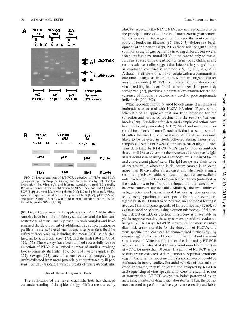
(85, 184, 200). Barriers to the application of RT-PCR to othersamples have been the inhibitory substances and the low con-centrations of virus usually present in such samples and haverequired the development of additional virus concentration orpurification steps. Several such assays have been described fordifferent food samples, including deli meats (224), salads (let-tuce, melons, and cole slaw) (78), and shellfish (10–12, 78, 84,120, 157). These assays have been applied successfully for thedetection of NLVs in a limited number of studies involvingfoods (primarily shellfish) (157, 158, 234), water samples (19,152), sewage (173), and other environmental samples (e.g.,swabs collected from areas potentially contaminated by ill per-sons) (36, 210) associated with outbreaks of viral gastroenteritis.
Use of Newer Diagnostic Tests
The application of the newer diagnostic tests has changedour understanding of the epidemiology of infections caused by
HuCVs, especially the NLVs. NLVs are now recognized to bethe principal cause of outbreaks of nonbacterial gastroenteri-tis, and new estimates suggest that they are the most commoncause of foodborne illnesses (67, 186, 243). Before the devel-opment of the newer assays, NLVs were not thought to be acommon cause of gastroenteritis in young children, but severalrecent studies have found NLVs to be second only to rotavi-ruses as a cause of viral gastroenteritis in young children, andseroprevalence studies suggest that infection in young childrenin developed countries is common (25, 82, 163, 205, 206).Although multiple strains may circulate within a community atone time, a single strain or strains within an antigenic clustermay predominate (106, 179, 196). In addition, the duration ofvirus shedding has been found to be longer than previouslyrecognized (79), providing a potential explanation for the oc-currence of foodborne outbreaks traced to postsymptomaticindividuals (209, 252).
What approach should be used to determine if an illness oroutbreak is associated with HuCV infection? Figure 6 is aschematic of an approach that has been proposed for thecollection and testing of specimens in the setting of an out-break (220). Guidelines for data and sample collection havebeen published previously (16, 162). Stool and serum samplesshould be collected from affected individuals as soon as possi-ble after the onset of clinical illness. Although virus is mostlikely to be detected in stools collected during illness, stoolsamples collected 1 or 2 weeks after illness onset may still havevirus detectable by RT-PCR. VLPs can be used in antibodydetection EIAs to determine the presence of virus-specific IgMin individual sera or rising total antibody levels in paired (acuteand convalescent phase) sera. The IgM assays are likely to beof greatest value when the initial serum sample is collectedmore than 10 days after illness onset and when only a singleserum sample is available. At present, these tests are availableonly in a limited number of research laboratories (indicated bythe shaded box in Fig. 6), but it is hoped that the reagents willbecome commercially available. Similarly, the availability ofantigen detection EIAs is limited, but fecal specimens can betested using hyperimmune sera specific for one or several an-tigenic clusters. If found to be positive, no additional testing isneeded. Similarly, some specialized laboratories may be able toevaluate stool specimens using electron microscopy. If the an-tigen detection EIA or electron microscopy is unavailable oryields negative results, these specimens should be evaluatedusing RT-PCR assays. RT-PCR is currently the most sensitivediagnostic assay available for the detection of HuCVs, andvirus-specific amplicons can be characterized further (e.g., bysequencing) to provide additional information about the virusstrain detected. Virus is stable and can be detected by RT-PCRin stool samples stored at 4°C for several months (at least) orat 270°C for more than 10 years. The ability of RT-PCR assaysto detect virus collected or stored under suboptimal conditions(e.g., in bacterial transport medium) is not known but could beevaluated in future studies. Potential vehicles of transmission(food and water) may be collected and analyzed by RT-PCRand sequencing of virus-specific amplicons to establish routesof transmission. RT-PCR assays are being performed by anincreasing number of diagnostic laboratories. Thus, the equip-ment needed to perform such assays is more readily available,
FIG. 5. Representation of RT-PCR detection of NLVs and SLVsby agarose gel electrophoresis (A) and confirmation by slot blot hy-bridization (B). Virus (V)- and internal standard control (IS)-specificRNAs are visible after amplification of NLVs (NV and SMA) and anSLV (Sapporo virus [Sa]) with primers NVp110 and p36 or p69. Virus-specific amplicons are detected by probes SR65 (NV), p117 (SMA),and p153 (Sapporo virus), while the internal standard control is de-tected by probe SR48 (3,159).
30 ATMAR AND ESTES CLIN. MICROBIOL. REV.
on May 19, 2018 by guest
http://cmr.asm
.org/D
ownloaded from
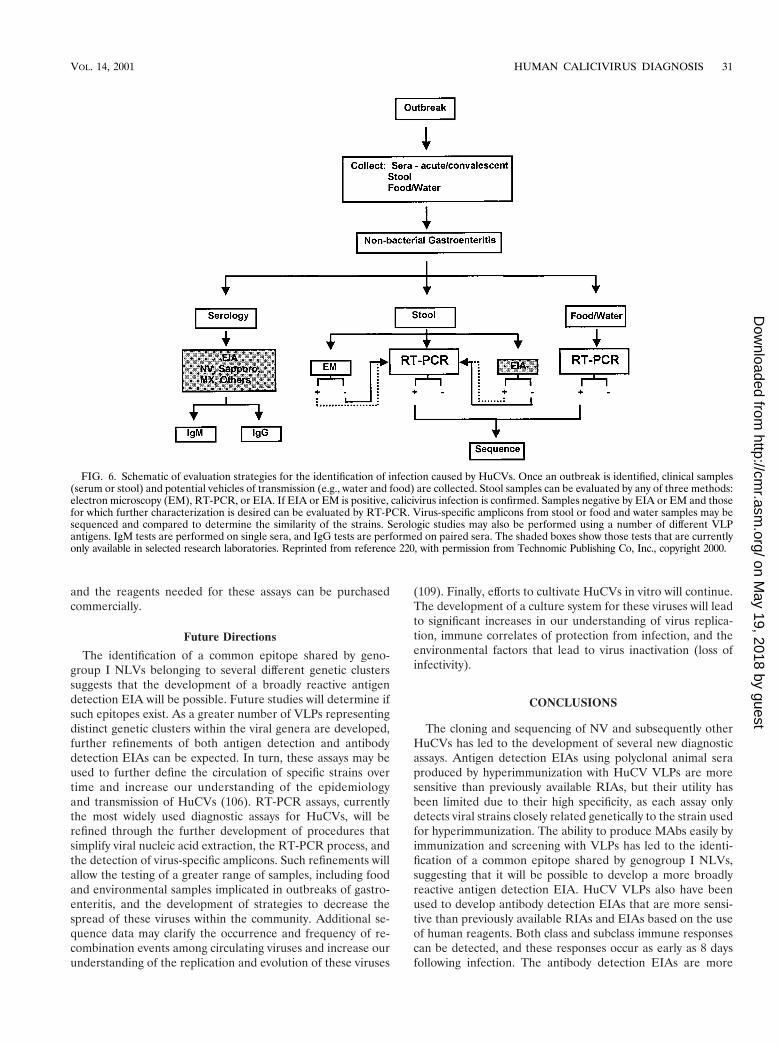
and the reagents needed for these assays can be purchasedcommercially.
Future Directions
The identification of a common epitope shared by geno-group I NLVs belonging to several different genetic clusterssuggests that the development of a broadly reactive antigendetection EIA will be possible. Future studies will determine ifsuch epitopes exist. As a greater number of VLPs representingdistinct genetic clusters within the viral genera are developed,further refinements of both antigen detection and antibodydetection EIAs can be expected. In turn, these assays may beused to further define the circulation of specific strains overtime and increase our understanding of the epidemiologyand transmission of HuCVs (106). RT-PCR assays, currentlythe most widely used diagnostic assays for HuCVs, will berefined through the further development of procedures thatsimplify viral nucleic acid extraction, the RT-PCR process, andthe detection of virus-specific amplicons. Such refinements willallow the testing of a greater range of samples, including foodand environmental samples implicated in outbreaks of gastro-enteritis, and the development of strategies to decrease thespread of these viruses within the community. Additional se-quence data may clarify the occurrence and frequency of re-combination events among circulating viruses and increase ourunderstanding of the replication and evolution of these viruses
(109). Finally, efforts to cultivate HuCVs in vitro will continue.The development of a culture system for these viruses will leadto significant increases in our understanding of virus replica-tion, immune correlates of protection from infection, and theenvironmental factors that lead to virus inactivation (loss ofinfectivity).
CONCLUSIONS
The cloning and sequencing of NV and subsequently otherHuCVs has led to the development of several new diagnosticassays. Antigen detection EIAs using polyclonal animal seraproduced by hyperimmunization with HuCV VLPs are moresensitive than previously available RIAs, but their utility hasbeen limited due to their high specificity, as each assay onlydetects viral strains closely related genetically to the strain usedfor hyperimmunization. The ability to produce MAbs easily byimmunization and screening with VLPs has led to the identi-fication of a common epitope shared by genogroup I NLVs,suggesting that it will be possible to develop a more broadlyreactive antigen detection EIA. HuCV VLPs also have beenused to develop antibody detection EIAs that are more sensi-tive than previously available RIAs and EIAs based on the useof human reagents. Both class and subclass immune responsescan be detected, and these responses occur as early as 8 daysfollowing infection. The antibody detection EIAs are more
FIG. 6. Schematic of evaluation strategies for the identification of infection caused by HuCVs. Once an outbreak is identified, clinical samples(serum or stool) and potential vehicles of transmission (e.g., water and food) are collected. Stool samples can be evaluated by any of three methods:electron microscopy (EM), RT-PCR, or EIA. If EIA or EM is positive, calicivirus infection is confirmed. Samples negative by EIA or EM and thosefor which further characterization is desired can be evaluated by RT-PCR. Virus-specific amplicons from stool or food and water samples may besequenced and compared to determine the similarity of the strains. Serologic studies may also be performed using a number of different VLPantigens. IgM tests are performed on single sera, and IgG tests are performed on paired sera. The shaded boxes show those tests that are currentlyonly available in selected research laboratories. Reprinted from reference 220, with permission from Technomic Publishing Co, Inc., copyright 2000.
VOL. 14, 2001 HUMAN CALICIVIRUS DIAGNOSIS 31
on May 19, 2018 by guest
http://cmr.asm
.org/D
ownloaded from
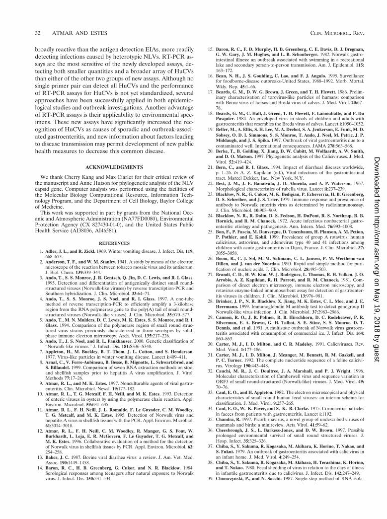
broadly reactive than the antigen detection EIAs, more readilydetecting infections caused by heterotypic NLVs. RT-PCR as-says are the most sensitive of the newly developed assays, de-tecting both smaller quantities and a broader array of HuCVsthan either of the other two groups of new assays. Although nosingle primer pair can detect all HuCVs and the performanceof RT-PCR assays for HuCVs is not yet standardized, severalapproaches have been successfully applied in both epidemio-logical studies and outbreak investigations. Another advantageof RT-PCR assays is their applicability to environmental spec-imens. These new assays have significantly increased the rec-ognition of HuCVs as causes of sporadic and outbreak-associ-ated gastroenteritis, and new information about factors leadingto disease transmission may permit development of new publichealth measures to decrease this common disease.
ACKNOWLEDGMENTS
We thank Cherry Kang and Max Ciarlet for their critical review ofthe manuscript and Anne Hutson for phylogenetic analysis of the NLVcapsid gene. Computer analysis was performed using the facilities ofthe Molecular Biology Computational Resource, Information Tech-nology Program, and the Department of Cell Biology, Baylor Collegeof Medicine.
This work was supported in part by grants from the National Oce-anic and Atmospheric Administration (NA77FD0080), EnvironmentalProtection Agency (CX 827430-01-0), and the United States PublicHealth Service (AI38036, AI46581).
REFERENCES
1. Adler, J. L., and R. Zickl. 1969. Winter vomiting disease. J. Infect. Dis. 119:668–673.
2. Anderson, T. F., and W. M. Stanley. 1941. A study by means of the electronmicroscope of the reaction between tobacco mosaic virus and its antiserum.J. Biol. Chem. 139:339–344.
3. Ando, T., S. S. Monroe, J. R. Gentsch, Q. Jin, D. C. Lewis, and R. I. Glass.1995. Detection and differentiation of antigenically distinct small round-structured viruses (Norwalk-like viruses) by reverse transcription-PCR andSouthern hybridization. J. Clin. Microbiol. 33:64–71.
4. Ando, T., S. S. Monroe, J. S. Noel, and R. I. Glass. 1997. A one-tubemethod of reverse transcription-PCR to efficiently amplify a 3-kilobaseregion from the RNA polymerase gene to the poly(A) tail of small round-structured viruses (Norwalk-like viruses). J. Clin. Microbiol. 35:570–577.
5. Ando, T., M. N. Mulders, D. C. Lewis, M. K. Estes, S. S. Monroe, and R. I.Glass. 1994. Comparison of the polymerase region of small round struc-tured virus strains previously characterized in three serotypes by solid-phase immune electron microscopy. Arch. Virol. 135:217–226.
6. Ando, T., J. S. Noel, and R. L. Fankhauser. 2000. Genetic classification of“Norwalk-like viruses.” J. Infect. Dis. 181:S336–S348.
7. Appleton, H., M. Buckley, B. T. Thom, J. L. Cotton, and S. Henderson.1977. Virus-like particles in winter vomiting disease. Lancet i:409–411.
8. Arnal, C., V. Ferre-Aubineau, B. Besse, B. Mignotte, L. Schwartzbrod, andS. Billaudel. 1999. Comparison of seven RNA extraction methods on stooland shellfish samples prior to hepatitis A virus amplification. J. Virol.Methods 77:17–26.
9. Atmar, R. L., and M. K. Estes. 1997. Nonculturable agents of viral gastro-enteritis. Clin. Microbiol. Newsl. 19:177–182.
10. Atmar, R. L., T. G. Metcalf, F. H. Neill, and M. K. Estes. 1993. Detectionof enteric viruses in oysters by using the polymerase chain reaction. Appl.Environ. Microbiol. 59:631–635.
11. Atmar, R. L., F. H. Neill, J. L. Romalde, F. Le Guyader, C. M. Woodley,T. G. Metcalf, and M. K. Estes. 1995. Detection of Norwalk virus andhepatitis A virus in shellfish tissues with the PCR. Appl. Environ. Microbiol.61:3014–3018.
12. Atmar, R. L., F. H. Neill, C. M. Woodley, R. Manger, G. S. Fout, W.Burkhardt, L. Leja, E. R. McGovern, F. Le Guyader, T. G. Metcalf, andM. K. Estes. 1996. Collaborative evaluation of a method for the detectionof Norwalk virus in shellfish tissues by PCR. Appl. Environ. Microbiol. 62:254–258.
13. Baker, J. C. 1987. Bovine viral diarrhea virus: a review. J. Am. Vet. Med.Assoc. 190:1449–1458.
14. Baron, R. C., H. B. Greenberg, G. Cukor, and N. R. Blacklow. 1984.Serological responses among teenagers after natural exposure to Norwalkvirus. J. Infect. Dis. 150:531–534.
15. Baron, R. C., F. D. Murphy, H. B. Greenberg, C. E. Davis, D. J. Bregman,G. W. Gary, J. M. Hughes, and L. B. Schonberger. 1982. Norwalk gastro-intestinal illness: an outbreak associated with swimming in a recreationallake and secondary person-to-person transmission. Am. J. Epidemiol. 115:163–172.
16. Bean, N. H., J. S. Goulding, C. Lao, and F. J. Angulo. 1995. Surveillancefor foodborne-disease outbreaks-United States, 1988–1992. Morb. Mortal.Wkly. Rep. 45:1–66.
17. Beards, G. M., D. W. G. Brown, J. Green, and T. H. Flewett. 1986. Prelim-inary characterisation of torovirus-like particles of humans: comparisonwith Berne virus of horses and Breda virus of calves. J. Med. Virol. 20:67–78.
18. Beards, G. M., C. Hall, J. Green, T. H. Flewett, F. Lamouliatte, and P. DuPasquier. 1984. An enveloped virus in stools of children and adults withgastroenteritis that resembles the Breda virus of calves. Lancet i:1050–1052.
19. Beller, M., A. Ellis, S. H. Lee, M. A. Drebot, S. A. Jenkerson, E. Funk, M. D.Sobsey, O. D. I. Simmons, S. S. Monroe, T. Ando, J. Noel, M. Petric, J. P.Middaugh, and J. S. Spika. 1997. Outbreak of viral gastroenteritis due to acontaminated well. International consequences. JAMA 278:563–568.
20. Berke, T., B. Golding, X. Jiang, D. W. Cubitt, M. Wolfaardt, A. W. Smith,and D. O. Matson. 1997. Phylogenetic analysis of the Caliciviruses. J. Med.Virol. 52:419–424.
21. Bern, C., and R. I. Glass. 1994. Impact of diarrheal diseases worldwide,p. 1–26. In A. Z. Kapikian (ed.), Viral infections of the gastrointestinaltract. Marcel Dekker, Inc., New York, N.Y.
22. Best, J. M., J. E. Banatvala, J. D. Almeida, and A. P. Waterson. 1967.Morphological characteristics of rubella virus. Lancet ii:237–239.
23. Blacklow, N. R., G. Cukor, M. K. Bedigian, P. Echeverria, H. B. Greenberg,D. S. Schreiber, and J. S. Trier. 1979. Immune response and prevalence ofantibody to Norwalk enteritis virus as determined by radioimmunoassay.J. Clin. Microbiol. 10:903–909.
24. Blacklow, N. R., R. Dolin, D. S. Fedson, H. DuPont, R. S. Northrup, R. B.Hornick, and R. M. Chanock. 1972. Acute infectious nonbacterial gastro-enteritis: etiology and pathogenesis. Ann. Intern. Med. 76:993–1008.
25. Bon, F., P. Fascia, M. Dauvergne, D. Tenenbaum, H. Planson, A. M. Petion,P. Pothier, and E. Kohli. 1999. Prevalence of group A rotavirus, humancalicivirus, astrovirus, and adenovirus type 40 and 41 infections amongchildren with acute gastroenteritis in Dijon, France. J. Clin. Microbiol. 37:3055–3058.
26. Boom, R., C. J. Sol, M. M. Salimans, C. L. Jansen, P. M. Wertheim-vanDillen, and J. van der Noordaa. 1990. Rapid and simple method for puri-fication of nucleic acids. J. Clin. Microbiol. 28:495–503.
27. Brandt, C. D., H. W. Kim, W. J. Rodriguez, L. Thomas, R. H. Yolken, J. O.Arrobio, A. Z. Kapikian, R. H. Parrott, and R. M. Chanock. 1981. Com-parison of direct electron microscopy, immune electron microscopy, androtavirus enzyme-linked immunosorbent assay for detection of gastroenter-itis viruses in children. J. Clin. Microbiol. 13:976–981.
28. Brinker, J. P., N. R. Blacklow, X. Jiang, M. K. Estes, C. L. Moe, and J. E.Herrmann. 1999. Immunoglobulin M antibody test to detect genogroup IINorwalk-like virus infection. J. Clin. Microbiol. 37:2983–2986.
29. Cannon, R. O., J. R. Poliner, R. B. Hirschhorn, D. C. Rodeheaver, P. R.Silverman, E. A. Brown, G. H. Talbot, S. E. Stine, S. S. Monroe, D. T.Dennis, and et al. 1991. A multistate outbreak of Norwalk virus gastroen-teritis associated with consumption of commercial ice. J. Infect. Dis. 164:860–863.
30. Carter, M. J., I. D. Milton, and C. R. Madeley. 1991. Caliciviruses. Rev.Med. Virol. 1:177–186.
31. Carter, M. J., I. D. Milton, J. Meanger, M. Bennett, R. M. Gaskell, andP. C. Turner. 1992. The complete nucleotide sequence of a feline calicivi-rus. Virology 190:443–448.
32. Cauchi, M. R., J. C. Doultree, J. A. Marshall, and P. J. Wright. 1996.Molecular characterization of Camberwell virus and sequence variation inORF3 of small round-structured (Norwalk-like) viruses. J. Med. Virol. 49:70–76.
33. Caul, E. O., and H. Appleton. 1982. The electron microscopical and physicalcharacteristics of small round human fecal viruses: an interim scheme forclassification. J. Med. Virol. 9:257–265.
34. Caul, E. O., W. K. Paver, and S. K. R. Clarke. 1975. Coronavirus particlesin faeces from patients with gastroenteritis. Lancet i:1192.
35. Chandra, R. 1997. Picorbirnavirus, a novel group of undescribed viruses ofmammals and birds: a minireview. Acta Virol. 41:59–62.
36. Cheesbrough, J. S., L. Barkess-Jones, and D. W. Brown. 1997. Possibleprolonged environmental survival of small round structured viruses. J.Hosp. Infect. 35:325–326.
37. Chiba, S., Y. Sakuma, R. Kogasaka, M. Akihara, K. Horino, T. Nakao, andS. Fukui. 1979. An outbreak of gastroenteritis associated with calicivirus inan infant home. J. Med. Virol. 4:249–254.
38. Chiba, S., Y. Sakuma, R. Kogasaka, M. Akihara, H. Terashima, K. Horino,and T. Nakao. 1980. Fecal shedding of virus in relation to the days of illnessin infantile gastroenteritis due to calicivirus. J. Infect. Dis. 142:247–249.
39. Chomczynski, P., and N. Sacchi. 1987. Single-step method of RNA isola-
32 ATMAR AND ESTES CLIN. MICROBIOL. REV.
on May 19, 2018 by guest
http://cmr.asm
.org/D
ownloaded from
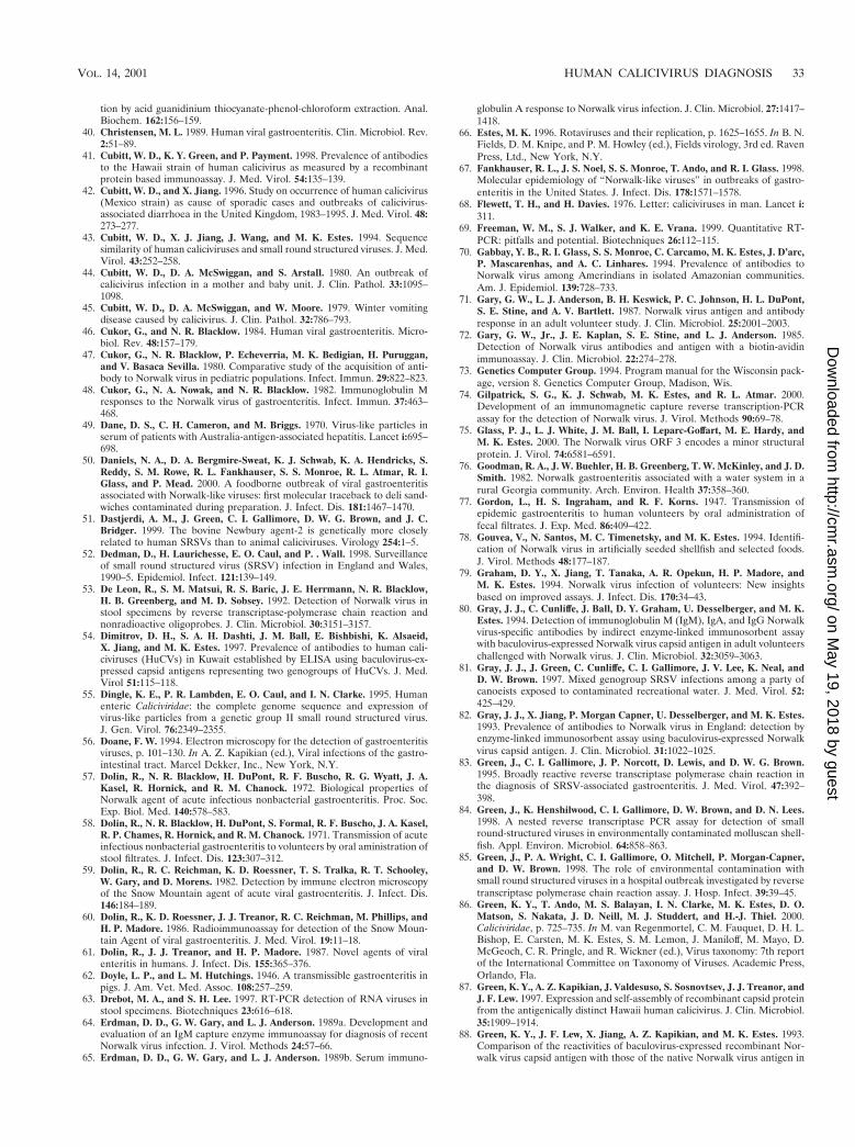
tion by acid guanidinium thiocyanate-phenol-chloroform extraction. Anal.Biochem. 162:156–159.
40. Christensen, M. L. 1989. Human viral gastroenteritis. Clin. Microbiol. Rev.2:51–89.
41. Cubitt, W. D., K. Y. Green, and P. Payment. 1998. Prevalence of antibodiesto the Hawaii strain of human calicivirus as measured by a recombinantprotein based immunoassay. J. Med. Virol. 54:135–139.
42. Cubitt, W. D., and X. Jiang. 1996. Study on occurrence of human calicivirus(Mexico strain) as cause of sporadic cases and outbreaks of calicivirus-associated diarrhoea in the United Kingdom, 1983–1995. J. Med. Virol. 48:273–277.
43. Cubitt, W. D., X. J. Jiang, J. Wang, and M. K. Estes. 1994. Sequencesimilarity of human caliciviruses and small round structured viruses. J. Med.Virol. 43:252–258.
44. Cubitt, W. D., D. A. McSwiggan, and S. Arstall. 1980. An outbreak ofcalicivirus infection in a mother and baby unit. J. Clin. Pathol. 33:1095–1098.
45. Cubitt, W. D., D. A. McSwiggan, and W. Moore. 1979. Winter vomitingdisease caused by calicivirus. J. Clin. Pathol. 32:786–793.
46. Cukor, G., and N. R. Blacklow. 1984. Human viral gastroenteritis. Micro-biol. Rev. 48:157–179.
47. Cukor, G., N. R. Blacklow, P. Echeverria, M. K. Bedigian, H. Puruggan,and V. Basaca Sevilla. 1980. Comparative study of the acquisition of anti-body to Norwalk virus in pediatric populations. Infect. Immun. 29:822–823.
48. Cukor, G., N. A. Nowak, and N. R. Blacklow. 1982. Immunoglobulin Mresponses to the Norwalk virus of gastroenteritis. Infect. Immun. 37:463–468.
49. Dane, D. S., C. H. Cameron, and M. Briggs. 1970. Virus-like particles inserum of patients with Australia-antigen-associated hepatitis. Lancet i:695–698.
50. Daniels, N. A., D. A. Bergmire-Sweat, K. J. Schwab, K. A. Hendricks, S.Reddy, S. M. Rowe, R. L. Fankhauser, S. S. Monroe, R. L. Atmar, R. I.Glass, and P. Mead. 2000. A foodborne outbreak of viral gastroenteritisassociated with Norwalk-like viruses: first molecular traceback to deli sand-wiches contaminated during preparation. J. Infect. Dis. 181:1467–1470.
51. Dastjerdi, A. M., J. Green, C. I. Gallimore, D. W. G. Brown, and J. C.Bridger. 1999. The bovine Newbury agent-2 is genetically more closelyrelated to human SRSVs than to animal caliciviruses. Virology 254:1–5.
52. Dedman, D., H. Laurichesse, E. O. Caul, and P. . Wall. 1998. Surveillanceof small round structured virus (SRSV) infection in England and Wales,1990–5. Epidemiol. Infect. 121:139–149.
53. De Leon, R., S. M. Matsui, R. S. Baric, J. E. Herrmann, N. R. Blacklow,H. B. Greenberg, and M. D. Sobsey. 1992. Detection of Norwalk virus instool specimens by reverse transcriptase-polymerase chain reaction andnonradioactive oligoprobes. J. Clin. Microbiol. 30:3151–3157.
54. Dimitrov, D. H., S. A. H. Dashti, J. M. Ball, E. Bishbishi, K. Alsaeid,X. Jiang, and M. K. Estes. 1997. Prevalence of antibodies to human cali-civiruses (HuCVs) in Kuwait established by ELISA using baculovirus-ex-pressed capsid antigens representing two genogroups of HuCVs. J. Med.Virol 51:115–118.
55. Dingle, K. E., P. R. Lambden, E. O. Caul, and I. N. Clarke. 1995. Humanenteric Caliciviridae: the complete genome sequence and expression ofvirus-like particles from a genetic group II small round structured virus.J. Gen. Virol. 76:2349–2355.
56. Doane, F. W. 1994. Electron microscopy for the detection of gastroenteritisviruses, p. 101–130. In A. Z. Kapikian (ed.), Viral infections of the gastro-intestinal tract. Marcel Dekker, Inc., New York, N.Y.
57. Dolin, R., N. R. Blacklow, H. DuPont, R. F. Buscho, R. G. Wyatt, J. A.Kasel, R. Hornick, and R. M. Chanock. 1972. Biological properties ofNorwalk agent of acute infectious nonbacterial gastroenteritis. Proc. Soc.Exp. Biol. Med. 140:578–583.
58. Dolin, R., N. R. Blacklow, H. DuPont, S. Formal, R. F. Buscho, J. A. Kasel,R. P. Chames, R. Hornick, and R. M. Chanock. 1971. Transmission of acuteinfectious nonbacterial gastroenteritis to volunteers by oral aministration ofstool filtrates. J. Infect. Dis. 123:307–312.
59. Dolin, R., R. C. Reichman, K. D. Roessner, T. S. Tralka, R. T. Schooley,W. Gary, and D. Morens. 1982. Detection by immune electron microscopyof the Snow Mountain agent of acute viral gastroenteritis. J. Infect. Dis.146:184–189.
60. Dolin, R., K. D. Roessner, J. J. Treanor, R. C. Reichman, M. Phillips, andH. P. Madore. 1986. Radioimmunoassay for detection of the Snow Moun-tain Agent of viral gastroenteritis. J. Med. Virol. 19:11–18.
61. Dolin, R., J. J. Treanor, and H. P. Madore. 1987. Novel agents of viralenteritis in humans. J. Infect. Dis. 155:365–376.
62. Doyle, L. P., and L. M. Hutchings. 1946. A transmissible gastroenteritis inpigs. J. Am. Vet. Med. Assoc. 108:257–259.
63. Drebot, M. A., and S. H. Lee. 1997. RT-PCR detection of RNA viruses instool specimens. Biotechniques 23:616–618.
64. Erdman, D. D., G. W. Gary, and L. J. Anderson. 1989a. Development andevaluation of an IgM capture enzyme immunoassay for diagnosis of recentNorwalk virus infection. J. Virol. Methods 24:57–66.
65. Erdman, D. D., G. W. Gary, and L. J. Anderson. 1989b. Serum immuno-
globulin A response to Norwalk virus infection. J. Clin. Microbiol. 27:1417–1418.
66. Estes, M. K. 1996. Rotaviruses and their replication, p. 1625–1655. In B. N.Fields, D. M. Knipe, and P. M. Howley (ed.), Fields virology, 3rd ed. RavenPress, Ltd., New York, N.Y.
67. Fankhauser, R. L., J. S. Noel, S. S. Monroe, T. Ando, and R. I. Glass. 1998.Molecular epidemiology of “Norwalk-like viruses” in outbreaks of gastro-enteritis in the United States. J. Infect. Dis. 178:1571–1578.
68. Flewett, T. H., and H. Davies. 1976. Letter: caliciviruses in man. Lancet i:311.
69. Freeman, W. M., S. J. Walker, and K. E. Vrana. 1999. Quantitative RT-PCR: pitfalls and potential. Biotechniques 26:112–115.
70. Gabbay, Y. B., R. I. Glass, S. S. Monroe, C. Carcamo, M. K. Estes, J. D’arc,P. Mascarenhas, and A. C. Linhares. 1994. Prevalence of antibodies toNorwalk virus among Amerindians in isolated Amazonian communities.Am. J. Epidemiol. 139:728–733.
71. Gary, G. W., L. J. Anderson, B. H. Keswick, P. C. Johnson, H. L. DuPont,S. E. Stine, and A. V. Bartlett. 1987. Norwalk virus antigen and antibodyresponse in an adult volunteer study. J. Clin. Microbiol. 25:2001–2003.
72. Gary, G. W., Jr., J. E. Kaplan, S. E. Stine, and L. J. Anderson. 1985.Detection of Norwalk virus antibodies and antigen with a biotin-avidinimmunoassay. J. Clin. Microbiol. 22:274–278.
73. Genetics Computer Group. 1994. Program manual for the Wisconsin pack-age, version 8. Genetics Computer Group, Madison, Wis.
74. Gilpatrick, S. G., K. J. Schwab, M. K. Estes, and R. L. Atmar. 2000.Development of an immunomagnetic capture reverse transcription-PCRassay for the detection of Norwalk virus. J. Virol. Methods 90:69–78.
75. Glass, P. J., L. J. White, J. M. Ball, I. Leparc-Goffart, M. E. Hardy, andM. K. Estes. 2000. The Norwalk virus ORF 3 encodes a minor structuralprotein. J. Virol. 74:6581–6591.
76. Goodman, R. A., J. W. Buehler, H. B. Greenberg, T. W. McKinley, and J. D.Smith. 1982. Norwalk gastroenteritis associated with a water system in arural Georgia community. Arch. Environ. Health 37:358–360.
77. Gordon, L., H. S. Ingraham, and R. F. Korns. 1947. Transmission ofepidemic gastroenteritis to human volunteers by oral administration offecal filtrates. J. Exp. Med. 86:409–422.
78. Gouvea, V., N. Santos, M. C. Timenetsky, and M. K. Estes. 1994. Identifi-cation of Norwalk virus in artificially seeded shellfish and selected foods.J. Virol. Methods 48:177–187.
79. Graham, D. Y., X. Jiang, T. Tanaka, A. R. Opekun, H. P. Madore, andM. K. Estes. 1994. Norwalk virus infection of volunteers: New insightsbased on improved assays. J. Infect. Dis. 170:34–43.
80. Gray, J. J., C. Cunliffe, J. Ball, D. Y. Graham, U. Desselberger, and M. K.Estes. 1994. Detection of immunoglobulin M (IgM), IgA, and IgG Norwalkvirus-specific antibodies by indirect enzyme-linked immunosorbent assaywith baculovirus-expressed Norwalk virus capsid antigen in adult volunteerschallenged with Norwalk virus. J. Clin. Microbiol. 32:3059–3063.
81. Gray, J. J., J. Green, C. Cunliffe, C. I. Gallimore, J. V. Lee, K. Neal, andD. W. Brown. 1997. Mixed genogroup SRSV infections among a party ofcanoeists exposed to contaminated recreational water. J. Med. Virol. 52:425–429.
82. Gray, J. J., X. Jiang, P. Morgan Capner, U. Desselberger, and M. K. Estes.1993. Prevalence of antibodies to Norwalk virus in England: detection byenzyme-linked immunosorbent assay using baculovirus-expressed Norwalkvirus capsid antigen. J. Clin. Microbiol. 31:1022–1025.
83. Green, J., C. I. Gallimore, J. P. Norcott, D. Lewis, and D. W. G. Brown.1995. Broadly reactive reverse transcriptase polymerase chain reaction inthe diagnosis of SRSV-associated gastroenteritis. J. Med. Virol. 47:392–398.
84. Green, J., K. Henshilwood, C. I. Gallimore, D. W. Brown, and D. N. Lees.1998. A nested reverse transcriptase PCR assay for detection of smallround-structured viruses in environmentally contaminated molluscan shell-fish. Appl. Environ. Microbiol. 64:858–863.
85. Green, J., P. A. Wright, C. I. Gallimore, O. Mitchell, P. Morgan-Capner,and D. W. Brown. 1998. The role of environmental contamination withsmall round structured viruses in a hospital outbreak investigated by reversetranscriptase polymerase chain reaction assay. J. Hosp. Infect. 39:39–45.
86. Green, K. Y., T. Ando, M. S. Balayan, I. N. Clarke, M. K. Estes, D. O.Matson, S. Nakata, J. D. Neill, M. J. Studdert, and H.-J. Thiel. 2000.Caliciviridae, p. 725–735. In M. van Regenmortel, C. M. Fauquet, D. H. L.Bishop, E. Carsten, M. K. Estes, S. M. Lemon, J. Maniloff, M. Mayo, D.McGeoch, C. R. Pringle, and R. Wickner (ed.), Virus taxonomy: 7th reportof the International Committee on Taxonomy of Viruses. Academic Press,Orlando, Fla.
87. Green, K. Y., A. Z. Kapikian, J. Valdesuso, S. Sosnovtsev, J. J. Treanor, andJ. F. Lew. 1997. Expression and self-assembly of recombinant capsid proteinfrom the antigenically distinct Hawaii human calicivirus. J. Clin. Microbiol.35:1909–1914.
88. Green, K. Y., J. F. Lew, X. Jiang, A. Z. Kapikian, and M. K. Estes. 1993.Comparison of the reactivities of baculovirus-expressed recombinant Nor-walk virus capsid antigen with those of the native Norwalk virus antigen in
VOL. 14, 2001 HUMAN CALICIVIRUS DIAGNOSIS 33
on May 19, 2018 by guest
http://cmr.asm
.org/D
ownloaded from
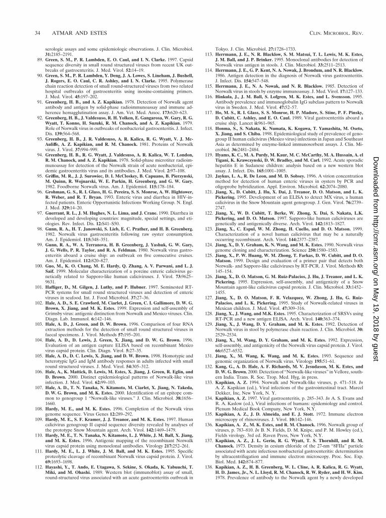
serologic assays and some epidemiologic observations. J. Clin. Microbiol.31:2185–2191.
89. Green, S. M., P. R. Lambden, E. O. Caul, and I. N. Clarke. 1997. Capsidsequence diversity in small round structured viruses from recent UK out-breaks of gastroenteritis. J. Med. Virol. 52:14–19.
90. Green, S. M., P. R. Lambden, Y. Deng, J. A. Lowes, S. Lineham, J. Bushell,J. Rogers, E. O. Caul, C. R. Ashley, and I. N. Clarke. 1995. Polymerasechain reaction detection of small round-structured viruses from two relatedhospital outbreaks of gastroenteritis using inosine-containing primers.J. Med. Virol. 45:197–202.
91. Greenberg, H. B., and A. Z. Kapikian. 1978. Detection of Norwalk agentantibody and antigen by solid-phase radioimmunoassay and immune ad-herence hemagglutination assay. J. Am. Vet. Med. Assoc. 173:620–623.
92. Greenberg, H. B., J. Valdesuso, R. H. Yolken, E. Gangarosa, W. Gary, R. G.Wyatt, T. Konno, H. Suzuki, R. M. Chanock, and A. Z. Kapikian. 1979.Role of Norwalk virus in outbreaks of nonbacterial gastroenteritis. J. Infect.Dis. 139:564–568.
93. Greenberg, H. B., J. R. Valdesuso, A. R. Kalica, R. G. Wyatt, V. J. Mc-Auliffe, A. Z. Kapikian, and R. M. Chanock. 1981. Proteins of Norwalkvirus. J. Virol. 37:994–999.
94. Greenberg, H. B., R. G. Wyatt, J. Valdesuso, A. R. Kalica, W. T. London,R. M. Chanock, and A. Z. Kapikian. 1978. Solid-phase microtiter radioim-munoassay for detection of the Norwalk strain of acute nonbacterial, epi-demic gastroenteritis virus and its antibodies. J. Med. Virol. 2:97–108.
95. Griffin, M. R., J. J. Surowiec, D. I. McCloskey, B. Capuano, B. Pierzynski,M. Quinn, R. Wojnarski, W. E. Parkin, H. Greenberg, and G. W. Gary.1982. Foodborne Norwalk virus. Am. J. Epidemiol. 115:178–184.
96. Grohman, G. S., R. I. Glass, H. G. Pereira, S. S. Monroe, A. W. Hightower,R. Weber, and R. T. Bryan. 1993. Enteric virus and diarrhea in HIV-in-fected patients. Enteric Opportunistic Infections Working Group. N. Engl.J. Med. 329:14–20.
97. Guerrant, R. L., J. M. Hughes, N. L. Lima, and J. Crane. 1990. Diarrhea indeveloped and developing countries: magnitude, special settings, and eti-ologies. Rev. Infect. Dis. 12:S41–S50.
98. Gunn, R. A., H. T. Janowski, S. Lieb, E. C. Prather, and H. B. Greenberg.1982. Norwalk virus gastroenteritis following raw oyster consumption.Am. J. Epidemiol. 115:348–351.
99. Gunn, R. A., W. A. Terranova, H. B. Greenberg, J. Yashuk, G. W. Gary,J. G. Wells, P. R. Taylor, and R. A. Feldman. 1980. Norwalk virus gastro-enteritis aboard a cruise ship: an outbreak on five consecutive cruises.Am. J. Epidemiol. 112:820–827.
100. Guo, M., K. O. Chang, M. E. Hardy, Q. Zhang, A. V. Parwani, and L. J.Saif. 1999. Molecular characterization of a porcine enteric calicivirus ge-netically related to Sapporo-like human caliciviruses. J. Virol. 73:9625–9631.
101. Hafliger, D., M. Gilgen, J. Luthy, and P. Hubner. 1997. Seminested RT-PCR systems for small round structured viruses and detection of entericviruses in seafood. Int. J. Food Microbiol. 37:27–36.
102. Hale, A. D., S. E. Crawford, M. Ciarlet, J. Green, C. I. Gallimore, D. W. G.Brown, X. Jiang, and M. K. Estes. 1999. Expression and self-assembly ofGrimsby virus: antigenic distinction from Norwalk and Mexico viruses. Clin.Diagn. Lab. Immunol. 6:142–146.
103. Hale, A. D., J. Green, and D. W. Brown. 1996. Comparison of four RNAextraction methods for the detection of small round structured viruses infaecal specimens. J. Virol. Methods 57:195–201.
104. Hale, A. D., D. Lewis, J. Green, X. Jiang, and D. W. G. Brown. 1996.Evaluation of an antigen capture ELISA based on recombinant Mexicovirus capsid protein. Clin. Diagn. Virol. 5:27–35.
105. Hale, A. D., D. C. Lewis, X. Jiang, and D. W. Brown. 1998. Homotypic andheterotypic IgG and IgM antibody responses in adults infected with smallround structured viruses. J. Med. Virol. 54:305–312.
106. Hale, A., K. Mattick, D. Lewis, M. Estes, X. Jiang, J. Green, R. Eglin, andD. Brown. 2000. Distinct epidemiological patterns of Norwalk-like virusinfection. J. Med. Virol. 62:99–103.
107. Hale, A. D., T. N. Tanaka, N. Kitamoto, M. Ciarlet, X. Jiang, N. Takeda,D. W. G. Brown, and M. K. Estes. 2000. Identification of an epitope com-mon to genogroup 1 “Norwalk-like viruses.” J. Clin. Microbiol. 38:1656–1660.
108. Hardy, M. E., and M. K. Estes. 1996. Completion of the Norwalk virusgenome sequence. Virus Genes 12:289–292.
109. Hardy, M. E., S. F. Kramer, J. J. Treanor, and M. K. Estes. 1997. Humancalicivirus genogroup II capsid sequence diversity revealed by analyses ofthe prototype Snow Mountain agent. Arch. Virol. 142:1469–1479.
110. Hardy, M. E., T. N. Tanaka, N. Kitamoto, L. J. White, J. M. Ball, X. Jiang,and M. K. Estes. 1996. Antigenic mapping of the recombinant Norwalkvirus capsid protein using monoclonal antibodies. Virology 217:252–261.
111. Hardy, M. E., L. J. White, J. M. Ball, and M. K. Estes. 1995. Specificproteolytic cleavage of recombinant Norwalk virus capsid protein. J. Virol.69:1693–1698.
112. Hayashi, Y., T. Ando, E. Utagawa, S. Sekine, S. Okada, K. Yabuuchi, T.Miki, and M. Ohashi. 1989. Western blot (immunoblot) assay of small,round-structured virus associated with an acute gastroenteritis outbreak in
Tokyo. J. Clin. Microbiol. 27:1728–1733.113. Herrmann, J. E., N. R. Blacklow, S. M. Matsui, T. L. Lewis, M. K. Estes,
J. M. Ball, and J. P. Brinker. 1995. Monoclonal antibodies for detection ofNorwalk virus antigen in stools. J. Clin. Microbiol. 33:2511–2513.
114. Herrmann, J. E., G. P. Kent, N. A. Nowak, J. Brondum, and N. R. Blacklow.1986. Antigen detection in the diagnosis of Norwalk virus gastroenteritis.J. Infect. Dis. 154:547–548.
115. Herrmann, J. E., N. A. Nowak, and N. R. Blacklow. 1985. Detection ofNorwalk virus in stools by enzyme immunoassay. J. Med. Virol. 17:127–133.
116. Hinkula, J., J. M. Ball, S. Lofgren, M. K. Estes, and L. Svensson. 1995.Antibody prevalence and immunoglobulin IgG subclass pattern to Norwalkvirus in Sweden. J. Med. Virol. 47:52–57.
117. Ho, M. S., R. I. Glass, S. S. Monroe, H. P. Madore, S. Stine, P. F. Pinsky,D. Cubitt, C. Ashley, and E. O. Caul. 1989. Viral gastroenteritis aboard acruise ship. Lancet ii:961–965.
118. Honma, S., S. Nakata, K. Numata, K. Kogawa, T. Yamashita, M. Oseto,X. Jiang, and S. Chiba. 1998. Epidemiological study of prevalence of geno-group II human calicivirus (Mexico virus) infections in Japan and SoutheastAsia as determined by enzyme-linked immunosorbent assays. J. Clin. Mi-crobiol. 36:2481–2484.
119. Hyams, K. C., M. A. Purdy, M. Kaur, M. C. McCarthy, M. A. Hussain, A. elTigani, K. Krawczynski, D. W. Bradley, and M. Carl. 1992. Acute sporadichepatitis E in Sudanese children: analysis based on a new western blotassay. J. Infect. Dis. 165:1001–1005.
120. Jaykus, L. A., R. De Leon, and M. D. Sobsey. 1996. A virion concentrationmethod for detection of human enteric viruses in oysters by PCR andoligoprobe hybridization. Appl. Environ. Microbiol. 62:2074–2080.
121. Jiang, X., D. Cubitt, J. Hu, X. Dai, J. Treanor, D. O. Matson, and L. K.Pickering. 1995. Development of an ELISA to detect MX virus, a humancalicivirus in the Snow Mountain agent genogroup. J. Gen. Virol. 76:2739–2747.
122. Jiang, X., W. D. Cubitt, T. Berke, W. Zhong, X. Dai, S. Nakata, L.K.Pickering, and D. O. Matson. 1997. Sapporo-like human caliciviruses aregenetically and antigenically diverse. Arch. Virol. 142:1813–1827.
123. Jiang, X., C. Espul, W. M. Zhong, H. Cuello, and D. O. Matson. 1999.Characterization of a novel human calicivirus that may be a naturallyoccurring recombinant. Arch. Virol. 144:2377–2387.
124. Jiang, X., D. Y. Graham, K. N. Wang, and M. K. Estes. 1990. Norwalk virusgenome cloning and characterization. Science 250:1580–1583.
125. Jiang, X., P. W. Huang, W. M. Zhong, T. Farkas, D. W. Cubitt, and D. O.Matson. 1999. Design and evaluation of a primer pair that detects bothNorwalk- and Sapporo-like caliciviruses by RT-PCR. J. Virol. Methods 83:145–154.
126. Jiang, X., D. O. Matson, G. M. Ruiz-Palacios, J. Hu, J. Treanor, and L. K.Pickering. 1995. Expression, self-assembly, and antigenicity of a SnowMountain agent-like calicivirus capsid protein. J. Clin. Microbiol. 33:1452–1455.
127. Jiang, X., D. O. Matson, F. R. Velazquez, W. Zhong, J. Hu, G. Ruiz-Palacios, and L. K. Pickering. 1995. Study of Norwalk-related viruses inMexican children. J. Med. Virol. 47:309–316.
128. Jiang, X., J. Wang, and M.K. Estes. 1995. Characterization of SRSVs usingRT-PCR and a new antigen ELISA. Arch. Virol. 140:363–374.
129. Jiang, X., J. Wang, D. Y. Graham, and M. K. Estes. 1992. Detection ofNorwalk virus in stool by polymerase chain reaction. J. Clin. Microbiol. 30:2529–2534.
130. Jiang, X., M. Wang, D. Y. Graham, and M. K. Estes. 1992. Expression,self-assembly, and antigenicity of the Norwalk virus capsid protein. J. Virol.66:6527–6532.
131. Jiang, X., M. Wang, K. Wang, and M. K. Estes. 1993. Sequence andgenomic organization of Norwalk virus. Virology 195:51–61.
132. Kang, G., A. D. Hale, A. F. Richards, M. V. Jesudason, M. K. Estes, andD. W. G. Brown. 2000. Detection of “Norwalk-like viruses” in Vellore, south-ern India. Trans. R. Soc. Trop. Med. Hyg, in press.
133. Kapikian, A. Z. 1994. Norwalk and Norwalk-like viruses, p. 471–518. InA. Z. Kapikian (ed.), Viral infections of the gastrointestinal tract. MarcelDekker, Inc, New York, N. Y.
134. Kapikian, A. Z. 1997. Viral gastroenteritis, p. 285–343. In A. S. Evans andR. A. Kaslow (ed.), Viral infections of humans: epidemiology and control.Plenum Medical Book Company, New York, N.Y.
135. Kapikian, A. Z., J. D. Almeida, and E. J. Stott. 1972. Immune electronmicroscopy of rhinoviruses. J. Virol. 10:142–146.
136. Kapikian, A. Z., M. K. Estes, and R. M. Chanock. 1996. Norwalk group ofviruses, p. 783–810. In B. N. Fields, D. M. Knipe, and P. M. Howley (ed.),Fields virology, 3rd ed. Raven Press, New York, N.Y.
137. Kapikian, A. Z., J. L. Gerin, R. G. Wyatt, T. S. Thornhill, and R. M.Chanock. 1973. Density in cesium chloride of the 27-nm “8FIIa” particleassociated with acute infectious nonbacterial gastroenteritis: determinationby ultracentrifugation and immune electron microscopy. Proc. Soc. Exp.Biol. Med. 142:874–877.
138. Kapikian, A. Z., H. B. Greenberg, W. L. Cline, A. R. Kalica, R. G. Wyatt,H. D. James, Jr., N. L. Lloyd, R. M. Chanock, R. W. Ryder, and H. W. Kim.1978. Prevalence of antibody to the Norwalk agent by a newly developed
34 ATMAR AND ESTES CLIN. MICROBIOL. REV.
on May 19, 2018 by guest
http://cmr.asm
.org/D
ownloaded from
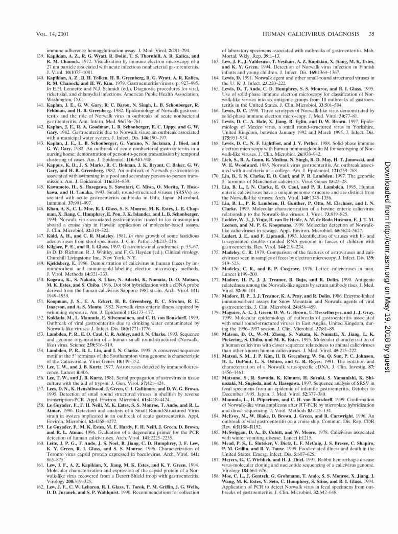
immune adherence hemagglutination assay. J. Med. Virol. 2:281–294.139. Kapikian, A. Z., R. G. Wyatt, R. Dolin, T. S. Thornhill, A. R. Kalica, and
R. M. Chanock. 1972. Visualization by immune electron microscopy of a27 nm particle associated with acute infectious nonbacterial gastorenteritis.J. Virol. 10:1075–1081.
140. Kapikian, A. Z., R. H. Yolken, H. B. Greenberg, R. G. Wyatt, A. R. Kalica,R. M. Chanock, and H. W. Kim. 1979. Gastroenteritis viruses, p. 927–995.In E.H. Lennette and N.J. Schmidt (ed.), Diagnostic procedures for viral,rickettsial, and chlamydial infections. American Public Health Association,Washington, D.C.
141. Kaplan, J. E., G. W. Gary, R. C. Baron, N. Singh, L. B. Schonberger, R.Feldman, and H. B. Greenberg. 1982. Epidemiology of Norwalk gastroen-teritis and the role of Norwalk virus in outbreaks of acute nonbacterialgastroenteritis. Ann. Intern. Med. 96:756–761.
142. Kaplan, J. E., R. A. Goodman, L. B. Schonberger, E. C. Lippy, and G. W.Gary. 1982. Gastroenteritis due to Norwalk virus: an outbreak associatedwith a municipal water system. J. Infect. Dis. 146:190–197.
143. Kaplan, J. E., L. B. Schonberger, G. Varano, N. Jackman, J. Bied, andG. W. Gary. 1982. An outbreak of acute nonbacterial gastroenteritis in anursing home: demonstration of person-to-person transmission by temporalclustering of cases. Am. J. Epidemiol. 116:940–948.
144. Kappus, K. D., J. S. Marks, R. C. Holman, J. K. Bryant, C. Baker, G. W.Gary, and H. B. Greenberg. 1982. An outbreak of Norwalk gastroenteritisassociated with swimming in a pool and secondary person-to-person trans-mission. Am. J. Epidemiol. 116:834–839.
145. Kawamoto, H., S. Hasegawa, S. Sawatari, C. Miwa, O. Morita, T. Hoso-kawa, and H. Tanaka. 1993. Small, round-structured viruses (SRSVs) as-sociated with acute gastroenteritis outbreaks in Gifu, Japan. Microbiol.Immunol. 37:991–997.
146. Khan, A. S., C. L. Moe, R. I. Glass, S. S. Monroe, M. K. Estes, L. E. Chap-man, X. Jiang, C. Humphrey, E. Pon, J. K. Islander, and L. B. Schonberger.1994. Norwalk virus-associated gastroenteritis traced to ice consumptionaboard a cruise ship in Hawaii: application of molecular-based assays.J. Clin. Microbiol. 32:318–322.
147. Kidd, A. H., and C. R. Madeley. 1981. In vitro growth of some fastidiousadenoviruses from stool specimens. J. Clin. Pathol. 34:213–216.
148. Kilgore, P. E., and R. I. Glass. 1997. Gastrointestinal syndromes, p. 55–67.In D. D. Richman, R. J. Whitley, and F. G. Hayden (ed.), Clinical virology.Churchill Livingstone Inc., New York, N.Y.
149. Kjeldsberg, E. 1986. Demonstration of calicivirus in human faeces by im-munosorbent and immunogold-labelling electron microscopy methods.J. Virol. Methods 14:321–333.
150. Kogawa, K., S. Nakata, S. Ukae, N. Adachi, K. Numata, D. O. Matson,M. K. Estes, and S. Chiba. 1996. Dot blot hybridization with a cDNA probederived from the human calicivirus Sapporo 1982 strain. Arch. Virol. 141:1949–1959.
151. Koopman, J. S., E. A. Eckert, H. B. Greenberg, B. C. Strohm, R. E.Isaacson, and A. S. Monto. 1982. Norwalk virus enteric illness acquired byswimming exposure. Am. J. Epidemiol 115:173–177.
152. Kukkula, M., L. Maunula, E. Silvennoinen, and C. H. von Bonsdorff. 1999.Outbreak of viral gastroenteritis due to drinking water contaminated byNorwalk-like viruses. J. Infect. Dis. 180:1771–1776.
153. Lambden, P. R., E. O. Caul, C. R. Ashley, and I. N. Clarke. 1993. Sequenceand genome organization of a human small round-structured (Norwalk-like) virus. Science 259:516–519.
154. Lambden, P. R., B. L. Liu, and I. N. Clarke. 1995. A conserved sequencemotif at the 59 terminus of the Southampton virus genome is characteristicof the Caliciviridae. Virus Genes 10:149–152.
155. Lee, T. W., and J. B. Kurtz. 1977. Astroviruses detected by immunofloures-cence. Lancet ii:406.
156. Lee, T. W., and J. B. Kurtz. 1981. Serial propagation of astrovirus in tissueculture with the aid of trypsin. J. Gen. Virol. 57:421–424.
157. Lees, D. N., K. Henshilwood, J. Green, C. I. Gallimore, and D. W. G. Brown.1995. Detection of small round structured viruses in shellfish by reversetranscription-PCR. Appl. Environ. Microbiol. 61:4418–4424.
158. Le Guyader, F., F. H. Neill, M. K. Estes, S. S. Monroe, T. Ando, and R. L.Atmar. 1996. Detection and analysis of a Small Round-Structured Virusstrain in oysters implicated in an outbreak of acute gastroenteritis. Appl.Environ. Microbiol. 62:4268–4272.
159. Le Guyader, F., M. K. Estes, M. E. Hardy, F. H. Neill, J. Green, D. Brown,and R. L. Atmar. 1996. Evaluation of a degenerate primer for the PCRdetection of human caliciviruses. Arch. Virol. 141:2225–2235.
160. Leite, J. P. G., T. Ando, J. S. Noel, B. Jiang, C. D. Humphrey, J. F. Lew,K. Y. Green, R. I. Glass, and S. S. Monroe. 1996. Characterization ofToronto virus capsid protein expressed in baculovirus. Arch. Virol. 141:865–875.
161. Lew, J. F., A. Z. Kapikian, X. Jiang, M. K. Estes, and K. Y. Green. 1994.Molecular characterization and expression of the capsid protein of a Nor-walk-like virus recovered from a Desert Shield troop with gastroenteritis.Virology 200:319–325.
162. Lew, J. F., C. W. Lebaron, R. I. Glass, T. Torok, P. M. Griffin, J. G. Wells,D. D. Juranek, and S. P. Wahlquist. 1990. Recommendations for collection
of laboratory specimens associated with outbreaks of gastroenteritis. Mab.Mortal. Wkly. Rep. 39:1–13.
163. Lew, J. F., J. Valdesuso, T. Vesikari, A. Z. Kapikian, X. Jiang, M. K. Estes,and K. Y. Green. 1994. Detection of Norwalk virus infection in Finnishinfants and young children. J. Infect. Dis. 169:1364–1367.
164. Lewis, D. 1991. Norwalk agent and other small-round structured viruses inthe U. K. J. Infect. 23:220–222.
165. Lewis, D., T. Ando, C. D. Humphrey, S. S. Monroe, and R. I. Glass. 1995.Use of solid-phase immune electron microscopy for classification of Nor-walk-like viruses into six antigenic groups from 10 outbreaks of gastroen-teritis in the United States. J. Clin. Microbiol. 33:501–504.
166. Lewis, D. C. 1990. Three serotypes of Norwalk-like virus demonstrated bysolid-phase immune electron microscopy. J. Med. Virol. 30:77–81.
167. Lewis, D. C., A. Hale, X. Jiang, R. Eglin, and D. W. Brown. 1997. Epide-miology of Mexico virus, a small round-structured virus in Yorkshire,United Kingdom, between January 1992 and March 1995. J. Infect. Dis.175:951–954.
168. Lewis, D. C., N. F. Lightfoot, and J. V. Pether. 1988. Solid-phase immuneelectron microscopy with human immunoglobulin M for serotyping of Nor-walk-like viruses. J. Clin. Microbiol. 26:938–942.
169. Lieb, S., R. A. Gunn, R. Medina, N. Singh, R. D. May, H. T. Janowski, andW. E. Woodward. 1985. Norwalk virus gastroenteritis. An outbreak associ-ated with a cafeteria at a college. Am. J. Epidemiol. 121:259–268.
170. Liu, B., I. N. Clarke, E. O. Caul, and P. R. Lambden. 1997. The genomic59 terminus of Manchester calicivirus. Virus Genes 15:25–28.
171. Liu, B. L., I. N. Clarke, E. O. Caul, and P. R. Lambden. 1995. Humanenteric caliciviruses have a unique genome structure and are distinct fromthe Norwalk-like viruses. Arch. Virol. 140:1345–1356.
172. Liu, B. L., P. R. Lambden, H. Gunther, P. Otto, M. Elschner, and I. N.Clarke. 1999. Molecular characterization of a bovine enteric calicivirus:relationship to the Norwalk-like viruses. J. Virol. 73:819–825.
173. Lodder, W. J., J. Vinje, R. van De Heide, A. M. de Roda Husman, E. J. T. M.Leenen, and M. P. G. Koopmans. 1999. Molecular detection of Norwalk-like caliciviruses in sewage. Appl. Environ. Microbiol. 65:5624–5627.
174. Ludert, J. E., and F. Liprandi. 1993. Identification of viruses with bi- andtrisegmented double-stranded RNA genome in faeces of children withgastroenteritis. Res. Virol. 144:219–224.
175. Madeley, C. R. 1979. Comparison of the features of astroviruses and cali-civiruses seen in samples of feces by electron microscopy. J. Infect. Dis. 139:519–523.
176. Madeley, C. R., and B. P. Cosgrove. 1976. Letter: caliciviruses in man.Lancet i:199–200.
177. Madore, H. P., J. J. Treanor, R. Buja, and R. Dolin. 1990. Antigenicrelatedness among the Norwalk-like agents by serum antibody rises. J. Med.Virol. 32:96–101.
178. Madore, H. P., J. J. Treanor, K. A. Pray, and R. Dolin. 1986. Enzyme-linkedimmunosorbent assays for Snow Mountain and Norwalk agents of viralgastroenteritis. J. Clin. Microbiol. 24:456–459.
179. Maguire, A. J., J. Green, D. W. G. Brown, U. Desselberger, and J. J. Gray.1999. Molecular epidemiology of outbreaks of gastroenteritis associatedwith small round-structured viruses in East Anglia, United Kingdom, dur-ing the 1996–1997 season. J. Clin. Microbiol. 37:81–89.
180. Matson, D. O., W.-M. Zhong, S. Nakata, K. Numata, X. Jiang, L. K.Pickering, S. Chiba, and M. K. Estes. 1995. Molecular characterization ofa human calicivirus with closer sequence relatedness to animal calicivirusesthan other known human caliciviruses. J. Med. Virol. 45:215–222.
181. Matsui, S. M., J. P. Kim, H. B. Greenberg, W. Su, Q. Sun, P. C. Johnson,H. L. DuPont, L. S. Oshiro, and G. R. Reyes. 1991. The isolation andcharacterization of a Norwalk virus-specific cDNA. J. Clin. Investig. 87:1456–1461.
182. Matsuno, S., R. Sawada, K. Kimura, H. Suzuki, S. Yamanishi, K. Shi-nozaki, M. Sugieda, and A. Hasegawa. 1997. Sequence analysis of SRSV infecal specimens from an epidemic of infantile gastroenteritis, October toDecember 1995, Japan. J. Med. Virol. 52:377–380.
183. Maunula, L., H. Piiparinen, and C. H. von Bonsdorff. 1999. Confirmationof Norwalk-like virus amplicons after RT-PCR by microplate hybridizationand direct sequencing. J. Virol. Methods 83:125–134.
184. McEvoy, M., W. Blake, D. Brown, J. Green, and R. Cartwright. 1996. Anoutbreak of viral gastroenteritis on a cruise ship. Commun. Dis. Rep. CDRRev. 6:R188–R192.
185. McSwiggan, D. A., D. Cubitt, and W. Moore. 1978. Calicivirus associatedwith winter vomiting disease. Lancet i:1215.
186. Mead, P. S., L. Slutsker, V. Dietz, L. F. McCaig, J. S. Bresee, C. Shapiro,P. M. Griffin, and R. V. Tauxe. 1999. Food-related illness and death in theUnited States. Emerg. Infect. Dis. 5:607–625.
187. Meyers, G., C. Wirblich, and H. J. Thiel. 1991. Rabbit hemorrhagic diseasevirus-molecular cloning and nucleotide sequencing of a calicivirus genome.Virology 184:664–676.
188. Moe, C. L., J. Gentsch, G. Grohmann, T. Ando, S. S. Monroe, X. Jiang, J.Wang, M. K. Estes, Y. Seto, C. Humphrey, S. Stine, and R. I. Glass. 1994.Application of PCR to detect Norwalk virus in fecal specimens from out-breaks of gastroenteritis. J. Clin. Microbiol. 32:642–648.
VOL. 14, 2001 HUMAN CALICIVIRUS DIAGNOSIS 35
on May 19, 2018 by guest
http://cmr.asm
.org/D
ownloaded from
189. Monroe, S. S., S. E. Stine, X. Jiang, M. K. Estes, and R. I. Glass. 1993.Detection of antibody to recombinant Norwalk virus antigen in specimensfrom outbreaks of gastroenteritis. J. Clin. Microbiol. 31:2866–2872.
190. Myrmel, M., E. Rimstad, M. Estes, E. Skjerve, and Y. Wasteson. 1996.Prevalence of serum antibodies to Norwalk virus among Norwegian militaryrecruits. Int. J. Food Microbiol. 29:233–240.
191. Nakata, S., S. Chiba, H. Terashima, Y. Sakuma, R. Kogasaka, and T.Nakao. 1983. Microtiter solid-phase radioimmunoassay for detection ofhuman calicivirus in stools. J. Clin. Microbiol. 17:198–201.
192. Nakata, S., M. K. Estes, and S. Chiba. 1988. Detection of human calicivirusantigen and antibody by enzyme-linked immunosorbent assays. J. Clin.Microbiol. 26:2001–2005.
193. Nakata, S., S. Honma, K. Numata, K. Kogawa, S. Ukae, N. Adachi, X.Jiang, M. K. Estes, Z. Gatheru, P. M. Tukei, and S. Chiba. 1998. Prevalenceof human calicivirus infections in Kenya as determined by enzyme immu-noassays for three genogroups of the virus. J. Clin. Microbiol. 36:3160–3163.
194. Narang, H. K., and A. A. Codd. 1981. Frequency of preclumped virus inroutine fecal specimens from patients with acute nonbacterial gastroenter-itis. J. Clin. Microbiol. 13:982–988.
195. Noel, J. S., T. Ando, J. P. Leite, K. Y. Green, K. E. Dingle, M. K. Estes, Y.Seto, S. S. Monroe, and R. I. Glass. 1997. The correlation of patientimmune responses with genetically characterized small round-structuredviruses involved in outbreaks of nonbacterial acute gastroenteritis in theUnited States, 1990 to 1995. J. Med. Virol. 53:372–383.
196. Noel, J. S., R. L. Fankhauser, T. Ando, S. S. Monroe, and R. I. Glass. 1999.Identification of a distinct common strain of “Norwalk-like viruses” havinga global distribution. J. Infect. Dis. 179:1334–1344.
197. Norcott, J. P., J. Green, D. Lewis, M. K. Estes, K. L. Barlow, and D. W.Brown. 1994. Genomic diversity of small round structured viruses in theUK. J. Med. Virol. 44:280–286.
198. Numata, K., M. E. Hardy, S. Nakata, S. Chiba, and M. K. Estes. 1997.Molecular characterization of human calicivirus Sapporo. Arch. Virol. 142:1469–1479.
199. Numata, K., S. Nakata, X. Jiang, M. K. Estes, and S. Chiba. 1994. Epide-miological study of Norwalk virus infections in Japan and Southeast Asia byenzyme-linked immunosorbent assays with Norwalk virus capsid proteinproduced by the baculovirus expression system. J. Clin. Microbiol. 32:121–126.
200. Ohyama, T., S. Yoshizumi, H. Sawada, Y. Uchiyama, Y. Katoh, N. Ha-maoka, and E. Utagawa. 1999. Detection and nucleotide sequence analysisof human caliciviruses (HuCVs) from samples in non-bacterial gastroen-teritis outbreaks in Hokkaido, Japan. Microbiol. Immunol. 43:543–550.
201. Oishi, I., K. Yamazaki, T. Kimoto, and Y. Minekawa. 1992. Demonstrationof low molecular weight polypeptides associated with small, round-struc-tured viruses by western immunoblot analysis. Microbiol. Immunol. 36:1105–1112.
202. Okada, S., S. Sekine, T. Ando, Y. Hayashi, M. Murao, K. Yabuuchi, T.Miki, and M. Ohashi. 1990. Antigenic characterization of small, round-structured viruses by immune electron microscopy. J. Clin. Microbiol. 28:1244–1248.
203. Okhuysen, P. C., X. Jiang, L. Ye, P. C. Johnson, and M. K. Estes. 1995.Viral shedding and fecal IgA response after Norwalk virus infection. J. In-fect. Dis. 171:566–569.
204. O’Ryan, M. L., P. A. Vial, N. Mamani, X. Jiang, M. K. Estes, C. Ferrecio,H. Lakkis, and D. O. Matson. 1998. Seroprevalence of Norwalk virus andMexico virus in Chilean individuals: assessment of independent risk factorsfor antibody acquisition. Clin. Infect. Dis. 27:789–795.
205. Pang, X. L., J. Joensuu, and T. Vesikari. 1999. Human calicivirus-associatedsporadic gastroenteritis in Finnish children less than two years of agefollowed prospectively during a rotavirus vaccine trial. Pediatr. Infect. Dis.J. 18:420–426.
206. Parker, S. P., W. D. Cubitt, and X. Jiang. 1995. Enzyme immunoassay usingbaculovirus-expressed human calicivirus (Mexico) for the measurement ofIgG responses and determining its seroprevalence in London, UK. J. Med.Virol. 46:194–200.
207. Parker, S. P., W. D. Cubitt, X. Jiang, and M. K. Estes. 1994. Seroprevalencestudies using a recombinant Norwalk virus protein enzyme immunoassay.J. Med. Virol. 42:146–150.
208. Parks, C. G., C. L. Moe, D. Rhodes, A. Lima, L. Barrett, F. Tseng, R. Baric,A. Talal, and R. Guerrant. 1999. Genomic diversity of “Norwalk-like vi-ruses” (NLVs): pediatric infections in a Brazilian shantytown. J. Med.Virol. 58:426–434.
209. Patterson, T., P. Hutchings, and S. Palmer. 1993. Outbreak of SRSVgastroenteritis at an international conference traced to food handled by apost-symptomatic caterer. Epidemiol. Infect. 111:157–162.
210. Pinto, R. M., J. M. Diez, and A. Bosch. 1994. Use of the colonic carcinomacell line CaCo-2 for in vivo amplification and detection of enteric viruses.J. Med. Virol. 44:310–315.
211. Prasad, B. V., M. E. Hardy, T. Dokland, J. Bella, M. G. Rossmann, andM. K. Estes. 1999. X-ray crystallographic structure of the Norwalk viruscapsid. Science 286:287–290.
212. Prasad, B. V. V., R. Rothnagel, X. Jiang, and M. K. Estes. 1994. Three-dimensional structure of baculovirus-expressed Norwalk virus capsids.J. Virol. 68:5117–5125.
213. Pujol, F. H., G. Vasquez, A. M. Rojas, M. E. Fuenmayor, C. L. Loureiro,I. Perez-Schael, M. K. Estes, and F. Liprandi. 1998. Norwalk virus infectionin Venezuela. Ann. Trop. Med. Parasitol. 92:205–211.
214. Reimann, H. A., A. H. Price, and J. H. Hodges. 1945. The cause of epidemicdiarrhea, nausea and vomiting (viral dysentery?). Proc. Soc. Exp. Biol. Med.59:8–9.
215. Resta, S., J. P. Luby, C. R. Rosenfeld, and J. D. Siegel. 1985. Isolation andpropagation of a human enteric coronavirus. Science 229:978–981.
216. Rosen, B. I., L. J. Saif, D. J. Jackwood, and M. Gorziglia. 1990. Serotypicdifferentiation of group A rotaviruses with porcine rotavirus gene 9 probes.J. Clin. Microbiol. 28:2526–2533.
217. Saito, H., S. Saito, K. Kamada, S. Harata, H. Sato, M. Morita, and Y.Miyajima. 1998. Application of RT-PCR designed from the sequence of thelocal SRSV strain to the screening in viral gastroenteritis outbreaks. Mi-crobiol. Immunol. 42:439–446.
218. Schreier, E., F. Doring, and U. Kunkel. 2000. Molecular epidemiology ofoutbreaks of gastroenteritis associated with small round structured virusesin Germany in 1997/98. Arch. Virol. 145:443–453.
219. Schwab, K. J., R. De Leon, and M. D. Sobsey. 1996. Immunoaffinity con-centration and purification of waterborne enteric viruses for detection byreverse transcriptase PCR. Appl. Environ. Microbiol. 62:2086–2094.
220. Schwab, K. J., M. K. Estes, and R. L. Atmar. 2000. Norwalk and otherhuman caliciviruses: molecular characterization, epidemiology and patho-genesis, p. 469–493. In J. W. Cary, J. E. Linz, and D. Bhatnagar (ed.),Microbial foodborne diseases: mechanisms of pathogenicity and toxin syn-thesis. Technomic Publishing Company, Inc., Lancaster, Pa.
221. Schwab, K. J., M. K. Estes, F. H. Neill, and R. L. Atmar. 1997. Use of heatrelease and an internal RNA standard control in reverse transcription-PCRdetection of Norwalk virus from stool samples. J. Clin. Microbiol. 35:511–514.
222. Schwab, K. J., F. H. Neill, M. K. Estes, and R. L. Atmar. 1998. Improve-ments in the RT-PCR detection of enteric viruses in environmental sam-ples. Water. Sci. Technol. 38:83–86.
223. Schwab, K. J., F. H. Neill, M. K. Estes, T. G. Metcalf, and R. L. Atmar.1998. Distribution of Norwalk virus within shellfish following bioaccumu-lation and subsequent depuration by detection using RT-PCR. J. FoodProt. 61:1674–1680.
224. Schwab, K. J., F. H. Neill, R. L. Fankhauser, N. A. Daniels, S. S. Monroe,D. A. Bergmire-Sweat, M. K. Estes, and R. L. Atmar. 2000. Development ofmethods to detect “Norwalk-like viruses” (NLVs) and hepatitis A virus indelicatessen foods: application to a food-borne NLV outbreak. Appl. En-viron. Microbiol. 66:213–218.
225. Seah, E. L., J. A. Marshall, and P. J. Wright. 1999. Open reading frame 1of the Norwalk-like virus Camberwell: completion of sequence and expres-sion in mammalian cells. J. Virol. 73:10531–10535.
226. Sharp, T. W., K. C. Hyams, D. Watts, A. F. Trofa, G. J. Martin, A. Z.Kapikian, K. Y. Green, X. Jiang, M. K. Estes, M. Waack, et al. 1995.Epidemiology of Norwalk virus during an outbreak of acute gastroenteritisaboard a US aircraft carrier. J. Med. Virol. 45:61–67.
227. Smit, T. K., P. Bos, I. Peenze, X. Jiang, M. K. Estes, and A. D. Steele. 1999.Seroepidemiological study of genogroup I and II calicivirus infections inSouth and southern Africa. J. Med. Virol. 59:227–231.
228. Smit, T. K., A. D. Steele, I. Peenze, X. Jiang, and M. K. Estes. 1997. Studyof Norwalk virus and Mexico virus infections at Ga-Rankuwa Hospital,Ga-Rankuwa, South Africa. J. Clin. Microbiol. 35:2381–2385.
229. Smith, A. W., E. S. Berry, D. E. Skilling, J. E. Barlough, S. E. Poet, T. Berke,J. Mead, and D. O. Matson. 1998. In vitro isolation and characterization ofa calicivirus causing a vesicular disease of the hands and feet. Clin. Infect.Dis. 26:434–439.
230. Snyder, J. D., and M. H. Merson. 1982. The magnitude of the globalproblem of acute diarrhoeal disease: a review of active surveillance data.Bull. WHO 60:605–613.
231. Steinhoff, M. C., R. G. Douglas, Jr., H. B. Greenberg, and D. R. Callahan.1980. Bismuth subsalicylate therapy of viral gastroenteritis. Gastroenterol-ogy 78:1495–1499.
232. Stene-Johansen, K., and B. Grinde. 1996. Sensitive detection of humanCaliciviridae by RT-PCR. J. Med. Virol. 50:207–213.
233. Sugieda, M., H. Nagaoka, Y. Kakishima, T. Ohshita, S. Nakamura, andS. Nakajima. 1998. Detection of Norwalk-like virus genes in the caecumcontents of pigs. Arch. Virol. 143:1215–1221.
234. Sugieda, M., K. Nakajima, and S. Nakajima. 1996. Outbreaks of Norwalk-like virus-associated gastroenteritis traced to shellfish: coexistence of twogenotypes in one specimen. Epidemiol. Infect. 116:339–346.
235. Taylor, J. W., G. W. Gary, Jr., and H. B. Greenberg. 1981. Norwalk-relatedviral gastroenteritis due to contaminated drinking water. Am. J. Epidemiol.114:584–592.
236. Taylor, M. B., S. Parker, W. O. Grabow, and W. D. Cubitt. 1996. Anepidemiological investigation of Norwalk virus infection in South Africa.Epidemiol. Infect. 116:203–206.
36 ATMAR AND ESTES CLIN. MICROBIOL. REV.
on May 19, 2018 by guest
http://cmr.asm
.org/D
ownloaded from
237. Thornhill, T. S., A. R. Kalica, R. G. Wyatt, A. Z. Kapikian, and R. M.Chanock. 1975. Pattern of shedding of the Norwalk particle in stools duringexperimentally induced gastroenteritis in volunteers as determined by im-mune electron microscopy. J. Infect. Dis. 132:28–34.
238. Thornhill, T. S., R. G. Wyatt, A. R. Kalica, R. Dolin, R. M. Chanock, andA. Z. Kapikian. 1977. Detection by immune electron microscopy of 26–27nm virus-like particles associated with two family outbreaks of gastroenter-itis. J. Infect. Dis. 135:20–27.
239. Treanor, J., R. Dolin, and H. P. Madore. 1988. Production of a monoclonalantibody against the Snow Mountain agent of gastroenteritis by in vitroimmunization of murine spleen cells. Proc. Natl. Acad. Sci. USA 85:3613–3617.
240. Treanor, J. J., X. Jiang, H. P. Madore, and M. K. Estes. 1993. Subclass-specific serum antibody responses to recombinant Norwalk virus capsidantigen (rNV) in adults infected with Norwalk, Snow Mountain, or Hawaiivirus. J. Clin. Microbiol. 31:1630–1634.
241. Treanor, J. J., H. P. Madore, and R. Dolin. 1988. Development of anenzyme immunoassay for the Hawaii agent of viral gastroenteritis. J. Virol.Methods 22:207–214.
242. Vial, P. A., K. L. Kotloff, B. D. Tall, J. G. Morris, Jr., and M. M. Levine.1990. Detection by immune elctron microscopy of 27-nm viral particlesassociated with community-acquired diarrhea in children. J. Infect. Dis.161:571–573.
243. Vinje, J., S. A. Altena, and M. Koopmans. 1997. The incidence and geneticvariability of small round-structured viruses in outbreaks of gastroenteritisin the Netherlands. J. Infect. Dis. 176:1374–1378.
244. Vinje, J., H. Deijl, R. van der Heide, D. Lewis, K. O. Hedlund, L. Svensson,and M. P. G. Koopmans. 2000. Molecular detection and epidemiology ofSapporo-like viruses. J. Clin. Microbiol. 38:530–536.
245. Vinje, J., J. Green, D. C. Lewis, C. I. Gallimore, D. W. G. Brown, andM. P. G. Koopmans. 2000. Genetic polymorphism across regions of thethree open reading frames of “Norwalk-like viruses.” Arch. Virol. 145:223–241.
246. Vinje, J., and M. P. G. Koopmans. 1996. Molecular detection and epide-miology of small round-structured viruses in outbreaks of gastroenteritis in
the Netherlands. J. Infect. Dis. 174:610–615.247. Vinje, J., and M. P. G. Koopmans. 2000. Simultaneous detection of “Nor-
walk-like viruses” by oligonucleotide array in a reverse line blot hybridiza-tion format. J. Clin. Microbiol. 38:2595–2601.
248. Wang, J., X. Jiang, H. P. Madore, J. Gray, U. Desselberger, T. Ando, Y.Seto, I. Oishi, J. F. Lew, K. Y. Green, and M. K. Estes. 1994. Sequencediversity of small round structured viruses. J. Virol. 68:5982–5990.
249. Warner, R. D., R. W. Carr, F. K. McCleskey, P. C. Johnson, L. M. Elmer,and V. E. Davison. 1991. A large nontypical outbreak of Norwalk virus.Gastroenteritis associated with exposing celery to nonpotable water andwith Citrobacter freundii. Arch. Intern. Med. 151:2419–2424.
250. Warren, K. S. 1990. Tropical medicine or tropical health: the Health Clarklectures, 1988. Rev. Infect. Dis. 12:142–156.
251. Weiss, M., F. Steck, and M. C. Horzinek. 1983. Purification and partialcharacterization of a new enveloped RNA virus (Berne virus). J. Gen.Virol. 64:1849–1858.
252. White, K. E., M. T. Osterholm, J. A. Mariotti, J. A. Korlath, D. H. Law-rence, T. L. Ristinen, and H. B. Greenberg. 1986. A foodborne outbreak ofNorwalk virus gastroenteritis: evidence for post-recovery transmission.Am. J. Epidemiol. 124:120–126.
253. Wilson, R., L. J. Anderson, R. C. Holman, G. W. Gary, and H. B. Green-berg. 1982. Waterborne gastroenteritis due to the Norwalk agent: clinicaland epidemiologic investigation. Am. J. Public Health 72:72–74.
254. Wolfaardt, M., M. B. Taylor, H. F. Booysen, L. Engelbrecht, W. O. Grabow,and X. Jiang. 1997. Incidence of human calicivirus and rotavirus infectionin patients with gastroenteritis in South Africa. J. Med. Virol. 51:290–296.
255. Wolfaardt, M., M. B. Taylor, O. K. Grabow, W. D. Cubitt, and X. Jiang.1995. Molecular characterisation of small round structured viruses associ-ated with gastroenteritis in South Africa. J. Med. Virol. 47:386–391.
256. Wright, P. J., I. C. Gunesekere, J. C. Doultree, and J. A. Marshall. 1998.Small round-structured (Norwalk-like) viruses and classical human calici-viruses in Southeastern Australia, 1980–1996. J. Med. Virol. 55:312–320.
257. Yolken, R. H., E. Dubovi, F. Leister, R. Reid, J. Almeido-Hill, and M.Santosham. 1989. Infantile gastroenteritis associated with excretion of pes-tivirus antigens. Lancet i:517–520.
VOL. 14, 2001 HUMAN CALICIVIRUS DIAGNOSIS 37
on May 19, 2018 by guest
http://cmr.asm
.org/D
ownloaded from
ERRATUM
Diagnosis of Noncultivatable Gastroenteritis Viruses,the Human Caliciviruses
ROBERT L. ATMAR AND MARY K. ESTES
Departments of Medicine and Molecular Virology & Microbiology,Baylor College of Medicine, Houston, Texas 77030
Volume 14, no. 1, p. 15–37, 2001. Page 15, Table of Contents, line 9: The page number for “Cross-protection and serologicstudies” should read “19.”
Page 15, Table of Contents, line 23: The page number for “EIA with VLPs” should read “23.”
446