diagnosis and management of midline posterior fossa...
TRANSCRIPT
DIAGNOSIS AND MANAGEMENT OF DIAGNOSIS AND MANAGEMENT OF DIAGNOSIS AND MANAGEMENT OF DIAGNOSIS AND MANAGEMENT OF MIDLINE POSTERIOR FOSSA TUMORS IN MIDLINE POSTERIOR FOSSA TUMORS IN MIDLINE POSTERIOR FOSSA TUMORS IN MIDLINE POSTERIOR FOSSA TUMORS IN
CHILDRENCHILDRENCHILDRENCHILDREN
Presented By : Dr. Manish K KasliwalPresented By : Dr. Manish K KasliwalPresented By : Dr. Manish K KasliwalPresented By : Dr. Manish K Kasliwal
INTRODUCTIONINTRODUCTIONINTRODUCTIONINTRODUCTION• MedulloblastomaMedulloblastomaMedulloblastomaMedulloblastoma• EpendymomaEpendymomaEpendymomaEpendymoma• AstrocytomaAstrocytomaAstrocytomaAstrocytoma• Brainstem gliomaBrainstem gliomaBrainstem gliomaBrainstem glioma• Brainstem gliomaBrainstem gliomaBrainstem gliomaBrainstem glioma• Choroid plexus papillomaChoroid plexus papillomaChoroid plexus papillomaChoroid plexus papilloma• DermoidDermoidDermoidDermoid
Medulloblastoma• Bailey and Cushing in 1925 first used the
term medulloblastoma.
• One of the most common tumors of posterior fossa(20 – 25 % all pediatric brain tumors)
• 5 –7 yrs – median age of diagnosis.
• 2 – 4 and 6 –8 yrs : two peaks in children
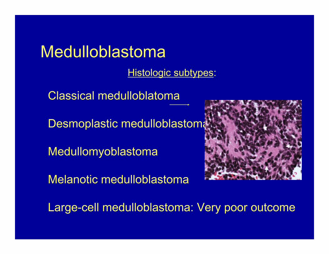
MedulloblastomaHistologic subtypes:
Classical medulloblatomaDesmoplastic medulloblastomaDesmoplastic medulloblastomaMedullomyoblastomaMelanotic medulloblastomaLarge-cell medulloblastoma: Very poor outcome
Medulloblastoma….origin
• Debatable:
– Origin from remnant of cells of the external – Origin from remnant of cells of the external granular layer of the cerebellum.
– Transformation of normal undifferentiated progenitor cells of superior medullary velum which migrate to the external granular layer.
Medulloblastoma….Clinical• Hydrocephalus : Raised ICP
• Behavioral change, listlessness, irritability, vomiting, and decreased social interactions.
• Headache, especially in the morning.
• Double vision.
• Head tilt : tonsillar herniation below the foramen magnum. (Can result from trochlear nerve palsy caused by direct tumor compression )
Medulloblastoma….Clinical
• Cerebellar symptoms
• Brain stem involvement• Brain stem involvement
• Leptomeningeal dissemination
Medulloblastoma….Clinical• Physical:
– Increasing head circumference , full anterior fontanalles with widely split cranial sutures.
• Fundus examination
– Papilledema can be present in as many as 90% of patients.
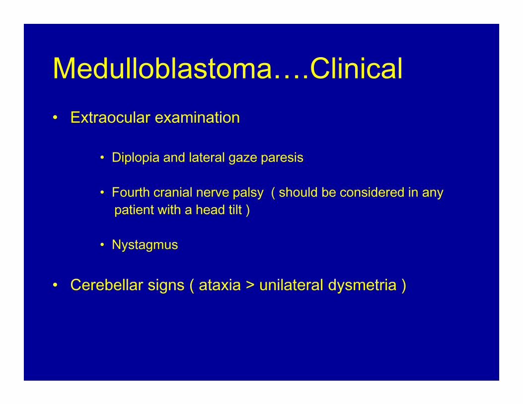
Medulloblastoma….Clinical• Extraocular examination
• Diplopia and lateral gaze paresis
• Fourth cranial nerve palsy ( should be considered in anypatient with a head tilt )patient with a head tilt )
• Nystagmus
• Cerebellar signs ( ataxia > unilateral dysmetria )
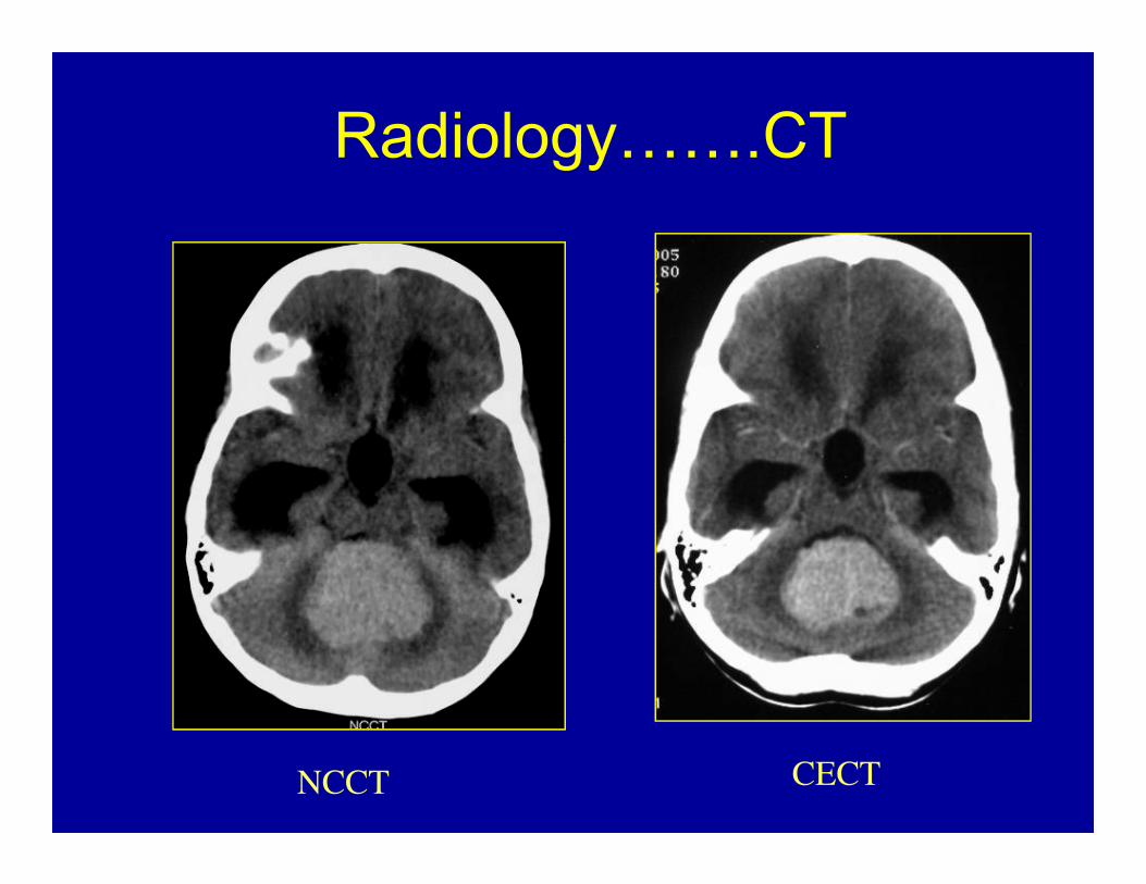
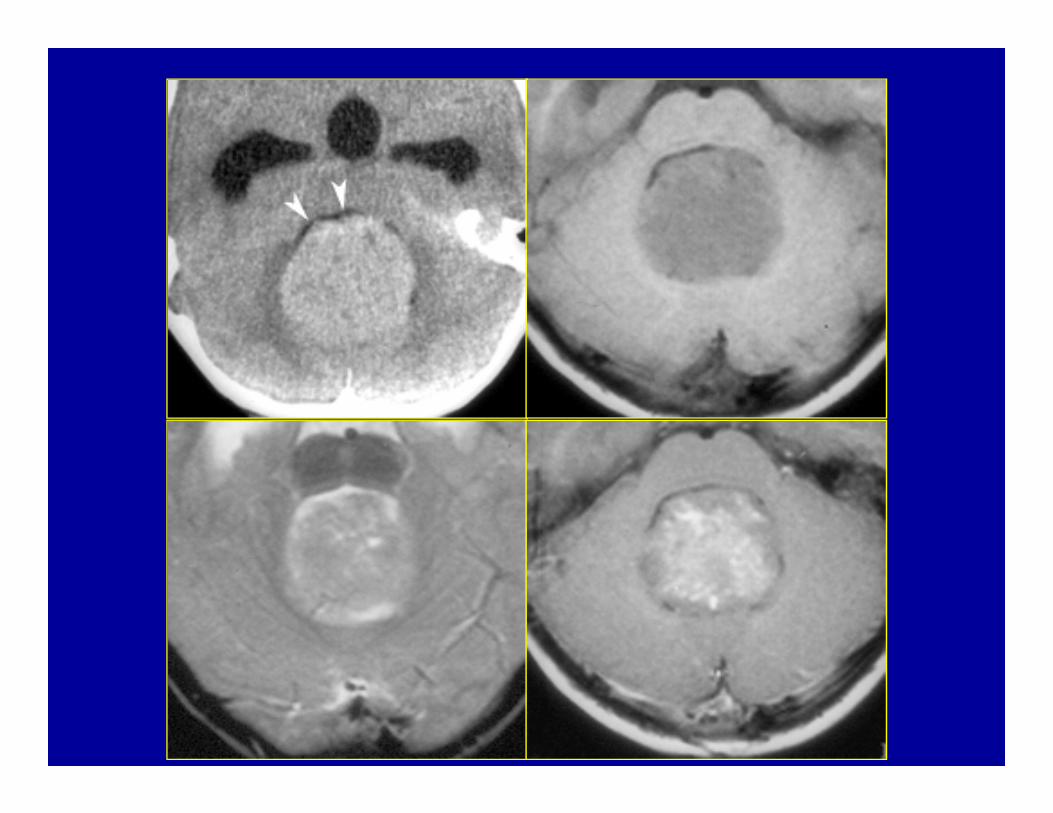
Radiology…….CT
NCCT CECT
Radiology…….MRI– Homogeneous enhancement ( may be absent in about
15 – 20 % )
– DWI shows restricted diffusion with increased ADC. – DWI shows restricted diffusion with increased ADC.
– MRI spine : Should be done at time of diagnosis.
– BEST : prior to surgery. If not possible Should bedelayed for at least 2 weeks after surgery.
M
E
D
U
L
L
O
BB
L
A
S
T
O
M
A
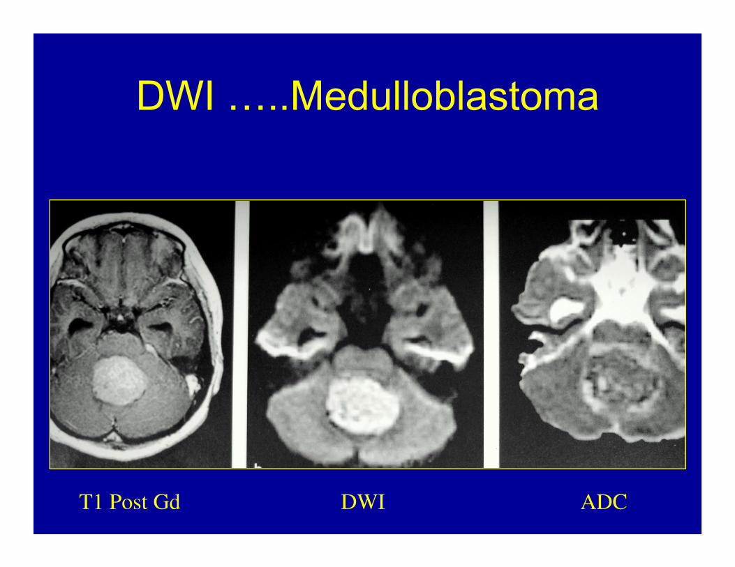
DWI …..Medulloblastoma
T1 Post Gd DWI ADC
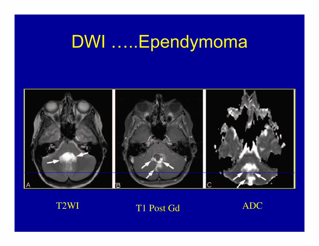
DWI …..Ependymoma
T2WI T1 Post Gd ADC
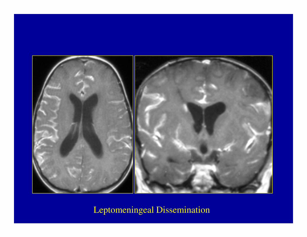
Leptomeningeal Dissemination
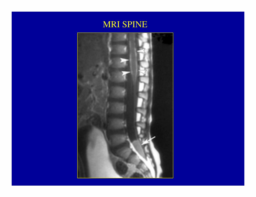
MRI SPINE
Radiology…….• Skeletal imaging
– Metastasis to the bone must be considered in any child with medulloblastoma and bone any child with medulloblastoma and bone pain.
– A skeletal survey helps elucidate lytic or sclerotic lesions.
Diagnosis …..CSF cytology• No standardized method: HOW and WHEN ??
• Lumbar puncture
• Ventricular drain
• Cisterna magna at the time of surgery from the for cytologic analysis.
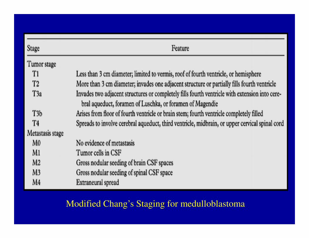
Modified Chang’s Staging for medulloblastoma
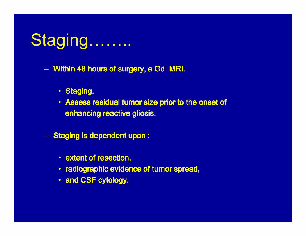
Staging……..– Within 48 hours of surgery, a Gd MRI. Within 48 hours of surgery, a Gd MRI. Within 48 hours of surgery, a Gd MRI. Within 48 hours of surgery, a Gd MRI.
• Staging.Staging.Staging.Staging.• Assess residual tumor size prior to the onset ofAssess residual tumor size prior to the onset ofAssess residual tumor size prior to the onset ofAssess residual tumor size prior to the onset of
enhancing reactive gliosis. enhancing reactive gliosis. enhancing reactive gliosis. enhancing reactive gliosis.
– Staging is dependent uponStaging is dependent uponStaging is dependent uponStaging is dependent upon :
• extent of resection,extent of resection,extent of resection,extent of resection,• radiographic evidence of tumor spread,radiographic evidence of tumor spread,radiographic evidence of tumor spread,radiographic evidence of tumor spread,• and CSF cytology.and CSF cytology.and CSF cytology.and CSF cytology.
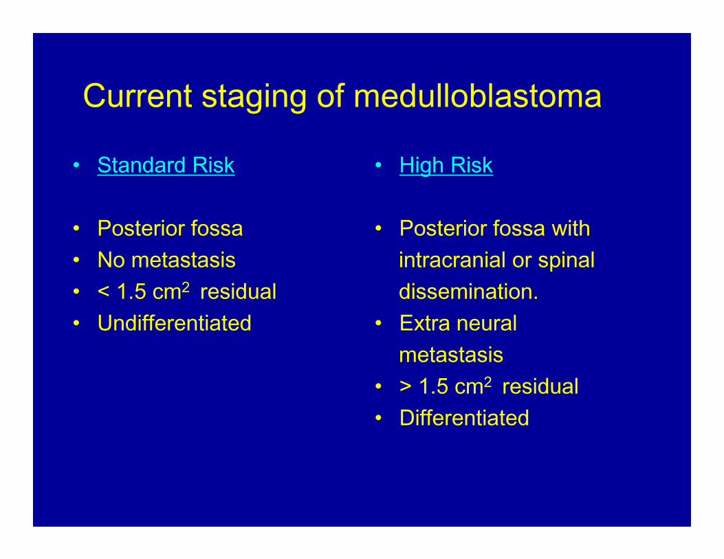
Current staging of medulloblastoma• Standard Risk
• Posterior fossa• No metastasis• < 1.5 cm2 residual
• High Risk
• Posterior fossa with intracranial or spinaldissemination.• < 1.5 cm2 residual
• Undifferentiateddissemination.
• Extra neural metastasis
• > 1.5 cm2 residual• Differentiated
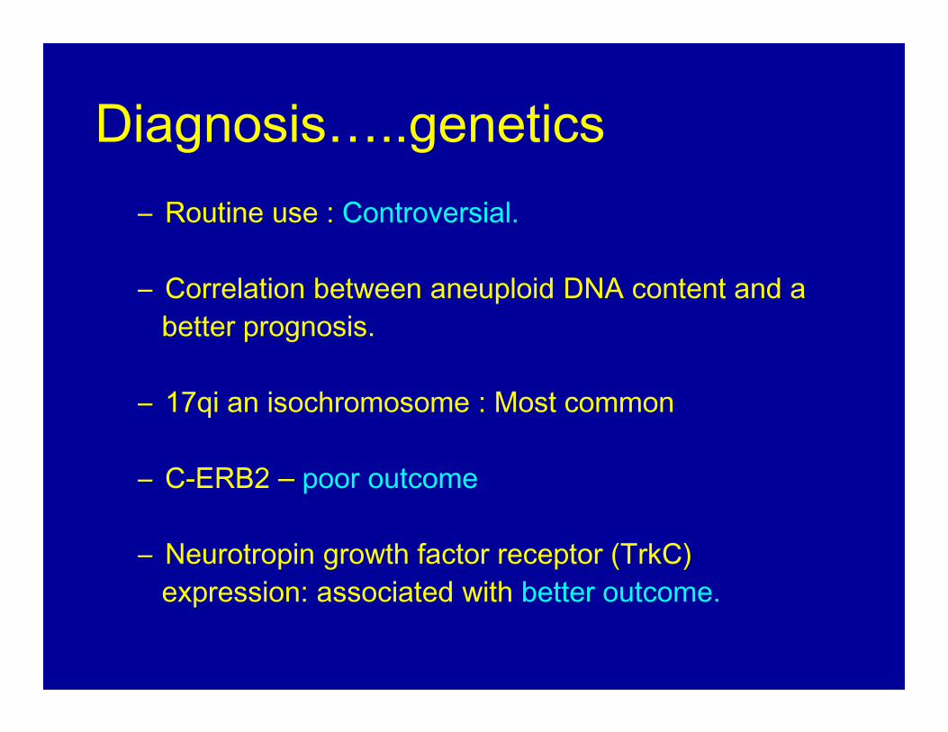
Diagnosis…..genetics– Routine use : Controversial.
– Correlation between aneuploid DNA content and abetter prognosis.
– 17qi an isochromosome : Most common
– C-ERB2 – poor outcome
– Neurotropin growth factor receptor (TrkC) expression: associated with better outcome.
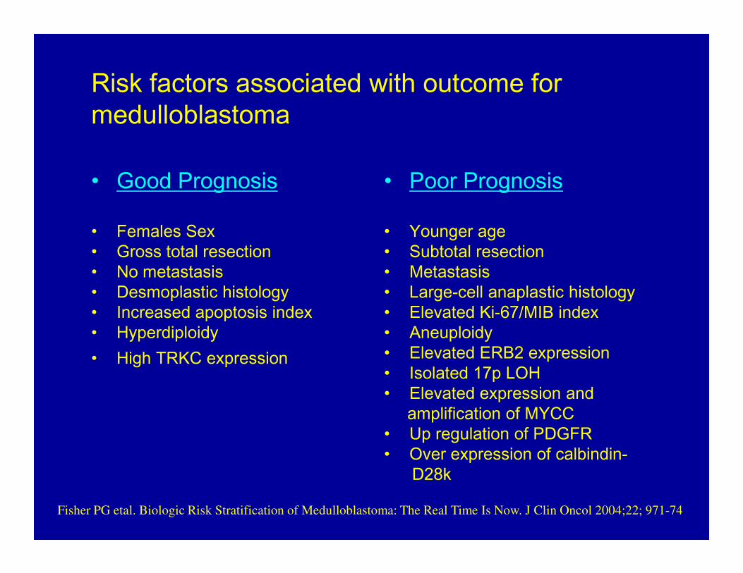
Risk factors associated with outcome for medulloblastoma
• Good Prognosis• Females Sex • Gross total resection • No metastasis • Desmoplastic histology
• Poor Prognosis• Younger age• Subtotal resection• Metastasis• Large-cell anaplastic histology • Desmoplastic histology
• Increased apoptosis index• Hyperdiploidy • High TRKC expression
• Large-cell anaplastic histology • Elevated Ki-67/MIB index • Aneuploidy • Elevated ERB2 expression • Isolated 17p LOH • Elevated expression and
amplification of MYCC• Up regulation of PDGFR • Over expression of calbindin-
D28k Fisher PG etal. Biologic Risk Stratification of Medulloblastoma: The Real Time Is Now. J Clin Oncol 2004;22; 971-74
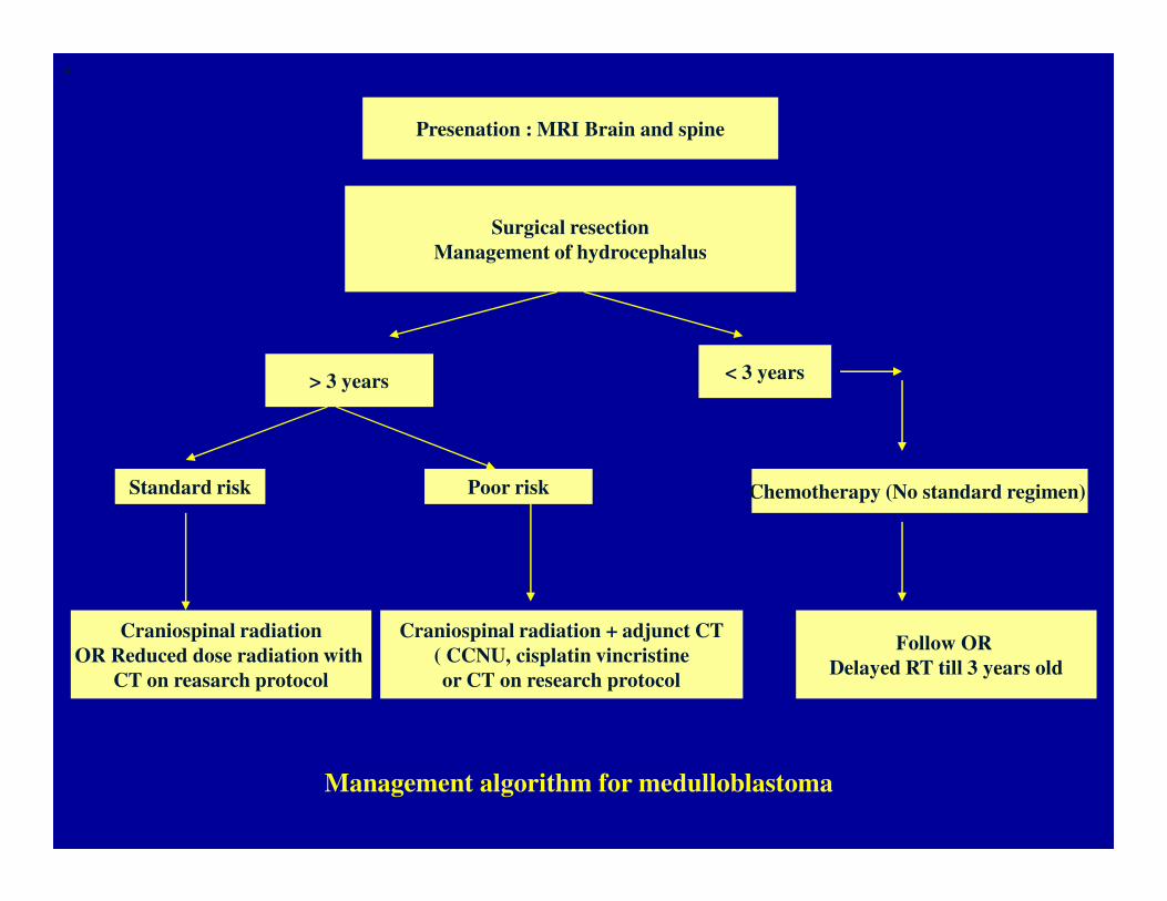
Presenation : MRI Brain and spine
•
Surgical resection
Management of hydrocephalus
> 3 years < 3 years
Standard risk Poor risk
Craniospinal radiation
OR Reduced dose radiation with
CT on reasarch protocol
Craniospinal radiation + adjunct CT
( CCNU, cisplatin vincristine
or CT on research protocol
Chemotherapy (No standard regimen)
Follow OR
Delayed RT till 3 years old
Management algorithm for medulloblastoma
Hydrocephalus
• The majority of children with posterior fossatumors have hydrocephalus at the time of presentation.presentation.
• There is no consensus regarding the management of HC in these children
Hydrocephalus• Treatment options:
– Ventriculoperitoneal shunt– Perioperative EVD– Perioperative EVD– Endoscopic third ventriculostomy – Direct surgical resection
Hydrocephalus……….• Recent studies have shown that ultimately 17 to 40% of children
have uncontrolled hydrocephalus and require shunt placementduring the postoperative period; and that this predominantlyoccurred within the 1st postoperative month.
• An expectant policy in these subgroup who ultimately require ashunt place them at risk of developing intracranial hypertension
,an increased rate of CSF leakage, and pseudomeningoceleformation, prolonged hospitalization.
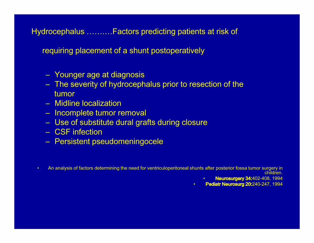
Hydrocephalus …….…Factors predicting patients at risk of requiring placement of a shunt postoperatively
– Younger age at diagnosis – The severity of hydrocephalus prior to resection of the
tumor – Midline localization – Incomplete tumor removal – Use of substitute dural grafts during closure– Use of substitute dural grafts during closure– CSF infection – Persistent pseudomeningocele
• An analysis of factors determining the need for ventriculoperitoneal shunts after posterior fossa tumor surgery in children.
• Neurosurgery 34:Neurosurgery 34:Neurosurgery 34:Neurosurgery 34:402-408, 1994• Pediatr Neurosurg 20:Pediatr Neurosurg 20:Pediatr Neurosurg 20:Pediatr Neurosurg 20:240-247, 1994
Management…….. SurgerySurgerySurgerySurgery
• Gross Total Resection, if possible.
• Brainstem damage should be avoided.
• Resolution of natural CSF pathways.
• Tumor adheres to the floor of the fourth ventricle,precluding gross total resection.( 1/3 rd of cases )
• Sugar coating – subarachnoid spread.
Management…….. Radiotherapy
• SURGERY alone : NOT CURATIVENOT CURATIVENOT CURATIVENOT CURATIVE• RADIOTHERAPY : cornerstone of adjuvant
therapy.therapy.
• 54 to 58 Gy to the primary site with 35Gy to the
entire craniospinal axis
Institution of presymptomatic craniospinal radiation
therapy is probably the single most important factor
responsible for the improved survival rates
Management…….. Radiotherapy
Complications of radiotherapy :
– lowered intelligence quotient (IQ),– small stature, endocrine dysfunction,– small stature, endocrine dysfunction,– behavioral abnormalities, – secondary neoplasms – white matter necrosis.– Reduction in IQ and neurobehavioral function.
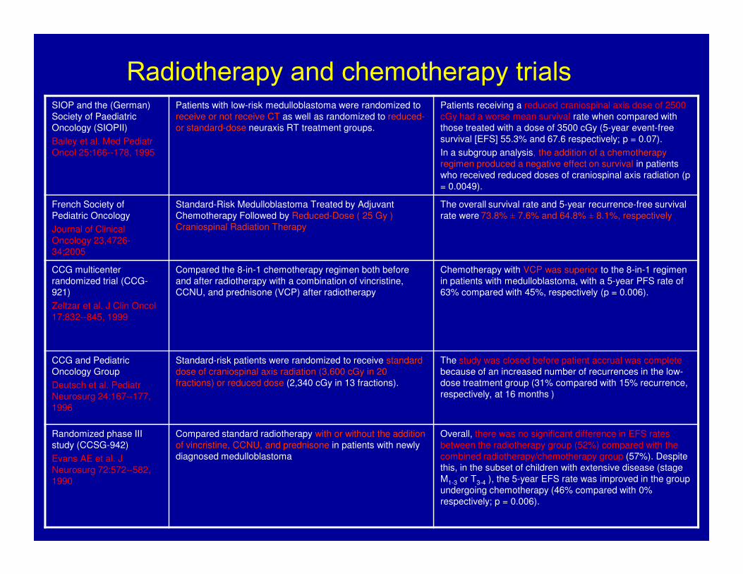
Radiotherapy and chemotherapy trialsSIOP and the (German)
Society of Paediatric
Oncology (SIOPII)
Bailey et al. Med Pediatr
Oncol 25:166--178, 1995
Patients with low-risk medulloblastoma were randomized to
receive or not receive CT as well as randomized to reduced-
or standard-dose neuraxis RT treatment groups.
Patients receiving a reduced craniospinal axis dose of 2500
cGy had a worse mean survival rate when compared with
those treated with a dose of 3500 cGy (5-year event-free
survival [EFS] 55.3% and 67.6 respectively; p = 0.07).
In a subgroup analysis, the addition of a chemotherapy
regimen produced a negative effect on survival in patients
who received reduced doses of craniospinal axis radiation (p
= 0.0049).
French Society of
Pediatric Oncology
Journal of Clinical
Oncology 23,4726-
34;2005
Standard-Risk Medulloblastoma Treated by Adjuvant
Chemotherapy Followed by Reduced-Dose ( 25 Gy )
Craniospinal Radiation Therapy
The overall survival rate and 5-year recurrence-free survival
rate were 73.8% ± 7.6% and 64.8% ± 8.1%, respectively
CCG multicenter
randomized trial (CCG-
921)
Compared the 8-in-1 chemotherapy regimen both before
and after radiotherapy with a combination of vincristine,
CCNU, and prednisone (VCP) after radiotherapy
Chemotherapy with VCP was superior to the 8-in-1 regimen
in patients with medulloblastoma, with a 5-year PFS rate of
63% compared with 45%, respectively (p = 0.006). 921)
Zeltzar et al. J Clin Oncol
17:832--845, 1999
CCNU, and prednisone (VCP) after radiotherapy 63% compared with 45%, respectively (p = 0.006).
CCG and Pediatric
Oncology Group
Deutsch et al. Pediatr
Neurosurg 24:167--177,
1996
Standard-risk patients were randomized to receive standard
dose of craniospinal axis radiation (3,600 cGy in 20
fractions) or reduced dose (2,340 cGy in 13 fractions).
The study was closed before patient accrual was complete
because of an increased number of recurrences in the low-
dose treatment group (31% compared with 15% recurrence,
respectively, at 16 months )
Randomized phase III
study (CCSG-942)
Evans AE et al. J
Neurosurg 72:572--582,
1990
Compared standard radiotherapy with or without the addition
of vincristine, CCNU, and prednisone in patients with newly
diagnosed medulloblastoma
Overall, there was no significant difference in EFS rates
between the radiotherapy group (52%) compared with the
combined radiotherapy/chemotherapy group (57%). Despite
this, in the subset of children with extensive disease (stage
M1-3 or T3-4 ), the 5-year EFS rate was improved in the group
undergoing chemotherapy (46% compared with 0%
respectively; p = 0.006).
Management….. Hyperfractionated radiotherapy
• Delivery of higher doses of radiation without increased toxicity.
• The typical hyperfractionated radiotherapy schedule • The typical hyperfractionated radiotherapy schedule consists of twice-daily fraction sizes of 100 to 120 cGy to a total dose of 7200 to 7800 cGy.
• In practice hyperfractionated therapy has shown no In practice hyperfractionated therapy has shown no In practice hyperfractionated therapy has shown no In practice hyperfractionated therapy has shown no advantage over the standard RT.advantage over the standard RT.advantage over the standard RT.advantage over the standard RT.
Management……. Chemotherapy– EXACT BENEFITS : UNCLEAR
– Delay the onset of radiation therapy in young children
( < 3 years )
– Increase in survival rates in high-risk children with
medulloblastoma
– Patients with recurrent or advanced disease
– Reduction in the RT dose to the neuraxis in patients with
nondisseminated disease
Management…….. New studies
– Sensitizing the tumor to irradiation with the concomitant useof chemotherapy.
– Presurgical chemotherapy to treat patients prior to surgery. – Presurgical chemotherapy to treat patients prior to surgery. – Intraventricular administration of cytotoxic agents,
– Newer drug combinations, and
– Immunotherapy based on genetics analysisImmunotherapy based on genetics analysisImmunotherapy based on genetics analysisImmunotherapy based on genetics analysis
ManagementManagementManagementManagement…….. Recurrent Medulloblastoma.. Recurrent Medulloblastoma.. Recurrent Medulloblastoma.. Recurrent Medulloblastoma• Recurrences : 30 to 40% of patients
• Chemotherapy : limited due to chemo resistance in those patients who have previously undergone CT
• Redosing with RT avoided due to radiation necrosis. ( Local RT using stereotactic techniques can be used can palliative )
ManagementManagementManagementManagement…….. Recurrent Medulloblastoma.. Recurrent Medulloblastoma.. Recurrent Medulloblastoma.. Recurrent Medulloblastoma
• High-dose chemotherapy with autologous SCR or autologous BMR : subject of intense investigation.
Stem cell rescue involves harvesting autologous bone Stem cell rescue involves harvesting autologous bone marrow or preferably, peripheral stem cells by using pheresis techniques and subsequently reinfusing them after provision of high-dose myeloablative chemotherapy.
• Int J Legal Med. 2001;114(6):331-7
Substantial toxicity :
Death, serious infection, and venoocclusive disease.
ManagementManagementManagementManagement…….. Recurrent Medulloblastoma.. Recurrent Medulloblastoma.. Recurrent Medulloblastoma.. Recurrent Medulloblastoma
• Though data suggests longer EFS. ( In the absence of RCT, the interpretation of the results remains limited )
• Benefits seen in a subset of patients, with locally recurrent disease (not involving thebrainstem) and without evidence of dissemination.
Management…….. Prognosis • 5 - year recurrence-free survival rates : 55% - 67%.
• Even after a good response to surgery and radiation, recurrence is common.• Most common site : PRIMARY TUMOR SITEPRIMARY TUMOR SITEPRIMARY TUMOR SITEPRIMARY TUMOR SITE• Most common site : PRIMARY TUMOR SITEPRIMARY TUMOR SITEPRIMARY TUMOR SITEPRIMARY TUMOR SITE
• Bone : most common site of systemic metastasis; followed by regional lymph node.
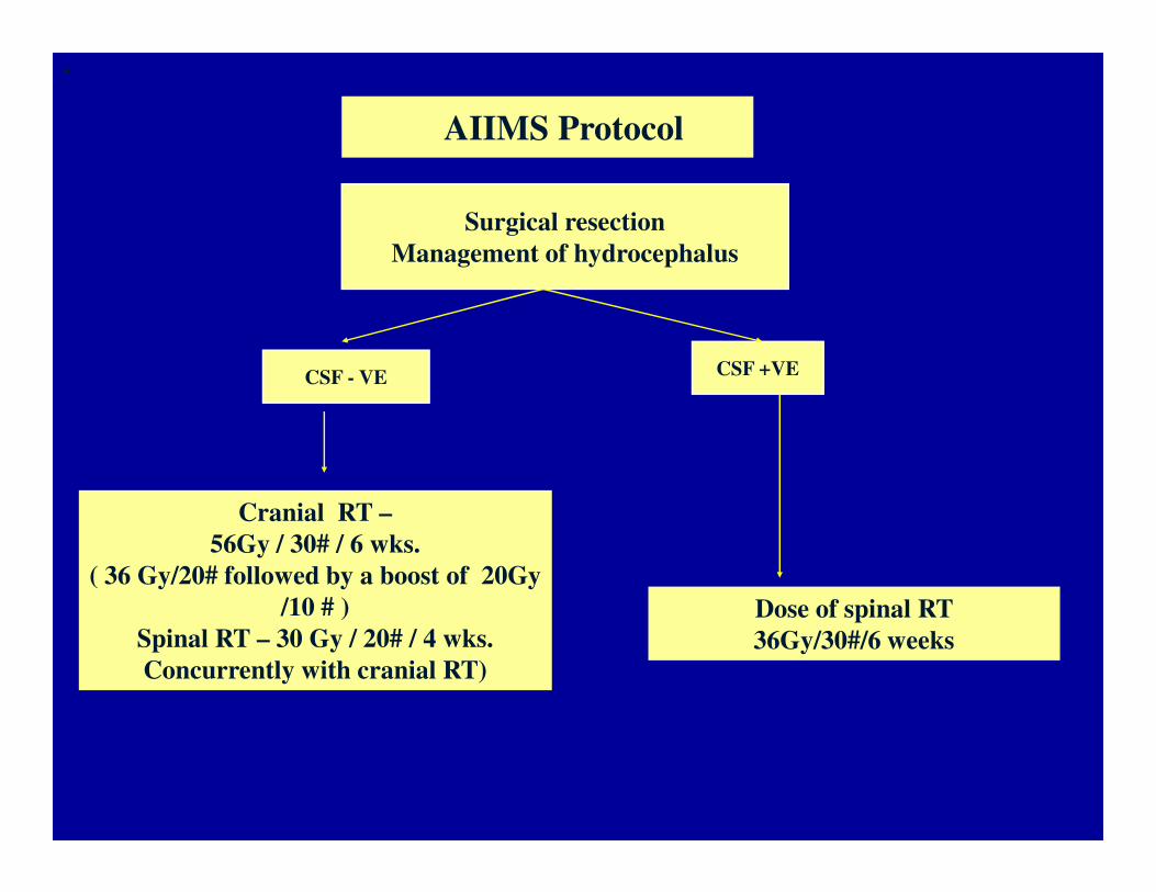
AIIMS Protocol
•
Surgical resection
Management of hydrocephalus
CSF - VE CSF +VE
Cranial RT –
56Gy / 30# / 6 wks.
( 36 Gy/20# followed by a boost of 20Gy
/10 # )
Spinal RT – 30 Gy / 20# / 4 wks.
Concurrently with cranial RT)
Dose of spinal RT
36Gy/30#/6 weeks
Cerebellar MutismCerebellar MutismCerebellar MutismCerebellar Mutism
• Cerebellar mutism was first reported in 1979 by Hirsh after a posterior fossa tumor resection.
• Also known as posterior fossa syndrome• Also known as posterior fossa syndrome
• Approximately 10 -1 5 % of children undergoing posterior fossa surgery for tumor.
Cerebellar MutismCerebellar MutismCerebellar MutismCerebellar Mutism
• Decreased or absent speech, irritability, hypotonia, ataxia.
• Onset : Immediate or delayed.
• Virtually all cases of mutism will occur within the firstweek of surgery ( 50% within the first two days )
• Most cases resolves in a week or two.( longest 52 months) with return of functional speech.
Factors associated with the development of Factors associated with the development of Factors associated with the development of Factors associated with the development of mutismmutismmutismmutism• Posterior fossa surgery for tumor. • Children • Midline tumor location• Cerebellar vermal incision• Cerebellar vermal incision• Large tumor size ( > 5cm )• Medulloblastoma
Cerebellar MutismCerebellar MutismCerebellar MutismCerebellar Mutism…. Pathophysiology. Pathophysiology. Pathophysiology. Pathophysiology....
• UNKNOWN. However not emotional.
• Focal decreased cerebral and cerebellar blood blow leading to decreased cell functioning in blow leading to decreased cell functioning in particular areas, dentatedentatedentatedentate----thalamithalamithalamithalami----cortical cortical cortical cortical pathwaypathwaypathwaypathway causing dysfunction. SPECT studies have lead support to this theory
Cerebellar MutismCerebellar MutismCerebellar MutismCerebellar Mutism…. Outcome. Outcome. Outcome. Outcome
• Speech almost always returns.
• The speech is virtually always becomes functional for communication, however it functional for communication, however it may not be the same as before surgery.
Cerebellar MutismCerebellar MutismCerebellar MutismCerebellar Mutism…. intervention. intervention. intervention. intervention
• Speech therapy• Assisting in some form of nonverbal
communication• Reassurance : usual course of cerebellar
mutism and what to expect in the recovery.• Practicing tongue and lip movements before
speech returns
Brain Stem Gliomas
• Brainstem tumors comprise 10–20% of all pediatric central nervous system tumors.
• Once considered uniformly fatal ; the perspective has changed now.
Clinical hallmark• Bilateral long tract signs• Bilateral multiple contiguous cranial nerve
palsies.• Horner’s syndrome• Horner’s syndrome• Inter Nuclear Ophthalmoplegia
BSG……Classification• The most recent classification system by
Choux et al based on both CT and MRI imaging– Type I – Diffuse– Type I – Diffuse– Type II – Intrinsic, focal– Type III – Exophytic, focal– Type IV – Cervicomedullary
– Pediatric Neurosurgery. New York, Churchill Livingstone, 2000, pp 471Pediatric Neurosurgery. New York, Churchill Livingstone, 2000, pp 471Pediatric Neurosurgery. New York, Churchill Livingstone, 2000, pp 471Pediatric Neurosurgery. New York, Churchill Livingstone, 2000, pp 471–491.491.491.491.
BSG……
• Type I : Diffuse brainstem gliomas• Appro. 75% of all tumors • Hypointense on CT • No significant enhancement on MRI.• No significant enhancement on MRI.• Characterized by diffuse infiltration and
swelling of the brainstem. • Typically, are malignant fibrillary
astrocytomas (WHO grade III or IV).
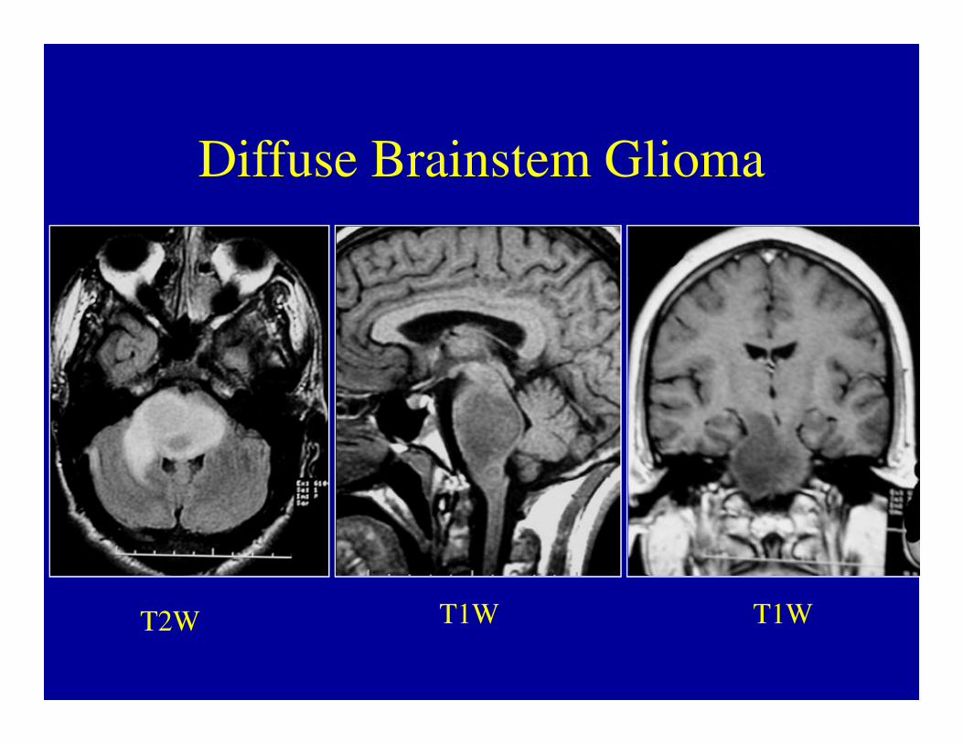
Diffuse Brainstem Glioma
T2W T1W T1W
BSG……
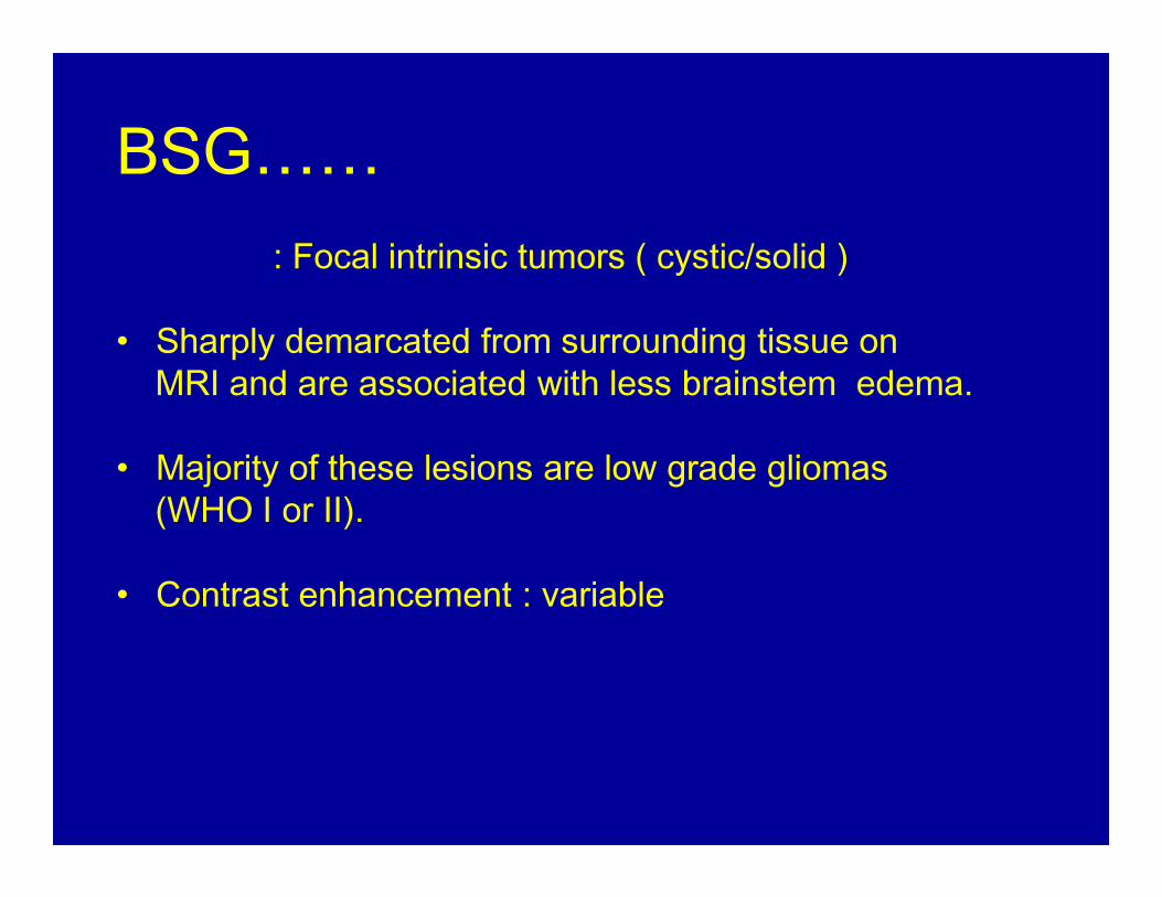
• Type II : Focal intrinsic tumors ( cystic/solid )
• Sharply demarcated from surrounding tissue on MRI and are associated with less brainstem edema.
• Majority of these lesions are low grade gliomas • Majority of these lesions are low grade gliomas (WHO I or II).
• Contrast enhancement : variable
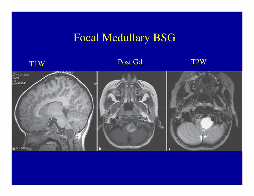
Focal Medullary BSG
T1W Post Gd T2W
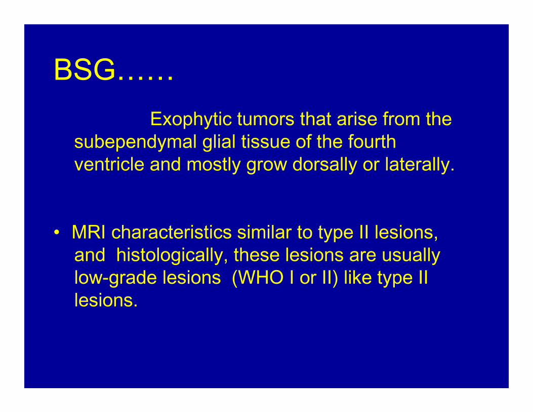
BSG……
• Type III : Exophytic tumors that arise from the subependymal glial tissue of the fourth ventricle and mostly grow dorsally or laterally.
• MRI characteristics similar to type II lesions,and histologically, these lesions are usually low-grade lesions (WHO I or II) like type II lesions.
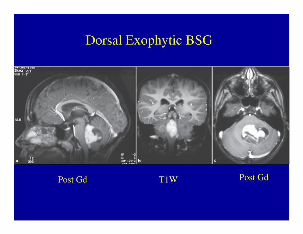
Dorsal Exophytic BSG
Post Gd T1W Post Gd
BSG……
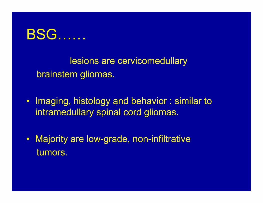
• Type IV lesions are cervicomedullary brainstem gliomas.
• Imaging, histology and behavior : similar to • Imaging, histology and behavior : similar to intramedullary spinal cord gliomas.
• Majority are low-grade, non-infiltrative tumors.
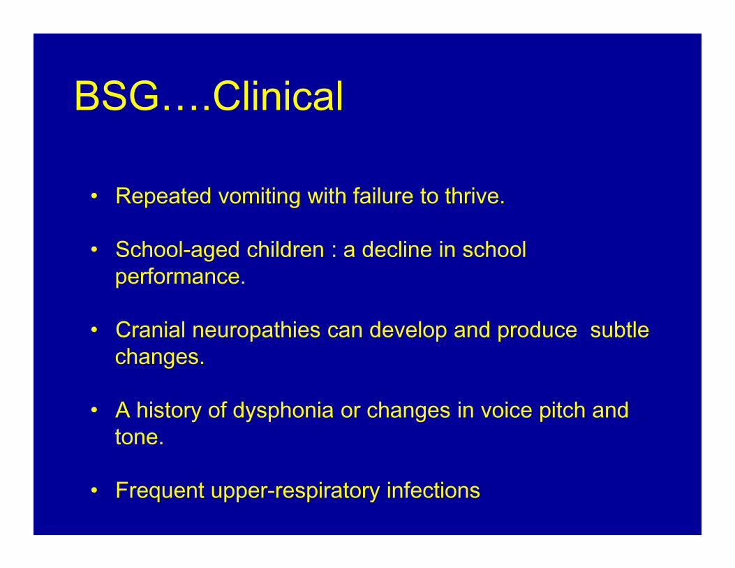
BSG….Clinical• Repeated vomiting with failure to thrive.
• School-aged children : a decline in school performance. performance.
• Cranial neuropathies can develop and produce subtle changes.
• A history of dysphonia or changes in voice pitch and tone.
• Frequent upper-respiratory infections
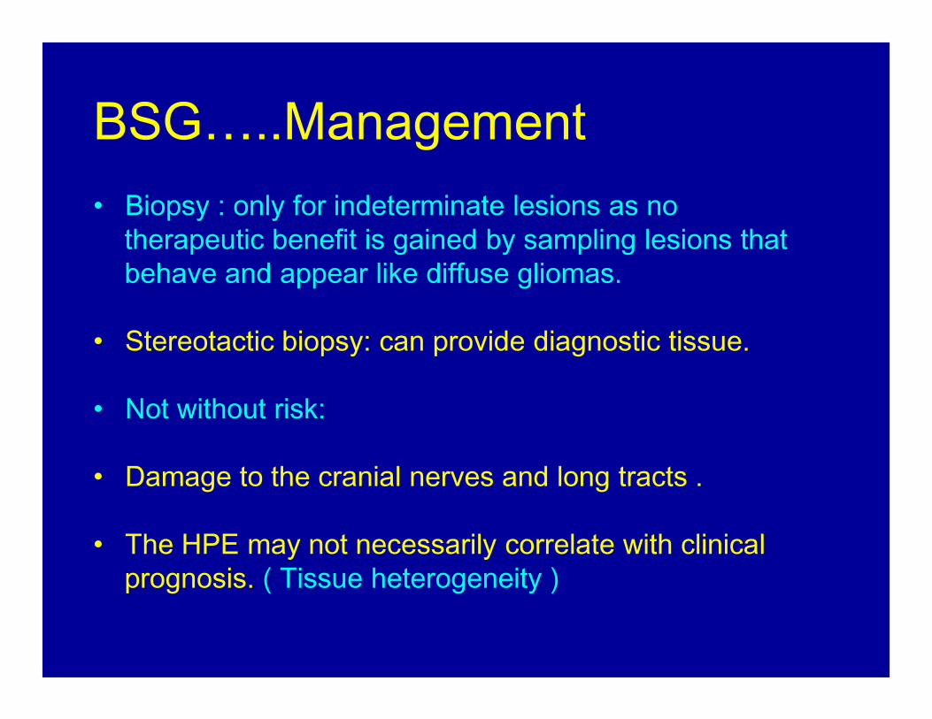
BSG…..Management• Biopsy : only for indeterminate lesions as no
therapeutic benefit is gained by sampling lesions thatbehave and appear like diffuse gliomas.
• Stereotactic biopsy: can provide diagnostic tissue.
• Not without risk:
• Damage to the cranial nerves and long tracts .
• The HPE may not necessarily correlate with clinical prognosis. ( Tissue heterogeneity )
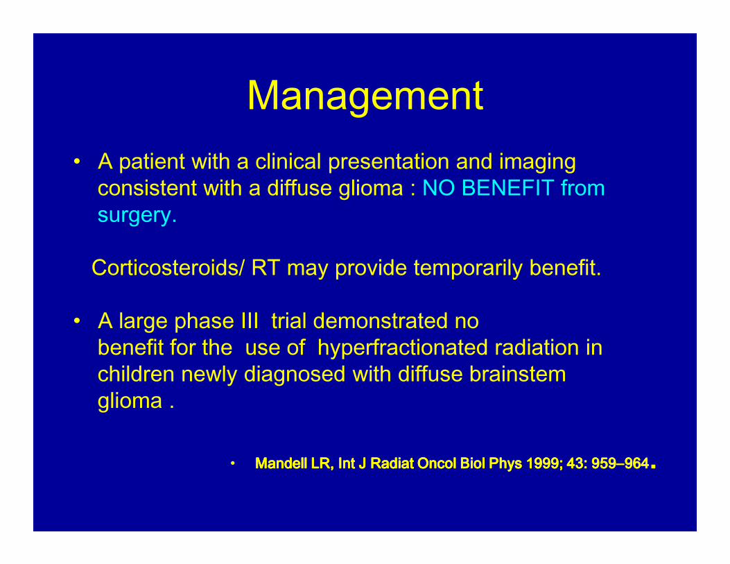
Management• A patient with a clinical presentation and imaging
consistent with a diffuse glioma : NO BENEFIT from surgery.
Corticosteroids/ RT may provide temporarily benefit.
• A large phase III trial demonstrated no benefit for the use of hyperfractionated radiation in children newly diagnosed with diffuse brainstem glioma .
• Mandell LR, Int J Radiat Oncol Biol Phys 1999; 43: 959Mandell LR, Int J Radiat Oncol Biol Phys 1999; 43: 959Mandell LR, Int J Radiat Oncol Biol Phys 1999; 43: 959Mandell LR, Int J Radiat Oncol Biol Phys 1999; 43: 959–964964964964....
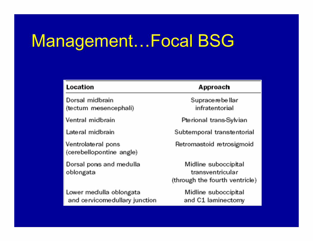
Management…Focal BSG
Management……..Postoperative Course
• Postoperative treatment and monitoring : on the location• Patients who have had a CSF diversion procedure : monitor
for reemergence of signs and symptoms of hydrocephalus.• Tumors of the pons carry the worst prognosis because the
majority are diffuse gliomas. ( survival rates are low with a1-year survival of 35–46% and 3- year survival of 11–17%.
Pediatr Neurosurg 1996; 24: 9Pediatr Neurosurg 1996; 24: 9Pediatr Neurosurg 1996; 24: 9Pediatr Neurosurg 1996; 24: 9–23232323.
Management……..Postoperative Course• The postoperative course of focal medullary neoplasms
depends on the tumor type.• Dorsal exophytic tumors treated with surgery have an excellent
prognosis with a 92% long-term survival some series.• Pediatr neurosurg 1994; 20: 2Pediatr neurosurg 1994; 20: 2Pediatr neurosurg 1994; 20: 2Pediatr neurosurg 1994; 20: 2–10101010• Pediatr neurosurg 1994; 20: 2Pediatr neurosurg 1994; 20: 2Pediatr neurosurg 1994; 20: 2Pediatr neurosurg 1994; 20: 2–10101010
• Pollack et al. reported a long-term survival of 94% in their series of 18 patients.
• J Neurosurg 1993; 78: 859J Neurosurg 1993; 78: 859J Neurosurg 1993; 78: 859J Neurosurg 1993; 78: 859–863863863863....
Management……..Postoperative Course
• However, significant lower cranial nervedysfunction can occur and may need prolonged postoperative need prolonged postoperative ventilation or a feeding gastrostomy postoperatively.
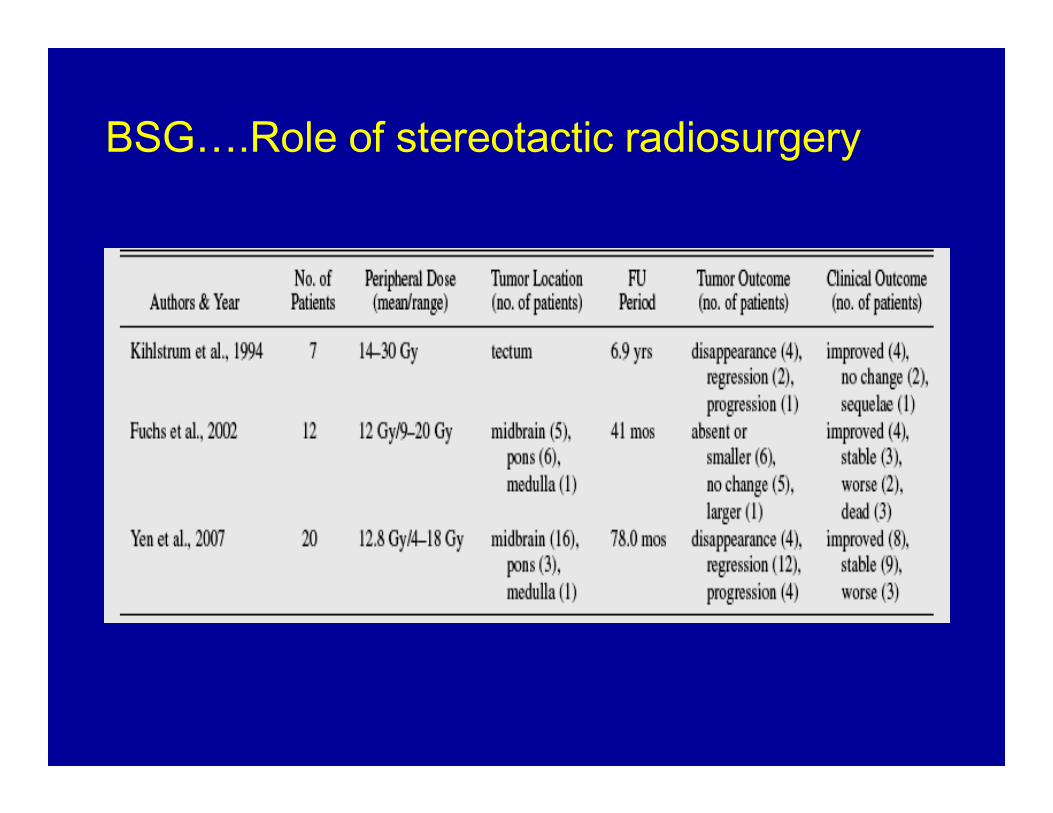
BSG….Role of stereotactic radiosurgery
Tectal plate gliomas• Unique subset of brainstem gliomas. • Presents with late onset obstructive
hydrocephalus that can be confused withbenign aqueductal stenosis.benign aqueductal stenosis.Tectal gliomas are believed to be low-grade astrocytic tumors that usually follow a benignclinical course.VP shunts or ETV for CSF diversion.
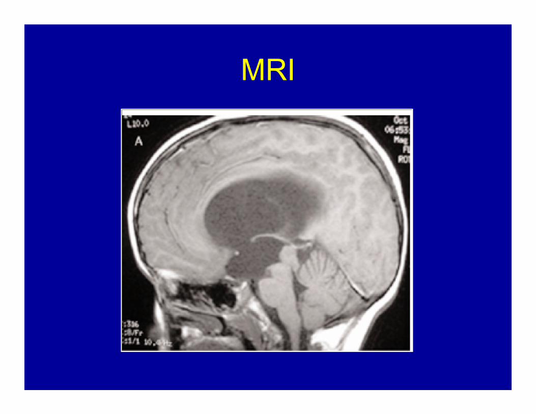
MRI
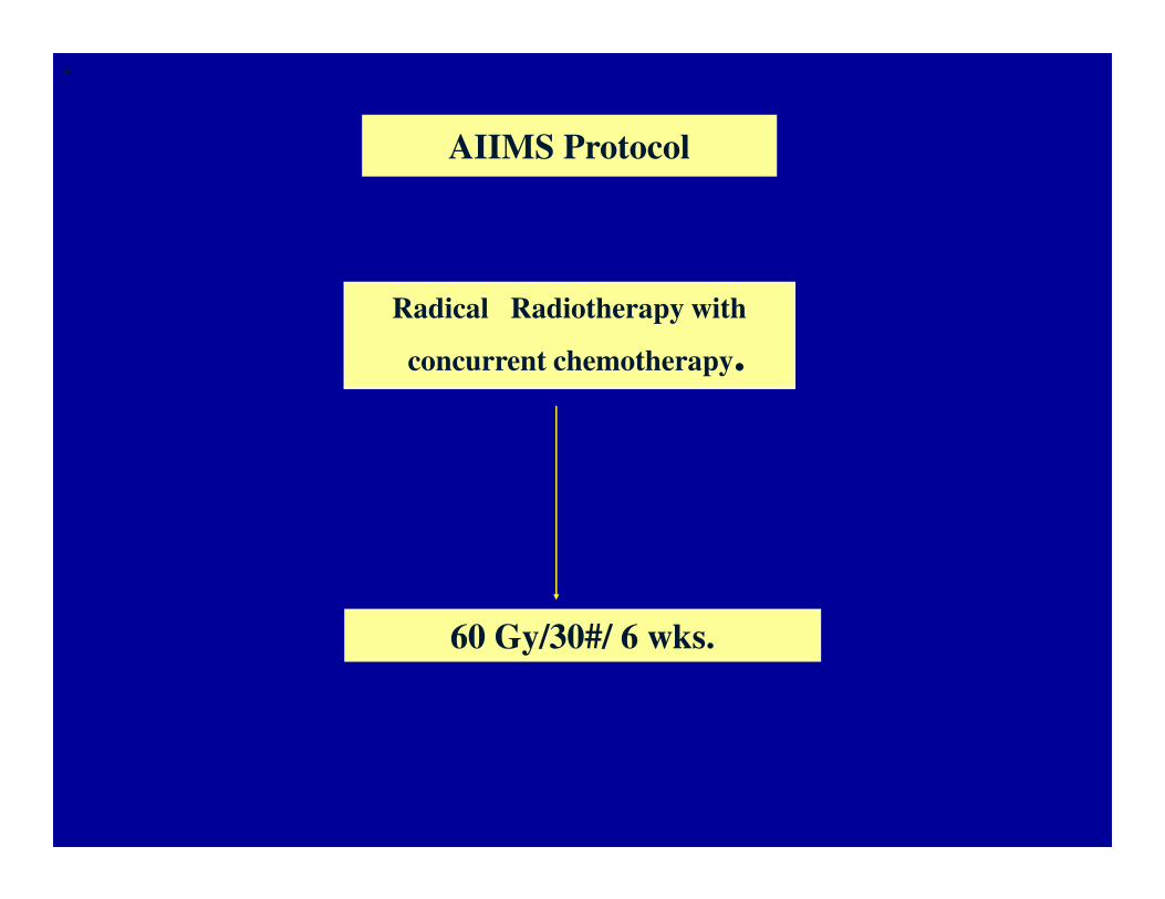
AIIMS Protocol
•
Radical Radiotherapy with
concurrent chemotherapy.
60 Gy/30#/ 6 wks.
Ependymoma• Ependymomas are glial tumors that arise from
ependymal cells within the CNS.
• WHO grade I : Myxopapillary ependymoma and subependymoma;subependymoma;
• WHO grade II : Ependymoma (with cellular,papillary and clear cell variants)
• WHO grade III : Anaplastic ependymoma. • Who grade IV : Ependymoblastomas
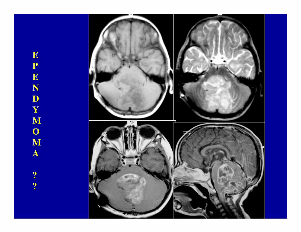
Ependymoma• In children : 90% of ependymomas are
intracranial, majority of these occuring in the posterior fossa usually arising from the roof of the fourth ventricle
• In adults : 75% of ependymomas arise within the spinal canal, with a significant minority occurring intracranially in the supratentorial compartment.
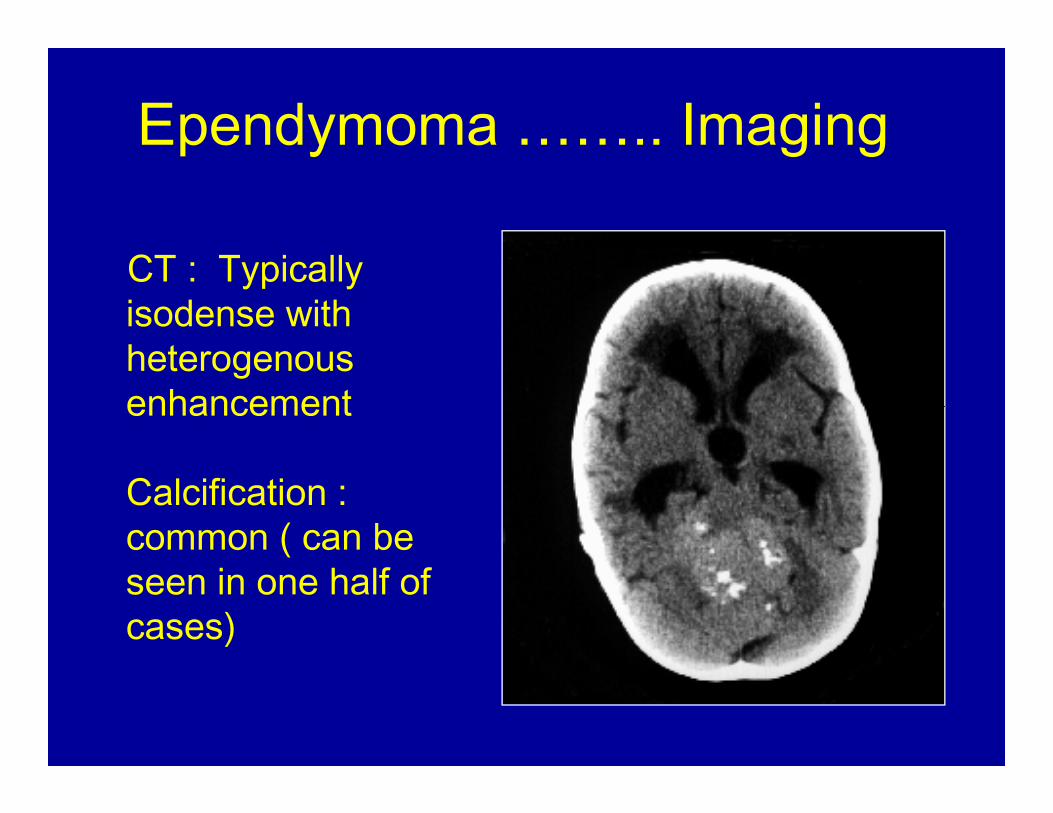
Ependymoma …….. ImagingCT : Typically isodense with heterogenous enhancementenhancementCalcification : common ( can be seen in one half ofcases)
Ependymoma…..MRI• On MRI, heterogeneous secondary to
necrosis, hemorrhage and calcification. • Heterogenous contrast enhancement• Plastic ependymomas.• Plastic ependymomas.• Extension to the cerebellopontine angle is
characteristic of ependymomas• Commonly found intraventricularly• Calcification common ( appro.45% of cases )
E
P
E
N
D
Y
MM
O
M
A
?
?

Ependymoma…..• Staging: Staging: Staging: Staging: No conventional staging criteria.
• Postoperative MRI is recommended within 48 hours of tumor resection to assess presence of hours of tumor resection to assess presence of residual tumor and to facilitate adjuvant treatment planning.
Ependymoma…..Surgery• The extent of surgical resection : most significant
factor associated with increased survival in almost every large series of pediatric ependymoma.
– Aggressive primary resection,Aggressive primary resection,Aggressive primary resection,Aggressive primary resection,– Immediate second look surgery if a postImmediate second look surgery if a postImmediate second look surgery if a postImmediate second look surgery if a post----operative operative operative operative
residual tumor is identified andresidual tumor is identified andresidual tumor is identified andresidual tumor is identified and– ReReReRe----surgery at time of recurrence.surgery at time of recurrence.surgery at time of recurrence.surgery at time of recurrence.
Ependymoma…Role of Radiotherapy
• Post-operative radiation recommended for patients older than 3 years.
• Stereotactic radiosurgery : therapeutic option in patients with residual, unresectable or recurrent tumor.
Ependymoma…Role of Chemotherapy
• May be useful < 3 years : Delay cranial radiation.
• Childhood intracranial ependymomas : in general chemo-resistant
over-expression of the multi-drug resistance-1 gene and the 06-methylguanine-DNA methyl transferase.
Children cancer group (CCG) 942: the only randomized trial, which compared survival after radiation alone, and survival after CT + RT did not show improved outcome
Med Pediatr Oncol 1996;27:8-14
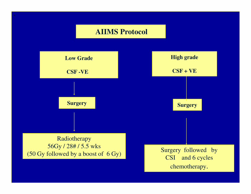
AIIMS Protocol
•
Low Grade
CSF -VE
High grade
CSF + VE
Surgery Surgery
Radiotherapy
56Gy / 28# / 5.5 wks
(50 Gy followed by a boost of 6 Gy)Surgery followed by
CSI and 6 cycles
chemotherapy.
Pilocytic astrocytoma• Pilocytic astrocytoma is the most common pediatric
central nervous system glial neoplasm
• Exceptional benign biologic behavior : extremely high survival rate 94% at 10 yearssurvival rate 94% at 10 years
• Most patients present in the first 2 decades
• Surgical resection is the treatment of choice.
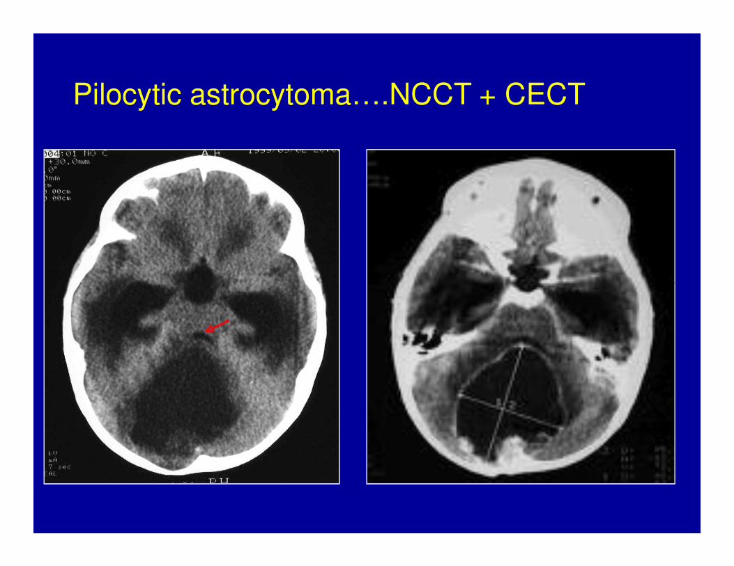
Pilocytic astrocytoma….NCCT + CECT
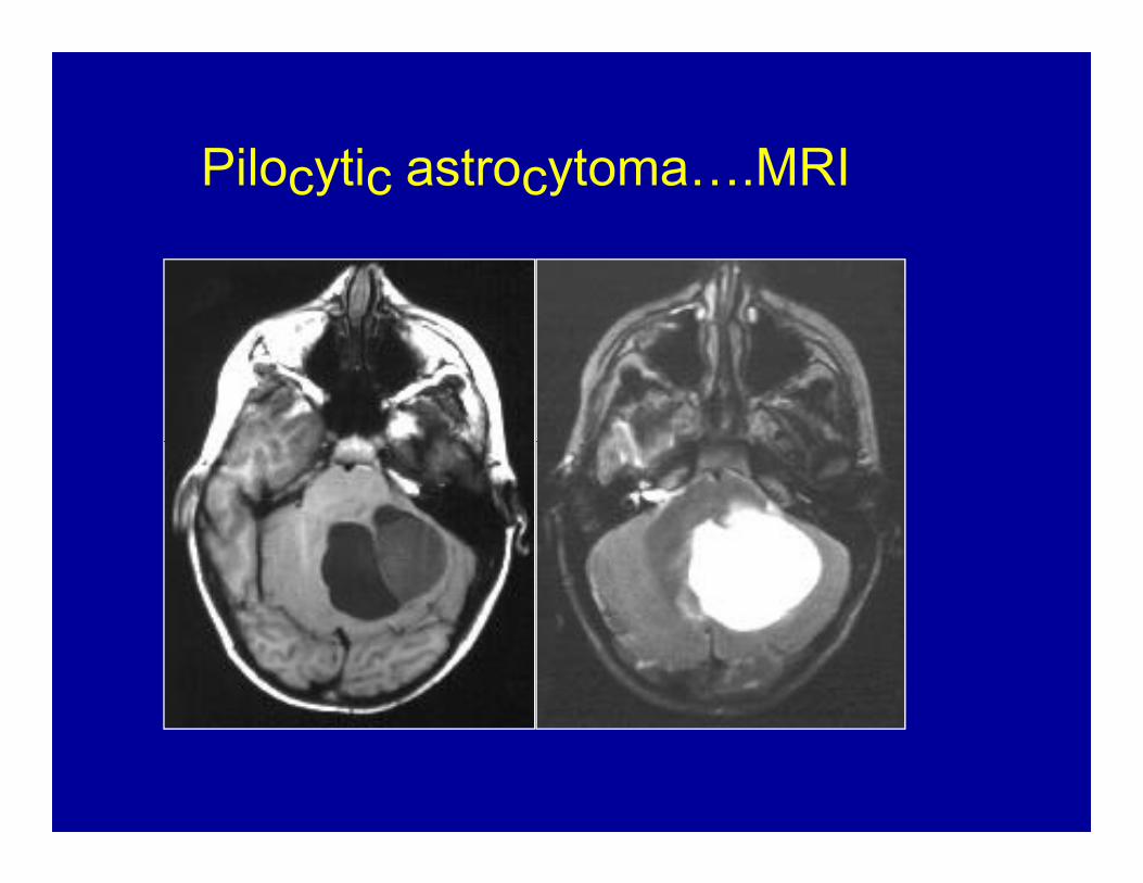
Pilocytic astrocytoma….MRI
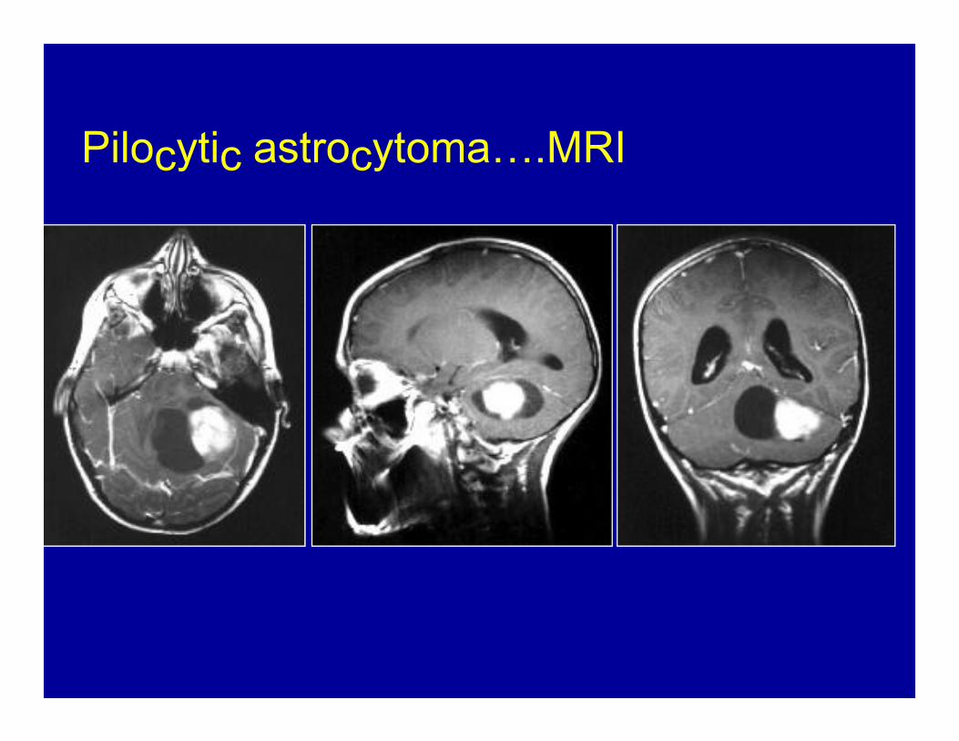
Pilocytic astrocytoma….MRI
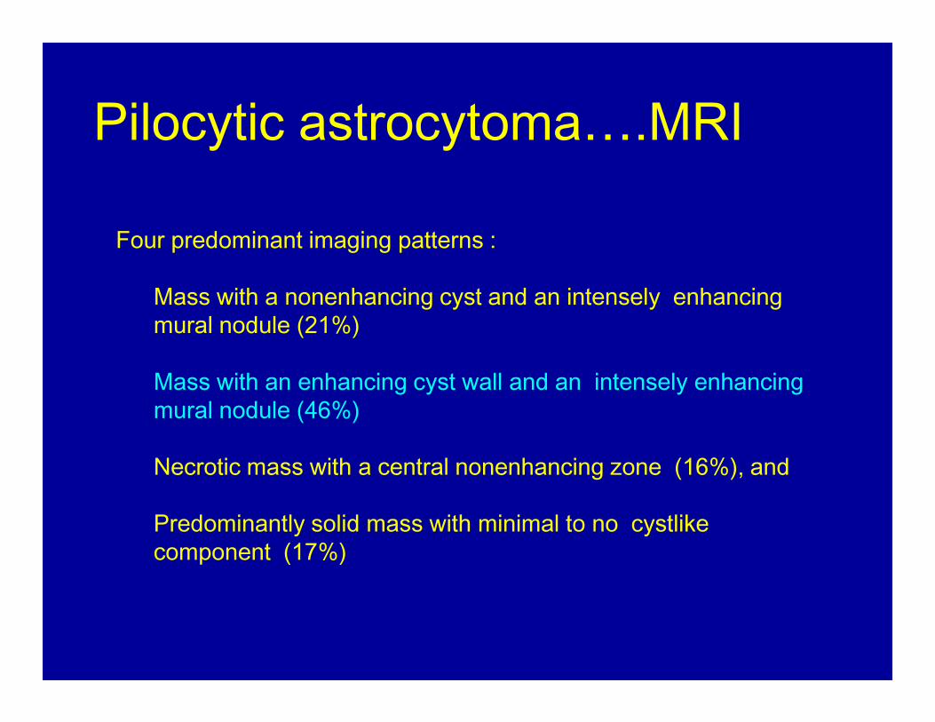
Pilocytic astrocytoma….MRIFour predominant imaging patterns :
Mass with a nonenhancing cyst and an intensely enhancing mural nodule (21%)Mass with an enhancing cyst wall and an intensely enhancing mural nodule (46%)Necrotic mass with a central nonenhancing zone (16%), and Predominantly solid mass with minimal to no cystlike component (17%)
Pilocytic astrocytoma….• Surgical resection of cerebellar pilocytic
astrocytomas is considered the treatment of choice.
• Radiation therapy is strictly avoided, given its risk of • Radiation therapy is strictly avoided, given its risk of causing significant morbidity in children younger than 5 years of age.
Pilocytic astrocytoma….• Resection of the mural nodule, when present, is the
key surgical objective, since the surrounding cyst occurs as a simple reactive change in most cases.
• Resection of the cyst wall : Controversial ??• Resection of the cyst wall : Controversial ??
NO STATISTICAL DIFFERENCE IN SURVIVALNO STATISTICAL DIFFERENCE IN SURVIVALNO STATISTICAL DIFFERENCE IN SURVIVALNO STATISTICAL DIFFERENCE IN SURVIVALhas been noted in patients who have undergone resection of the cyst wall compared with those in which the cyst is left alone.
Pilocytic astrocytomaPilocytic astrocytomaPilocytic astrocytomaPilocytic astrocytoma…. Prognosis . Prognosis . Prognosis . Prognosis
• EXCELLENT: 10-year survival rate : up to 94%
• In contrast to the generally poor outcome (a 5-year survival of usually only 30%) for patients with an infiltrating brainstem glioma (WHO grade II), those withwith
Dorsally exophytic brainstem pilocytic astrocytomahas a much better prognosis, with stable neurologic status and long term survival.
Pilocytic astrocytoma….Recurrence• Can occur many years after surgery• Repeat surgery : Desired treatment• Radiotherapy can be avoided if complete• Radiotherapy can be avoided if complete
resection possible.• Residual / Unresectable recurrence : RT
preferably SRS.
Choroid Plexus PapillomaChoroid Plexus PapillomaChoroid Plexus PapillomaChoroid Plexus Papilloma
• CPP are benign neoplasms of the choroid plexus.
• Lateral ventricles : most common location in children.
• 4-6% of the intracranial neoplasms in children youngerthan 2 years.
• 12-13% of intracranial neoplasms in children younger than 1 year.
Choroid Plexus PapillomaChoroid Plexus PapillomaChoroid Plexus PapillomaChoroid Plexus Papilloma…..Clinical..Clinical..Clinical..Clinical
• Hydrocephalus and raised ICT
• The tumor itself can cause mass effect.• The tumor itself can cause mass effect.
• SURGERY does not guarantees resolution of HCP , possibly because of derangement of reabsorption mechanisms or blockage at other sites in the ventricular system.
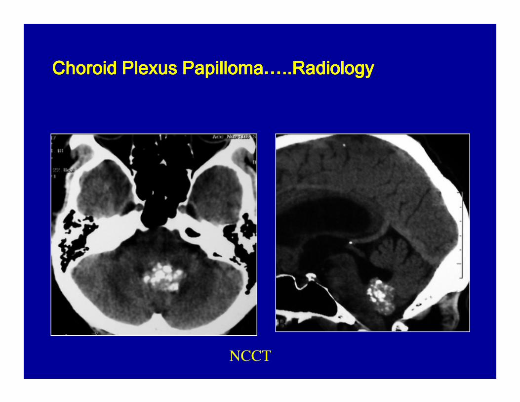
Choroid Plexus PapillomaChoroid Plexus PapillomaChoroid Plexus PapillomaChoroid Plexus Papilloma…..Radiology..Radiology..Radiology..Radiology
NCCT
Choroid Plexus PapillomaChoroid Plexus PapillomaChoroid Plexus PapillomaChoroid Plexus Papilloma…..Radiology..Radiology..Radiology..Radiology
On MRI : lobulated frond like intermediate-to-strong intensity on both T1- and T2 - weighted images with dense enhancement.images with dense enhancement.
Choroid plexus carcinoma appears more heterogeneous than the papilloma and often shows adjacent parenchymal invasion orsurrounding edema.
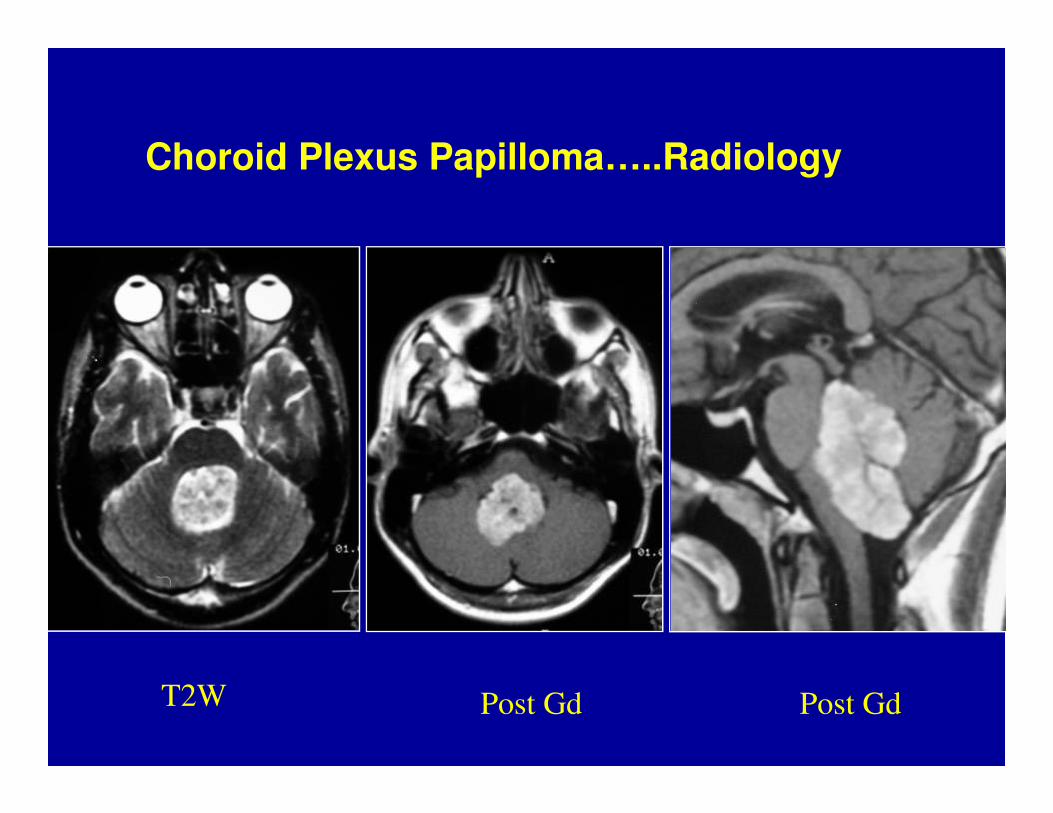
Choroid Plexus Papilloma…..Radiology
T2W Post Gd Post Gd

Choroid Plexus PapillomaChoroid Plexus PapillomaChoroid Plexus PapillomaChoroid Plexus Papilloma…ManagementManagementManagementManagement
• Treatment of hydrocephalus must be considered both before and after any surgical procedures.
• An acute increase in ICP : V P Shunt. • An acute increase in ICP : V P Shunt.
• Hydrocephalus often resolves following removal of the mass.
Choroid Plexus PapillomaChoroid Plexus PapillomaChoroid Plexus PapillomaChoroid Plexus Papilloma…ManagementManagementManagementManagement
• Total surgical resection is the goal.
• Complete removal: generally curative in CPP.
• Even in choroid plexus carcinoma, total resection leads • Even in choroid plexus carcinoma, total resection leads to the best possible outcome.
• Adjuvant CT and RT have been demonstrated to increase survival in the treatment of choroid plexus carcinoma, although gross total resection remains the primary treatment.
Dermoid cyst• Congenital ectodermal inclusion cysts.• Extremely rare, constituting fewer than 0.5% of
primary intracranial tumors .primary intracranial tumors .• Midline sellar, parasellar, or frontonasal regions :
most common sites.• Posterior fossa ( vermis or within the 4th
ventricle)
Dermoid cyst• Origin : ectodermal.( inclusion of ectodermally
committed cells at the time of neural tube closure (3rd–5th week of embryogenesis.)
• Glandular secretion and epithelial desquamation.
• Growth can lead to rupture of the cyst contents, causing a chemical meningitis that may lead to vasospasm, infarction, and even death.
Dermoid cyst• Well - defined, lobulated, “pearly” mass of
variable size.
• Characteristically,the cyst contains thick, • Characteristically,the cyst contains thick, disagreeable, foul-smelling, yellow material due to the secretion of sebaceous glands and desquamated epithelium.
• The cysts may also contain hair and/or teeth
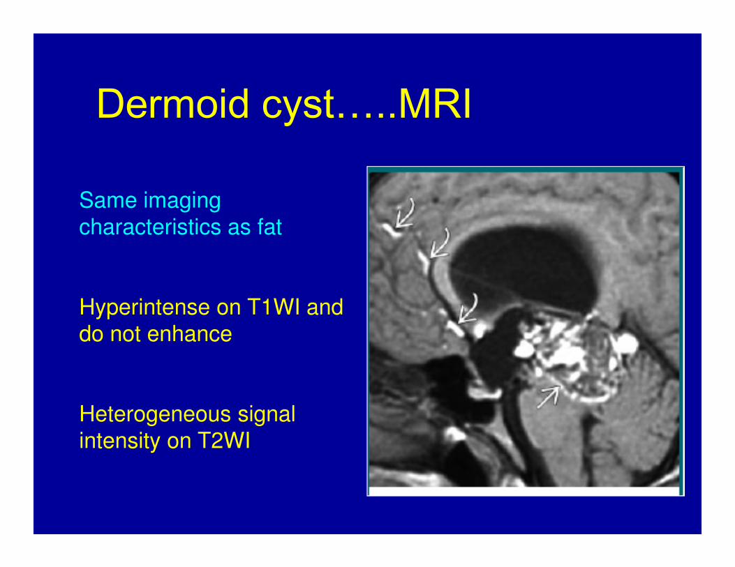
Dermoid cyst…..MRISame imaging
characteristics as fat
Hyperintense on T1WI and
do not enhance
Heterogeneous signal
intensity on T2WI
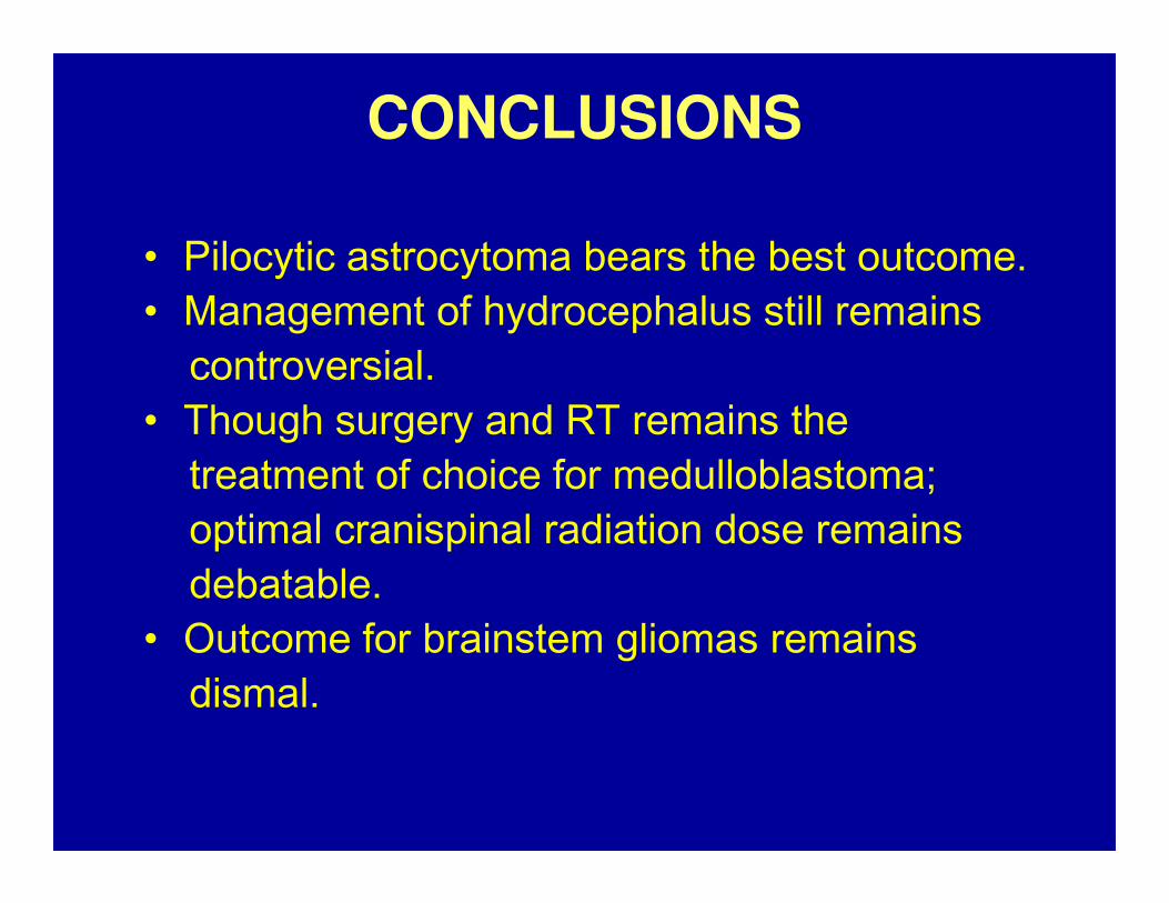
CONCLUSIONS
• Pilocytic astrocytoma bears the best outcome.• Management of hydrocephalus still remains
controversial.• Though surgery and RT remains the
treatment of choice for medulloblastoma; treatment of choice for medulloblastoma; optimal cranispinal radiation dose remains debatable.
• Outcome for brainstem gliomas remains dismal.

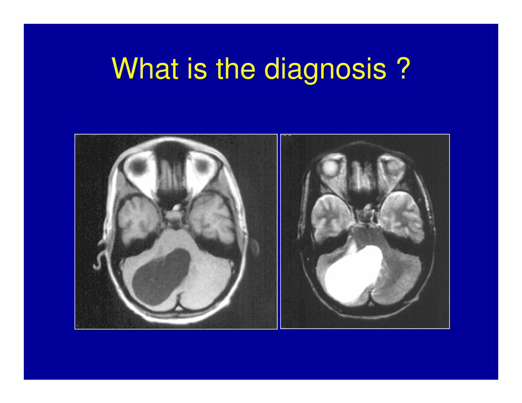
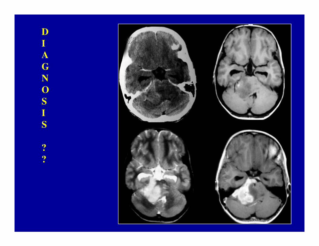
What is the diagnosis ?
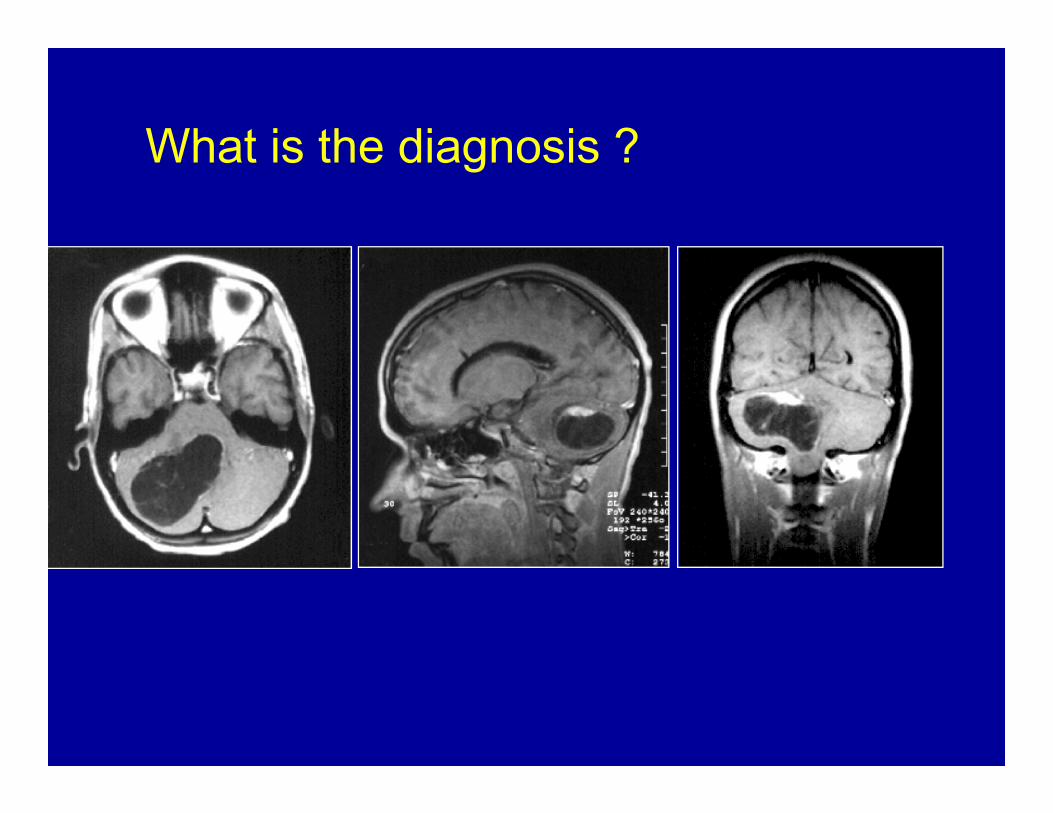
What is the diagnosis ?
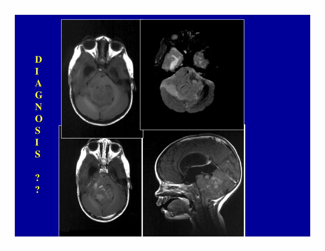
D
I
A
G
N
O
SS
I
S
?
?
D
I
A
G
N
O
S
I
SS
?
?
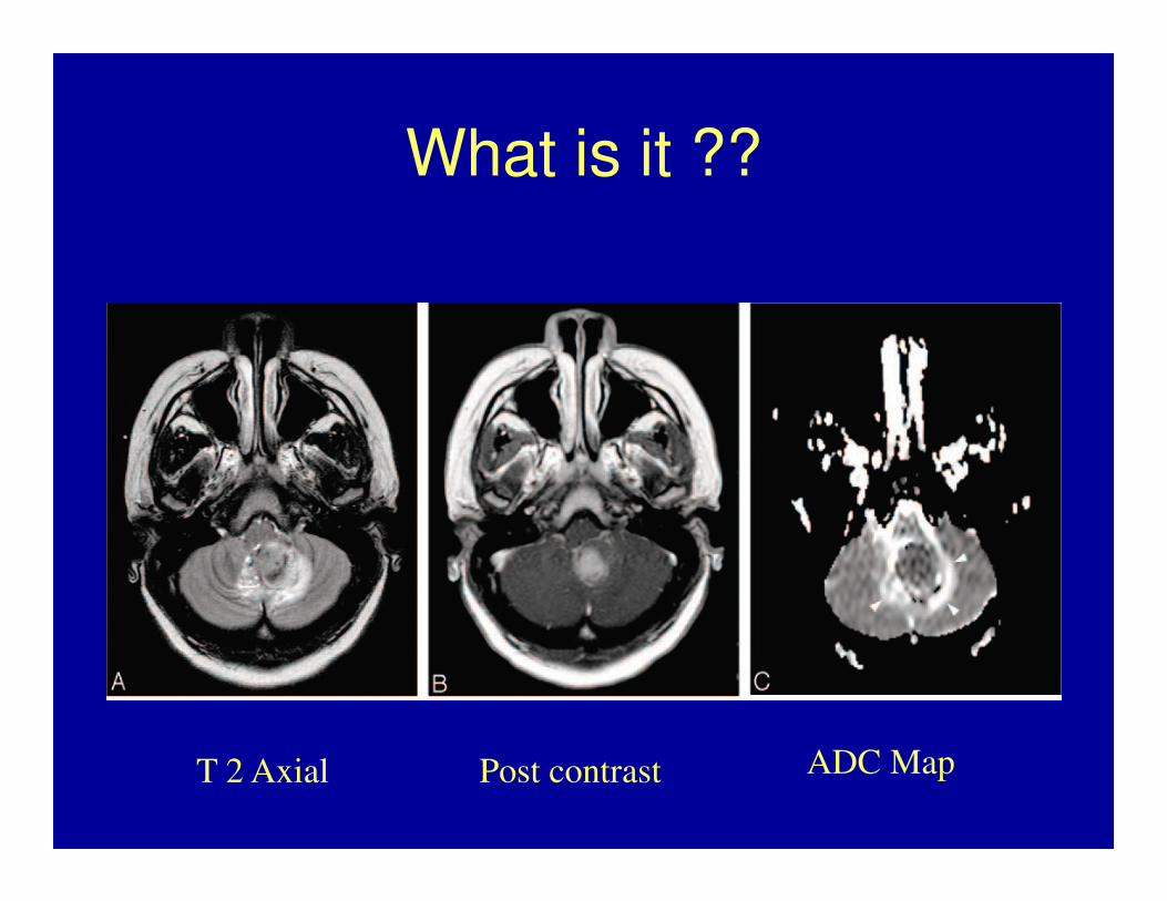
What is it ??
Post contrast T 2 Axial ADC Map
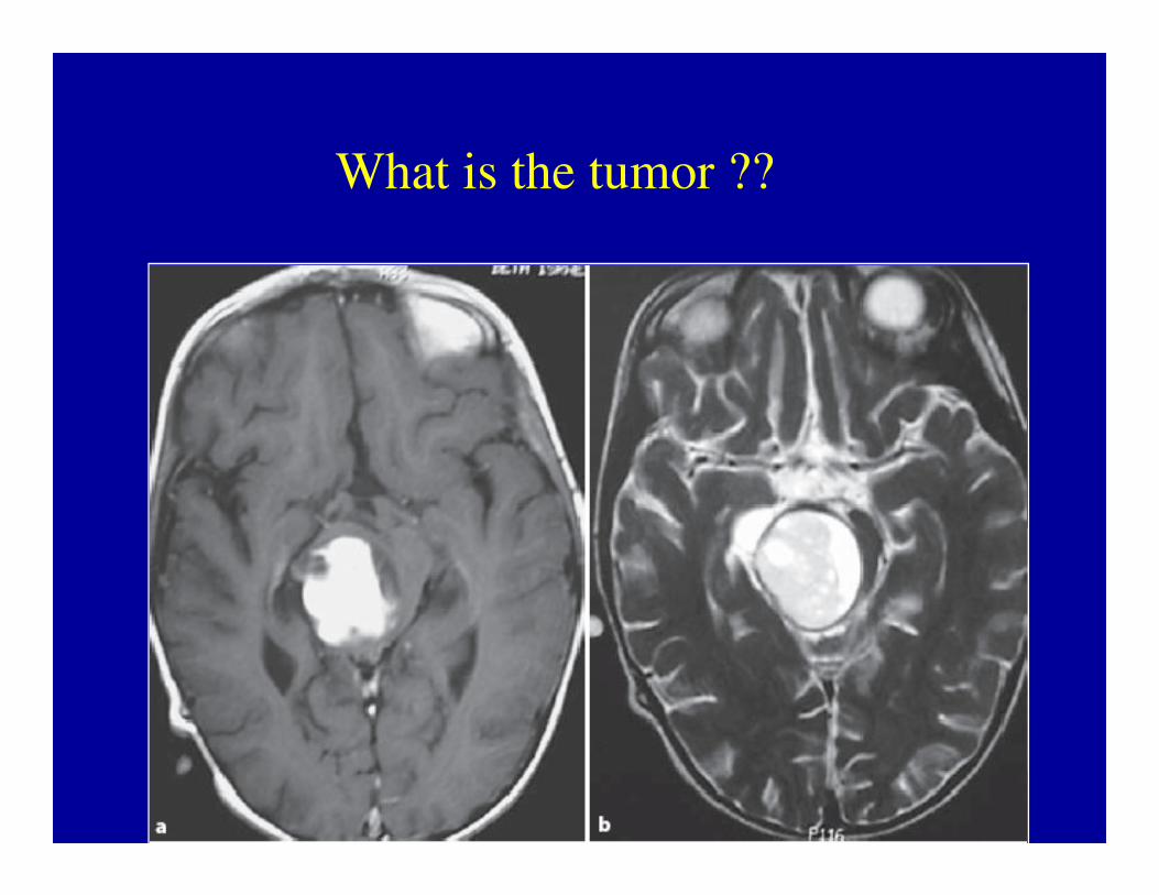
What is the tumor ??
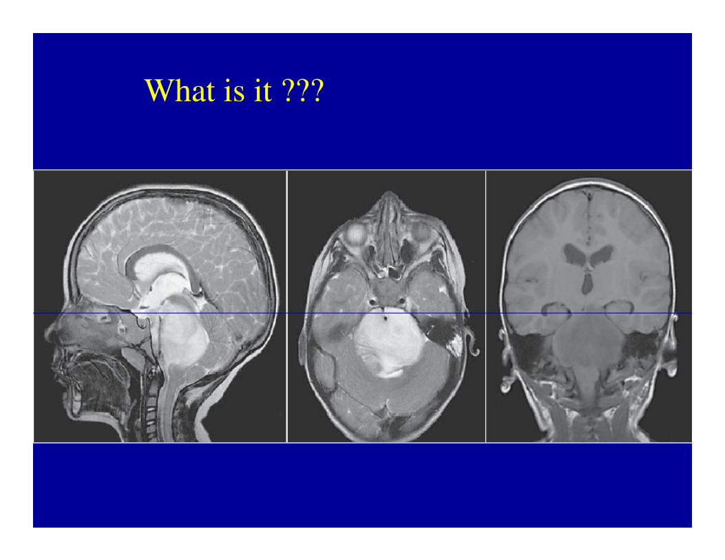
What is it ???