diagnosis and management of cardiac … · of cardiac tamponade and constrictive pericarditis...
TRANSCRIPT
DIAGNOSIS AND MANAGEMENT OF CARDIAC TAMPONADE AND CONSTRICTIVE PERICARDITIS ACCORDING TO THE ESC GUIDELINES ON PERICARDIAL DISEASE
Professor Dr. Bernhard Maisch
Philipps-University and HGZ Marburg
Disclosures
• Prof. Maisch holds several patents for pericardial access
Learning objectives
• Aims
-The participant should be able to diagnose the major pericardialdisease entities.
• Objectives
-is aware of the pathology, etiology and pathophysiology underlyingthe different pericardial syndromes.
- interprets relevant diagnostic measures such as relevant laboratoryinvestigations and imaging methods e.g. echocardiography for thediagnosis of dry pericarditis, pericardial effusion with and withouttamponade, constrictive and constrictive-effusive pericarditis byechocardiography and MRI
-knows medical (symptomatic and etiologcal) and interventionaltreatment options (pericardiocentesis, pericardioscopy, surgery)
-respects the limits of management in his practice and be able torefer the individual patient to specialized centers with a heartteam.
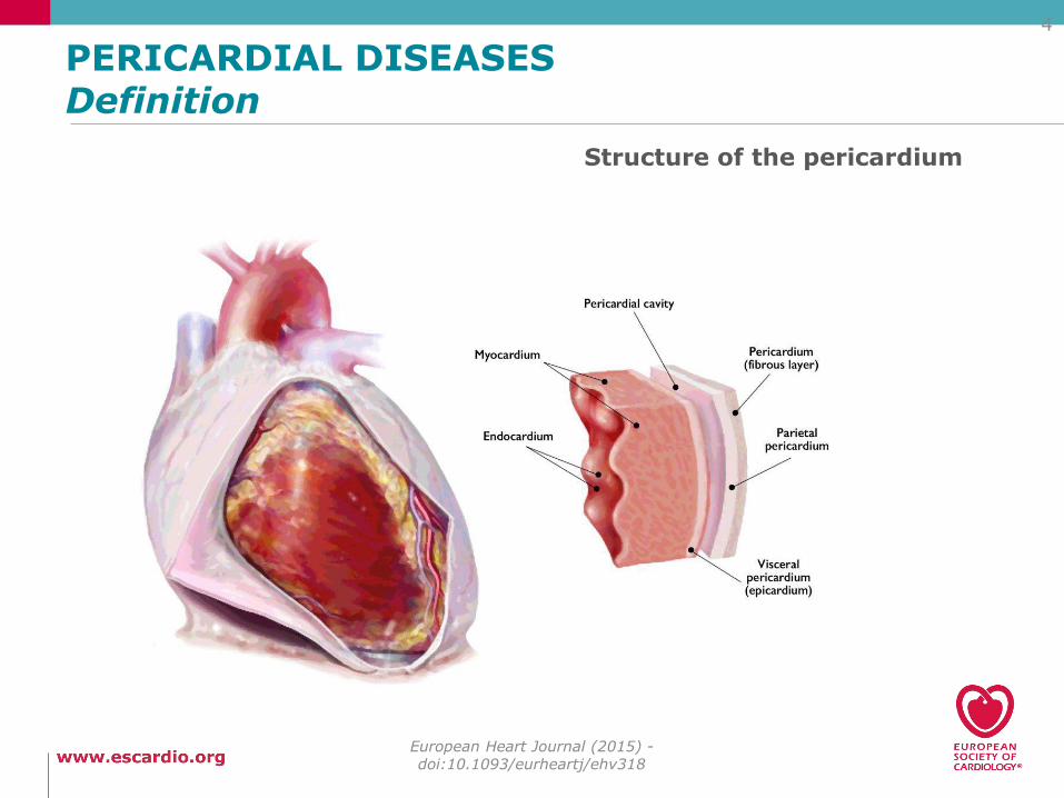
PERICARDIAL DISEASESDefinition
European Heart Journal (2015) -doi:10.1093/eurheartj/ehv318
4
Structure of the pericardium

What is new in the 2015 ESC guidelines since 2004?
5
1) Triage strategy in pericarditis and pericardial effusion.
2) Specific diagnostic criteria for acute and recurrent pericarditis.
3) Update in immunopathogenesis.
4) Multimodality imaging
5) Enlarged epidemiologic data pool
2004 2015
What is new in the 2015 ESC guidelines since 2004?
European Heart Journal (2015) -doi:10.1093/eurheartj/ehv318
6
6. Multicenter treatment trials
7. New treatment options in refractory pericarditis.
8. Prospective and retrospective studies (>100 patients) on prognosis.
9. Imaging of (reversible) pericardial inflammation.
10. Age and gender specific data
Which is the most frequently mentionedcause for a pericardial effusion in
internal medicine ?
a) Idiopathic pericarditis
b) Viral pericarditis
c) malignant pericarditis
d) Radiation induced pericarditis
Correct is a)
But is this a satisfying response ?
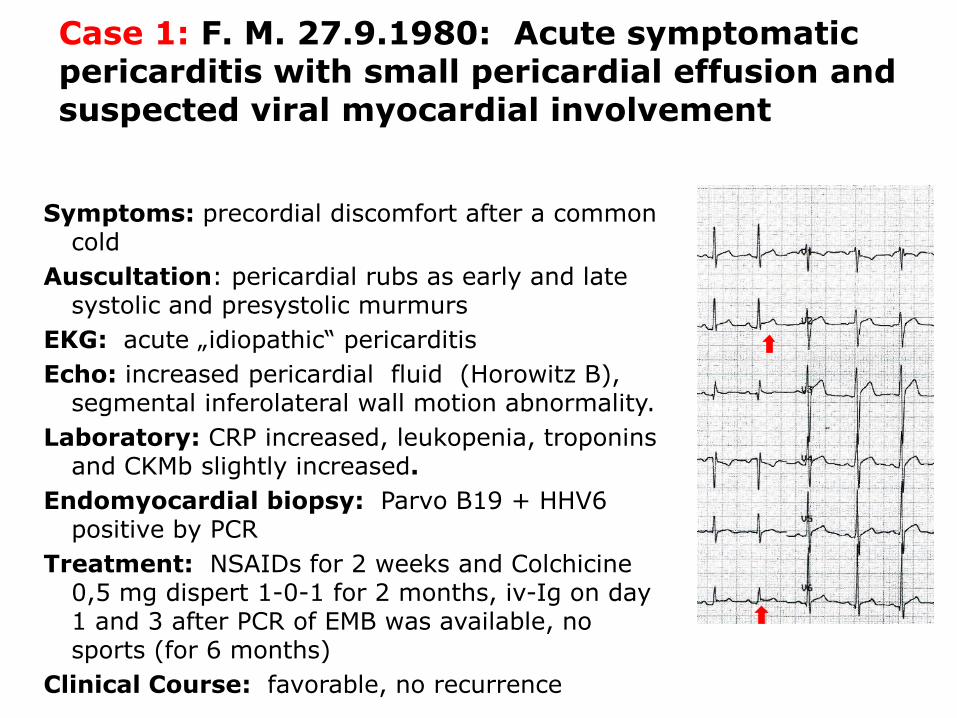
Case 1: F. M. 27.9.1980: Acute symptomaticpericarditis with small pericardial effusion and suspected viral myocardial involvement
Symptoms: precordial discomfort after a commoncold
Auscultation: pericardial rubs as early and latesystolic and presystolic murmurs
EKG: acute „idiopathic“ pericarditis
Echo: increased pericardial fluid (Horowitz B), segmental inferolateral wall motion abnormality.
Laboratory: CRP increased, leukopenia, troponinsand CKMb slightly increased.
Endomyocardial biopsy: Parvo B19 + HHV6 positive by PCR
Treatment: NSAIDs for 2 weeks and Colchicine0,5 mg dispert 1-0-1 for 2 months, iv-Ig on day1 and 3 after PCR of EMB was available, nosports (for 6 months)
Clinical Course: favorable, no recurrence
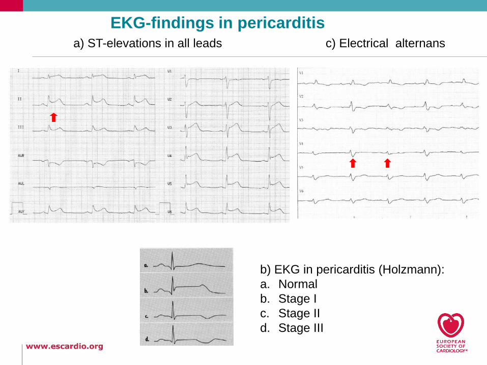
a) ST-elevations in all leads
b) EKG in pericarditis (Holzmann):
a. Normal
b. Stage I
c. Stage II
d. Stage III
c) Electrical alternans
EKG-findings in pericarditis
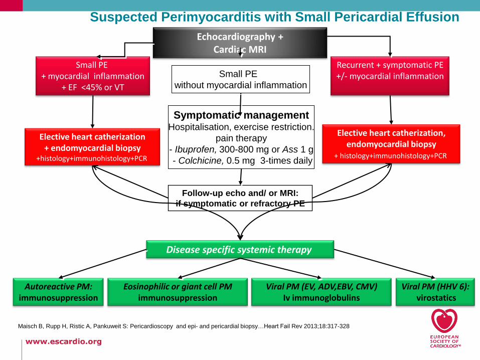
Suspected Perimyocarditis with Small Pericardial Effusion
Echocardiography + Cardiac MRI
Symptomatic managementHospitalisation, exercise restriction.
pain therapy
- Ibuprofen, 300-800 mg or Ass 1 g
- Colchicine, 0.5 mg 3-times daily
Small PE + myocardial inflammation
+ EF <45% or VT
Recurrent + symptomatic PE+/- myocardial inflammationSmall PE
without myocardial inflammation
Follow-up echo and/ or MRI:
if symptomatic or refractory PE
Elective heart catherization+ endomyocardial biopsy
+histology+immunohistology+PCR
Elective heart catherization,endomyocardial biopsy
+ histology+immunohistology+PCR
Autoreactive PM:immunosuppression
Eosinophilic or giant cell PMimmunosuppression
Viral PM (EV, ADV,EBV, CMV)Iv immunoglobulins
Disease specific systemic therapy
Viral PM (HHV 6):virostatics
Maisch B, Rupp H, Ristic A, Pankuweit S: Pericardioscopy and epi- and pericardial biopsy…Heart Fail Rev 2013;18:317-328
11
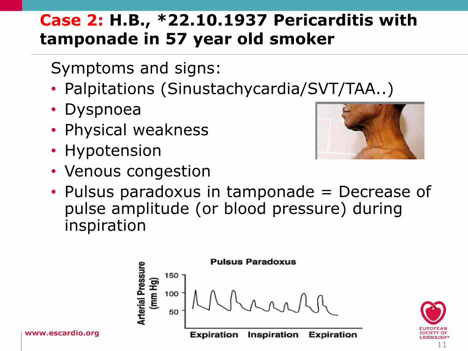
Case 2: H.B., *22.10.1937 Pericarditis withtamponade in 57 year old smoker
Symptoms and signs:
• Palpitations (Sinustachycardia/SVT/TAA..)
• Dyspnoea
• Physical weakness
• Hypotension
• Venous congestion
• Pulsus paradoxus in tamponade = Decrease of pulse amplitude (or blood pressure) duringinspiration
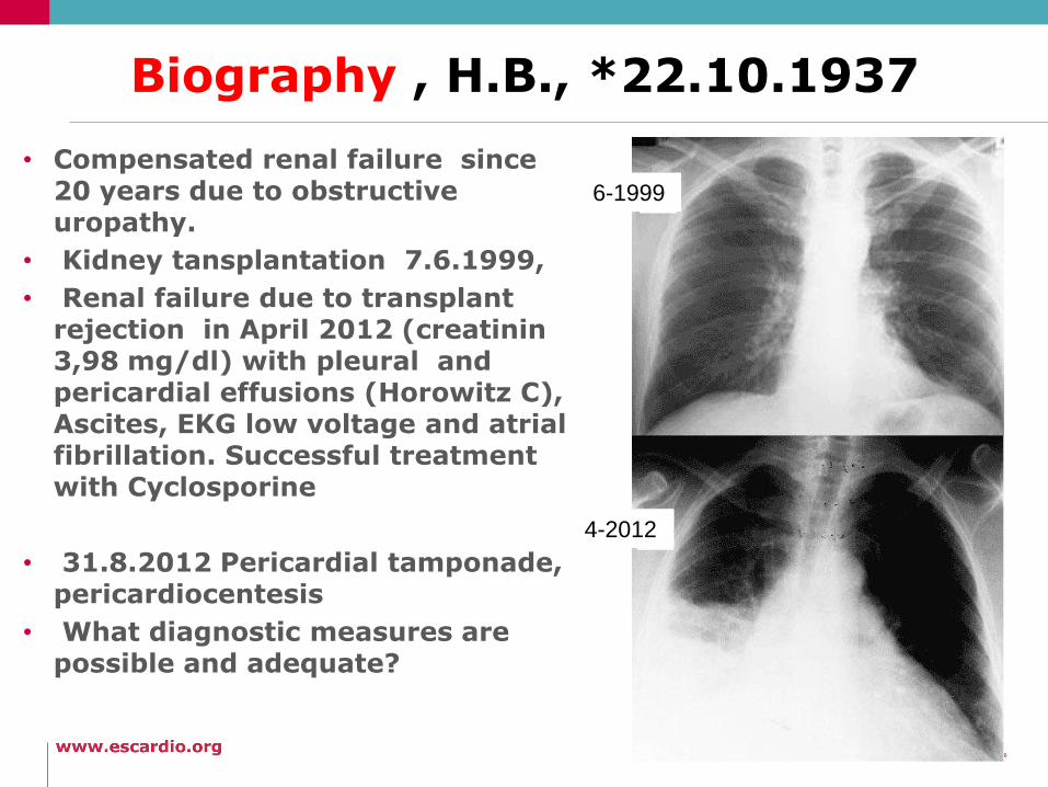
Biography , H.B., *22.10.1937
• Compensated renal failure since20 years due to obstructiveuropathy.
• Kidney tansplantation 7.6.1999,
• Renal failure due to transplant rejection in April 2012 (creatinin3,98 mg/dl) with pleural and pericardial effusions (Horowitz C), Ascites, EKG low voltage and atrialfibrillation. Successful treatmentwith Cyclosporine
• 31.8.2012 Pericardial tamponade, pericardiocentesis
• What diagnostic measures arepossible and adequate?
6-1999
4-2012
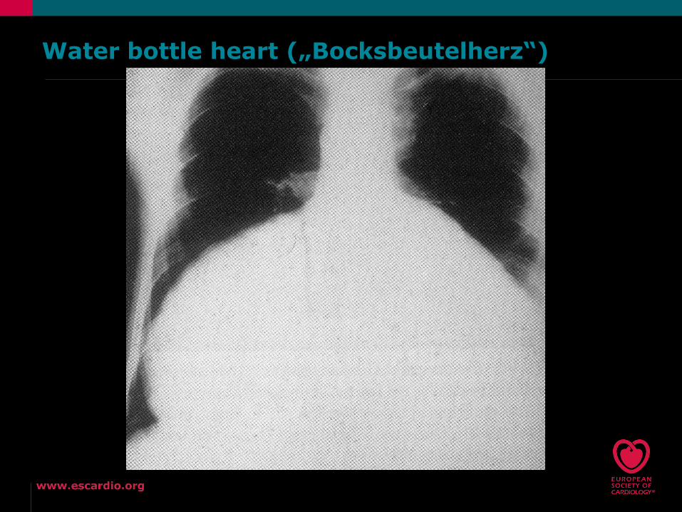
Water bottle heart („Bocksbeutelherz“)
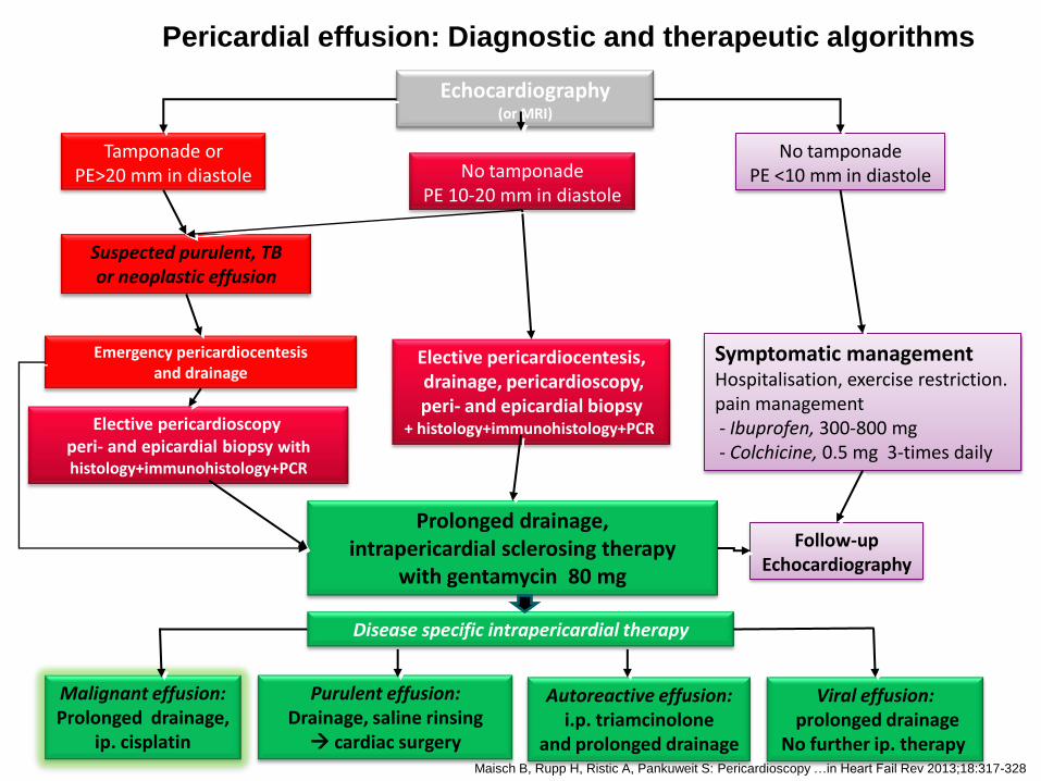
Echocardiography (or MRI)
Symptomatic managementHospitalisation, exercise restriction.pain management- Ibuprofen, 300-800 mg - Colchicine, 0.5 mg 3-times daily
Emergency pericardiocentesisand drainage
Tamponade orPE>20 mm in diastole No tamponade
PE 10-20 mm in diastole
Suspected purulent, TBor neoplastic effusion
No tamponadePE <10 mm in diastole
Elective pericardioscopy, peri- and epicardial biopsy with histology+immunohistology+PCR
Follow-upEchocardiography
Prolonged drainage, intrapericardial sclerosing therapy
with gentamycin 80 mg
Elective pericardiocentesis,drainage, pericardioscopy,peri- and epicardial biopsy
+ histology+immunohistology+PCR
Malignant effusion:Prolonged drainage,
ip. cisplatin
Purulent effusion:Drainage, saline rinsing cardiac surgery
Autoreactive effusion:i.p. triamcinolone
and prolonged drainage
Disease specific intrapericardial therapy
Viral effusion:prolonged drainage
No further ip. therapy
Pericardial effusion: Diagnostic and therapeutic algorithms
Maisch B, Rupp H, Ristic A, Pankuweit S: Pericardioscopy …in Heart Fail Rev 2013;18:317-328
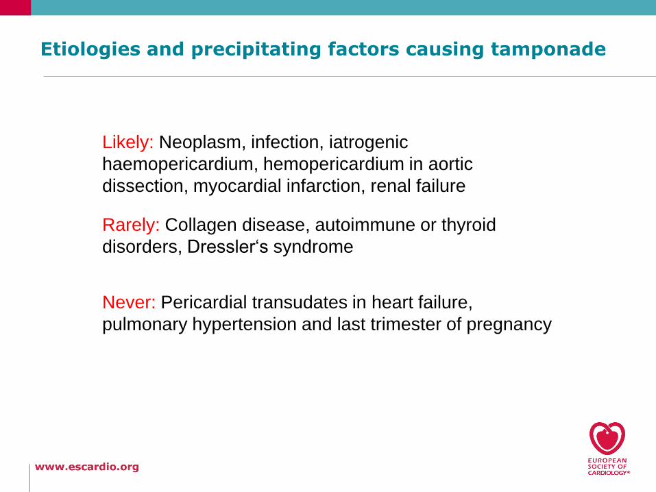
Etiologies and precipitating factors causing tamponade
Likely: Neoplasm, infection, iatrogenic
haemopericardium, hemopericardium in aortic
dissection, myocardial infarction, renal failure
Rarely: Collagen disease, autoimmune or thyroid
disorders, Dressler‘s syndrome
Never: Pericardial transudates in heart failure,
pulmonary hypertension and last trimester of pregnancy
17
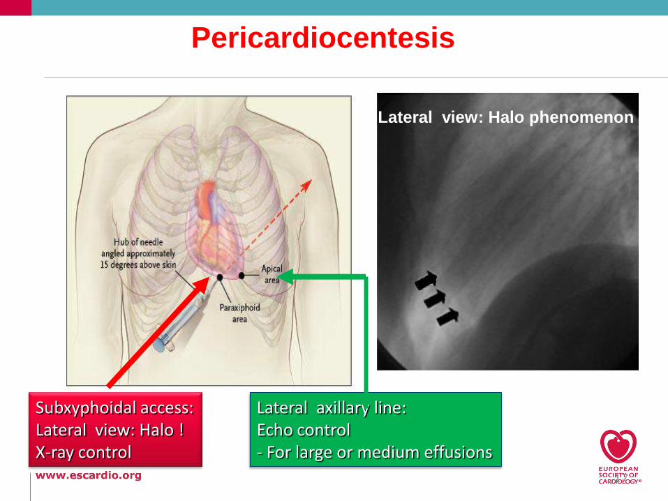
Pericardiocentesis
Subxyphoidal access:Lateral view: Halo !X-ray control
Lateral axillary line:Echo control- For large or medium effusions
Lateral view: Halo phenomenon
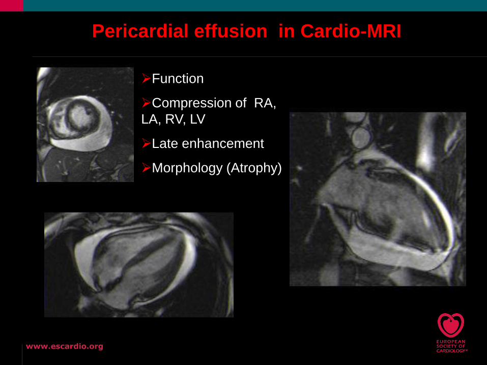
Function
Compression of RA,
LA, RV, LV
Late enhancement
Morphology (Atrophy)
Pericardial effusion in Cardio-MRI
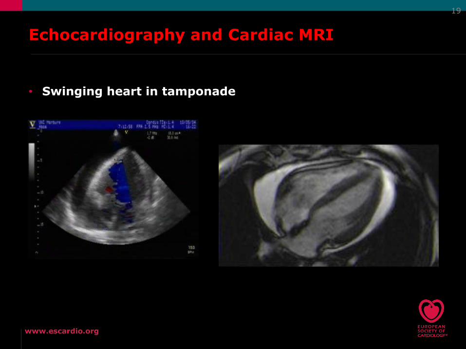
Echocardiography and Cardiac MRI
• Swinging heart in tamponade
19
20
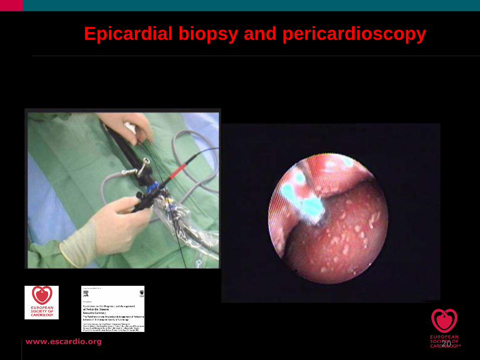
Epicardial biopsy and pericardioscopy
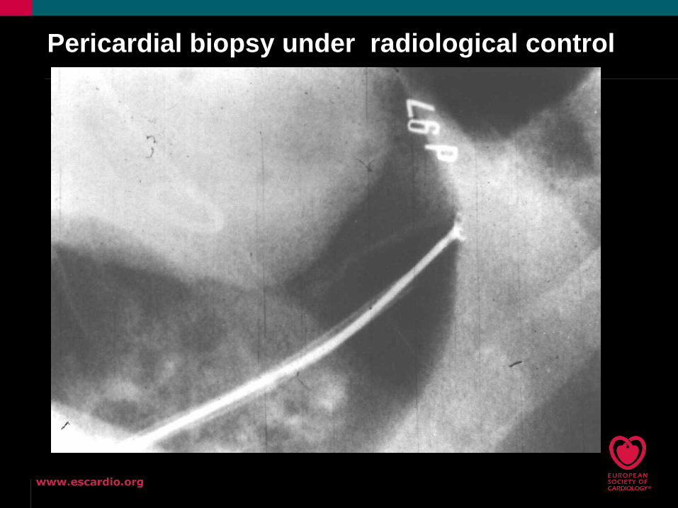
Pericardial biopsy under radiological control
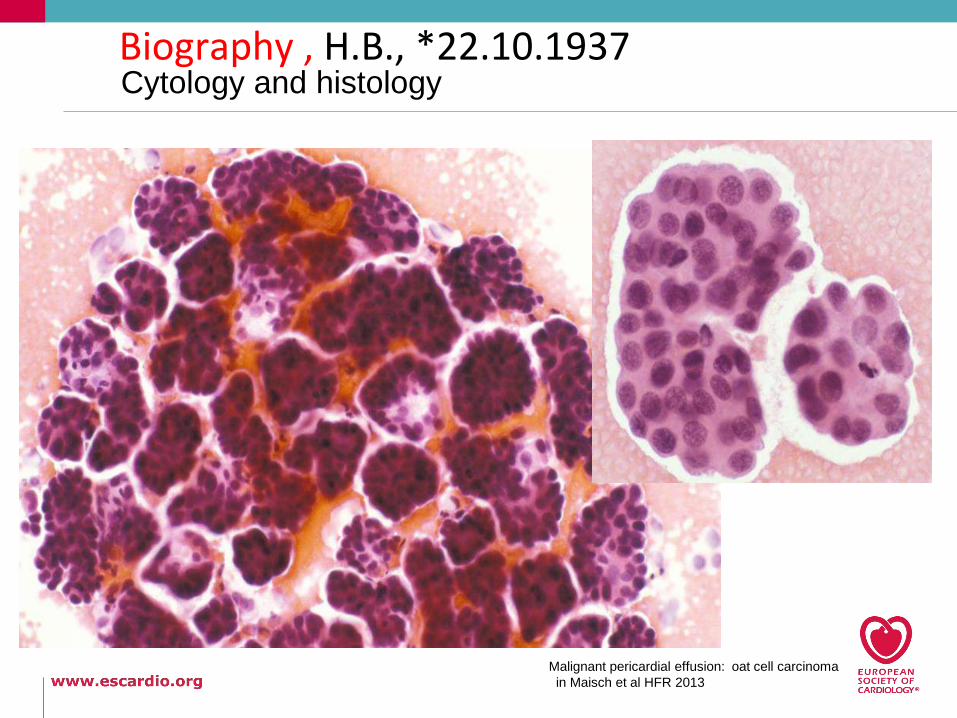
Cytology and histology
Malignant pericardial effusion: oat cell carcinoma
in Maisch et al HFR 2013
Biography , H.B., *22.10.1937
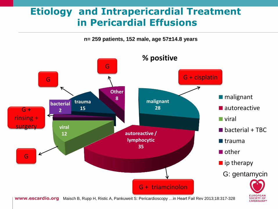
n= 259 patients, 152 male, age 57±14.8 years
malignant 28
autoreactive / lymphocytic
35
viral12
bacterial 2
trauma15
Other8
0
% positive
malignant
autoreactive
viral
bacterial + TBC
trauma
other
ip therapy
G + cisplatin
G
G + triamcinolon
G
G + rinsing + surgery
G
G: gentamycin
Maisch B, Rupp H, Ristic A, Pankuweit S: Pericardioscopy …in Heart Fail Rev 2013;18:317-328
Etiology and Intrapericardial Treatment in Pericardial Effusions
24
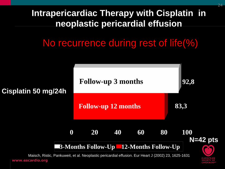
92,8
83,3
Cisplatin 50 mg/24h
0 20 40 60 80 100
3-Months Follow-Up 12-Months Follow-Up
Follow-up 12 months
Follow-up 3 months
No recurrence during rest of life(%)
N=42 pts
Intrapericardiac Therapy with Cisplatin in
neoplastic pericardial effusion
Maisch, Ristic, Pankuweit, et al. Neoplastic pericardial effusion. Eur Heart J (2002) 23, 1625-1631
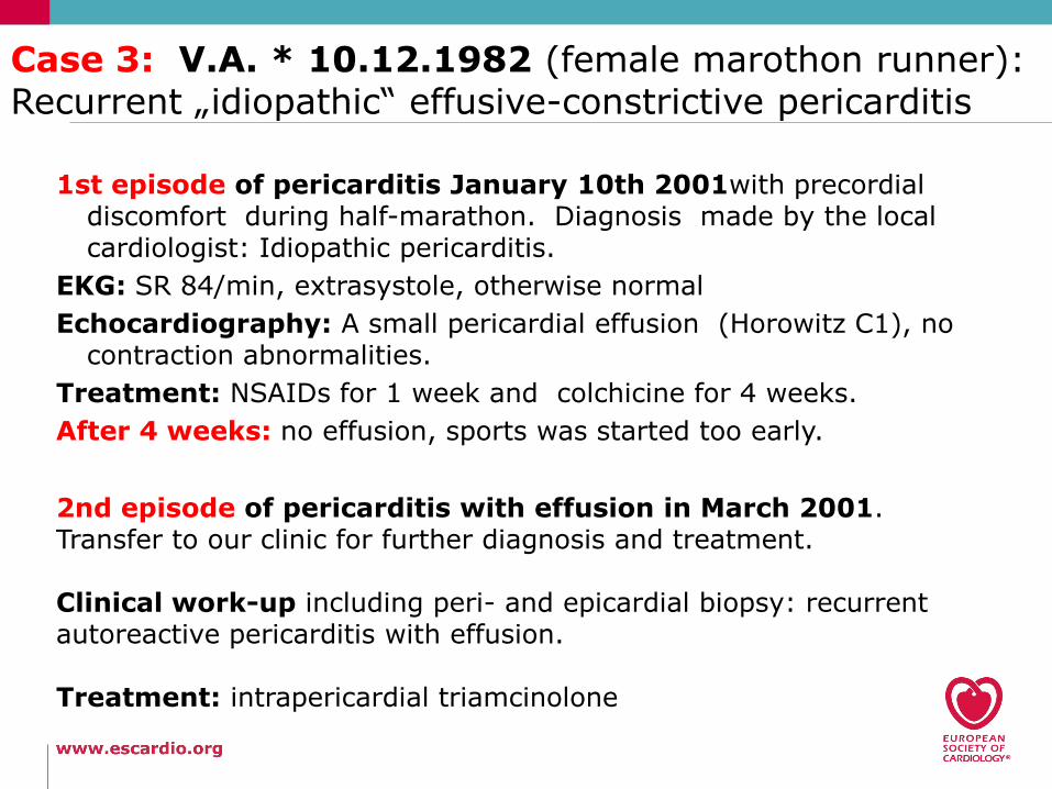
Case 3: V.A. * 10.12.1982 (female marothon runner): Recurrent „idiopathic“ effusive-constrictive pericarditis
1st episode of pericarditis January 10th 2001with precordialdiscomfort during half-marathon. Diagnosis made by the localcardiologist: Idiopathic pericarditis.
EKG: SR 84/min, extrasystole, otherwise normal
Echocardiography: A small pericardial effusion (Horowitz C1), nocontraction abnormalities.
Treatment: NSAIDs for 1 week and colchicine for 4 weeks.
After 4 weeks: no effusion, sports was started too early.
2nd episode of pericarditis with effusion in March 2001. Transfer to our clinic for further diagnosis and treatment.
Clinical work-up including peri- and epicardial biopsy: recurrentautoreactive pericarditis with effusion.
Treatment: intrapericardial triamcinolone
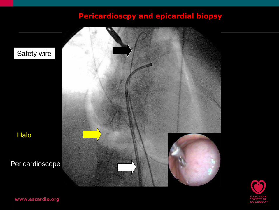
Pericardioscpy and epicardial biopsy
Halo
Pericardioscope
Safety wire
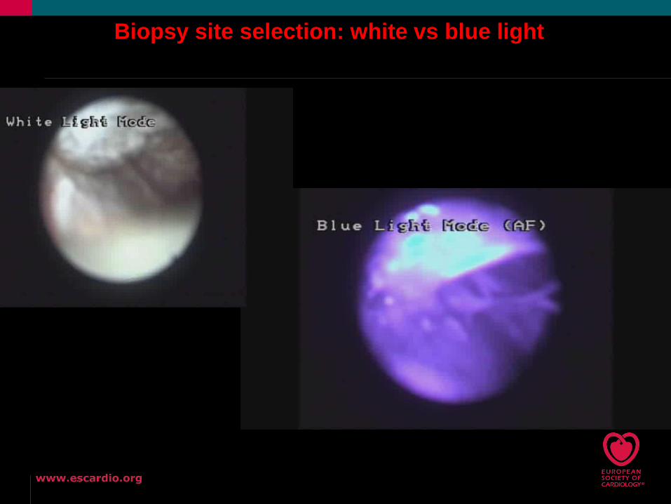
Biopsy site selection: white vs blue light
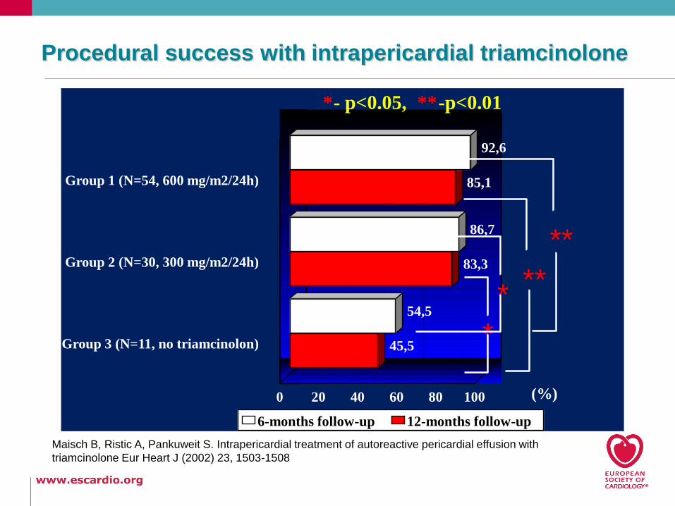
Procedural success with intrapericardial triamcinolone
92,6
86,7
54,5
85,1
83,3
45,5
Group 1 (N=54, 600 mg/m2/24h)
Group 2 (N=30, 300 mg/m2/24h)
Group 3 (N=11, no triamcinolon)
0 20 40 60 80 100
6-months follow-up 12-months follow-up
(%)
*
***
**
*- p<0.05, **-p<0.01
Maisch B, Ristic A, Pankuweit S. Intrapericardial treatment of autoreactive pericardial effusion with
triamcinolone Eur Heart J (2002) 23, 1503-1508
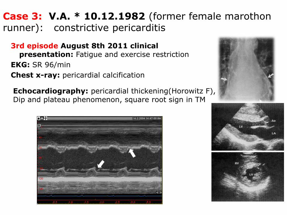
Case 3: V.A. * 10.12.1982 (former female marothonrunner): constrictive pericarditis
3rd episode August 8th 2011 clinicalpresentation: Fatigue and exercise restriction
EKG: SR 96/min
Chest x-ray: pericardial calcification
Echocardiography: pericardial thickening(Horowitz F),Dip and plateau phenomenon, square root sign in TM
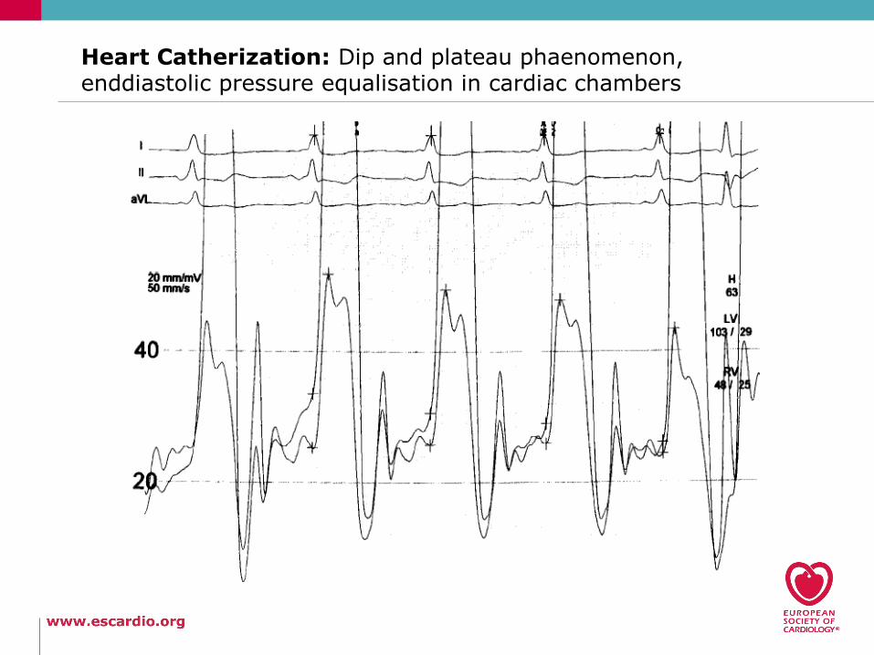
Heart Catherization: Dip and plateau phaenomenon, enddiastolic pressure equalisation in cardiac chambers
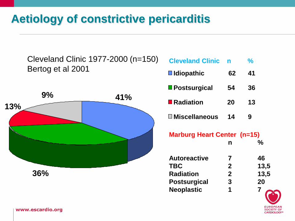
Cleveland Clinic 1977-2000 (n=150)
Bertog et al 2001
41%
36%
13%
9%
Idiopathic 62 41
Postsurgical 54 36
Radiation 20 13
Miscellaneous 14 9
Cleveland Clinic n %
Marburg Heart Center (n=15)
n %
Autoreactive 7 46
TBC 2 13,5
Radiation 2 13,5
Postsurgical 3 20
Neoplastic 1 7
Aetiology of constrictive pericarditis
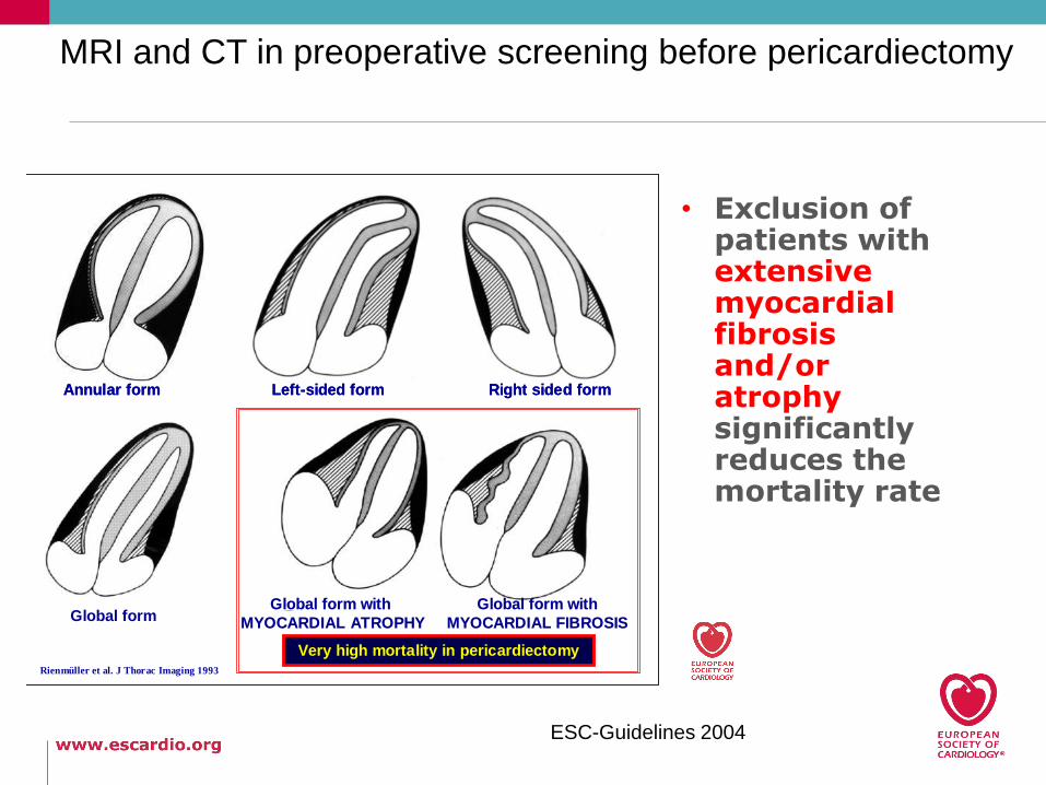
Rienmüller et al. J Thorac Imaging 1993
Annular form Left-sided form Right sided form
Global form Global form with Global form with
MYOCARDIAL ATROPHYMYOCARDIAL ATROPHY
Global form withGlobal form with
MYOCARDIAL FIBROSISMYOCARDIAL FIBROSIS
Very high mortality in Very high mortality in pericardiectomypericardiectomyRienmüller et al. J Thorac Imaging 1993
Annular form Left-sided form Right sided formAnnular form Left-sided form Right sided form
Global form Global form with Global form with
MYOCARDIAL ATROPHYMYOCARDIAL ATROPHY
Global form withGlobal form with
MYOCARDIAL FIBROSISMYOCARDIAL FIBROSIS
Very high mortality in Very high mortality in pericardiectomypericardiectomy
• Exclusion of patients with extensive myocardial fibrosis and/or atrophy significantly reduces the mortality rate
ESC-Guidelines 2004
MRI and CT in preoperative screening before pericardiectomy
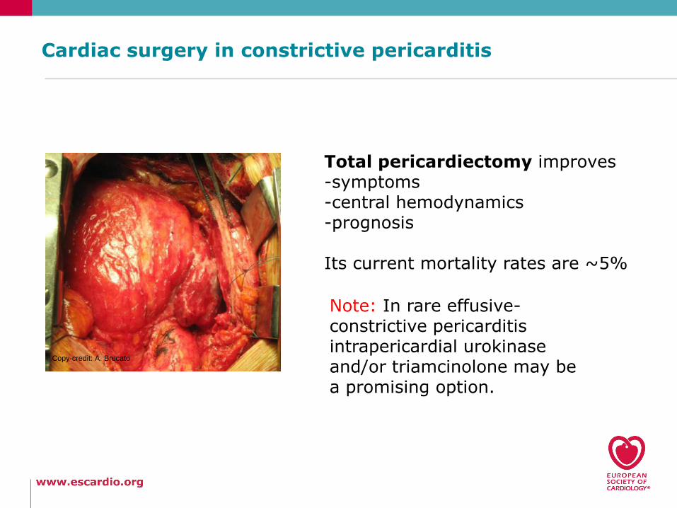
Cardiac surgery in constrictive pericarditis
Copy-credit: A. Brucato
Total pericardiectomy improves-symptoms-central hemodynamics-prognosis
Its current mortality rates are ~5%
Note: In rare effusive-constrictive pericarditisintrapericardial urokinaseand/or triamcinolone may bea promising option.
36
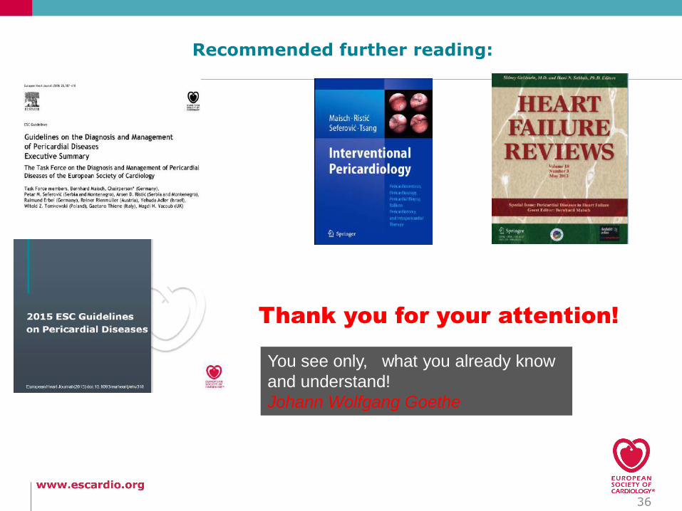
Thank you for your attention!
You see only, what you already know
and understand!
Johann Wolfgang Goethe
Recommended further reading: