diabetic foot ulcers – prevention and treatment · diabetic foot ulcers – prevention and...
TRANSCRIPT

Biatain® – the simple choice
Diabetic foot ulcers – prevention and treatment A Coloplast quick guide
Table of content
Diabetic foot ulcers have a considerable negative impact on patients’ lives, and are highly susceptible to infection that all too often leads to amputation. It is essential that diabetic foot ulcers receive the best possible wound management. Successfully treating a diabetic foot ulcer requires a comprehensive understanding of the wound: its cause, progression, risk, and treatment. But more than this, it takes a cross functional approach, where the patient also has an active role in the treatment process.
The information provided here is intended as a general guideline. Please consult diabetic foot ulcer guidelines applicable in your area. For further study, please refer to the International Consensus on the Diabetic Foot, 2011.2
We hope that this quick guide will help you diagnose, assess and treat diabetic foot ulcers in clinical practice, as well as identify opportunities for prevention and minimising the risk of infection and amputation.
Developed byFaculty panel: Dr. Christian Münter, Germany; Professor Patricia Price, UK; Wilma Ruigrok van der Werven, MA, RN, Netherlands; Professor Gary Sibbald, Canada
Review panel: Patricia Coutts, RN, Canada; Mike Edmonds, Consultant Diabetologist, UK; Professor Keith Harding UK; Maria Mousley, AHP, Consultant, Podiatrist, UK
This Coloplast quick guide was updated in March 2012 in collaboration with Dr. Christian Münter.
Introduction
Introduction ................................................................................... 3The diabetic foot – a clinical challenge ........................................... 5Pathway to clinical care and clinical evidence ................................ 6How to prevent DFU’s .................................................................... 7
Prevention and education ........................................................ 7Prevention of ulcer formation ................................................... 8An interprofessional team approach ......................................... 9
The patient’s role ......................................................................... 10Consider the whole patient to ensure effective care ..................... 11How to diagnose and assess a diabetic foot ulcer ....................... 12
“The VIPS” of diabetic foot management ................................ 12Local wound assessment ...................................................... 13Types of neuropathy .............................................................. 1410g monofilament testing ...................................................... 15Areas at risk for neuropathic, ischaemic and neuoro-ischaemic ulcers ........................................................ 16Clinical symptoms of neuropathic and ischmaemic foot ulcers ................................................... 17Ulcer assessment .................................................................. 18Wound bed ............................................................................ 19Superficial and deep infection symptoms ............................... 20Wagner classification ............................................................. 21
How to treat a diabetic foot ulcer ................................................. 22Treatment of diabetic foot ulcers ............................................ 22Local wound treatment .......................................................... 23Coloplast solutions for diabetic foot ulcers ............................. 24Coloplast antimicrobial dressings for infected diabetic foot ulcers and ulcers at risk of infection ................... 26
References .................................................................................. 28Biatain® – superior absorption for faster healing ........................... 30Other Coloplast products for diabetic foot ulcers ......................... 32
2 3
“I marvel that society would pay a surgeon a fortune to remove a person’s leg – but nothing to save it!”George Bernard Shaw
Diabetes is a serious chronic disease that needs attention. Approximately 15% of all people with diabetes will be affected by a foot ulcer during their lifetime.1
Diabetic foot ulcers (DFUs) often co-exist with vascular insufficien-cy and are the major cause of gangrene and amputation in people with diabetes. Risk of developing diabetic foot ulcers is greatly in-creased by reduced sensation and blood pressure.
Diabetic foot ulcers represent a huge risk to the patient’s quality of life, escalating wound/infection management and costs, and account for a large proportion of all national healthcare budgets
· Five-year recurrence rates of foot ulcers are 70%2
· Up to 85% of all amputations in relation to people with diabetes are preceded by a foot ulcer1-2
· People with diabetes with one lower limb amputation have a 50% risk of developing a serious lesion in the second limb within 2 years3
· People with diabetes have a 50% mortality rate in the 5 years following the initial amputation4
It is possible to reduce amputation rates by 49-85% through a care strategy that combines prevention, the interprofessional diabetes care team, appropriate organisation, close monitoring and education.1
The diabetic foot – a clinical challenge
4 5

Diabetic foot ulcers
Evidence-based wound management
Patient-centred concerns
Local wound care Treat the cause
Clinicalresearch
Real life studiesHealth economic
analysis
Painmanagement
Tissuedebridement
Bacterialbalance
Exudatemanagement
Pathway to clinical care and clinical evidence
Prevention and education
“49-85% of all diabetic foot related problems are preventable.” Spraul, M., 2000.6
“This can be achieved through a combination of good foot care, provided by an interprofessional diabetes care team, and appropriate education for people with diabetes.”Modified from Bakker, K. et al., 2005.1
“Education of patients, carers, and healthcare providers is an essential component of an effective, interprofessional team approach, …but effective systems and structures for screening, provision of chiropody and footwear, and prompt treatment when required must be in place.”Modified from Spraul, M., 2000.6
“The most important aspects, for example, danger signs which require prompt action by the patient, should be summarized and repeated.”Spraul, M., 2000.6
“Successful diagnosis and treatment of patients with chronic wounds involve holistic care and a team approach. The integration of the work of an interprofessional care team that includes doctors, nurses and allied health professionals with the patient, family and caregivers offers an optimal formula for achieving wound resolution.”Sibbald, R.G., et al, 2001.18
How to prevent DFUs
6 7
Prevention of ulcer formation
People with diabetes must inspect their feet regularly, or have a family member or care provider do it on their behalf. Daily inspection is the foundation of diabetic foot ulcer prevention. All wounds and sores should be taken seriously early on.
Regular, gentle cleansing with soapy water, followed by the application of topical moisturizers, helps to keep the skin healthy and better able to resist breakdown and injury.
Shoes should be checked to ensure that they fit properly and offer adequate support. Consider athletic/sports shoes and thick, padded socks. Diabetic socks (unrestrictive on circulation) are also available. In the case of foot deformities or special support needs, custom shoes should be considered.
Minor foot injuries and infections, such as cuts, scrapes, blisters and tinea pedis (athletes foot), can be unintentionally worsened by home treatments that impede healing. Patients should be reminded to avoid hot soaks, heating pads and harsh topical agents such as hydrogen peroxide, iodine and astringents. A moist wound environment will help prevent ulcer formation. Minor wounds should be gently cleansed and treated with topical antiseptics. In addition, a physician should inspect any minor wounds that do not heal quickly.
By reinforcing preventive advice and inspecting the patient’s feet at routine follow-ups, the physician can help the patient develop and maintain good foot-care practices.
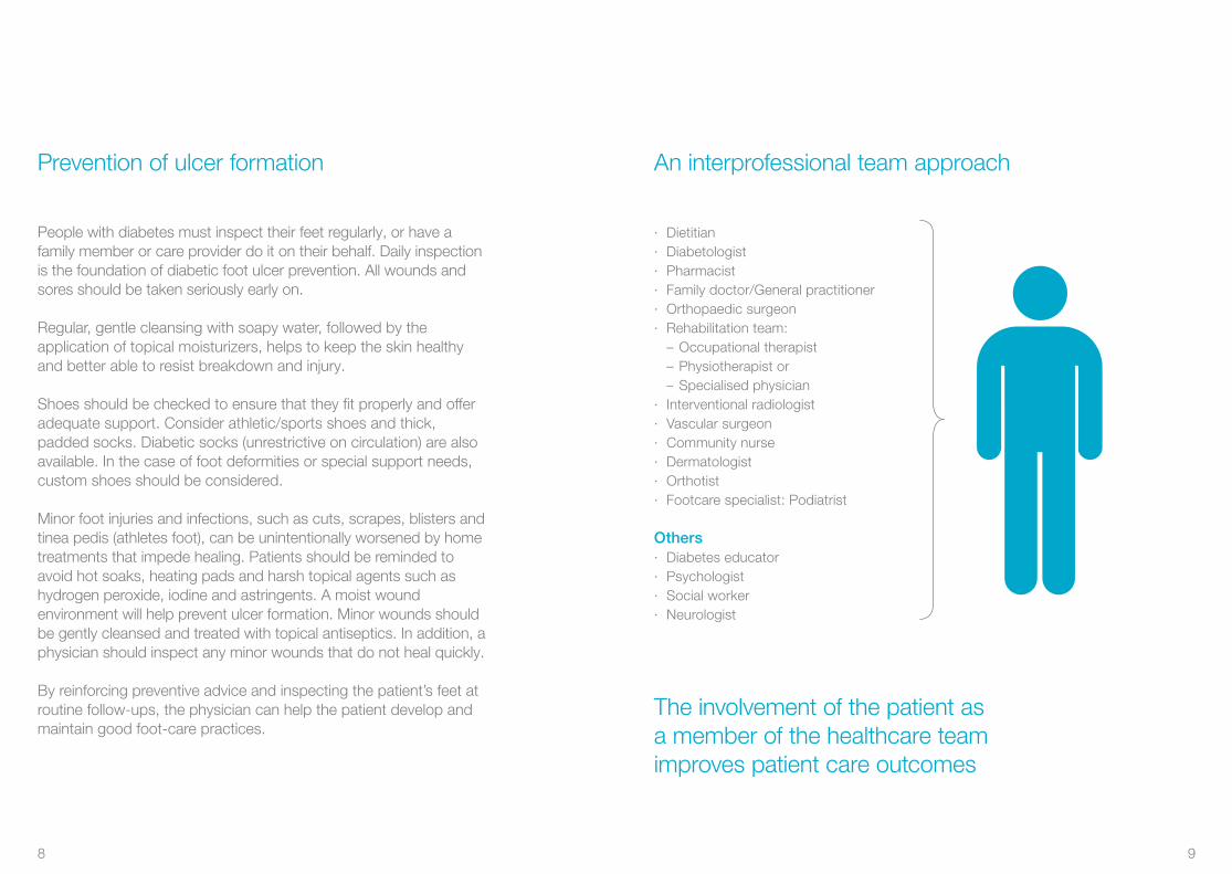
An interprofessional team approach
· Dietitian· Diabetologist· Pharmacist· Family doctor/General practitioner· Orthopaedic surgeon· Rehabilitation team:
– Occupational therapist– Physiotherapist or – Specialised physician
· Interventional radiologist· Vascular surgeon· Community nurse· Dermatologist· Orthotist· Footcare specialist: Podiatrist
Others· Diabetes educator· Psychologist· Social worker· Neurologist
The involvement of the patient as a member of the healthcare team improves patient care outcomes
8 9
Education of patient, family and healthcare providers, such as using an easy to understand patient leaflet for education, must be a priority.
· Any cut or open skin should be treated by a qualified healthcare provider immediately
· Inspect and examine the feet and shoes on a daily basis
· Appropriate footwear
· Nails should be cared for by a qualified foot specialist (podiatrist or related disciplines)
· Dry skin should be treated with appropriate moisturizing, such as (humectant) creams containing urea or lactid acid18
· Fungal infections, especially of the toe webs require topical antifungal agents
Patient self-exam needs to be part ofdiabetic foot care and follow-up
Patients should always remember to remove socks and shoes for regular inspection of both feet
The patient’s role
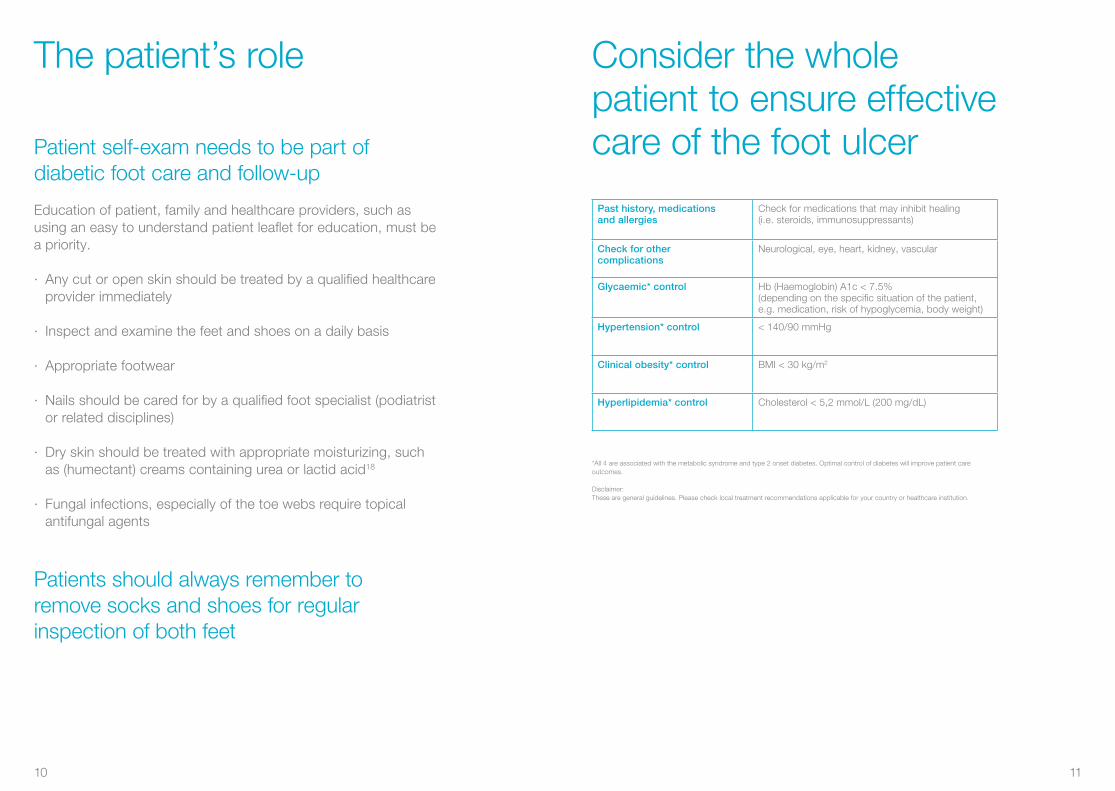
*All 4 are associated with the metabolic syndrome and type 2 onset diabetes. Optimal control of diabetes will improve patient care outcomes.
Disclaimer: These are general guidelines. Please check local treatment recommendations applicable for your country or healthcare institution.
Past history, medicationsand allergies
Check for medications that may inhibit healing (i.e. steroids, immunosuppressants)
Check for othercomplications
Neurological, eye, heart, kidney, vascular
Glycaemic* control Hb (Haemoglobin) A1c < 7.5% (depending on the specific situation of the patient, e.g. medication, risk of hypoglycemia, body weight)
Hypertension* control < 140/90 mmHg
Clinical obesity* control BMI < 30 kg/m2
Hyperlipidemia* control Cholesterol < 5,2 mmol/L (200 mg/dL)
Consider the whole patient to ensure effective care of the foot ulcer
10 11
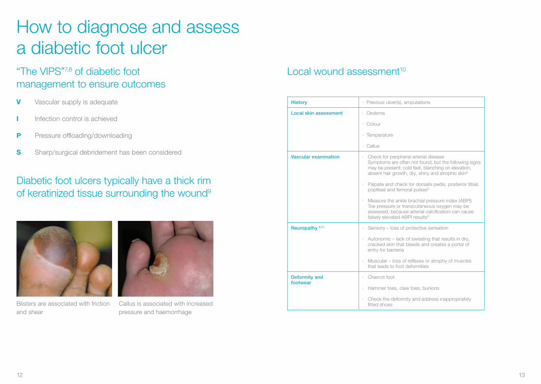
“The VIPS”7,8 of diabetic footmanagement to ensure outcomes
Diabetic foot ulcers typically have a thick rim of keratinized tissue surrounding the wound9
V Vascular supply is adequate
I Infection control is achieved
P Pressure offloading/downloading
S Sharp/surgical debridement has been considered
Callus is associated with increased pressure and haemorrhage
Blisters are associated with friction and shear
How to diagnose and assess a diabetic foot ulcer
Local wound assessment10
History · Previous ulcer(s), amputations
Local skin assessment · Oedema
· Colour
· Temperature
· Callus
Vascular examination · Check for peripheral arterial disease Symptoms are often not found, but the following signs
may be present: cold feet, blanching on elevation, absent hair growth, dry, shiny and atrophic skin9
· Palpate and check for dorsalis pedis, posterior tibial, popliteal and femoral pulses9
· Measure the ankle brachial pressure index (ABPI) Toe pressure or transcutaneous oxygen may be
assessed, because arterial calcification can cause falsely elevated ABPI results9
Neuropathy 8,11 · Sensory – loss of protective sensation
· Autonomic – lack of sweating that results in dry, cracked skin that bleeds and creates a portal of entry for bacteria
· Muscular – loss of reflexes or atrophy of muscles that leads to foot deformities
Deformity andfootwear
· Charcot foot
· Hammer toes, claw toes, bunions
· Check the deformity and address inappropriately fitted shoes
12 13
Types of neuropathy10
Etiology Sensoryneuropathy
Autonomicneuropathy
Motorneuropathy
Characteristics · Loss of protective sensation
· No perception of shoes rubbing or temperature changes
· Reduced sweating results in dry cracked skin
· Increased blood flow leads to a warm foot
· Dysfunction of the motor nerves that control the movement of the foot. Limited joint mobility may increase plantar pressure
· Foot deformities develop
· Hammer toes
Clinicalpresentations
· Unaware of a foot ulcer or lack of discomfort when a wound is being probed
· Dry skin with cracks and fissures
· Bounding pulses
· Dilated dorsal veins
· Warm feet
· High medial longitudinal arch, leading to prominent metatarsal heads and pressure points over the plantar forefoot
· Clawed toes
· Altered gait
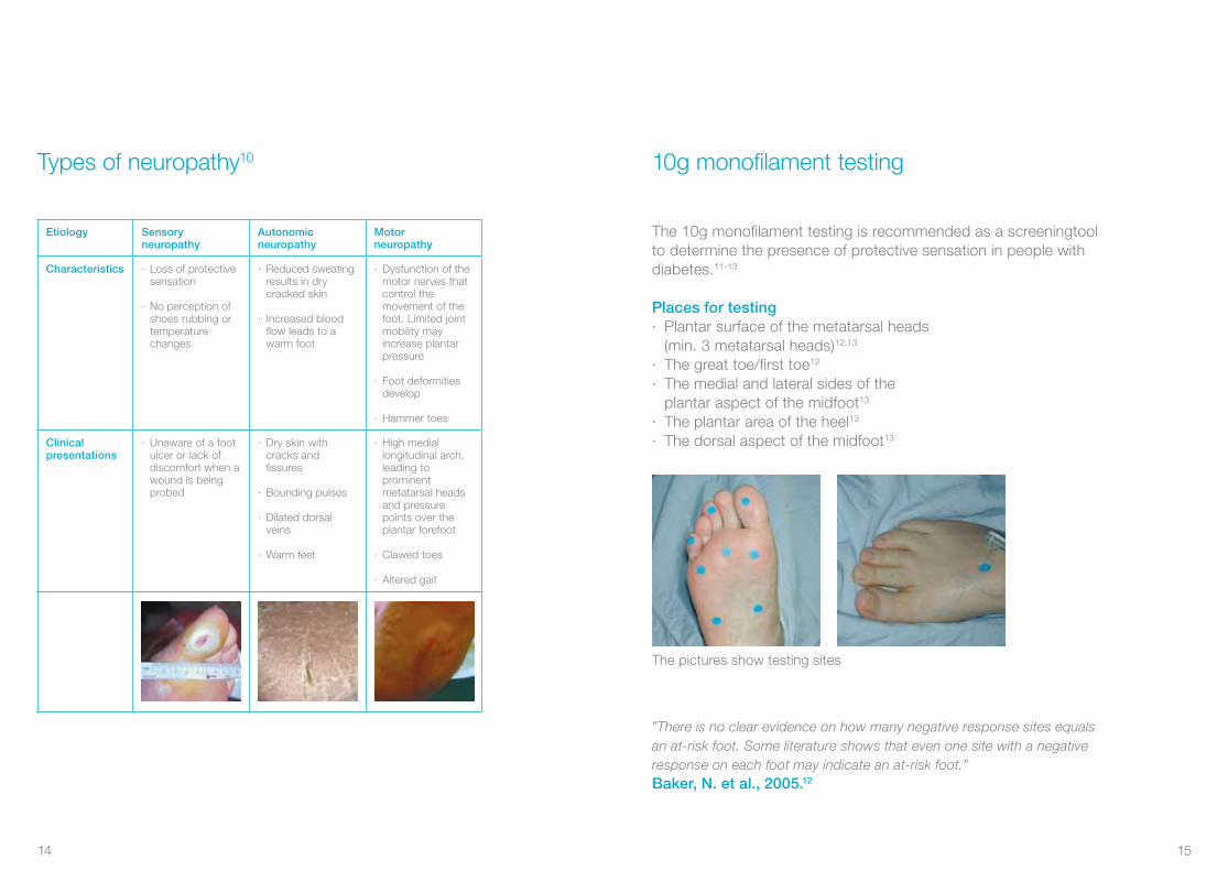
10g monofilament testing
The 10g monofilament testing is recommended as a screeningtool to determine the presence of protective sensation in people with diabetes.11-13
Places for testing· Plantar surface of the metatarsal heads (min. 3 metatarsal heads)12,13
· The great toe/first toe12
· The medial and lateral sides of the plantar aspect of the midfoot13
· The plantar area of the heel13
· The dorsal aspect of the midfoot13
”There is no clear evidence on how many negative response sites equals an at-risk foot. Some literature shows that even one site with a negative response on each foot may indicate an at-risk foot.”
The pictures show testing sites
Baker, N. et al., 2005.12
14 15
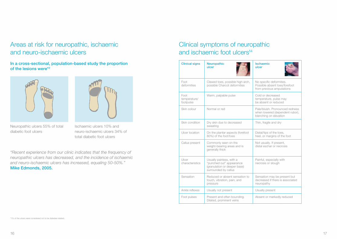
Areas at risk for neuropathic, ischaemicand neuro-ischaemic ulcers
In a cross-sectional, population-based study the proportion of the lesions were*2
“Recent experience from our clinic indicates that the frequency of neuropathic ulcers has decreased, and the incidence of ischaemic and neuro-ischaemic ulcers has increased, equaling 50-50%.”Mike Edmonds, 2005.
*1% of the ulcers were considered not to be diabetes-related.
Neuropathic ulcers 55% of total diabetic foot ulcers
Ischaemic ulcers 10% and neuro-ischaemic ulcers 34% of total diabetic foot ulcers
Clinical symptoms of neuropathicand ischaemic foot ulcers14
Clinical signs Neuropathiculcer
Ischaemiculcer
Foot deformities
Clawed toes, possible high arch, possible Charcot deformities
No specific deformities.Possible absent toes/forefoot from previous amputations
Foot temperature/footpulse
Warm, palpable pulse Cold or decreasedtemperature, pulse maybe absent or reduced
Skin colour Normal or red Pale/bluish. Pronounced redness when lowered (dependent rubor), blanching on elevation
Skin condition Dry skin due to decreasedsweating
Thin, fragile and dry
Ulcer location On the plantar aspects (forefoot 80%) of the foot/toes
Distal/tips of the toes,heel, or margins of the foot
Callus present Commonly seen on theweight-bearing areas and isgenerally thick
Not usually. If present,distal eschar or necrosis
Ulcercharacteristics
Usually painless, with a“punched out” appearance(granulation or deeper base) surrounded by callus
Painful, especially withnecrosis or slough
Sensation Reduced or absent sensation to touch, vibration, pain, and pressure
Sensation may be present but decreased if there is associated neuropathy
Ankle reflexes Usually not present Usually present
Foot pulses Present and often bounding.Dilated, prominent veins
Absent or markedly reduced
16 17
Ulcer assessment
Neuropathic pain
Burning, stinging, shooting andstabbing (non-stimulus dependent)
Local pain Deep infection or Charcot joint
Size Length, width, depth and location, preferably with clinical photograph
Wound bed Appearance· Black (necrosis) · Yellow, red, pink · Undermined
Infection signs OdourBe aware that some signs (fever, pain, increased white blood count/ ESR) may be absent. Evaluate the ulcer for signs of infection, inflammation and oedema. For more information, please see page 20
Exudate Copious, moderate, mild, none
Wound edge Callus and scale, maceration, erythema, oedema
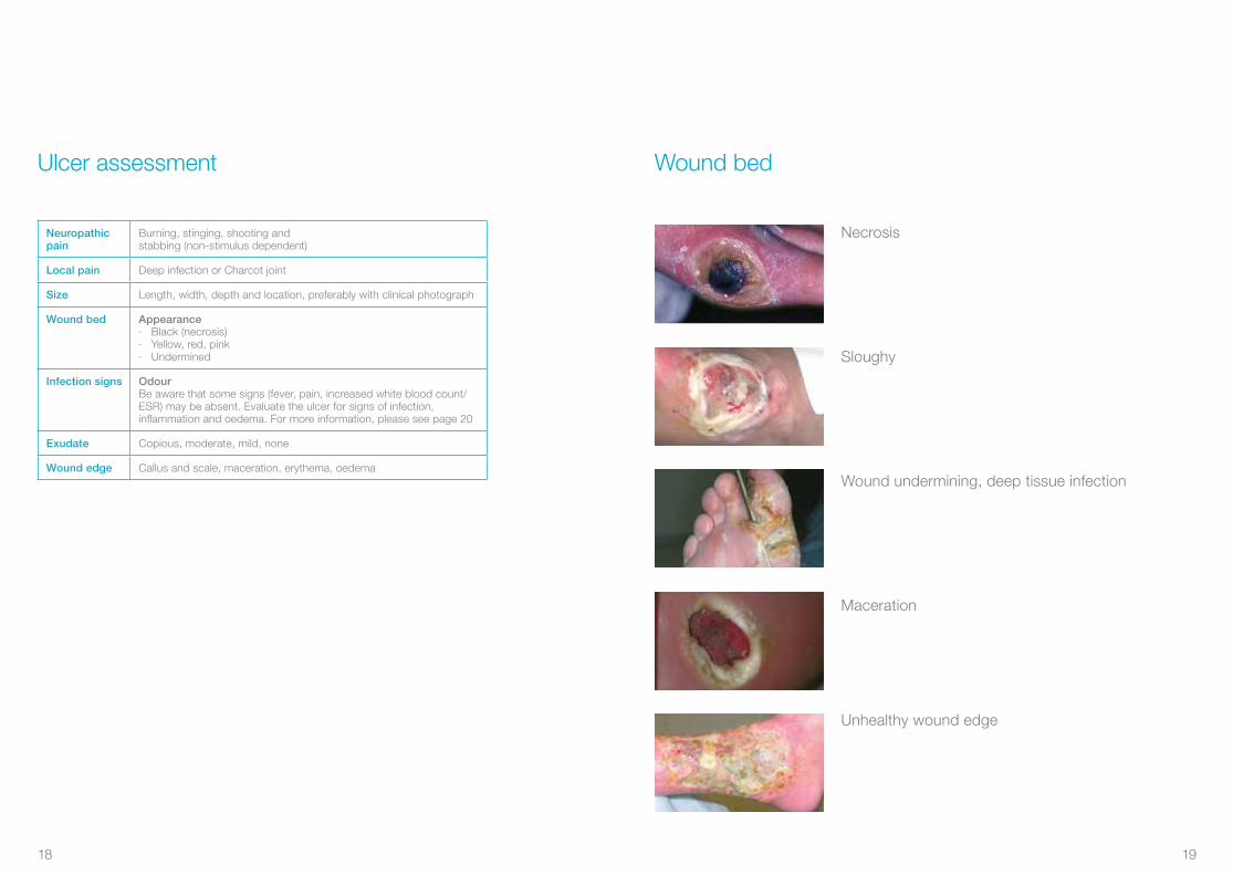
Wound bed
Necrosis
Sloughy
Wound undermining, deep tissue infection
Maceration
Unhealthy wound edge
18 19
Superficial and deep infection symptoms10,15,16
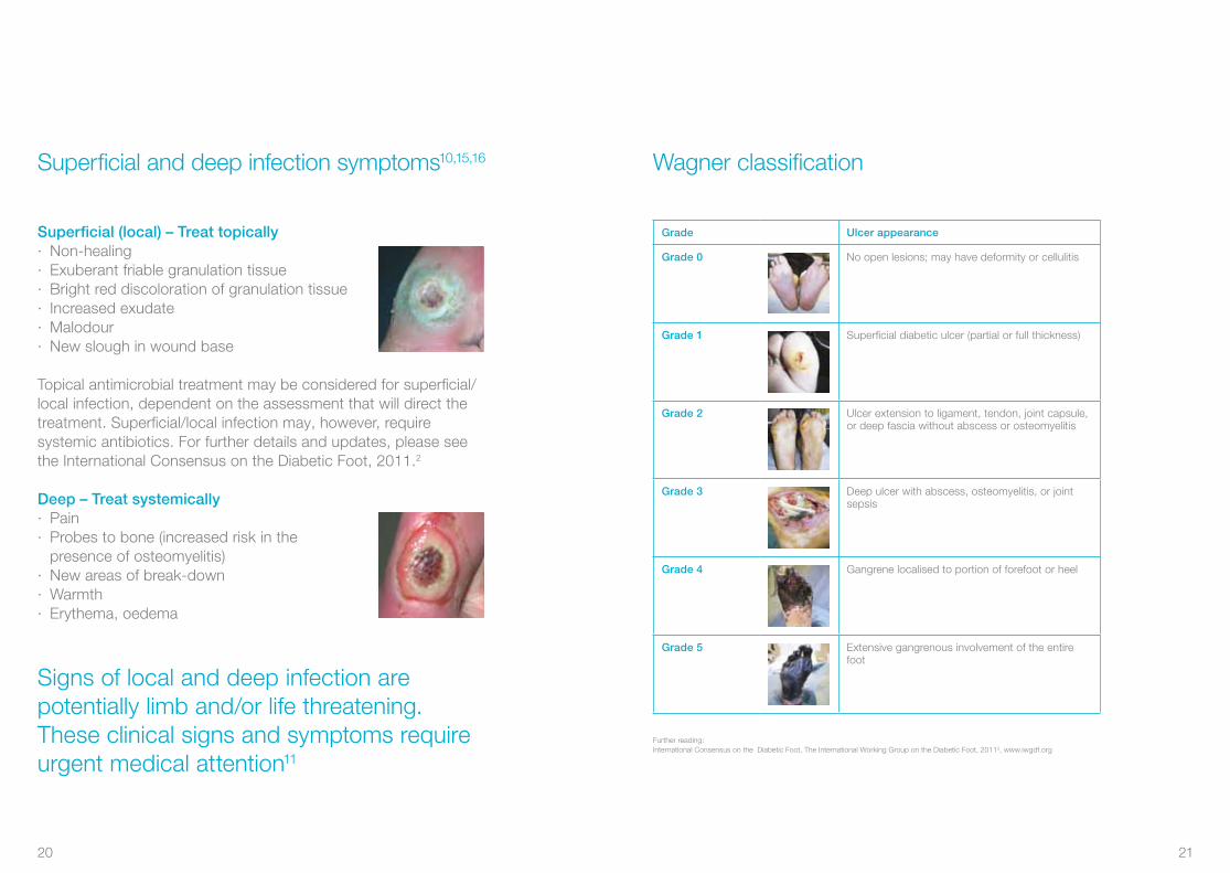
Superficial (local) – Treat topically· Non-healing· Exuberant friable granulation tissue· Bright red discoloration of granulation tissue· Increased exudate· Malodour· New slough in wound base
Topical antimicrobial treatment may be considered for superficial/local infection, dependent on the assessment that will direct the treatment. Superficial/local infection may, however, require systemic antibiotics. For further details and updates, please see the International Consensus on the Diabetic Foot, 2011.2
Deep – Treat systemically· Pain· Probes to bone (increased risk in the
presence of osteomyelitis)· New areas of break-down· Warmth· Erythema, oedema
Signs of local and deep infection are potentially limb and/or life threatening. These clinical signs and symptoms require urgent medical attention11
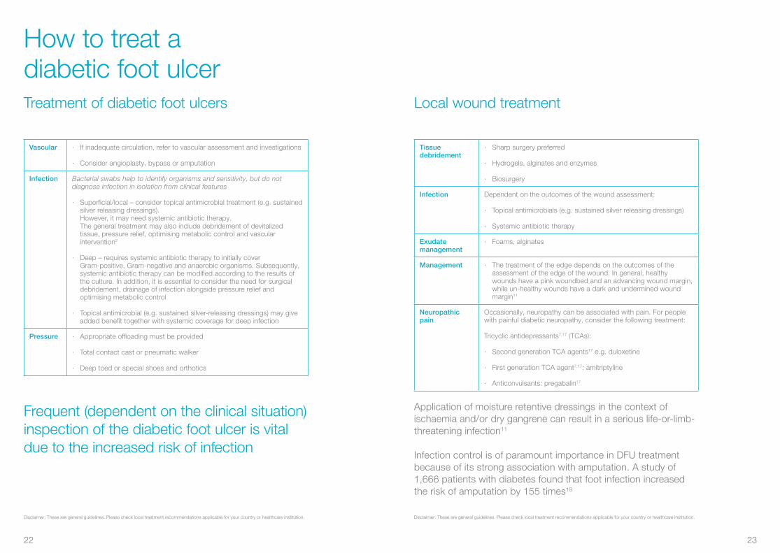
Wagner classification
Further reading: International Consensus on the Diabetic Foot, The International Working Group on the Diabetic Foot, 20112, www.iwgdf.org
Grade Ulcer appearance
Grade 0 No open lesions; may have deformity or cellulitis
Grade 1 Superficial diabetic ulcer (partial or full thickness)
Grade 2 Ulcer extension to ligament, tendon, joint capsule, or deep fascia without abscess or osteomyelitis
Grade 3 Deep ulcer with abscess, osteomyelitis, or joint sepsis
Grade 4 Gangrene localised to portion of forefoot or heel
Grade 5 Extensive gangrenous involvement of the entire foot
20 21
Treatment of diabetic foot ulcers
Disclaimer: These are general guidelines. Please check local treatment recommendations applicable for your country or healthcare institution.
Vascular · If inadequate circulation, refer to vascular assessment and investigations
· Consider angioplasty, bypass or amputation
Infection Bacterial swabs help to identify organisms and sensitivity, but do not diagnose infection in isolation from clinical features
· Superficial/local – consider topical antimicrobial treatment (e.g. sustained silver releasing dressings). However, it may need systemic antibiotic therapy. The general treatment may also include debridement of devitalized tissue, pressure relief, optimising metabolic control and vascular intervention2
· Deep – requires systemic antibiotic therapy to initially cover Gram-positive, Gram-negative and anaerobic organisms. Subsequently, systemic antibiotic therapy can be modified according to the results of the culture. In addition, it is essential to consider the need for surgical debridement, drainage of infection alongside pressure relief and optimising metabolic control
· Topical antimicrobial (e.g. sustained silver-releasing dressings) may give added benefit together with systemic coverage for deep infection
Pressure · Appropriate offloading must be provided
· Total contact cast or pneumatic walker
· Deep toed or special shoes and orthotics
Frequent (dependent on the clinical situation) inspection of the diabetic foot ulcer is vital due to the increased risk of infection
How to treat a diabetic foot ulcer
Disclaimer: These are general guidelines. Please check local treatment recommendations applicable for your country or healthcare institution.
Local wound treatment
Tissue debridement
· Sharp surgery preferred
· Hydrogels, alginates and enzymes
· Biosurgery
Infection Dependent on the outcomes of the wound assessment:
· Topical antimicrobials (e.g. sustained silver releasing dressings)
· Systemic antibiotic therapy
Exudatemanagement
· Foams, alginates
Management · The treatment of the edge depends on the outcomes of the assessment of the edge of the wound. In general, healthy wounds have a pink woundbed and an advancing wound margin, while un-healthy wounds have a dark and undermined wound margin11
Neuropathic pain
Occasionally, neuropathy can be associated with pain. For people with painful diabetic neuropathy, consider the following treatment:
Tricyclic antidepressants7,17 (TCAs):
· Second generation TCA agents17 e.g. duloxetine
· First generation TCA agent7,17: amitriptyline
· Anticonvulsants: pregabalin17
Application of moisture retentive dressings in the context of ischaemia and/or dry gangrene can result in a serious life-or-limb-threatening infection11
Infection control is of paramount importance in DFU treatment because of its strong association with amputation. A study of 1,666 patients with diabetes found that foot infection increased the risk of amputation by 155 times19
22 23
Coloplast solutions for diabetic foot ulcers
Biatain Non-Adhesive – superior absorption for wounds with extra fragile skinBiatain Non-Adhesive is a soft and flexible absorbent polyurethane foam dressing with bevelled edges
Biatain Silicone – superior absorption for general purposesBiatain Silicone is a soft and flexible absorbent foam dressing with a gentle silicone adhesive only on the border leaving the foam free to absorb exudate and heal the wound
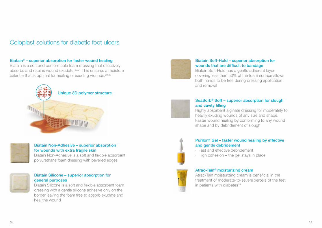
Unique 3D polymer structure
Biatain® – superior absorption for faster wound healingBiatain is a soft and conformable foam dressing that effectively absorbs and retains wound exudate.20,21 This ensures a moisture balance that is optimal for healing of exuding wounds.22,23
Biatain Soft-Hold – superior absorption for wounds that are difficult to bandageBiatain Soft-Hold has a gentle adherent layer covering less than 50% of the foam surface allows both hands to be free during dressing application and removal
SeaSorb® Soft – superior absorption for slough and cavity fillingHighly absorbent alginate dressing for moderately to heavily exuding wounds of any size and shape. Faster wound healing by conforming to any wound shape and by debridement of slough
Purilon® Gel – faster wound healing by effectiveand gentle debridement· Fast and effective debridement· High cohesion – the gel stays in place
Atrac-Tain® moisturizing creamAtrac-Tain moisturizing cream is beneficial in the treatment of moderate-to-severe xerosis of the feet in patients with diabetes24
24 25

Coloplast antimicrobial dressings for infected diabetic foot ulcers and ulcers at risk of infection
Biatain® Ag – superior absorption for infected woundsSustained release of silver during the entire wear time(up to 7 days)25
· Optimal healing environment26-27
· Rapid killing of bacteria28
· Designed to prevent wound infection
Biatain Ag Non-Adhesive – superior absorption for infected wounds with extra fragile skinBiatain Ag is a soft and conformable silver foam dressing that is proven to help infected wounds heal faster26,27
Biatain Silicone Ag – superior absorption for infected woundsBiatain Silicone Ag is a soft and flexible absorbent silver foam dressing with a gentle silicone adhesive border
SeaSorb® Ag – superior absorption for slough and cavity filling on infected woundsHighly absorbent antimicrobial alginate dressing for moderately to heavily exuding infected wounds or wounds at risk of infection. Faster wound healing by conforming to any wound shape and by debridement of slough.· Designed to fight cavity wound infection· Effect on a broad range of bacteria
Physiotulle® AgPhysiotulle Ag is a silver-containing, non-occlusive, hydrocolloid-based wound contact layer
26 27
1. Bakker, K. et al. The year of the diabetic foot, Diabetes Voice, March 2005, Vol. 50(1): 11-14.
2. International Working Group on the Diabetic Foot, International Consensus on the Diabetic Foot, 2007, 2011.
3. Jude, E. et al. Assessment of the diabetic foot. Chronic Wound Care: Chapter 58, In: Krasner, D.L. et al., A Clinical Sourcebook for Healthcare Professionals, Third Edition, HMP Communications Inc. 2001: 589-597.
4. Armstrong, D.G. et al. Diabetic foot infections: stepwise medical and surgical management. International Wound Journal, 2004, Vol. 1(2): 123-132.
5. Williams, R. et al. The size of the problem: Epidemiological and economic aspects of foot problems in diabetes. In: Boulton, A.J.M. et al., The Foot in Diabetes, John Wiley & Sons, Ltd., 2000: 3-17.
6. Spraul, M. Education – can it prevent diabetic foot ulcers and amputations? In: Boulton, A.J.M. et al., The Foot in Diabetes, John Wiley & Sons, Ltd., 2000: 111-120.
7. Reddy, M. Wound healing: The next milennium. Diabetic Microvascular Complications Today, May/June 2005: 25-27.
8. Inlow, S. et al. Best practices for the prevention, diagnosis, and treatment of diabetic foot ulcers, Ostomy/Wound Management 2000, Vol. 46(11): 55-68.
9. Frykberg, R.G. et al. A summary of guidelines for managing the diabetic foot. Advances in Skin & Wound Care 2005, Vol. 18(4): 209-213.
10. Edmonds, M. et al. A Practical Manual of Diabetic Foot Care, Blackwell Science, Oxford 2004.
11. Registered Nurses’ Association of Ontario 2005. Assessment and management of foot ulcers for people with diabetes. Toronto, Canada: Registered, Nurses’ Association of Ontario.
12. Baker, N. et al. A user’s guide to foot screening. Part 1: Peripheral neuropathy, The Diabetic Foot 2005, Vol. 8(1): 28-37.
13. Browne, A.C. et al. The diabetic neuropathic ulcer: An overview. Ostomy/Wound Management, 1999. Vol. 45 (No. 1A: Suppl).
14. Edmonds, M.E. et al. Managing the Diabetic Foot, Blackwell Science, Oxford 2005.
15. Sibbald, R.G. et al. Preparing the Wound Bed 2003: Focus on infection and inflammation, Ostomy/Wound Management, November 2003, Vol. 49(1): 24-51.
16. Sibbald, R.G. et al. Cost–effective faster wound healing of critically colonized wounds with a sustained release silver foam dressing, based upon the symposium ”Bacteria, sustained release of silver and improved healing”, An official satellite symposium of the WUWHS 2004. Published at www.worldwidewounds.com December 2005.
References
17. CG96 Neuropathic pain - pharmacological management: full guideline, NHS, National Institute for Health and Clinical Excellence, 27 May 2010 (http://guidance.nice.org.uk/CG96/Guidance/pdf/English).
18. Sibbald, R.G. et al. Dermatological aspects of wound care, Chapter 30, In: Krasner, D.L. et al., A Clinical Sourcebook for Healthcare Professionals, Third Edition, HMP Communications Inc., 2001: 273-285.
19. Lavery et al. Diabetes Care 2006;29(6):1288–93.
20. Andersen et al. A randomized, controlled study to compare the effectiveness of two foam dressings in the management of lower leg ulcers. Ostomy/Wound Management 2002;(48)8:34-41.
21. Thomas et al. www.dressings.org/TechnicalPublications/PDF/Coloplast-Dressings-Testing-2003-2004.pdf
22. White R and Cutting KF. Modern exudate management: a review of wound treatments. WorldWideWounds 2006.
23. Romanelli et al. Exudate management made easy. Wounds International 2010;1(2).
24. Pham et al. A prospective, randomized, controlled double-blind study of a moisturizer for xerosis of the feet in patients with diabetes. OstomyWound Management 2002;48(5):30-36.
25. Buchholtz. An in-vitro comparison of antimicrobial activity and silver release from foam dressings. Wounds UK 2009.
26. Jørgensen et al. The silver-releasing foam dressing, Contreet Foam, promotes faster healing of critically colonised venous leg ulcers: a randomised, controlled trial. International Wound Journal 2005;2(1):64-73.
27. Münter et al. Effect of a sustained silver-releasing dressing on ulcers with delayed healing: the CONTOP study. Journal of Wound Care. 2006;15(5):199-206.
28. Ip et al. Antimicrobial activities of silver dressings: an in vitro comparison. Journal of Medical Microbiology 2006;55:59-63.
28 29

Biatain SiliconeItem no.
National code
7½x7½ 3343410x10 3343512½x12½ 3343615x15 3343717½x17½ 33438
Biatain Silicone LiteItem no.
National code
7½x7½ 3344410x10 3344512½x12½ 33446
Biatain Soft-HoldItem no.
National code
5x7 347310x10 347010x20 347215x15 3475
Biatain Non-AdhesiveItem no.
National code
5x7 610510x10 341010x20 341215x15 341320x20 34165x8 Cavity 3451
Biatain AdhesiveItem no.
National code
7½x7½ 346210x10 343012½x12½ 342015x15 342118x18 342318x28 342617x17 Sacral jun.
3483
23x23 Sacral
3485
Ø17 Contour
3486
19x20 Heel 3488 Biatain Ag AdhesiveItem no.
National code
7½x7½ 963112½x12½ 963215x15 346418x18 963523x23 Sacral
9641
19x20 Heel
9643
Biatain Ag Non-AdhesiveItem no.
National code
5x7 510510x10 962210x20 962315x15 962520x20 96265x8 Cavity 9628
Biatain Silicone AgItem no.
National code
7½x7½ 3963610x10 3963712½x12½ 39638
Biatain Ibu Soft-HoldItem no.
National code
10x10 414010x20 4142
Biatain Ibu Non-AdhesiveItem no.
National code
5x7 410510x10 411010x20 411215x15 411520x20 4120
Superior absorption fornon-infected wounds*
Superior absorption forinfected wounds
Superior absorption forpainful wounds
Biatain® – superior absorption for faster healing
* Can be used for all types of exuding wounds.
30 31
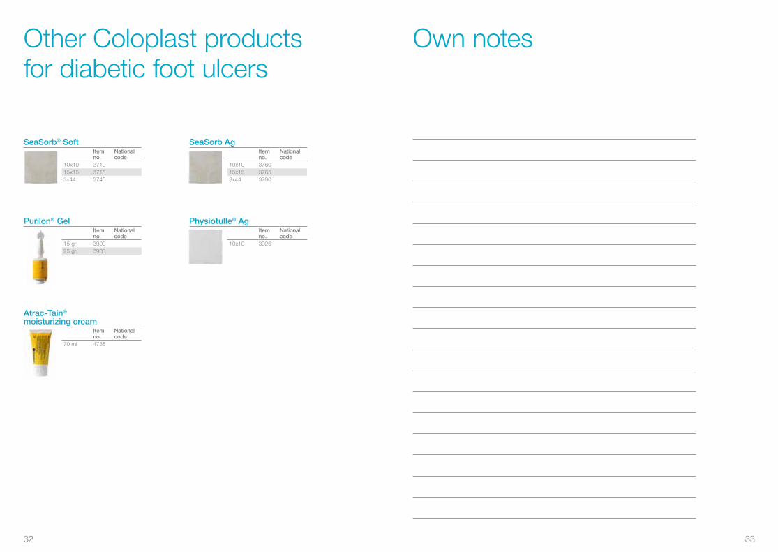
Other Coloplast products for diabetic foot ulcers
SeaSorb® SoftItem no.
National code
10x10 371015x15 37153x44 3740
Purilon® Gel Item no.
National code
15 gr 390025 gr 3903
SeaSorb AgItem no.
National code
10x10 376015x15 37653x44 3780
Atrac-Tain®
moisturizing creamItem no.
National code
70 ml 4738
Physiotulle® AgItem no.
National code
10x10 3926
Own notes
32 33

Own notes
34 35

Coloplast develops products and services that make life easier for people with very personal and private medical conditions. Working closely with the people who use our products, we create solutions that are sensitive to their special needs. We call this intimate healthcare. Our business includes ostomy care, urology and continence care and wound and skin care. We operate globally and employ more than 7,000 people.
The Coloplast logo is a registered trademark of Coloplast A/S. © [YYYY-MM.] All rights reserved Coloplast A/S, 3050 Humlebæk, Denmark.
Coloplast A/S Holtedam 1
3050 Humlebæk Denmark
www.coloplast.com
After 30 years in wound care,we at Coloplast believe that absorption is the key to better healing. Our Biatain® portfolio brings superior absorption to daily wound care needs, making Biatain the simple choice for faster healing.