diabetic foot ulcer - molnlycke.nl · diabetic foot ulcer (dfu) prevalence data estimates that,...
TRANSCRIPT

Diabetic foot ulcers A guide to assessment and management
Managing diabetic foot ulcers with specialist care Your patients with diabetes face challenges every day. We understand how these become your challenges too. Managing long-term conditions involves being able to balance eating, physical activity, medication, and injections. It’s a team effort that can involve a lot of resources and a mix of specialist care.
How common are diabetic foot problems? Diabetic foot problems are among the most serious and costly complications of diabetes. Diabetic foot ulcer (DFU) prevalence data estimates that, annually, foot ulcers develop in 9.1 million to 26.1 million diabetes patients worldwide1. Other research has shown that more than half of DFUs become infected; and the risk of death for diabetes patients with foot ulcers is 2.5 times
Up to one in every four patients with diabetes risk developing
a DFU in their lifetime3
Up to
80% of diabetic foot amputations are
preceded by a DFU5
higher than patients without a foot ulcer1. The rising prevalence of diabetes worldwide has seen an increase in the number of resulting lower limb amputations2. Both ulcers and amputations have an enormous impact on people’s lives, often leading to reduced independence, social isolation and psychological stress.
Globally, one leg is lost every
20 seconds as a result of diabetes4
Up to 85% of amputations can be avoided when
an effective care plan is adopted6
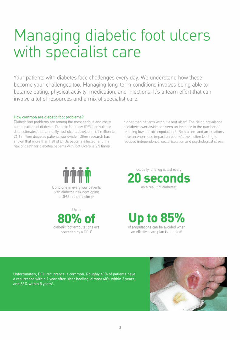
Unfortunately, DFU recurrence is common. Roughly 40% of patients have a recurrence within 1 year after ulcer healing, almost 60% within 3 years, and 65% within 5 years1.
2

A holistic approach to DFU care Diabetes is a complex disease. We understand that managing DFUs requires input from a range of specialities throughout the organisation. A multidisciplinary footcare team (MDFT) can provide comprehensive specialist foot and wound care, calling on the expertise of7:
• Doctors with a special interest in diabetes
• Podiatrists
• Diabetes specialist nurses
• Infection specialists
• Vascular surgeons
• Orthopaedic surgeons
• Orthotists
• Social workers
• Psychologists
What about your patients’ physical, psychological and social health situation? A MDFT’s holistic approach is important, not only to focus on evaluating and managing the wound, but diagnosing and treating underlying diseases8. By adopting a holistic approach to wound healing, with appropriate referrals and multidisciplinary involvement, DFUs can be healed and lives saved7-9:
Assessment of the patient and the ulcer should include the evaluation of:8
• Diabetes, management and blood glucose control
• Previous history of foot ulceration and surgery
• Underlying conditions e.g diabetes renal impairment
• Symptoms and signs of peripheral artery or venous disease
• All sensory, motor and autonomic neuropathy and the need for pressure off-loading
• Systemic signs of infection
• Pain such as neuropathic and/or wound-related pain
• Local wound assessment for appropriate management approach. See page 7–9.
• Socioeconomic circumstances, dexterity, visual acuity and insight
• Smoking status
If a person has a limb-threatening or life-threatening diabetic foot problem, they should be referred immediately to acute services and a MDFT informed. For all other active diabetic foot problems, the person should be referred within 1 working day to a MDFT.10
What about prevention? You and your team care about the outcomes for your patients. So prevention strategies make sense as a crucial step in avoiding an ulcer. It’s all part of effective foot care – a partnership between you, your patients and their carers.
Appropriate information that enables patients and carers to participate in decision making is often at the heart of all effective prevention strategies. We’ve heard how your patients like to have an understanding of the rationale behind some of the clinical decisions – it’s information that supports good self-care – so we’ve included patient education and self-care advice on page 10.
3
Aetiology of diabetic foot ulcers Did you know there are three key aetiologies that influence assessment, treatment of the underlying condition and management of a DFU?
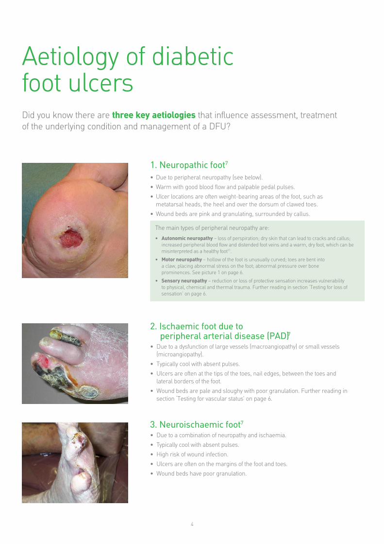
1. Neuropathic foot7
• Due to peripheral neuropathy (see below).
• Warm with good blood flow and palpable pedal pulses.
• Ulcer locations are often weight-bearing areas of the foot, such as metatarsal heads, the heel and over the dorsum of clawed toes.
• Wound beds are pink and granulating, surrounded by callus.
The main types of peripheral neuropathy are:
• Autonomic neuropathy – loss of perspiration; dry skin that can lead to cracks and callus; increased peripheral blood flow and distended foot veins and a warm, dry foot, which can be misinterpreted as a healthy foot11.
• Motor neuropathy – hollow of the foot is unusually curved; toes are bent into a claw, placing abnormal stress on the foot; abnormal pressure over bone prominences. See picture 1 on page 6.
• Sensory neuropathy – reduction or loss of protective sensation increases vulnerability to physical, chemical and thermal trauma. Further reading in section ‘Testing for loss of sensation’ on page 6.
2. Ischaemic foot due to peripheral arterial disease (PAD)7
• Due to a dysfunction of large vessels (macroangiopathy) or small vessels (microangiopathy).
• Typically cool with absent pulses.
• Ulcers are often at the tips of the toes, nail edges, between the toes and lateral borders of the foot.
• Wound beds are pale and sloughy with poor granulation. Further reading in section ‘Testing for vascular status’ on page 6.
3. Neuroischaemic foot7
• Due to a combination of neuropathy and ischaemia.
• Typically cool with absent pulses.
• High risk of wound infection.
• Ulcers are often on the margins of the foot and toes.
• Wound beds have poor granulation.
4
5
-
Diabetic foot ulcer classifcations How is your team classifying each wound? Did you know it’s important that each wound is classifed according to a validated clinical tool9? For example:
• Wagner12 • University of Texas13-14 • PEDIS15 • SINBAD16 • WIfI (WiFi)17
Wagner classifcation of diabetic foot ulcers
Grade 0 No ulcer in a high risk foot
Grade 1 Superfcial ulcer involving the full skin thickness but not underlying tissues
Grade 2 Deep ulcer, penetrating down to ligaments and muscle, but no bone involvement or abscess formation
Grade 3 Deep ulcer with cellulitis or abscess formation, often with osteomyelitis
Grade 4 Localised gangrene
Grade 5 Extensive gangrene involving the whole foot
University of Texas classifcation of diabetic foot ulcers Ulcer stage Ulcer grade (depth)
0 I II III
A Pre/post ulcerative lesion completely epithelialised
Superfcial ulcer, not involving tendon, capsule or bone
Ulcer penetrating to tendon or capsule
Ulcer penetrating to bone or joint
B Infection Infection Infection Infection
C Ischaemia Ischaemia Ischaemia Ischaemia
D Infection and ischaemia Infection and ischaemia Infection and ischaemia Infection and ischaemia
To ensure holistic assessment and treatment of DFUs, the wound should be classified according to a validated clinical tool9.
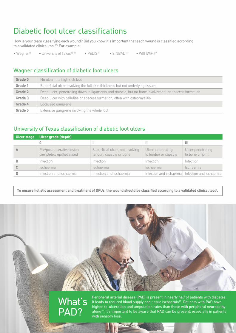
Peripheral arterial disease (PAD) is present in nearly half of patients with diabetes. It leads to reduced blood supply and tissue ischaemia18. Patients with PAD have What’s higher re ulceration and amputation rates than those with peripheral neuropathy alone19. It’s important to be aware that PAD can be present, especially in patients PAD? with sensory loss.
A guide to assessing DFUs Inspecting foot deformities
Excessive or abnormal plantar pressure, resulting from limited joint mobility, often combined with foot deformities, is a common underlying cause of DFUs in individuals with neuropathy3.
Common foot deformities7,11: • Prominent metatarsal heads • Hammer toes • Clawed toes • A high-arch foot • Hallux valgus (bunion), hallux rigidus (stiff big toe)
and plantar fat pad atrophy • Charcot deformity (read more below)
Patients also develop atypical walking patterns and this can result in calluses, which increase the abnormal pressure and can cause subcutaneous haemorrhage and ulcers. At the same time with neuropathy and the loss of sensation, the patient continues to walk on the foot, increasing the risk of further problems7.
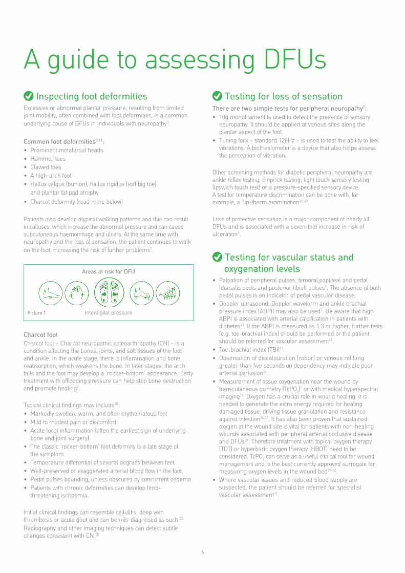
Areas at risk for DFU
Interdigital pressure Picture 1
Charcot foot Charcot foot – Charcot neuropathic osteoarthropathy (CN) – is a condition affecting the bones, joints, and soft tissues of the foot and ankle. In the acute stage, there is inflammation and bone reabsorption, which weakens the bone. In later stages, the arch falls and the foot may develop a ‘rocker-bottom’ appearance. Early treatment with offloading pressure can help stop bone destruction and promote healing7.
Typical clinical findings may include20: • Markedly swollen, warm, and often erythematous foot • Mild to modest pain or discomfort. • Acute local inflammation (often the earliest sign of underlying
bone and joint surgery). • The classic ‘rocker-bottom’ foot deformity is a late stage of
the symptom. • Temperature differential of several degrees between feet. • Well-preserved or exaggerated arterial blood flow in the foot. • Pedal pulses bounding, unless obscured by concurrent oedema. • Patients with chronic deformities can develop limb-
threatening ischaemia.
Initial clinical findings can resemble cellulitis, deep vein thrombosis or acute gout and can be mis-diagnosed as such.20
Radiography and other imaging techniques can detect subtle changes consistent with CN.20
Testing for loss of sensation There are two simple tests for peripheral neuropathy7: • 10g monofilament is used to detect the presence of sensory
neuropathy. It should be applied at various sites along the plantar aspect of the foot.
• Tuning fork – standard 128Hz – is used to test the ability to feel vibrations. A biothesiometer is a device that also helps assess the perception of vibration.
Other screening methods for diabetic peripheral neuropathy are ankle reflex testing, pinprick testing, light touch sensory testing (Ipswich touch test) or a pressure-specified sensory device. A test for temperature discrimination can be done with, for example, a Tip-therm examination21, 22.
Loss of protective sensation is a major component of nearly all DFUs and is associated with a seven-fold increase in risk of ulceration3.
Testing for vascular status and oxygenation levels
• Palpation of peripheral pulses: femoral,popliteal and pedal (dorsalis pedis and posterior tibial) pulses7. The absence of both pedal pulses is an indicator of pedal vascular disease.
• Doppler ultrasound, Doppler waveform and ankle brachial pressure index (ABPI) may also be used7. Be aware that high ABPI is associated with arterial calcification in patients with diabetes23. If the ABPI is measured as 1.3 or higher, further tests (e.g. toe-brachial index) should be performed or the patient should be referred for vascular assessment11.
• Toe-brachial index (TBI)11. • Observation of discolouration (robur) or venous refilling
greater than five seconds on dependency may indicate poor arterial perfusion24.
• Measurement of tissue oxygenation near the wound by transcutaneous oximetry (TcPO2)
7 or with medical hyperspectral imaging25. Oxygen has a crucial role in wound healing, it is needed to generate the extra energy required for healing damaged tissue, driving tissue granulation and resistance against infection26,27. It has also been proven that sustained oxygen at the wound site is vital for patients with non-healing wounds associated with peripheral arterial occlusive disease and DFUs28. Therefore treatment with topical oxygen therapy (TOT) or hyperbaric oxygen therapy (HBOT) need to be considered. TcPO2 can serve as a useful clinical tool for wound management and is the best currently approved surrogate for measuring oxygen levels in the wound bed29-32.
• Where vascular issues and reduced blood supply are suspected, the patient should be referred for specialist vascular assessment11.
6
Examination of the wound and Identifying infection surrounding skin
A physical examination should determine7,8: • Whether the wound is predominantly neuropathic, ischaemic or
neuroischaemic. • Critical limb ischaemia (if ischaemic). • Any musculoskeletal deformities. • Size/depth/location of the wound. • Tissue types present (colour/status of the wound bed):
– Black/necrotic. – Yellow/slough. – Red/granulation. – Pink/epithelialisation.
• Exposed bones, tendons, joint capsules or orthopaedic implants.
• Signs of infection (See Identifying infection page 7). • Odour: presence and nature. • Local pain: e.g. location, type, cause, intensity and duration. • Exudate: amount (high, moderate, low, none) colour and
consistency and whether it is purulent. • Status of the wound edge: e.g. callus, maceration, erythema,
oedema, undermining/tracks/sinuses and raised edge. • Condition of surrounding skin: e.g. maceration/excoriation,
erythema, oedema or dry skin.
Is your team documenting wound size and status with digital photographs and/or apps? The placement of a paper ruler in the vicinity of the wound can help to indicate its size. For consistency, always measure in centimetres, listing in the order of length x width x depth.33
Approximately
56% of DFUs become infected34
A study has identified 3 factors associated with developing a foot infection: a previous amputation, loss of protective sensation and peripheral vascular disease (defined as any missing pedal pulsation or an ABPI of <0.8)35.
Approximately 56% of DFUs become infected and about 20% of patients with an infected wound on the foot will undergo a lower extremity amputation34. If infection is suspected, the DFU should be sampled after debridement for microbiological analysis and the result should be used to guide antibiotic selection15.
The diagnosis of diabetic foot infection (DFI) should be based on the presence of two or more of the following: local swelling or induration, erythema, local tenderness/pain, local warmth or purulent secretions15. Some DFIs may not exhibit these signs, especially in the case of patients who have peripheral neuropathy or limb ischemia15.
It is important to recognise the subtle signs of infection which may include just one of these signs above, combined with two local signs of infection (from the list below)8.
Additional or secondary signs, e.g8,15: • Increased exudate • Nonpurulent secretions • Friable or discolored, granulation tissue • Undermining of wound edges • Malodour
DFI is classified into mild (superficial with minimal cellulitis), moderate (deeper or more extensive), or severe. In severe infections, fever or hypothermia, increased heart and respiratory rates, and high or low white cell counts may occur15,35,36.
Other causes of an inflammatory response of the skin should be excluded (e.g trauma, gout, acute Charcot neuro-osteoarthropathy, fracture, thrombosis, venous stasis)15,36.
In case of an acute spreading infection, critical limb ischaemia, wet gangrene or an unexplained hot, red, swollen foot with or without the presence of pain, the patient urgently needs to be referred to the specialist foot care team7.
Osteomyelitis Osteomyelitis – an infection in a bone – can be difficult to diagnose in the early stages. It should be consider as a potential complication of any infected, deep, or large foot ulcer, especially one that is chronic or overlies a bony prominence. A probe-to-bone (PTB) test should be done for any DFI with an open wound15. The National Institute for Health, Care and Excellence (NICE) in the United Kingdom recommend that if initial x-rays do not confirm the presence of osteomyelitis and suspicion remains high, consider magnetic resonance imaging (MRI)10,15. The most definitive way to diagnose osteomyelitis is by the combined findings of culture and histology from a bone specimen obtained during deep debridement or by biopsy15.
7
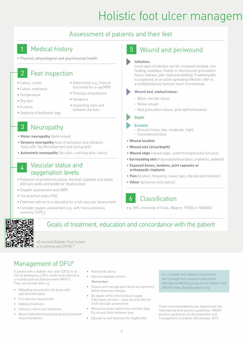
Holistic foot ulcer managem Assessment of patients and their feet
1 Medical history • Physical, physiological and psychosocial health
2 Feet inspection • Callus, cracks • Deformities e.g. Charcot
foot (need for x-ray/MRI) • Colour, erythema • Previous amputations • Temperature • Gangrene • Dry skin • Inspecting nails and• Eczema
between the toes • Oedema of feet/lower legs
Neuropathy • Motor neuropathy (deformities)
• Sensory neuropathy (loss of sensation and vibration. Tests with 10g Monoflament and tuning fork)
• Autonomic neuropathy (dry skin, cracking skin, callus)
4 Vascular status and oxygenation levels
• Palpation of peripheral pulses: femoral, popliteal and pedal (dorsalis pedis and posterior tibial) pulses
• Doppler assessment and ABPI
• Toe-brachial index (TBI)
• Potential referral to a specialist for a full vascular assessment
• Consider oxygen assessment e.g. with transcutaneous oximetry (TcPO2)
3
5 Wound and periwound
Infection: Local signs of infection can be: increased exudate, non-healing, malodour, friable or discoloured granulation tissue, redness, pain, heat and swelling. If osteomyelitis is suspected, or an active spreading infection, refer to a multidisciplinary footcare team immediately.
Wound bed, status/colour:
– Black necrotic tissue
– Yellow slough
– Red granulation tissue, pink epithelialisation
Depth
Exudate – Amount (none, low, moderate, high) – Consistency/colour
• Wound location
• Wound size (area/depth)
• Wound edge (raised edge, undermining/tracks/ sinuses)
• Surrounding skin (maceration/excoriation, erythema, oedema)
• Exposed bones, tendons, joint capsules or orthopaedic implants
• Pain (location, frequency, cause, type, intensity and duration)
• Odour (presence and nature)
6 Classifcation e.g. WIfI, University of Texas, Wagner, PEDIS or SINBAD
Goals of treatment, education and concordance with the patient
60-second Diabetic Foot Screen a Screening tool (2018).37
Management of DFU8
A patient with a diabetic foot ulcer (DFU) or at • Nutritional advice. For complete and updated assessment risk of developing a DFU needs to be referral to • Optimal diabetes control.
a multidisciplinary footcare team (MDFT). and management guidance please visit Remember:They can provide with e.g. International Working Group on the Diabetic Foot
• Assess and manage pain (local and systemic) (IWGDF) https://iwgdfguidance.org • Offloading wound and risk areas with before dressing changes.
specialist foot wear. • Be aware of the arterial blood supply. • Full vascular assessment. If dry black necrosis – keep dry and refer for • Oedema treatment. a full vascular assessment.
These recommendations are aligned with the • Infection control and treatment. • Moisturize lower extremities and feet daily. International best practice guidelines: IWGDF
Do not put lotion between toes. • Wound debridement/cleansing and treatment practical guidelines on the prevention and recommendation. • Educate on self-treatment for healthy feet. management of diabetic foot disease, 2019.
8
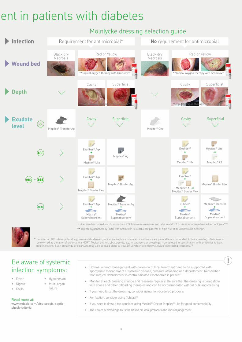
ent in patients with diabetes Mölnlycke dressing selection guide
Infection Requirement for antimicrobial* No requirement for antimicrobial
Black dry Necrosis
Wound bed
Depth
Exudate level Mepilex® Transfer Ag
–
Red or Yellow Black dry Red or Yellow Necrosis
**Topical oxygen therapy with Granulox® **Topical oxygen therapy with Granulox®
Cavity Superfcial Cavity Superfcial
**Topical oxygen therapy with Granulox® **Topical oxygen therapy with Granulox®
Cavity Superfcial Cavity Superfcial
Mepitel® One
Exufber® Mepilex® Lite Exufber® Ag+ + or+
Mepilex® Ag
Mepilex® Lite Mepilex® Lite Mepilex® XT
Exufber®Exufber® Ag+ ++ Mepilex® Border Ag Mepilex® Border Flex
Mepilex® XT or Mepilex® Border Flex Mepilex® Border Flex
Exufber® Ag+ Mepilex® Transfer Ag +
Exufber® Mepilex® Transfer + + +
Mextra® Mextra® Mextra® Mextra®
Superabsorbent Superabsorbent Superabsorbent Superabsorbent
If ulcer size has not reduced by more than 50% by 4 weeks reassess and refer to a MDFT or consider other/advanced technologies8,9,11.
** Topical oxygen therapy (TOT) with Granulox® is suitable for patients at high risk of delayed wound healing38.
* For infected DFUs (see picture), aggressive debridement, topical antiseptics and systemic antibiotics are generally recommended. Active spreading infection must be referred as a matter of urgency to a MDFT. Topical antimicrobial agents, e.g. in cleansers or dressings, may be used in combination with antibiotics to treat
mild infections. Such dressings or cleansers may also be used alone to treat DFUs which are highly at risk of developing infections.7,8
!Be aware of systemic infection symptoms: • Fever • Hypotension
• Rigour • Multi-organ failure • Chills
Read more at: www.mdcalc.com/sirs-sepsis-septic-shock-criteria
• Optimal wound management with provision of local treatment need to be supported with appropriate management of systemic disease, pressure offloading and debridement. Remember that surgical debridement is contraindicated if ischaemia is present11
• Monitor at each dressing change and reassess regularly. Be sure that the dressing is compatible with shoes and other offloading therapies and can be accommodated without bulk and creasing
• If you need to cut the dressing, consider using non-bordered products
• For fxation, consider using Tubifast®
• If you need to dress a toe, consider using Mepitel® One or Mepilex® Lite for good conformability
• The choice of dressings must be based on local protocols and clinical judgement
9

Patient education for self-care
Educating your patients on proper foot care and periodic examinations can help prevent foot problems and ulceration. Education should be presented in a structured and organised manner; the aim is to enhance motivation and skills. Have your patients understood the messages? Are they motivated to act? Do they have sufficient self-care skills?
Here’s a checklist to share with your patients, to help them keep their feet healthy39.
1. Take care of your diabetes and your health. 7. Wear shoes and socks at all times. Never walk Maintenance of good blood glucose control can help barefoot. Wear comfortable shoes that fit well and reduce the risk of both developing neuropathy and protect your feet both indoor and outdoor. Check circulation damage. If you have a foot problem, inside your shoes before wearing them. Make sure keeping your blood sugars well controlled can help the lining is smooth and there are no objects inside. the healing process. Change socks daily and use socks without constraining
cuffs or seams (or with the seams inside out).
2. Check your feet every day. Look at your bare feet for 8. Stay active to maintain healthy blood circulation. red spots, cuts, swelling and blisters. If you cannot
Be active each day for example: walking, dancing, see the bottoms of your feet, use a mirror or ask someone for help.
3. Have your feet examined for sensitivity and pulses at least annually by a professional (such as a podiatrist). If your clinician identifies your feet as being at risk for ulceration, you should be examined more often.
swimming, or going bike riding. Put your feet up when sitting. Two or three times per day, wiggle your toes and move your ankles up and down for five minutes. Don’t cross your legs for long periods of time. Give up smoking, it can damage your circulation.
9. Protect your feet from extreme temperatures. Wear shoes at the beach or on hot pavements. Don’t
4. Wash your feet every day with lukewarm water. Dry put your feet into hot water. Test water before putting them carefully, especially between to toes. your feet in. Never use hot-water bottles, heating
pads, or electric blankets. You could burn your feet without realising it.
5. Keep your skin soft and smooth. Rub a thin coat of skin lotion over the tops and bottoms of your feet, but 10. Pick the right shoes. Proper shoes are an not between your toes. important part of keeping your feet healthy. Buy
your shoes in the late afternoon or evening, when 6. Trim your toenails straight across and file the feet are at their largest. Pick comfortable footwear
edges with an emery board or nail file. with enough room for your toes. Avoid opentoed shoes. If you need more advice or help, consult an orthopaedic shoemaker.
Call or see your healthcare provider if you have cuts or breaks in the skin, or have an ingrown nail. Also, tell your healthcare provider if your foot changes colour, shape, or just feels different; for example, becomes less sensitive or hurts. If you have corns or calluses, your healthcare provider can trim them for you. Your healthcare provider can also trim your toenails if you cannot do so safely.
10

Dressing information
Mepitel® One
• Soft silicone wound contact layer • For dry to highly exuding wounds • Highly transparent for quick and easy
wound inspection
Mepilex® XT Mepilex® Ag
• Foam dressings with soft silicone wound contact layers with (Mepilex Ag) and without silver (Mepilex XT)
• For low to moderately exuding wounds, designed to maintain a moist wound environment
• Soft and conformable foam dressing • Can easily be cut to size • Mepilex XT can handle both low and
high viscosity fluid44
Exufber®
• Gelling fber dressing • Transforms into a gel that provide a
moist wound environment 53,54
• High tensile strength to enable dressing removal in one piece54
• Absorbs and retains exudate, blood and bacteria54
• Soft and conformable which make it easy to apply53
Mextra® Superabsorbent
• Superabsorbent dressing with fluid-repellent backing
• For highly exuding wounds • Superabsorbent particles for high
absorption and retention69
• Soft and conformable • Fluid repellent backing layer protects
against fluid strike-through
• Can remain in place for up to 14 days depending on the wound condition40
• Minimises skin damage and pain at dressing changes40-42
• Mepilex Ag kills wound-related pathogens within 30 minutes; and carries on doing so for up to 7 days (in vitro studies)45
• Minimise skin damage and pain at dressing changes43
Exufber® Ag+
• Gelling fbre dressing containing silver
• Transforms into a gel and softly conforms to the wound bed55,56
• For moderately to highly exuding wounds
• The Hydrolock® Technology absorbs and locks in exudate, blood and bacteria. The high structural integrity enables one-piece dressing removal57-62
• Silver kills a broad range of pathogens (in vitro) and reduce bioflm, the antimicrobial effect is kept for up to seven days (in vivo)63-65
• Can easily be cut and used in cavities
Proven choice for a better outcome Safetac® is the original less-pain contact layer with silicone adhesion. We designed it to mould softly to skin without sticking to the moist wound71 – so you can remove it easily without damaging the skin72. That means less pain for your patients43.
Safetac also protects new tissue and intact skin – so wounds remain undisturbed to support faster natural healing41,42,73,74. And it seals the wound margins to protect skin from damaging leaks and maceration75,76. This combination of less pain43 and less skin damage42,72-75,77
– to support faster healing41,42,73,74 – can also reduce the cost of treatment42,64,68.
You can trust Mölnlycke® dressings with Safetac, for better patient and economic outcomes.
Mepilex® Lite
• Light foam dressing with soft silicone wound contact layer
• For non to low-exuding wounds; designed to maintain a moist wound environment
Mepilex® Border Flex
• All-in-one bordered foam dressing with flex cuts
• For moderately to highly exuding wounds; designed to maintain a moist wound environment
• Enables 360 degree stretch to enhance stay-on-ability and conformability46-49
• Contains superabsorbent fbres for high absorption and retention50
• Minimise skin damage and pain at dressing changes43,50
Mepilex® Transfer
• Thin, soft, and highly conformable • Can easily be cut to size • Minimises pain and damage at
dressing change43
Mepilex® Border Ag
• All-in-one bordered foam dressing containing silver
• For moderately to highly exuding wounds; designed to maintain a moist wound environment
• Combines excellent exudate management properties with antimicrobial action51,52
• Minimise skin damage and pain at dressing changes43
• Exudate transfer dressings with • Mepilex Transfer Ag inactivates a
Mepilex® Transfer Ag
(Mepilex Transfer Ag) and without silver (Mepilex Transfer)
• Effectively transfer exudate to a secondary layer66
• Very thin and conformable foam for diffcult-to-dress locations
• Can easily be cut to size
Tubifast®
• Tubular retention bandage • Holds dressings securely, without
constriction or compression • A variety of lengths are available • Available in a range of quick reference,
colour-coded sizes to ft everything from small limbs to adult trunks
broad range of microorganisms (in vitro studies)67
• Mepilex Transfer Ag combines a rapid antimicrobial effect within 30 min and a sustained effect up to 14 days (in vitro studies)67
• Minimise skin damage and pain at dressing changes43,68
Granulox®
• Topical haemoglobin-based spray • The haemoglobin spray acts by
facilitating the diffusion of oxygen from the atmosphere into the wound bed
• Time to heal diabetic foot ulcers 50% shorter than with standard of care70
• Granulox® is easy to handle and to apply
Skin stripping occurs with traditional adhesive72
No skin stripping occurs with Safetac technology72
11
Proving it every day At Mölnlycke®, we deliver innovative solutions for managing wounds, improving surgical safety and effciency and preventing pressure ulcers. Solutions that help achieve better outcomes and are backed by clinical and health-economic evidence.
In everything we do, we are guided by a single purpose: to help healthcare professionals perform at their best. And we’re committed to proving it every day.
Mölnlycke would like to acknowledge Dr. Paul Chadwick for reviewing this guide.
Please note: This guide is not comprehensive and the reader should always refer to local guidelines.
References: 1. Armstrong, D.G., Boulton, A.J.M., Bus, S.A. Diabetic foot ulcers and their recurrence. New Engl J Med 2017;376:2367-75. 2. Rodrigues, B.T., Vangaveti, V.N., Malabu, U.H. Prevalence and risk factors for diabetic lower limb amputation: a clinic-based case control study. J Diabetes Res 2016: 5941957. Available at: http://dx.doi.org/10.1155/2016/5941957 (Accessed 8 November 2018). 3. Singh, N., Armstrong, D.G., Lipsky, B.A. Preventing foot ulcers in patients with diabetes. JAMA 2005;293(2):217-28. 4. Hinchliffe, R.J., Andros, G., Apelqvist, J., et al. A systematic review of the effectiveness of revascularization of the ulcerated foot in patients with diabetes and peripheral arterial disease. Diabetes Metab Res Rev 2012;28 (Supplement 1): 179-217. 5. Boulton, A.J.M. The pathway to foot ulceration in diabetes. Med Clin N Am 2013;97:775-90. 6. Pecoraro, R.E., Reiber, G.E., Burgess, E.M. Pathways to diabetic limb amputation. Basis for prevention. Diabetes Care 1990; 13(5): 513-21. 7. International Best Practice Guidelines. Wound Management in Diabetic Foot Ulcers. Wounds International 2013. Available at: http://www.woundsinternational.com (Accessed 8 November 2018). 8. World Union of Wound Healing Societies (WUWHS). Florence Congress. Position Document. Local Management of Diabetic Foot Ulcers. Wounds International 2016. Available at: http://www.woundsinternational.com (Accessed 8 November 2018). 9. Frykberg, R.G., Banks, J. Challenges in the treatment of chronic wounds. Adv Wound Care (New Rochelle) 2015;4:560-82. 10. National Institute for Health and Care Excellence. Diabetic foot problems: prevention and management. NICE guideline 19 2015. Available at: https://www.nice.org.uk/guidance/ ng19/diabetic-footproblems- prevention-and management-pdf-183729829933 (Accessed 8 November 2018). 11. Ousey, K., Chadwick, P., Jawien, A., et al. Identifying and treating foot ulcers in patients with diabetes: saving feet, legs and lives. J Wound Care 2018;27 (Suppl 5):S1-S52. 12. Wagner, R.W. The dysvascular foot: a system for diagnosis and treatment. Foot Ankle 1981;2(2):64-122. 13. Lavery, L.A., Armstrong, D.G., Harkless, L.B. Classifcation of diabetic foot wounds. J Foot Ankle Surg 1996;35:528-31. 14. Armstrong, D.G., Lavery, L.A., Harkless, L.B. Validation of a diabetic wound classifcation system. The contribution of depth, infection, and ischemia to risk of amputation. Diabetes Care 1998;21:855-9. 15. Lipsky, B., Berendt, A., Cornia, P.B. Infectious Diseases Society of America clinical practice guideline for the diagnosis and treatment of diabetic foot infections. IDSA guidelines. Clin Infect Dis 2012;54:132-73. 16. Ince, P., Abbas, Z.G., Lutale, J.K., et al. Use of the SINBAD classifcation system and score in comparing outcome of foot ulcer management on three continents. Diabetes Care 2008;31:964-67. 17. Mills, J.L., Conte, M.S., Armstrong D.G., et al. Society for Vascular Surgery Lower Extremity Guidelines Committee. The Society for Vascular Surgery Lower Extremity Threatened Limb Classifcation System: risk stratifcation based on wound, ischemia, and foot infection (WiFi). J Vasc Surg 2013;59(1), 220–34.e1–2. 18. Prompers, L., Huijberts, M., Apelqvist, J., et al. High prevalence of ischaemia, infection and serious comorbidity in patients with diabetic foot disease in Eu-rope. Baseline results from the Eurodiale study. Diabetologica 2007;50(1):18-25. 19. Apelqvist, J., Elgzyri, T., Larsson, J., et al. Factors related to outcome of neuroischaemic / ischemic foot ulcer in diabetic patients. J Vasc Surg 2011;53:1582-8. 20. Rogers, L.C., Frykberg, R.G., Armstrong, D.G. The Charcot Foot in Diabetes. Diabetes Care 2011;34:2123-9. 21. Craig, A.B., Strauss, M.B., Daniller, A. Miller,S.S. Foot sensation testing in the patient with diabetes: introduction of the quick & easy assessment tool. Wounds 2014;26(8):221-231. 22. Viswanathan, V., Snehalatha, C., Seena, R., Ramachandran, A. Early recognition of diabetic neuropathy: evaluation of a simple outpatient procedure using thermal perception. Postgrad Med J 2002;78:541–542. 23. Bailey, M.A., Griffn, K.J., Scott, D.J.A. Clinical assessment of patients with peripheral arterial disease. Semin Intervent Radiol 2014;31:292-9. 24. LoGerfo, F.W., Coffman, J.D. Current concepts. Vascular and microvascular disease of the foot in diabetes. Implications for foot care. New Engl J Med 1984;311:1615-19. 25. Lua, G., Fei, B. Medical hyperspectral imaging: a review. Biomed Opt. 2014;19(1):010901. 26. Sen, C.K. Wound healing essentials: let there be oxygen. Wound Repair Regen 2009;17(1):1–18. 27. Gottrup, F. Oxygen in wound healing and infection. Wound J Surg 2004;28(3):312–5. 28. Dissemond, J., Kroger, K., Storck, M., et al. Topical oxygen wound therapies for chronic wounds: a review. J Wound Care 2015;24(2):53–63. 29. Ruangsetakit, C., Chinsakchai, K., Mahawongkajit, P. et al (2010) Transcutaneous oxygen tension: a useful predictor of ulcer healing in critical limb ischaemia. J Wound Care 2010;19(5):202–6. 30. Arsenault, K.A., Al-Otaibi, A., Devereaux, P.J. et al. The use of transcutaneous oximetry to predict healing complicationsof lower limb amputations. Eur J Vasc Endovasc Surg 2012;43:329–36. 31. Zulec, M. Transcutaneous oximetry – between theory and practice. Acta Med Croatica 2014;68 Suppl 1:S59–S61. 32. Gottrup, F., Dissemond, J., Baines. et al. Use of oxygen therapies in wound healing, with special focus on topical and hyperbaric oxygen treatment. J Wound Care, 2017;26(5), Suppl, S1–S42. 33. Nichols, E. Wound assessment part 1: how to measure a wound. Wound Essentials 2015;10:51-5. 34. Wu, S.C., Driver, V.R., Wrobel, J.S., Armstrong, D.G. Foot ulcers in the diabetic patient, prevention and treatment. Vasc Health Risk Manage 2007;3:65-76. 35. Peters, E.J., Lavery, L.A., Armstrong, D.G. Diabetic lower extremity infection: influence of physical, psychological, and social factors. J Diabetes Complications 2005;19:107-12. 36. Lipsky BA, Aragon-Sanchez J, Diggle M, et al. IWDGF Guidance on the diagnosis and management of foot infections in persons with diabetes. International Working Group on the Diabetic Foot, 2015. 37. INLOW’s 60-second Diabetic Foot Screen. Screening tool. Canadian Association of Wound Care. www.cawc.net. 2011. 38. Chadwick, P.M., McCardle, J., Luxmi, M., et al. Appropriate use of topical haemoglobin in chronic wound management: consensus recommendations. Wounds UK 2015;EWMA Special: 30–35. 39. National Institute of Diabetes and Digestive and Kidney Diseases. Preventing Diabetes Problems. Diabetes and Foot Problems. Available at: https://www.niddk.nih.gov/health-information/diabetes/overview/ preventing-problems/foot-problems (Accessed 8 November 2018). 40. Patton, M.L., Mullins, R.F., Smith, D., Korentager, R. An open, prospective, randomized pilot investigation evaluating pain with the use of a soft silicone wound contact layer vs bridal veil and staples on split thickness skin grafts as a primary dressing. J Burn Care Res 2013;34:674-81. 41. David, F., Wutze, J-L., Breton, N., et al. A randomised, controlled, non-inferiority trial comparing the performance of a soft silicone-coated wound contact layer (Mepitel One) with a lipidocolloid wound contact layer (UrgoTul) in the treatment of acute wounds. Int Wound J 2017 doi:10.1111/iwj.12853. 42. Gotschall, C.S., Morrison, M.I., Eichelberger, M.R. Prospective, randomized study of the effcacy of Mepitel on children with partial-thickness scalds. J Burn Care Rehabil 1998;19:279-83. 43. White, R. A multinational survey of the assessment of pain when removing dressings. Wounds UK 2008;4:14-22. 44. Mölnlycke Health Care data on fle, report 20160105-002. 45. Chadwick, P., Taherinejad, F., Hamberg, K., Waring, M. Clinical and scientifc data on a silver-containing soft-silicone foam dressing: an overview. J Wound Care 2009;18:483-91. 46. ProDerm study report 16.0180-23. Assessment of Wearing Properties of Wound Dressings on the Knees. Data on fle. 47. ProDerm study report 16.0180-23. Assessment of Wearing Properties of Wound Dressings on the Elbows. Data on fle. 48. ALTEN Finite Element Modelling simulation. Laboratory report no. PD-530246. 49. Haycocks, S., Chadwick, P., Davies, P. Case series: Mepilex Border Comfort in the treatment of diabetic foot ulcers with exudate. Diabetic Foot Journal 2018;21:265-71. 50. External test lab report SMTL15/4863/2. 51. External lab report: NAMSA 09C 29253 01/09C 29253 02. 52. Kles C.L., Murrah, C.P., Smith, K., et al. Achieving and sustaining zero. Preventing surgical site infections after isolated coronary artery bypass with saphenous vein harvest through implementation of a staff-driven quality improvement process. Dimensions Crit Care Nurs 2015;34:265-72. 53. Smet, S., Beele, H., Saine, L., Suys, E., Henrickx, B. Open, noncomparative, multi-centre post market clinical follow-up investigation to evaluate performance and safety on pressure ulcers when using a gelling fbre dressing as intend-ed. Poster Presentation at European Pressure Ulcer Advisory Panel Confer-ence, 2015, Ghent, Belgium. 54. Chadwick P, McCardle J. Open, non-comparative, multicentre post clinical study of the performance and safety of a gelling fbre wound dressing on diabetic foot ulcers. J Wound Care 2016;25:290-300. 55. Davies P, McCarty S. An in-use product evaluation of a gelling f bre dressing in wound management. E-poster presentation at Wounds UK Conference, 2017, Harrogate, United Kingdom. 56. Lev-Tov et al. An interim analysis of clinical investigation to evaluate exudate management and comfort of use of an antimicrobial gelling fber dressing* in medium to highly exudative wounds. Poster presented at the Symposium of Advanced Wound Care, Fall meeting 2018, Las Vegas, NV, USA. 57. Mölnlycke Health Care Laboratory Report PD-521248 (unpublished). 58. Mölnlycke Health Care Laboratory Report PD-556978 (unpublished). 59. Mölnlycke Health Care Laboratory Report PD-520425 (unpublished). 60. Mölnlycke Health Care Laboratory Report PD- 521232 (unpublished). 61. Mölnlycke Health Care Laboratory Report PD- 522900 (unpublished). 62. Mölnlycke Health Care Laboratory Report PD- 521245 (unpublished). 63. Hamberg K et al. Antimicrobial effect of a new silver-containing gelling f bre dressing against common wound pathogens. Poster presented at the Symposium on Advanced Wound Care Spring meeting/ Wound Healing Society (WHS) Annual Meeting 2017, Apr 05–09, 2017, San Diego, CA, USA. 64. Gil J et al. 2017. Evaluation of a Gelling Fiber Dressing with Silver to Eliminate MRSA Bioflm Infections and Enhance the Healing. Poster presented at the Symposium on Advanced Wound Care Spring meeting/ Wound Healing Society (WHS) Annual Meeting 2017, Apr 05–09, 2017, San Diego, CA, USA. 65. Valdes et al. 2017. Evaluation of a Gelling Fiber Dressing with Silver to Eliminate P. a. Biof lm Infections and Enhance the Healing. Poster presented at the Symposium on Advanced Wound Care Spring meeting/Wound Healing Society (WHS) Annual Meeting 2017, Apr 05–09, 2017, San Diego, CA, USA. 66. Grocott Patricia Clinical investigation Mepilex®
Transfer, Clinical Investigation of a silicone dressing in product development phase in the palliative management of patients with pressure sores and malignant wounds, study id MIN101 UK, London UK,2000. 67. External lab report; NAMSA 11C_51788_01. 68. Meuleneire, F. Management of diabetic foot ulcers using dressings with Safetac: a review of case studies. Wounds UK 2008;4:16-30. 69. Tickle, J., Fletcher, J. Mextra Superabsorbent made easy. Wounds UK 2012;8: 1-4. 70. Hunt SD, Elg F. Clinical effectiveness of hemoglobin spray (Granulox®) as adjunctive therapy in the treatment of chronic diabetic foot ulcers. Diabetic Foot & Ankle 2016;7:33101. 71. White R. Evidence for atraumatic soft silicone wound dressing use. Wounds UK 2005;1(3):104-109. 72. Waring, M., Biefeldt, S., Matzold, K.P., Butcher, M. An evaluation of the skin stripping of wound dressing adhesives. J Wound Care 2011;20:412-22. 73. Silverstein, P., Heimbach, D., Meites, H., et al. An open, parallel, randomized, comparative, multicenter study to evaluate the cost-effectiveness, performance, tolerance, and safety of a silver-containing soft silicone foam dressing (intervention) vs silver sulfadiazine cream. J Burn Care Res 2011;32:617-26. 74. Gee Kee, E.L., Kimble, R.M., Cuttle, L., Khan, A., Stockton, K.A. Randomized controlled trial of three burns dressings for partial thickness burns in children. Burns 2015;41:946-55. 75. Meaume, S., Van De Looverbosch, D., Heyman, H., Romanelli, M., Ciangherotti, A., Charpin, S. A study to compare a new self-adherent soft silicone dressing with a self-adherent polymer dressing in stage II pressure ulcers. Ostomy Wound Manage 2003; 49 (9): 44-51. 76. Wiberg, A-B., Feili, F., Daun, E-K. Preventing maceration with a soft silicone dressing: in-vitro evaluation. Poster presentation at the 3rd Congress of the World Union of Wound Healing Societies, Toronto, Canada, 2008. 77. Bredow, J., Hoffmann, K., Hellmich, M. Eysel, P., Zarghooni, K. Randomized clinical trial to evaluate performance of flexible self-adherent absorbent dressing coated with silicone layer after hip, knee or spinal surgery in comparison to standard wound dressing. Poster presentation at the 5th Congress of the World Union of Wound Healing Societies, Florence, Italy, 2016.
Find out more at www.molnlycke.com Mölnlycke Health Care AB, Box 13080, Gamlestadsvägen 3C, SE-402 52 Göteborg, Sweden. Phone +46 31 722 30 00. The Mölnlycke, Mepilex, Mextra, Tubifast, Exufber, Epaderm, Mepitel, TwoWay Stretch, Granulox and Safetac trademarks, names and logotypes are registered globally to one or more of the Mölnlycke Health Care group of companies. © 2019 Mölnlycke Health Care AB. All rights reserved. HQIM001160