dexmedetomidine inhibits lps-induced …...hypoxia inducible factor 1α (hif1α) is a common...
TRANSCRIPT
www.aging-us.com 9534 AGING
INTRODUCTION
Macrophages are the frontline cells of innate immunity
[1]. They sense and immediately respond to invading
pathogens, thus providing an early defense against
external attacks. The stimulation of Toll-like receptors
(TLRs) on the surface of these cells by microbial
products leads to the activation of signaling cascades
that result in the induction of antimicrobial genes and
inflammatory cytokines [2–5]. These biological factors
drive further inflammation and induce the adaptive
immune response, which is mediated by effector
lymphocytes and is more specific for the particularly
invading pathogen [6–8].
Recent studies of cellular metabolism in macrophages
have shown profound alterations in metabolic profiles
during macrophage activation [9–11]. For example,
classically activated macrophages require glycolysis for
their survival and polarization [12, 13], whereas
oxidative phosphorylation (OXPHOS) favors the
differentiation of alternatively activated macrophages
[14, 15]. Thus, metabolic reprogramming during
macrophage activation is crucial to its function in
inflammation and tissue remodeling [16].
Hypoxia inducible factor 1α (HIF1α) is a common
component of pathways involved in the control of
cellular metabolism and plays an important role in
www.aging-us.com AGING 2020, Vol. 12, No. 10
Research Paper
Dexmedetomidine inhibits LPS-induced proinflammatory responses via suppressing HIF1α-dependent glycolysis in macrophages
Qingyuan Meng1,*, Pinhao Guo1,*, Zhengyu Jiang1, Lulong Bo1, Jinjun Bian1 1Faculty of Anesthesiology, Changhai Hospital, Naval Medical University, Shanghai 200433, China *Equal contribution
Correspondence to: Lulong Bo, Jinjun Bian; email: [email protected], [email protected] Keywords: dexmedetomidine, glycolysis, HIF1α, macrophage Received: April 15, 2019 Accepted: April 13, 2020 Published: May 20, 2020
Copyright: Meng et al. This is an open-access article distributed under the terms of the Creative Commons Attribution License (CC BY 3.0), which permits unrestricted use, distribution, and reproduction in any medium, provided the original author and source are credited.
ABSTRACT
Dexmedetomidine, a highly selective α2-adrenoceptor agonist, has been reported to exert an anti-inflammatory effect in several animal models, but the mechanism remains unclear. Previous studies have shown that hypoxia inducible factor 1α-induced glycolysis is essential for the activation of inflammatory macrophages. However, whether dexmedetomidine influences hypoxia inducible factor 1α-induced glycolysis and thus exerts an anti-inflammatory effect has been poorly investigated. This study aims to elucidate the anti-inflammatory mechanism of dexmedetomidine involving the hypoxia inducible factor 1α-dependent glycolytic pathway. We showed that dexmedetomidine could suppress lipopolysaccharide-induced inflammatory cytokine production; inhibit the extracellular acidification rate, glucose consumption and lactate production; and decrease the expression of glycolytic genes in macrophages. The enhancement of glycolysis by the granulocyte-macrophage colony-stimulating factor or higher concentration of glucose could reverse the anti-inflammatory effect of dexmedetomidine on lipopolysaccharide-treated macrophages. Moreover, dexmedetomidine significantly inhibited the upregulation of hypoxia inducible factor 1α at the mRNA and protein levels. Genetic inhibition of hypoxia inducible factor 1α expression could reverse the anti-inflammatory effect of dexmedetomidine. Taken together, our results indicate that dexmedetomidine attenuates lipopolysaccharide-induced proinflammatory responses partially by suppressing hypoxia inducible factor 1α-dependent glycolysis in macrophages.
www.aging-us.com 9535 AGING
regulating immune cell effector functions [17]. HIF1α
facilitates the metabolic switch to glycolysis so that
immune cells can continue to produce adenosine
triphosphate (ATP) when oxygen is limited, as oxygen
is not required for glycolysis. HIF1α promotes this
metabolic switch by binding to hypoxia response
elements in target genes [18], such as genes encoding
the glucose transporter 1 (GLUT1) and glycolytic
enzymes [19–21]. HIF1α expression is induced in
lipopolysaccharide (LPS)-activated macrophages, where
it is critically involved in glycolysis and the induction
of proinflammatory gene expression [22].
Dexmedetomidine (DEX), a highly selective agonist of
α2-adrenoceptor, is clinically used for sedation and
analgesia [23, 24]. Mounting evidence suggests that
DEX exhibits anti-inflammatory properties in various
sepsis-associated disorders, such as acute lung injury
[25], encephalopathy [26], acute kidney injury [27] and
microcirculatory dysfunction [28]. However, the
mechanism by which DEX exerts an anti-inflammatory
effect remains uncertain. Therefore, the aim of the
present study was to evaluate the pharmacological
effect of DEX on LPS-induced proinflammatory
responses in macrophages and explore whether DEX
inhibits HIF1α-mediated glycolytic pathway.
RESULTS
DEX inhibits the proinflammatory response in LPS-
treated macrophages
In this study, different types of macrophages were used
to test whether DEX has anti-inflammatory effect as
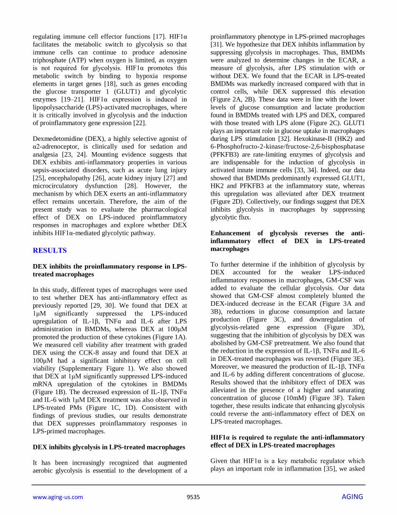
previously reported [29, 30]. We found that DEX at
1μM significantly suppressed the LPS-induced
upregulation of IL-1β, TNFα and IL-6 after LPS
administration in BMDMs, whereas DEX at 100μM
promoted the production of these cytokines (Figure 1A).
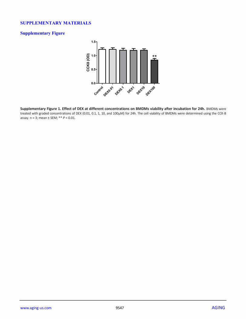
We measured cell viability after treatment with graded
DEX using the CCK-8 assay and found that DEX at
100μM had a significant inhibitory effect on cell
viability (Supplementary Figure 1). We also showed
that DEX at 1μM significantly suppressed LPS-induced
mRNA upregulation of the cytokines in BMDMs
(Figure 1B). The decreased expression of IL-1β, TNFα
and IL-6 with 1μM DEX treatment was also observed in
LPS-treated PMs (Figure 1C, 1D). Consistent with
findings of previous studies, our results demonstrate
that DEX suppresses proinflammatory responses in
LPS-primed macrophages.
DEX inhibits glycolysis in LPS-treated macrophages
It has been increasingly recognized that augmented
aerobic glycolysis is essential to the development of a
proinflammatory phenotype in LPS-primed macrophages
[31]. We hypothesize that DEX inhibits inflammation by
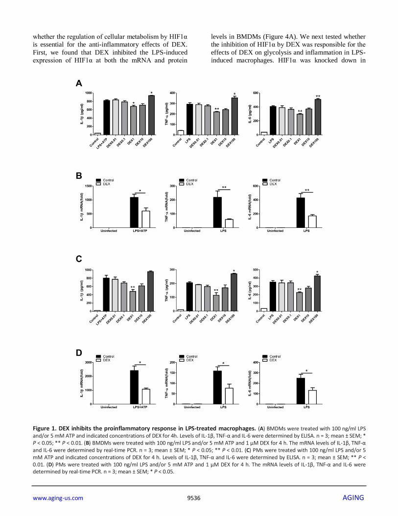
suppressing glycolysis in macrophages. Thus, BMDMs
were analyzed to determine changes in the ECAR, a
measure of glycolysis, after LPS stimulation with or
without DEX. We found that the ECAR in LPS-treated
BMDMs was markedly increased compared with that in
control cells, while DEX suppressed this elevation
(Figure 2A, 2B). These data were in line with the lower
levels of glucose consumption and lactate production
found in BMDMs treated with LPS and DEX, compared
with those treated with LPS alone (Figure 2C). GLUT1
plays an important role in glucose uptake in macrophages
during LPS stimulation [32]. Hexokinase-II (HK2) and
6-Phosphofructo-2-kinase/fructose-2,6-bisphosphatase
(PFKFB3) are rate-limiting enzymes of glycolysis and
are indispensable for the induction of glycolysis in
activated innate immune cells [33, 34]. Indeed, our data
showed that BMDMs predominantly expressed GLUT1,
HK2 and PFKFB3 at the inflammatory state, whereas
this upregulation was alleviated after DEX treatment
(Figure 2D). Collectively, our findings suggest that DEX
inhibits glycolysis in macrophages by suppressing
glycolytic flux.
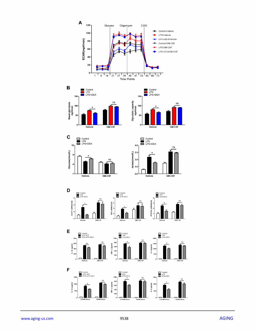
Enhancement of glycolysis reverses the anti-
inflammatory effect of DEX in LPS-treated
macrophages
To further determine if the inhibition of glycolysis by
DEX accounted for the weaker LPS-induced
inflammatory responses in macrophages, GM-CSF was
added to evaluate the cellular glycolysis. Our data
showed that GM-CSF almost completely blunted the
DEX-induced decrease in the ECAR (Figure 3A and
3B), reductions in glucose consumption and lactate
production (Figure 3C), and downregulation of
glycolysis-related gene expression (Figure 3D),
suggesting that the inhibition of glycolysis by DEX was
abolished by GM-CSF pretreatment. We also found that
the reduction in the expression of IL-1β, TNFα and IL-6
in DEX-treated macrophages was reversed (Figure 3E).
Moreover, we measured the production of IL-1β, TNFα
and IL-6 by adding different concentrations of glucose.
Results showed that the inhibitory effect of DEX was
alleviated in the presence of a higher and saturating
concentration of glucose (10mM) (Figure 3F). Taken
together, these results indicate that enhancing glycolysis
could reverse the anti-inflammatory effect of DEX on
LPS-treated macrophages.
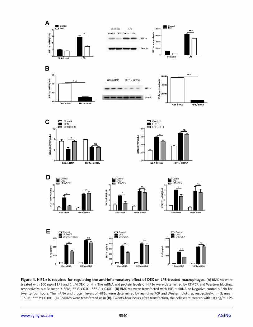
HIF1α is required to regulate the anti-inflammatory
effect of DEX in LPS-treated macrophages
Given that HIF1α is a key metabolic regulator which
plays an important role in inflammation [35], we asked
www.aging-us.com 9536 AGING
whether the regulation of cellular metabolism by HIF1α
is essential for the anti-inflammatory effects of DEX.
First, we found that DEX inhibited the LPS-induced
expression of HIF1α at both the mRNA and protein
levels in BMDMs (Figure 4A). We next tested whether
the inhibition of HIF1α by DEX was responsible for the
effects of DEX on glycolysis and inflammation in LPS-
induced macrophages. HIF1α was knocked down in
Figure 1. DEX inhibits the proinflammatory response in LPS-treated macrophages. (A) BMDMs were treated with 100 ng/ml LPS and/or 5 mM ATP and indicated concentrations of DEX for 4h. Levels of IL-1β, TNF-α and IL-6 were determined by ELISA. n = 3; mean ± SEM; * P < 0.05; ** P < 0.01. (B) BMDMs were treated with 100 ng/ml LPS and/or 5 mM ATP and 1 μM DEX for 4 h. The mRNA levels of IL-1β, TNF-α and IL-6 were determined by real-time PCR. n = 3; mean ± SEM; * P < 0.05; ** P < 0.01. (C) PMs were treated with 100 ng/ml LPS and/or 5 mM ATP and indicated concentrations of DEX for 4 h. Levels of IL-1β, TNF-α and IL-6 were determined by ELISA. n = 3; mean ± SEM; ** P < 0.01. (D) PMs were treated with 100 ng/ml LPS and/or 5 mM ATP and 1 μM DEX for 4 h. The mRNA levels of IL-1β, TNF-α and IL-6 were determined by real-time PCR. n = 3; mean ± SEM; * P < 0.05.
www.aging-us.com 9537 AGING
Figure 2. DEX inhibits glycolysis in LPS-treated macrophages. (A and B) BMDMs were seeded in Seahorse XFe96 cell culture microplates and treated with 100 ng/ml LPS and 1 μM DEX for 4 h. The real-time ECAR was recorded, and basal glycolysis and glycolytic capacity values were plotted. n = 5; mean ± SEM; * P < 0.05. (C) BMDMs were treated with 100 ng/ml LPS and 1 μM DEX for 4 h. Supernatants were collected, and the levels of glucose and lactate were measured. n = 3; mean ±SEM; * P < 0.05. (D) BMDMs were treated with 100 ng/ml LPS and 1 μM DEX for 4 h. The mRNA levels of GLUT1, HK2 and PFKFB3 were determined by RT-PCR. n = 3; mean ± SEM; * P < 0.05.
www.aging-us.com 9538 AGING
www.aging-us.com 9539 AGING
Figure 3. Enhancement of glycolysis reverses the anti-inflammatory effect of DEX on LPS-treated macrophages. (A and B) BMDMs were seeded in Seahorse XFe96 cell culture microplates and treated with 25 ng/ml GM-CSF for 24 h before being treated with 100 ng/ml LPS and 1 μM DEX for 4 h. The real-time ECAR was recorded, and basal glycolysis and glycolytic capacity values were plotted. n = 5; mean ± SEM; * P < 0.05. (C) BMDMs were treated with 25 ng/ml GM-CSF for 24 h before being treated with 100 ng/ml LPS and 1 μM DEX for 4 h. Supernatants were collected, and the levels of glucose and lactate were measured. n = 3; mean ±SEM; * P < 0.05. (D) BMDMs were treated with 25 ng/ml GM-CSF for 24 h before being treated with 100 ng/ml LPS and 1 μM DEX for 4 h. The mRNA levels of GLUT1, HK2 and PFKFB3 were determined by RT-PCR. n = 3; mean ± SEM; * P < 0.05. (E) BMDMs were treated with 25 ng/ml GM-CSF for 24 h before being treated with 100 ng/ml LPS and/or 5 mM ATP and 1 μM DEX for 4 h. Levels of IL-1β, TNF-α and IL-6 were measured by ELISA. n = 3; mean ± SEM; * P < 0.05; ** P < 0.01. (F) BMDMs were treated with 0.5mM or 10mM glucose before being treated with 100 ng/ml LPS and/or 5 mM ATP and 1 μM DEX for 4 h. Levels of IL-1β, TNF-α and IL-6 were measured by ELISA. n = 3; mean ± SEM; * P < 0.05.
BMDMs by special small-interfering RNA (siRNA) and
the success of the knockdown was confirmed (Figure
4B). We found that HIF1α knockdown reversed the
inhibitory effects of DEX on LPS-induced increases in
glucose consumption, lactate production (Figure 4C)
and glycolysis-associated gene expression (Figure 4D).
In addition, HIF1α knockdown reversed the inhibition
of IL-1β, TNFα and IL-6 expression by DEX in LPS-
treated macrophages (Figure 4E). Collectively, these
data suggest that DEX inhibits the production of
inflammatory cytokines partially by suppressing HIF1α
activation in macrophages.
DISCUSSION
Macrophages respond to microbial stimuli by triggering
the expression of an array of inflammatory cytokines,
which in turn cause the infiltration and activation
of other types of immune cells to orchestrate a full-
fledged immune-inflammatory response [36, 37].
Proinflammatory stimuli induces a metabolic switch in
macrophages, leading to a Warburg-like upregulation of
aerobic glycolysis to regulate the balance between
inflammatory and regulatory immune phenotypes [12,
38]. Here, we provide evidence for the DEX-mediated
regulation of glucose metabolism in activated
macrophages and suggest that DEX acts to inhibit
inflammatory responses in part by controlling the
HIF1α-dependent glycolytic pathway.
DEX has been regarded as a highly selective α2-
adrenoceptor agonist and is mostly applied in different
clinical settings for sedative or analgesic requirements.
Along with its beneficial effects, DEX has been
reported to potentially exert anti-inflammatory effects
during endotoxemia. A previous study revealed that
DEX significantly reduces mortality and decreases the
levels of inflammatory cytokines during endotoxemia in
rats [39]. DEX reduces sepsis-related acute lung injury
and has a protective effect on ischemia-reperfusion
injury of the heart, brain, kidneys, and intestine in
animal model [40–43]. DEX affects the immune cell
ratio and suppresses inflammatory cytokine production
in spleen and lymphocytes [44]. Our results were
consistent with findings of previous studies, showing
that DEX at 1μM significantly reduces the production
and release of proinflammatory cytokines by LPS-
induced macrophages. The dose of DEX used in our
study is much higher than its clinical use. Because of
interspecies variability, drug doses needed in animal
studies are usually much higher than that used clinically
(often up to 10-fold higher). Our data showed that the
dose of DEX used did not cause any damage to cell
viability. However, the safety of using such a high
dosage of DEX in septic patients remains to be
elucidated. Nevertheless, it was reported that some
patients, especially pediatric patients, required higher
dosages of DEX (up to approximately 5 to 10 times of
clinical dosages in adults) to achieve adequate sedation.
Moreover, such dosages were well-tolerated by those
patients [29, 30].
The activation of macrophages by proinflammatory
stimuli causes them to undergo a metabolic switch
towards glycolysis and away from OXPHOS. The
importance of glycolysis in the proinflammatory response
of macrophages has been demonstrated in a previous
study, whereas inhibition of glycolysis using 2-
deoxyglucose (2-DG) decreased the proinflammatory
response [13]. Gong et al. indicated that blockade of
glycolysis with 3-(3-pyridinyl)-1-(4-pyridinyl)-2-propen-
1-one (3PO) could alleviate sepsis-related acute lung
injury via suppressing inflammation and apoptosis of
alveolar epithelial cells [45]. Xie et al. indicated that
inhibition of pyruvate kinase 2 (PKM2), which catalyzes
the final and rate-limiting reaction of the glycolytic
pathway, could attenuate the NOD-, leucine rich region-
and pyrin domain-containing-3 (NLRP3) and absent in
melanoma 2 (AIM2) inflammasome activation and
consequently suppress the release of IL-1β and high
mobility group box 1 (HMGB1) [46]. Based on studies
mentioned above, we speculate that DEX might regulate
glycolysis in activated macrophages. Our data showing
the restraint of glycolysis by DEX via the suppression of
glucose uptake, lactate production and glycolytic gene
expression suggest that DEX might act to reverse the
metabolic program associated with the inflammatory
response.
www.aging-us.com 9540 AGING
Figure 4. HIF1α is required for regulating the anti-inflammatory effect of DEX on LPS-treated macrophages. (A) BMDMs were treated with 100 ng/ml LPS and 1 μM DEX for 4 h. The mRNA and protein levels of HIF1α were determined by RT-PCR and Western blotting, respectively. n = 3; mean ± SEM; ** P < 0.01, *** P < 0.001. (B) BMDMs were transfected with HIF1α siRNA or Negative control siRNA for twenty-four hours. The mRNA and protein levels of HIF1α were determined by real-time PCR and Western blotting, respectively. n = 3; mean ± SEM; *** P < 0.001. (C) BMDMs were transfected as in (B). Twenty-four hours after transfection, the cells were treated with 100 ng/ml LPS
www.aging-us.com 9541 AGING
and 1 μM DEX for 4 h. Supernatants were collected, and the levels of glucose and lactate were measured. n = 3; mean ± SEM; * P < 0.05. (D) BMDMs were transfected as in (B). Twenty-four hours after transfection, the cells were treated with 100 ng/ml LPS and 1 μM DEX for 4 h. The mRNA levels of GLUT1, HK2 and PFKFB3 were determined by RT-PCR. n = 3; mean ± SEM; * P < 0.05. (E) BMDMs were transfected as in (B). Twenty-four hours after transfection, the cells were treated with 100 ng/ml LPS and/or 5 mM ATP and 1 μM DEX for 4 h. Levels of IL-1β, TNF-α and IL-6 were determined by ELISA. n = 3; mean ± SD; ** P < 0.01.
We next asked whether the inhibition of aerobic
glycolysis mediated the immunological actions of DEX.
GM-CSF has been reported to augment glycolytic flux via
a mechanism that depends on PFKFB3 in vitro [47]. Na et
al. showed that GM-CSF increases macrophage glycolytic
capacity by upregulating GLUT expression [48]. In this
study, we showed that GM-CSF pretreatment almost
completely reversed the attenuating effects of DEX on the
LPS-induced enhancement of glycolysis and release of
inflammatory cytokines. It should be noted that this result
could not be completely attributed to the acceleration of
glycolysis, for that GM-CSF might have other effects
through which it can promote LPS-induced inflammatory
cytokine expression. Therefore, we next measured the
production of IL-1β, TNFα and IL-6 in the presence of a
saturating concentration of glucose and found that DEX
was much less effective under this condition, suggesting
that the anti-inflammatory effect of DEX can be
attenuated by enhancing glycolysis.
It is increasingly recognized that HIF1α acts as a central
regulator of cellular metabolism and promotes
inflammatory gene expression. Blouin et al. were the first
to show that the stimulation of macrophages with LPS
increases HIF1α protein levels, leading to the formation of
a functional HIF-1 complex that binds to hypoxic
response elements in target genes, including GLUT1,
HK2 and PFKFB3 [49]. It was later found that HIF1α-
mediated glycolytic reprogramming of activated
macrophages plays a significant role in monocyte-derived
macrophage migration into tissues [50]. HIF1α also
induces the transcription of the inflammatory cytokines.
Our results indicated that DEX inhibited HIF1α
expression, which implied that DEX controlled metabolic
processes by engaging in the regulation of HIF1α
signaling. Our conjecture described above was supported
by the finding that the DEX-mediated inhibition of
glycolysis was reversed after HIF1α knockdown. In
addition, we showed that the restraint of IL-1β, TNFα and
IL-6 production by DEX was abolished by decreasing
HIF1α levels genetically with HIF1α knockdown. DEX
may inhibit cerebral ischemia-reperfusion (I/R) injury by
inhibiting the HIF1α pathway and inhibiting apoptosis in
I/R rat brain. The inhibition of HIF1α by DEX restored
the balance between catabolic aerobic processes and
catabolic anaerobic processes [51]. Our results indicate
that DEX inhibits glycolysis by suppressing HIF1α, which
was consistent with the previous study [51]. Collectively,
these data suggest that HIF1α is necessary for DEX to
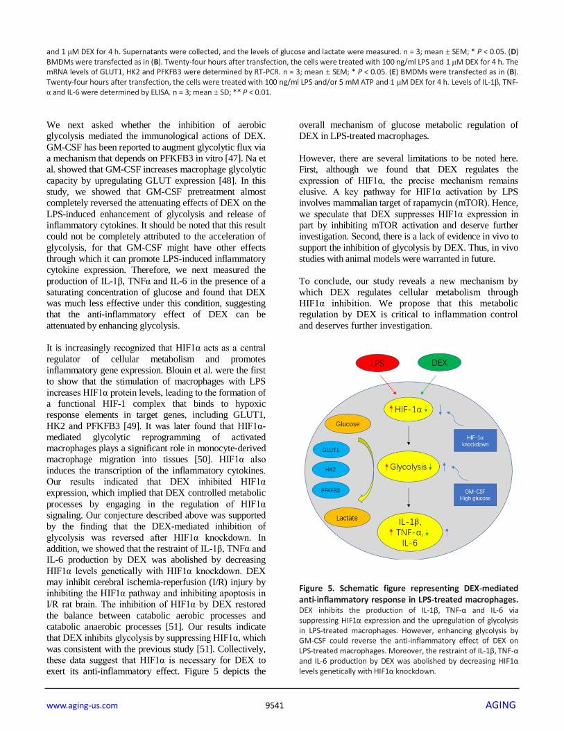
exert its anti-inflammatory effect. Figure 5 depicts the
overall mechanism of glucose metabolic regulation of
DEX in LPS-treated macrophages.
However, there are several limitations to be noted here.
First, although we found that DEX regulates the
expression of HIF1α, the precise mechanism remains
elusive. A key pathway for HIF1α activation by LPS
involves mammalian target of rapamycin (mTOR). Hence,
we speculate that DEX suppresses HIF1α expression in
part by inhibiting mTOR activation and deserve further
investigation. Second, there is a lack of evidence in vivo to
support the inhibition of glycolysis by DEX. Thus, in vivo
studies with animal models were warranted in future.
To conclude, our study reveals a new mechanism by
which DEX regulates cellular metabolism through
HIF1α inhibition. We propose that this metabolic
regulation by DEX is critical to inflammation control
and deserves further investigation.
Figure 5. Schematic figure representing DEX-mediated anti-inflammatory response in LPS-treated macrophages. DEX inhibits the production of IL-1β, TNF-α and IL-6 via suppressing HIF1α expression and the upregulation of glycolysis in LPS-treated macrophages. However, enhancing glycolysis by GM-CSF could reverse the anti-inflammatory effect of DEX on LPS-treated macrophages. Moreover, the restraint of IL-1β, TNF-α and IL-6 production by DEX was abolished by decreasing HIF1α levels genetically with HIF1α knockdown.
www.aging-us.com 9542 AGING
MATERIALS AND METHODS
Reagents
LPS from Escherichia coli O111:B4, ATP (A2383)
and DEX (SML0956) were obtained from Sigma-
Aldrich. GM-CSF and M-CSF were purchased from
PeproTech.
Cell culture and animals
Bone marrow-derived cells from C57BL/6J mice were
cultured in RPMI 1640 medium (Invitrogen)
supplemented with 10% FBS and 1% penicillin/
streptomycin and differentiated into bone marrow-
derived macrophages (BMDMs) with murine
macrophage colony-stimulating factor (M-CSF)
treatment for 5 d. Peritoneal macrophages (PMs) were
elicited in C57BL/6J mice 3 d after the intraperitoneal
injection of 4% thioglycolate (Sigma-Aldrich) and
cultured in RPMI 1640 medium without glucose. Male
C57BL/6 mice were obtained from the Laboratory
Animal Center of Naval Medical University and housed
in a specific pathogen-free environment at the optimal
temperature with a 12 h light/dark cycle.
Small-interfering RNA (siRNA) treatment
The siRNA sequences were designed at GeneChem
(Shanghai, China). The following siRNA sequences were
used: HIF1α siRNA: 5’-GCUCACCAUCAGUUAUUUA
TT-3’, and Negative control siRNA: 5’-UUCUCCGAA
CGUGUCACGUTT-3’. Murine BMDMs were
transfected with the siRNAs using Lipofectamine 2000
(Invitrogen) according to the manufacturer’ instructions.
The supernatant was replaced with complete culture
medium after 24 h.
Extracellular acidification rate (ECAR)
The XFe96 Extracellular Flux Analyzer (Agilent) was
used for real-time recording of the ECAR. In brief,
BMDMs were seeded in Seahorse XFe96 microplates
(3×10⁴ cells per well) and treated with LPS, DEX, or
both. Before analysis, the cells were incubated in
ECAR medium for 1 h at 37°C in room air. The cells
were sequentially treated with 10 mM glucose, 1 μM
oligomycin, and 50 mM 2-DG. Real-time ECARs
were recorded according to the manufacturer’s
manual.
Metabolite measurements
The glucose and lactate concentrations within cell
medium were determined by the Glucose and Lactate
Colorimetric Assay Kits (BioVision), respectively.
Cell Counting Kit-8 assay
The viability of BMDMs were determined using the
Cell Counting Kit-8 assay kit (Dojindo) as previously
reported [52].
Real-time polymerase chain reaction (PCR)
Total RNA was extracted from cells using RNAiso
(TaKaRa) according to the manufacturer’s instructions.
cDNA was synthesized using a PrimeScriptTMRT
reagent kit (TaKaRa). Real-time PCR was performed
using SYBR green master mix (TaKaRa). The real-time
PCR primers used in this study are listed in
Supplementary Table 1 of the Supplementary Materials.
Enzyme-linked immunosorbent assay (ELISA)
Levels of IL-1β, TNFα and IL-6 in cell supernatants were
measured with commercial ELISA kits (eBioscience)
according to the manufacturer’s instructions.
Western blotting
Cells were homogenized in lysis buffer, and protein
lysates were separated on 10% SDS gels and transferred
to polyvinylidene fluoride membranes (Millipore). After
blocking, the membranes were incubated with a primary
antibody (anti-HIF1α, Cell Signaling Technology)
overnight, followed by a 1 h incubation with a
horseradish peroxidase-conjugated secondary antibody
(Cell Signaling Technology) at room temperature. The
band intensity was quantified by densitometric analyses
using ImageJ software.
Statistical analysis
Data are presented as the mean ± standard deviation.
Significant differences between multiple groups were
detected using ANOVA. Differences between two
groups were detected using a t test. All analyses were
performed using GraphPad Prism 5 statistical software.
A value of P < 0.05 was considered statistically
significant.
Abbreviations
DEX: dexmedetomidine; HIF1α: hypoxia inducible
factor 1α; ECAR: extracellular acidification rate; GM-
CSF: granulocyte-macrophage colony-stimulating
factor; TLRs: toll-like receptors; OXPHOS: oxidative
phosphorylation; GLUT1: glucose transporter 1;
HK2: Hexokinase-II; PFKFB3: 6-Phosphofructo-
2-kinase/fructose-2,6-bisphosphatase; 2-DG: 2-
deoxyglucose; 3PO: 3-(3-pyridinyl)-1-(4-pyridinyl)-
2-propen-1-one; PKM2: pyruvate kinase 2; NLRP3:
www.aging-us.com 9543 AGING
NOD-, leucine rich region- and pyrin domain-
containing-3; AIM2: absent in melanoma 2; HMGB1:
high mobility group box 1; mTOR: mammalian target
of rapamycin.
AUTHOR CONTRIBUTIONS
MQY and GPH performed most experiments. BJJ
designed experiments and provided the financial
support. MQY performed Seahorse XF Glycolysis
Stress Test. BLL and GPH contributed to the statistical
analysis. MQY and JZY wrote the paper.
ACKNOWLEDGMENTS
The authors would like to thank Y Zhang, N Li and J
WANG for excellent technical assistance.
CONFLICTS OF INTEREST
The authors have no financial conflicts of interest.
FUNDING
This work was supported by the National Natural Science
Foundation of China (no.81671939, 81871579,
81671887) and the Shanghai Outstanding Youth Medical
Professionals Training Program (no.2017YQ015), and
Shanghai Science and Technology Committee Rising-Star
Program (19QA1408500).
REFERENCES
1. Akira S, Uematsu S, Takeuchi O. Pathogen recognition and innate immunity. Cell. 2006; 124:783–801.
https://doi.org/10.1016/j.cell.2006.02.015 PMID:16497588
2. Janeway CA Jr, Medzhitov R. Innate immune recognition. Annu Rev Immunol. 2002; 20:197–216.
https://doi.org/10.1146/annurev.immunol.20.083001.084359 PMID:11861602
3. Takeuchi O, Akira S. Pattern recognition receptors and inflammation. Cell. 2010; 140:805–20.
https://doi.org/10.1016/j.cell.2010.01.022 PMID:20303872
4. Lawrence T, Natoli G. Transcriptional regulation of macrophage polarization: enabling diversity with identity. Nat Rev Immunol. 2011; 11:750–61.
https://doi.org/10.1038/nri3088 PMID:22025054
5. Murray PJ, Wynn TA. Obstacles and opportunities for understanding macrophage polarization. J Leukoc Biol. 2011; 89:557–63.
https://doi.org/10.1189/jlb.0710409 PMID:21248152
6. Wynn TA, Chawla A, Pollard JW. Macrophage biology in development, homeostasis and disease. Nature. 2013; 496:445–55.
https://doi.org/10.1038/nature12034 PMID:23619691
7. Mosser DM, Edwards JP. Exploring the full spectrum of macrophage activation. Nat Rev Immunol. 2008; 8:958–69.
https://doi.org/10.1038/nri2448 PMID:19029990
8. Sica A, Mantovani A. Macrophage plasticity and polarization: in vivo veritas. J Clin Invest. 2012; 122:787–95.
https://doi.org/10.1172/JCI59643 PMID:22378047
9. Pearce EL, Pearce EJ. Metabolic pathways in immune cell activation and quiescence. Immunity. 2013; 38:633–43.
https://doi.org/10.1016/j.immuni.2013.04.005 PMID:23601682
10. O’Neill LA, Pearce EJ. Immunometabolism governs dendritic cell and macrophage function. J Exp Med. 2016; 213:15–23.
https://doi.org/10.1084/jem.20151570 PMID:26694970
11. Ganeshan K, Chawla A. Metabolic regulation of immune responses. Annu Rev Immunol. 2014; 32:609–34.
https://doi.org/10.1146/annurev-immunol-032713-120236 PMID:24655299
12. Kelly B, O’Neill LA. Metabolic reprogramming in macrophages and dendritic cells in innate immunity. Cell Res. 2015; 25:771–84.
https://doi.org/10.1038/cr.2015.68 PMID:26045163
13. Tannahill GM, Curtis AM, Adamik J, Palsson-McDermott EM, McGettrick AF, Goel G, Frezza C, Bernard NJ, Kelly B, Foley NH, Zheng L, Gardet A, Tong Z, et al. Succinate is an inflammatory signal that induces IL-1β through HIF-1α. Nature. 2013; 496:238–42.
https://doi.org/10.1038/nature11986 PMID:23535595
14. Vats D, Mukundan L, Odegaard JI, Zhang L, Smith KL, Morel CR, Wagner RA, Greaves DR, Murray PJ, Chawla A. Oxidative metabolism and PGC-1beta attenuate macrophage-mediated inflammation. Cell Metab. 2006; 4:13–24.
https://doi.org/10.1016/j.cmet.2006.05.011 PMID:16814729
15. Rodríguez-Prados JC, Través PG, Cuenca J, Rico D, Aragonés J, Martín-Sanz P, Cascante M, Boscá L. Substrate fate in activated macrophages: a comparison
www.aging-us.com 9544 AGING
between innate, classic, and alternative activation. J Immunol. 2010; 185:605–14.
https://doi.org/10.4049/jimmunol.0901698 PMID:20498354
16. Galván-Peña S, O'Neill LA. Metabolic reprograming in macrophage polarization. Front Immunol. 2014; 5:420.
https://doi.org/10.3389/fimmu.2014.00420 PMID:25228902
17. Corcoran SE, O’Neill LA. HIF1α and metabolic reprogramming in inflammation. J Clin Invest. 2016; 126:3699–707.
https://doi.org/10.1172/JCI84431 PMID:27571407
18. Mole DR, Blancher C, Copley RR, Pollard PJ, Gleadle JM, Ragoussis J, Ratcliffe PJ. Genome-wide association of hypoxia-inducible factor (HIF)-1alpha and HIF-2alpha DNA binding with expression profiling of hypoxia-inducible transcripts. J Biol Chem. 2009; 284:16767–75.
https://doi.org/10.1074/jbc.M901790200 PMID:19386601
19. Chen C, Pore N, Behrooz A, Ismail-Beigi F, Maity A. Regulation of glut1 mRNA by hypoxia-inducible factor-1. Interaction between H-ras and hypoxia. J Biol Chem. 2001; 276:9519–25.
https://doi.org/10.1074/jbc.M010144200 PMID:11120745
20. Obach M, Navarro-Sabaté A, Caro J, Kong X, Duran J, Gómez M, Perales JC, Ventura F, Rosa JL, Bartrons R. 6-phosphofructo-2-kinase (pfkfb3) gene promoter contains hypoxia-inducible factor-1 binding sites necessary for transactivation in response to hypoxia. J Biol Chem. 2004; 279:53562–70.
https://doi.org/10.1074/jbc.M406096200 PMID:15466858
21. Riddle SR, Ahmad A, Ahmad S, Deeb SS, Malkki M, Schneider BK, Allen CB, White CW. Hypoxia induces hexokinase II gene expression in human lung cell line A549. Am J Physiol Lung Cell Mol Physiol. 2000; 278:L407–16.
https://doi.org/10.1152/ajplung.2000.278.2.L407 PMID:10666126
22. Wang T, Liu H, Lian G, Zhang SY, Wang X, Jiang C. HIF1 α-induced glycolysis metabolism is essential to the activation of inflammatory macrophages. Mediators Inflamm. 2017; 2017:9029327.
https://doi.org/10.1155/2017/9029327 PMID:29386753
23. Kawazoe Y, Miyamoto K, Morimoto T, Yamamoto T, Fuke A, Hashimoto A, Koami H, Beppu S, Katayama Y, Itoh M, Ohta Y, Yamamura H, and Dexmedetomidine for Sepsis in Intensive Care Unit Randomized Evaluation (DESIRE) Trial Investigators. Effect of
dexmedetomidine on mortality and ventilator-free days in patients requiring mechanical ventilation with sepsis: a randomized clinical trial. JAMA. 2017; 317:1321–28.
https://doi.org/10.1001/jama.2017.2088 PMID:28322414
24. Su X, Meng ZT, Wu XH, Cui F, Li HL, Wang DX, Zhu X, Zhu SN, Maze M, Ma D. Dexmedetomidine for prevention of delirium in elderly patients after non-cardiac surgery: a randomised, double-blind, placebo-controlled trial. Lancet. 2016; 388:1893–902.
https://doi.org/10.1016/S0140-6736(16)30580-3 PMID:27542303
25. Wu Y, Liu Y, Huang H, Zhu Y, Zhang Y, Lu F, Zhou C, Huang L, Li X, Zhou C. Dexmedetomidine inhibits inflammatory reaction in lung tissues of septic rats by suppressing TLR4/NF-κB pathway. Mediators Inflamm. 2013; 2013:562154.
https://doi.org/10.1155/2013/562154 PMID:23690665
26. Ning Q, Liu Z, Wang X, Zhang R, Zhang J, Yang M, Sun H, Han F, Zhao W, Zhang X. Neurodegenerative changes and neuroapoptosis induced by systemic lipopolysaccharide administration are reversed by dexmedetomidine treatment in mice. Neurol Res. 2017; 39:357–66.
https://doi.org/10.1080/01616412.2017.1281197 PMID:28173746
27. Kang K, Gao Y, Wang SC, Liu HT, Kong WL, Zhang X, Huang R, Qi ZD, Zheng JB, Qu JD, Liu RJ, Liu YS, Wang HL, Yu KJ. Dexmedetomidine protects against lipopolysaccharide-induced sepsis-associated acute kidney injury via an α7 nAChR-dependent pathway. Biomed Pharmacother. 2018; 106:210–16.
https://doi.org/10.1016/j.biopha.2018.06.059 PMID:29960167
28. Miranda ML, Balarini MM, Bouskela E. Dexmedetomidine attenuates the microcirculatory derangements evoked by experimental sepsis. Anesthesiology. 2015; 122:619–30.
https://doi.org/10.1097/ALN.0000000000000491 PMID:25313879
29. Lai YC, Tsai PS, Huang CJ. Effects of dexmedetomidine on regulating endotoxin-induced up-regulation of inflammatory molecules in murine macrophages. J Surg Res. 2009; 154:212–19.
https://doi.org/10.1016/j.jss.2008.07.010 PMID:19181340
30. Chang Y, Huang X, Liu Z, Han G, Huang L, Xiong YC, Wang Z. Dexmedetomidine inhibits the secretion of high mobility group box 1 from lipopolysaccharide-activated macrophages in vitro. J Surg Res. 2013; 181:308–14.
www.aging-us.com 9545 AGING
https://doi.org/10.1016/j.jss.2012.07.017 PMID:22939552
31. Cui H, Banerjee S, Guo S, Xie N, Liu G. IFN regulatory factor 2 inhibits expression of glycolytic genes and lipopolysaccharide-induced proinflammatory responses in macrophages. J Immunol. 2018; 200:3218–30.
https://doi.org/10.4049/jimmunol.1701571 PMID:29563175
32. Freemerman AJ, Johnson AR, Sacks GN, Milner JJ, Kirk EL, Troester MA, Macintyre AN, Goraksha-Hicks P, Rathmell JC, Makowski L. Metabolic reprogramming of macrophages: glucose transporter 1 (GLUT1)-mediated glucose metabolism drives a proinflammatory phenotype. J Biol Chem. 2014; 289:7884–96.
https://doi.org/10.1074/jbc.M113.522037 PMID:24492615
33. Li Y, Lu B, Sheng L, Zhu Z, Sun H, Zhou Y, Yang Y, Xue D, Chen W, Tian X, Du Y, Yan M, Zhu W, et al. Hexokinase 2-dependent hyperglycolysis driving microglial activation contributes to ischemic brain injury. J Neurochem. 2018; 144:186–200.
https://doi.org/10.1111/jnc.14267 PMID:29205357
34. Jiang H, Shi H, Sun M, Wang Y, Meng Q, Guo P, Cao Y, Chen J, Gao X, Li E, Liu J. PFKFB3-driven macrophage glycolytic metabolism is a crucial component of innate antiviral defense. J Immunol. 2016; 197:2880–90.
https://doi.org/10.4049/jimmunol.1600474 PMID:27566823
35. Zhang W, Petrovic JM, Callaghan D, Jones A, Cui H, Howlett C, Stanimirovic D. Evidence that hypoxia-inducible factor-1 (HIF-1) mediates transcriptional activation of interleukin-1beta (IL-1beta) in astrocyte cultures. J Neuroimmunol. 2006; 174:63–73.
https://doi.org/10.1016/j.jneuroim.2006.01.014 PMID:16504308
36. Biswas SK, Chittezhath M, Shalova IN, Lim JY. Macrophage polarization and plasticity in health and disease. Immunol Res. 2012; 53:11–24.
https://doi.org/10.1007/s12026-012-8291-9 PMID:22418728
37. Shapouri-Moghaddam A, Mohammadian S, Vazini H, Taghadosi M, Esmaeili SA, Mardani F, Seifi B, Mohammadi A, Afshari JT, Sahebkar A. Macrophage plasticity, polarization, and function in health and disease. J Cell Physiol. 2018; 233:6425–40.
https://doi.org/10.1002/jcp.26429 PMID:29319160
38. Kornberg MD, Bhargava P, Kim PM, Putluri V, Snowman AM, Putluri N, Calabresi PA, Snyder SH. Dimethyl fumarate targets GAPDH and aerobic
glycolysis to modulate immunity. Science. 2018; 360:449–53.
https://doi.org/10.1126/science.aan4665 PMID:29599194
39. Taniguchi T, Kidani Y, Kanakura H, Takemoto Y, Yamamoto K. Effects of dexmedetomidine on mortality rate and inflammatory responses to endotoxin-induced shock in rats. Crit Care Med. 2004; 32:1322–26.
https://doi.org/10.1097/01.ccm.0000128579.84228.2a PMID:15187514
40. Wang Y, Wang C, Zhang D, Wang H, Bo L, Deng X. Dexmedetomidine protects against traumatic brain injury-induced acute lung injury in mice. Med Sci Monit. 2018; 24:4961–67.
https://doi.org/10.12659/MSM.908133 PMID:30013022
41. Shen M, Wang S, Wen X, Han XR, Wang YJ, Zhou XM, Zhang MH, Wu DM, Lu J, Zheng YL. Dexmedetomidine exerts neuroprotective effect via the activation of the PI3K/Akt/mTOR signaling pathway in rats with traumatic brain injury. Biomed Pharmacother. 2017; 95:885–93.
https://doi.org/10.1016/j.biopha.2017.08.125 PMID:28903184
42. Qiu R, Yao W, Ji H, Yuan D, Gao X, Sha W, Wang F, Huang P, Hei Z. Dexmedetomidine restores septic renal function via promoting inflammation resolution in a rat sepsis model. Life Sci. 2018; 204:1–8.
https://doi.org/10.1016/j.lfs.2018.05.001 PMID:29733849
43. Chen Y, Miao L, Yao Y, Wu W, Wu X, Gong C, Qiu L, Chen J. Dexmedetomidine ameliorate CLP-induced rat intestinal injury via inhibition of inflammation. Mediators Inflamm. 2015; 2015:918361.
https://doi.org/10.1155/2015/918361 PMID:26273145
44. Xianbao L, Hong Z, Xu Z, Chunfang Z, Dunjin C. Dexmedetomidine reduced cytokine release during postpartum bleeding-induced multiple organ dysfunction syndrome in rats. Mediators Inflamm. 2013; 2013:627831.
https://doi.org/10.1155/2013/627831 PMID:23840096
45. Gong Y, Lan H, Yu Z, Wang M, Wang S, Chen Y, Rao H, Li J, Sheng Z, Shao J. Blockage of glycolysis by targeting PFKFB3 alleviates sepsis-related acute lung injury via suppressing inflammation and apoptosis of alveolar epithelial cells. Biochem Biophys Res Commun. 2017; 491:522–29.
https://doi.org/10.1016/j.bbrc.2017.05.173 PMID:28576491
46. Xie M, Yu Y, Kang R, Zhu S, Yang L, Zeng L, Sun X, Yang M, Billiar TR, Wang H, Cao L, Jiang J, Tang D. PKM2-

www.aging-us.com 9546 AGING
dependent glycolysis promotes NLRP3 and AIM2 inflammasome activation. Nat Commun. 2016; 7:13280.
https://doi.org/10.1038/ncomms13280 PMID:27779186
47. Singh P, González-Ramos S, Mojena M, Rosales-Mendoza CE, Emami H, Swanson J, Morss A, Fayad ZA, Rudd JH, Gelfand J, Paz-García M, Martín-Sanz P, Boscá L, Tawakol A. GM-CSF enhances macrophage glycolytic activity in vitro and improves detection of inflammation in vivo. J Nucl Med. 2016; 57:1428–35.
https://doi.org/10.2967/jnumed.115.167387 PMID:27081166
48. Na YR, Gu GJ, Jung D, Kim YW, Na J, Woo JS, Cho JY, Youn H, Seok SH. GM-CSF induces inflammatory macrophages by regulating glycolysis and lipid metabolism. J Immunol. 2016; 197:4101–09.
https://doi.org/10.4049/jimmunol.1600745 PMID:27742831
49. Blouin CC, Pagé EL, Soucy GM, Richard DE. Hypoxic gene activation by lipopolysaccharide in macrophages: implication of hypoxia-inducible factor 1alpha. Blood. 2004; 103:1124–30.
https://doi.org/10.1182/blood-2003-07-2427 PMID:14525767
50. Semba H, Takeda N, Isagawa T, Sugiura Y, Honda K, Wake M, Miyazawa H, Yamaguchi Y, Miura M, Jenkins DM, Choi H, Kim JW, Asagiri M, et al. HIF-1α-PDK1 axis-induced active glycolysis plays an essential role in macrophage migratory capacity. Nat Commun. 2016; 7:11635.
https://doi.org/10.1038/ncomms11635 PMID:27189088
51. Wang YQ, Tang YF, Yang MK, Huang XZ. Dexmedetomidine alleviates cerebral ischemia-reperfusion injury in rats via inhibition of hypoxia-inducible factor-1α. J Cell Biochem. 2018. [Epub ahead of print].
https://doi.org/10.1002/jcb.28058 PMID:30456861
52. Zhou JR, Xu Z, Jiang CL. Neuropeptide Y promotes TGF-beta1 production in RAW264.7 cells by activating PI3K pathway via Y1 receptor. Neurosci Bull. 2008; 24:155–59.
https://doi.org/10.1007/s12264-008-0130-6 PMID:18500388
www.aging-us.com 9547 AGING
SUPPLEMENTARY MATERIALS
Supplementary Figure
Supplementary Figure 1. Effect of DEX at different concentrations on BMDMs viability after incubation for 24h. BMDMs were treated with graded concentrations of DEX (0.01, 0.1, 1, 10, and 100μM) for 24h. The cell viability of BMDMs were determined using the CCK-8 assay. n = 3; mean ± SEM; ** P < 0.01.
www.aging-us.com 9548 AGING
Supplementary Table
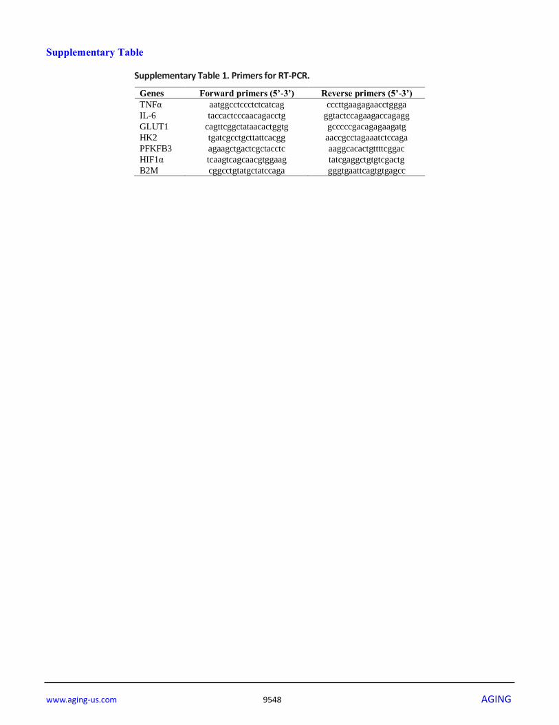
Supplementary Table 1. Primers for RT-PCR.
Genes Forward primers (5’-3’) Reverse primers (5’-3’)
TNFα aatggcctccctctcatcag cccttgaagagaacctggga
IL-6 taccactcccaacagacctg ggtactccagaagaccagagg
GLUT1 cagttcggctataacactggtg gcccccgacagagaagatg
HK2 tgatcgcctgcttattcacgg aaccgcctagaaatctccaga
PFKFB3 agaagctgactcgctacctc aaggcacactgttttcggac
HIF1α tcaagtcagcaacgtggaag tatcgaggctgtgtcgactg
B2M cggcctgtatgctatccaga gggtgaattcagtgtgagcc