development of an isolated, in vitro c. elegans gonad preparation adam broslat advisor: dr. kevin...
TRANSCRIPT

Development of an Isolated, in Vitro C. elegans Gonad Preparation
Adam Broslat
Advisor: Dr. Kevin Strange
Professor of Anesthesiology and Pharmacology

What are C. elegans ?
• nematode ~1 mm long
•hermaphroditic
•completely sequenced genome (97Mb or 19K distinct genes)
•simple body plan (959 somatic cells)

Design Project
Goal:
design an isolated, in vitro C. elegans gonad preparation protocol.
Purposes for Project
• for the purpose of characterizing the molecular mechanisms of heterologous cell-to-cell communication using quantitative microscopy
This includes: Voltage sensitive dyes, pH sensitive dyes, Ca+ sensitive dyes, etc.

Research Description
We recently identified a ClC-type anion channel encoded by clh-3 that is functionally expressed in C. elegans oocytes. CLH-3 is activated uring oocyte meiotic maturation suggesting that the channel plays a role in meiotic cell cycle progression, ovulation, fertilization, and/or early development. Disruption of channel expression by RNA interference has little effect on various reproductive events. However, in worms injected with clh-3 dsRNA, we observed that ovulatory contractions of the gonadal sheath cells were initiated prematurely This suggests that CLH-3 functions in inhibitory signaling pathways that modulate sheath cell contractile activity (Rutledge et al., Curr. Biol.11: 161-170, 2001). Oocytes are coupled to sheath cells by gap junctions (Hall et al., Dev. Biol. 212:101-123, 1999) indicating that the two cell types may communicate via electrical and chemical signals. We postulated that activation of CLH-3 during meiotic maturation depolarizes the oocyte and electrically-coupled sheath cell plasma membranes. We also postulated that depolarization modulates sheath cell contractile activity by regulating calcium influx via receptor-activated calcium channels that are triggered by depletion of IP3-sensitive intracellular calcium stores. To begin testing this model, we have developed an isolated gonad preparation.

Project Phases
• Phase 1– Physical extraction of gonad
• Where, when, and how
• Phase 2– Functional buffer development
• allow normal function
• Phase 3– Imaging
• Staging setup for gonad positioning and stabilization
1st Phase – Micro-dissection procedure
• The nematode's gonad will be isolated in such a way not to harm the physiology of the gonad.
• Gonad operates independently of the worm. Problems:
The intestines “cloud” the view of gonad after dissection. Gonad does not completely remove itself without manipulation.Transport of isolated gonad is difficult.

Solutions to Date
• Dissection is made in the scope chamber filled with buffer
• Incisions made with a modified injection needle (guillotined at red line)
• Gonad is half extracted through depressurization
• The other half is forced by suction using micropipettes or cutting past spermatheca in uterus

2nd Phase – Functional Buffer
• The worm and/or gonad must be placed in a buffer solution that promotes normal gonad function while being observed.
Problems:Worms are extremely active in buffer.Buffer allows floating and movement.Buffer evaporates from scope chamber
Solutions to Date
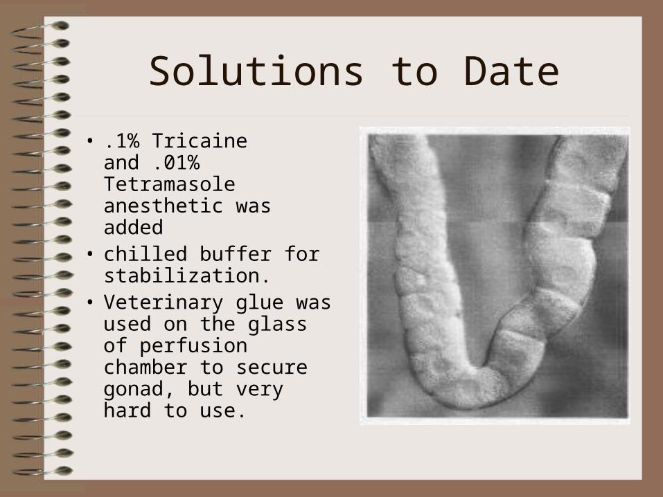
• .1% Tricaine and .01% Tetramasole anesthetic was added
• chilled buffer for stabilization.
• Veterinary glue was used on the glass of perfusion chamber to secure gonad, but very hard to use.
Buffer Recipes
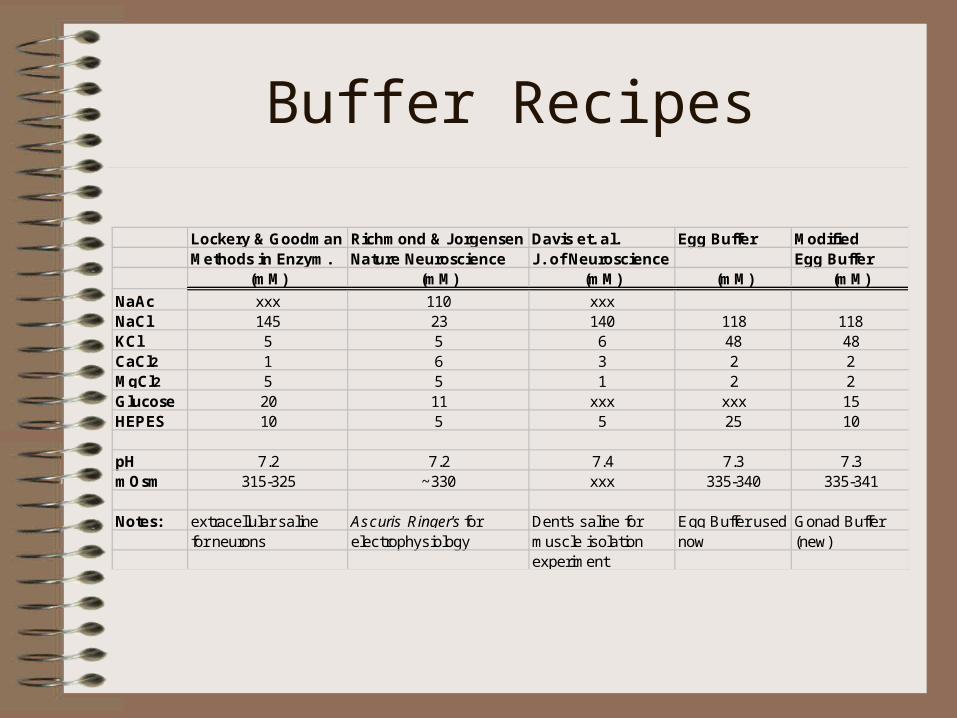
Lockery & Goodman Richmond & Jorgensen Davis et. al. Egg Buffer Modified Methods in Enzym. Nature Neuroscience J. of Neuroscience Egg Buffer
(mM) (mM) (mM) (mM) (mM)
NaAc xxx 110 xxxNaCl 145 23 140 118 118KCl 5 5 6 48 48CaCl2 1 6 3 2 2MgCl2 5 5 1 2 2Glucose 20 11 xxx xxx 15HEPES 10 5 5 25 10
pH 7.2 7.2 7.4 7.3 7.3mOsm 315-325 ~330 xxx 335-340 335-341
Notes: extracellular saline Ascuris Ringer's for Dent's saline for Egg Buffer used Gonad Buffer for neurons electrophysiology muscle isolation now (new)
experiment

Worm Response to Buffer
• Any of the previous listed buffers would sustain function to a point.
• The worm gonad needs the the salts to mimic the interstitial fluid of the worm.
• The gonad must also have an energy source . . .this is where the glucose comes in.

Actual Response to BufferLockery & Goodman Richmond & Jorgensen Davis et. al. Egg Buffer Modified Methods in Enzym. Nature Neuroscience J. of Neuroscience Egg Buffer
NormalContraction yes yes yes yes yes
Length of normal contractile activityTrial 1 x 38 minutes 49 minutes 58 minutes 96 minutesTrial 2 x 45 minutes 49 minutes 55 minutes 88 minutesTrial 3 x 44 minutes 44 minutes 64 minutes 85 minutesTrial 4 x x x 66 minutes 107 minutes
Notes: extracellular saline Ascuris Ringer'sfor Dent's saline for Egg Buffer used Gonad Buffer for neurons electrophysiology muscle isolation now (new)
experiment

3rd Phase - Imaging
• Gonad stabilization
• DIC image acquisition
• Imaging with argon laser confocal microscope (time allowing)
• Tie the process together and formalize protocol

3rd Phase Problems
• Glue mentioned earlier does not work well– Prevents further slide use– Immediately solidifies under liquid buffer
• Any movement under 63x DIC scope causes focal plane change or field change
• Protocol must work for various microscope setups (DIC, laser confocal, etc)

3rd Phase Solutions
• Using similar micro-pipettes as in dissection, gonad can be held on both ends.
• Pipettes must be mounted on both sides and allow for x,y, and z motion for initial placement to accommodate gonad.
• Pipette tip must be modified to restrict complete entry of gonad

Stage / Rig Setup
Pipette Holder
Y- motion Translation stage
Dampened post
X- motion Translation stage
Z- motion adjustable platform
Magnetic Base