determination of methamphetamine by gc-mass based on ... synthesis of mips for methamphetamine...
TRANSCRIPT

Eurasian Journal of Analytical Chemistry ISSN: 1306-3057 OPEN ACCESS 2019 14 (2): 71-83
Received: 09 March 2019 ▪ Revised: 14 April 2019 ▪ Accepted: 20 April 2019
Abstract: A novel method characterized by high sensitivity, low cost and high stability was
developed. This method based on a molecularly imprinted polymer(MIP) using a functional
monomer which is 2-vinylpyridine (2-VP) and 2-hydroxyethyl methacrylate (2-HEMA),
suitable cross-linker and the template which is MAMP to fabricate a monolithic solid-phase
micro extraction (SPME) fiber. A polymer was also prepared without selective binding sites
this was referred to as a non-imprinted polymer (NIP). (SPME) with gas chromatography
and mass spectrometry (GC/MS); all these analytical methods used to extraction, pre-
concentration and selective determination of methamphetamine (MAMP) and its
derivatives. Firmness, stability and duration of the fabricated fiber give it its fundamental
and indispensable role in SPME.The samples were collected from methamphetamine
suspected abuser donate provided from the medico legal Directorate (Baghdad,
Iraq).Monitoring of the analytes was performed using a (GC MS) and using UV-vis and
Scanning electron microscopy (SEM) and FTIR. The relative standard deviations ) RSD%)
for five Patients repeated experiments for three measurements are range of (at 30-60 ppm
of MAMP) is (1.20-3.61) %. The relative recoveries obtained for MAMP in spiked human
urine samples are in the range of (93.63-99.71).
Keywords: Methamphetamines/Molecularly imprinted polymer / GC / MS.
INTRODUCTION
Methamphetamine (MAMP) is one of the stimulants of the central nervous system. These drugs affect orgasm and hallucinations and enhance the ability to stay awake [1].The powerful potential of the drug is addictive and addictive, as is the effect of toxicity tolerance on users. Because of the widespread use of this drug, this clinical lead and forensic laboratory routinely conducts some tests on MAMP and its metabolites. Typically, samples must be processed through one of the following methods: liquid fluid extraction (LLE) and solid phase extraction (SPE) [2-6].Because of time and organic solvents that are heavily used to adjust and remove the sample, this multi-step procedure results in the analyst being wasted. Precision extraction at SPME is easy, strong, and fast. This method does not require any solvent. These characteristics solve many difficulties in sampling and sample injection into an analytical device. The newly developed method, such as direct SPME [7, 8], headspace SPME [9-12] and SPME [13] were used in the tube to determine MAMP.
There is a real need to get rid of the least selectivity which is a major drawback of this method which leads to significant obstacles in sample analysis. On the other hand, commercially available fibers are characterized by low stability, low selectivity, not strong enough and very expensive, so there is a need to improve the properties of these fibers. [14-17].MIP is based on the use of materials with high-ability properties to identify specifically the analytical molecules that make up the identification sites specified in the polymer matrix by structure in an analytical presence such as particle printing. Through the use of
Muhamed Farhan Abd, Chemistry Department, College of Science, University of Baghdad, Al-Jaderia, Baghdad, IRAQ. E-mail: [email protected]
Yehya Kamal Al-Bayati, Chemistry Department, College of Science, University of Baghdad, Al-Jaderia, Baghdad, IRAQ. E-mail: [email protected]
Determination of Methamphetamine by
GC-Mass based on Molecularly Imprinted
Solid-phase Used 2-hydroxy Ethyl Methacrylate
as Functional Monomer Muhamed Farhan Abd, Yehya Kamal Al-Bayati
72 Muhamed Farhan Abd et.al
a common polymerization process for monomer, we can obtain cross-linked synthetic polymers, which are performed in the presence of a template molecule. Both the polymer and the mold are washed, and contain specific sites that are recognized as sites of recognition. These sites and model particles complement each other in shape, size and chemical function.MIP shows the ability to selectively select the template and its derivatives which was applied for determine several drugs as ibuprofen[18], warfarin[19] and Mebeverine Hydrochloride [20].
The aim of this study is to identify methamphetamine (MAMP) by preparing new MIPs that are used as solid-phase recovery and mass-spectrometry (GC-Ms) as a detector.
Fig.1: Structure of methamphetamine (MAMP)
Experimental Reagents and Chemicals
2-Vinyl pyridine (2-VP), (2-hydroxy ethyl methacrylate)(2-HEM),divinyl benzene (DVB), Trimethylpropane trimethacrylate (TMPTMA)and benzoyl peroxide were purchased from Sigma–Aldrich (St. Louis, MO, USA, www.sigma-aldrich.com), methanol, chloroform, acetonitrile, acetic acid, formic acid and trifluoroacetic (TFA)anhydride were purchased from Merck (Darmstadt, Germany, www.merck.com).) Methamphetamine (MAMP) was provided from the medico legal institution (Baghdad, Iraq).nitrogen gas (99.99) from Arab gulf factory Baghdad.
Instrumentation
The control was performed using GC MC (Ategilent chnologies (7890A) (United States of America) and the use of UV (Shimadzu uv spectrophotometer 1800 pc) and scanning electron microscopy (SEM) (JSM.6390A). FTIR Shimadzu (FTIR) - 8000 (Japan), heating / series. Chromatography is the separation of a mixture of compounds mixed into monoclonal components and then estimated by following three steps using GC.
1. Injecting a sample into the GC. 2. Separate the sample into individual components. 3. Detection of compounds in the sample. During this process, status messages are displayed from Agilent 7890A GC, and user changes can be
made to parameter settings through the operator panel. Ultraviolet radiation was used to measure pure methamphetamine uptake of 258 nm and was then used again to measure MIP-methamphetamine uptake, which was pre-washed after washing to ensure that all methamphetamine was removed. Sonerx (W.GERMANY) was used to stir up the prepolymer solution.
MIP procedure
2.0 mmol template (MAMP) was dissolved in 20 mL porogen (acetonitrile) and 2 mmol of functional monomer 2-Vinyl pyridine (2-VP) was added. After the ultrasonic flipping, the resulting mixture was added for 10 minutes, 10.5 mmol via the DVB link and 10 mg initiator (benzoyl peroxide) to the solution. The solution was bubbled with nitrogen for 15 min and used as bulk solution. Sealed the tub by the rubber. Then the tub was leaved in the water bath at 55c overnight. After completion of the polymerization, the wire was completely withdrawn.
The coating was performed with a NIP layer in the same manner as described above except that MAMP was not included in the polymerization process.MIP and NIP coated tubes were washed several times with an excess amount of nitric acid / acetic acid / distilled water multiplier (30:5:15, v / v / v) in the ssucsulait for 48 hours until Remove the mold and the non-reacting compounds as much as possible and dry them for 2 hour in a vacuum. MIP and NIP were prepared in the oven to examine MIP and NIP prepared in the oven for its dosage. Before extraction, from the sampling device and used as extraction needles.
Daring. Before extraction, from the sampling device and use it as extraction wells. The plastic injector (Colum) was filled with the MIP using a plastic syringe.
The solution (urine or standard solution) was poured from the top end of the Colombian. The solution movement was at an electric discharge at 70 rpm.
73 Eurasian Journal of Analytical Chemistry
Sampling Procedure
Prepare a stock solution at concentration (30, 60, 90, 120, 150 ppm) of MAMP at pH 8 passed through Colum at a flow rate of 70 rpm. The colum was washed twice with 2 mL distilled water to remove the matrix interference and then removed from the MIP.
The Sampling Device
A 3 ml plastic syringe was used and each syringe was filled with different weights ranging (0.2, 0.4, 0.6 gm)from MIP which was previously grinded and sifted (0.75 microns).
Real Sample
Urine samples of suspected methamphetamine were collected and sent at the request of the judge to forensic medicine (Bagda, Iraq). The centrifuge sample was at 5000 rpm for 10 minutes to get rid of any precipitated material. The urine supernatant was directly impregnated with methamphetamine, and the non-pointed and squid samples were subjected to extraction by Colom.
Extraction Procedure
Methamphetamine was extracted from urine using MIP methamphetamine solid phase extraction (SPE) colum. This Colum was prepared by packing it with a machete, 0.4 gm, the size of its container, 3 ml .The SPE vacuum was loaded with floating material from the urine sample centrifuged at a flow rate (70 rpm). 1 mL of distilled water was then added 1 ml of acetonitrile / distilled (70:30, v / v) to the Colum. After the light of the ceiling was collected from Colum in the small beaker. Then it was dried for 10 minutes. 1 ml of acetic acid / acetonitrile (1: 100, v / v) was added and the plates were also collected in the same cup and dried again in a water bath at 50c. To the remaining in the beaker; 2 ml of Formic Acid Methanol (1: 100, v / v) was added. Evaporates were evaporated to dry under the nitrogen stream. The solution was prepared from TFA Anhydride - Ethyl Acetate (5: 1, v / v) was prepared, 100μl of this solution was added to the remainder and then mixed and heated the sample at 80c for 10 minutes in heating / flipping. The solution was cooled to room temperature and later the solvent evaporated to dry under the nitrogen stream. 1 ml of methanol was added to the residue. The sample was ready for injection in GC MC.
RESULTS AND DISCUSSION
Synthesis of MIPs for Methamphetamine (MAMP)
Two MIPPs of MAMP were installed by self-assembly (non-covalent) bulk polymerization method. Functional monomers have been instrumental in studying interactions with template, Two monomers were used2-Vinyl pyridine (2-VP), (2-hydroxy ethyl methacrylate)(2-HEM)for the synthesized the MIPs and NIPs.
FTIR Analysis
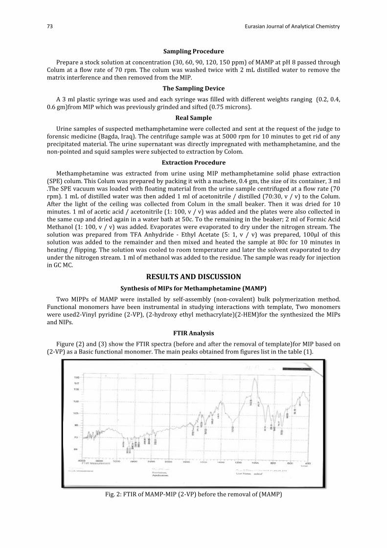
Figure (2) and (3) show the FTIR spectra (before and after the removal of template)for MIP based on (2-VP) as a Basic functional monomer. The main peaks obtained from figures list in the table (1).
Fig. 2: FTIR of MAMP-MIP (2-VP) before the removal of (MAMP)

74 Muhamed Farhan Abd et.al
Fig. 3: FTIR of MAMP-MIP (2-VP) after the removal of (MAMP)
Table 1: The most identified peaks of FTIR spectra for MAMP-imprinted polymer and NIP using ( 2-VP) as a functional monomer
Functional Group
MAMP MAMP –MIP(2-VP) before template removal
MAMP–MIP(2-VP)after template removal
1 N-H str. (cm-1) 3456 3047 ----
2 C-H aliphatic.(cm-1) ,28352968 2918,2846 2923,2873 3 C=O str.ester.(cm-1) ---- 1726 1718 4 C=C str. (cm-1) 1596 1581 1595 5 C-H aromatic.(cm-1) ,30163159 3047 3010 6 C-H bending (cm-1) 1483 1471 1456
8 Out-of plane-mono-sub
748,700 794,707 ----
9 Out-of plane-para-sub ---- 833 833 10 C-O str.ester.(cm-1) ---- 1271 1271 11 C=C str.olefin (cm-1) ----- 1633 1631
The Fourier transmission infrared spectrometry spectra of leached and un leached methamphetamine (MAMP) imprinted polymers MIP and NIP were recorded in the range of 400–4000 cm-1 by the KBr pellet method (Table 1).From the table1,the FTIR spectrum of the MAMP shows the following bands: (3456, 3016, 2968, 2835, 1596, 1483, 748 and 700) cm-1 for N-H stretching , C-H aromatic stretching, C-H. Aliphatic stretching, C=C stretching, C-H bending and out of plan bending for mono substituted ring. The FTIR spectrum of the MAMP –MIP (2-VP) before template removal shows the following bands (3047,2918,2846,1726,1581,3047,1475,794,707,833 and 1271)cm-1 for N-H stretching, C-H aliphatic stretching,, C=O stretching ester, , C=C stretching, C-H aromatic stretching, C-H bending and out of plan bending for mono substituted ring,out of plan bending for para substituted ring , C-O-C stretching and C=C stretching olefin . The FTIR spectrum of the MIP (2-VP) after template removal shows the absence of N-H stretching, C=C stretching and disappear the band of C=O stretching ester, out of plan bending for mono, para substituted ring and C=C str.olefin which excise in template (MAMP) spectrum which indicate the extracted of drug from template.
Figure (4) and (5) show the FTIR spectra (before and after the removal of template) for MIP based on (2-HEMA) as acidic functional monomer. The main peaks obtained from figures list in the table (2).

75 Eurasian Journal of Analytical Chemistry
Fig. 4: FTIR of MAMP-MIP (2-HEMA) before the removal of (MAMP)
Fig. 5: FTIR of MAMP-MIP (2-HEMA) after the removal of (MAMP)
Table 2: The most identified peaks of FT-IR spectra for MAMP-imprinted polymer and NIP using 2-HEMA as a functional monomer
Functional Group
MAMP MAMP –MIP(2-HEMA) before template removal
MAMP–MIP(2-hydroxy ethyl methacrylate)after template removal
1 N-H str. (cm-1) 3456 3114 ----
2 C-H aliphatic.(cm-1) ,28352968 2954,2850 2923,2873
3 O-H str.(cm-1) ---- 3114 3429
4 C=O str.ester.(cm-1) ---- 1735,1718 1731 5 C=C str. (cm-1) 1596 1602 ----
6 C-H aromatic.(cm-1) ,30163159 3047 3010 7 C=C str.olefin (cm-1) 1647 1631
8 Out-of plane-mono-sub
748,700 750,707 -----
9 C-O str.ester.(cm-1) ---- 1265 1265
76 Muhamed Farhan Abd et.al
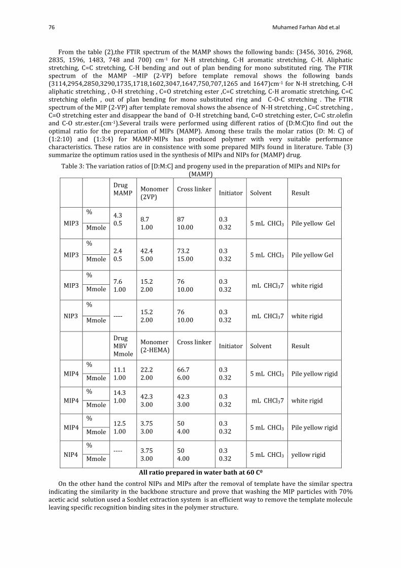
From the table (2),the FTIR spectrum of the MAMP shows the following bands: (3456, 3016, 2968, 2835, 1596, 1483, 748 and 700) cm-1 for N-H stretching, C-H aromatic stretching, C-H. Aliphatic stretching, C=C stretching, C-H bending and out of plan bending for mono substituted ring. The FTIR spectrum of the MAMP –MIP (2-VP) before template removal shows the following bands (3114,2954,2850,3290,1735,1718,1602,3047,1647,750,707,1265 and 1647)cm-1 for N-H stretching, C-H aliphatic stretching, , O-H stretching , C=O stretching ester ,C=C stretching, C-H aromatic stretching, C=C stretching olefin , out of plan bending for mono substituted ring and C-O-C stretching . The FTIR spectrum of the MIP (2-VP) after template removal shows the absence of N-H stretching , C=C stretching , C=O stretching ester and disappear the band of O-H stretching band, C=O stretching ester, C=C str.olefin and C-O str.ester.(cm-1).Several trails were performed using different ratios of (D:M:C)to find out the optimal ratio for the preparation of MIPs (MAMP). Among these trails the molar ratios (D: M: C) of (1:2:10) and (1:3:4) for MAMP-MIPs has produced polymer with very suitable performance characteristics. These ratios are in consistence with some prepared MIPs found in literature. Table (3) summarize the optimum ratios used in the synthesis of MIPs and NIPs for (MAMP) drug.
Table 3: The variation ratios of [D:M:C] and progeny used in the preparation of MIPs and NIPs for (MAMP)
Result Solvent Initiator Cross linker
Monomer (2VP)
Drug MAMP
Pile yellow Gel 5 mL CHCl3 0.3
0.32
87 10.00
8.7 1.00
4.3
0.5
%
MIP3 Mmole
Pile yellow Gel 5 mL CHCl3 0.3 0.32
73.2 15.00
42.4
5.00 2.4
0.5
%
MIP3 Mmole
white rigid 7 mL CHCl3 0.3 0.32
76
10.00 15.2
2.00 7.6
1.00
%
MIP3 Mmole
white rigid 7 mL CHCl3 0.3 0.32
76 10.00
15.2 2.00
----
%
NIP3 Mmole
Result Solvent Initiator Cross linker
Monomer (2-HEMA)
Drug MBV Mmole
Pile yellow rigid 5 mL CHCl3 0.3
0.32
66.7 6.00
22.2 2.00
11.1
1.00
%
MIP4 Mmole
white rigid 7 mL CHCl3 0.3
0.32
42.3 3.00
42.3 3.00
14.3
1.00
%
MIP4 Mmole
Pile yellow rigid 5 mL CHCl3 0.3
0.32 50
4.00 3.75
3.00 12.5
1.00
%
MIP4 Mmole
yellow rigid 5 mL CHCl3 0.3 0.32
50
4.00 3.75
3.00 ----
%
NIP4 Mmole
All ratio prepared in water bath at 60 C0
On the other hand the control NIPs and MIPs after the removal of template have the similar spectra indicating the similarity in the backbone structure and prove that washing the MIP particles with 70% acetic acid solution used a Soxhlet extraction system is an efficient way to remove the template molecule leaving specific recognition binding sites in the polymer structure.

77 Eurasian Journal of Analytical Chemistry
Adsorption Isotherm The absorption of isoterm in the understanding of the adsorption mechanism of the adsorption mold
with polymer surface. The data obtained from isothermal equilibrium was analyzed to show the isochromatic type of LANGMUIR or Freundlich models [21]. This is determined by plotting the binding capacity (Q) versus the free concentration of the drug, and Q is calculated according to the following equation:
Q = [(Ci – Cf) Vs *1000] / MMIP Ci = initial drug concentration (µmol / mL) Cf = final drug concentration (µmol / mL) Vs = volume of solution tested (mL) MMIP = mass of dried polymer (mg) Than measuring binding parameter
MIP/drug binding calculated by Scatchard analysis using the equation Q/ Cf = (Qmax - Q) / Kd Qmax = maximum capacity Kd = dissociation constant at binding side.
Isotherm adsorption obtained after shaking different concentrations of MAMP with a synthesis particle for 2 hours in a thermal water bath at 25 ° C as given in in fig (6) . Experimental data for regrouping experiments were included in Table (4).
Table 4: Rebinding values of (MAMP) using MAMP -MIP particles based on (2VP) and (2-HEMA) MAMP-MIP(2VP)
MAMP-MIP(2-HEMA)
Mass of MIP g
Ci mM
Cfree mM
Q μMole /g
Q/Cfree L/g
Ci mM
Cfree mM
Q μMole /g
Q/Cfree L/g
0.2
0.2010 0.1574 2.18 13.85 0.2010 0.1605 2.025 12.6168 0.4020 0.3102 4.59 14.796 0.4020 0.3128 4.46 14.2583 0.6030 0.518 2.25 8.204 0.6030 0.5312 3.59 6.7582 0.8040 0.726 3.9 5.3719 0.8040 0.7419 3.105 4.1852
0.4
0.2010 0.1472 1.345 9.1372 0.2010 0.1307 1.7575 13.4468 0.4020 0.2993 2.5675 8.5783 0.4020 0.2793 3.0675 10.9828 0.6030 0.4605 3.5625 7.7361 0.6030 0.4727 3.2575 6.8912 0.8040 0.671 3.325 4.9552 0.8040 0.6831 3.0225 4.4246
Fig. 6: Binding isotherm of 2-VP and 2-HEMA monomers by plotting Q against Ci
78 Muhamed Farhan Abd et.al
Plots in figure (6) for(2-VP and 2-HEMA ) monomers indicates that the binding capacity increase with increasing the concentration of the drug.
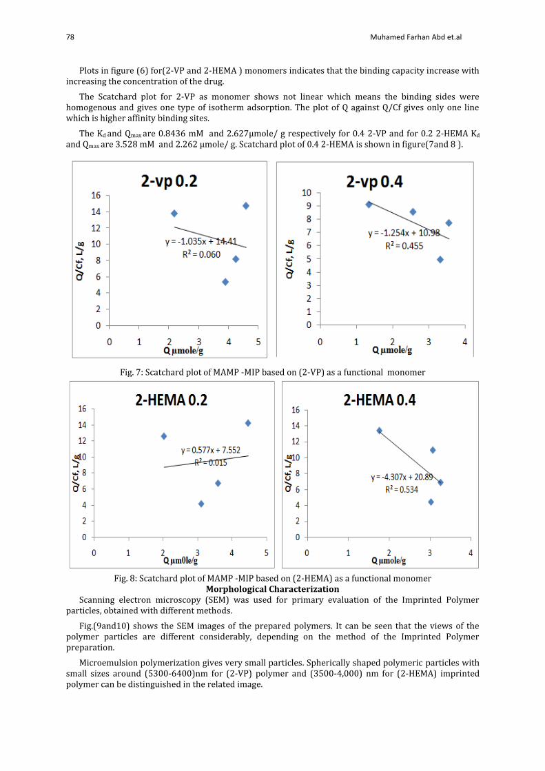
The Scatchard plot for 2-VP as monomer shows not linear which means the binding sides were homogenous and gives one type of isotherm adsorption. The plot of Q against Q/Cf gives only one line which is higher affinity binding sites.
The Kd and Qmax are 0.8436 mM and 2.627μmole/ g respectively for 0.4 2-VP and for 0.2 2-HEMA Kd
and Qmax are 3.528 mM and 2.262 μmole/ g. Scatchard plot of 0.4 2-HEMA is shown in figure(7and 8 ).
Fig. 7: Scatchard plot of MAMP -MIP based on (2-VP) as a functional monomer
Fig. 8: Scatchard plot of MAMP -MIP based on (2-HEMA) as a functional monomer
Morphological Characterization Scanning electron microscopy (SEM) was used for primary evaluation of the Imprinted Polymer
particles, obtained with different methods.
Fig.(9and10) shows the SEM images of the prepared polymers. It can be seen that the views of the polymer particles are different considerably, depending on the method of the Imprinted Polymer preparation.
Microemulsion polymerization gives very small particles. Spherically shaped polymeric particles with small sizes around (5300-6400)nm for (2-VP) polymer and (3500-4,000) nm for (2-HEMA) imprinted polymer can be distinguished in the related image.

79 Eurasian Journal of Analytical Chemistry
Fig. 9: SEM of [MAMP-MIP(2-VP)](10μm)obtained by bulk polymerization
Fig. 10: SEM of [MAMP –MIP (2-HEMA)] (50μm)obtained by bulk polymerization
Effect of Flow Rate As we noticed that the flow rate of peristaltic pump used for extraction of the MAMP from the
extraction needle is an important for the time needed for extraction .The flow rate of the sample solution through the fabricated extraction needle is an important factor because it controls the total analysis time and must be enough to prevent waste of time. The flow rate, on the one hand, must be low enough to make an effective retention of the analyte. Therefore, to evaluate the influence of the time of contact

80 Muhamed Farhan Abd et.al
between the MIP and the sample solution on the recovery, the effect of the sample loading flow rate has been studied in the range of 10-100 rpm.show the figures(11and12).
Fig. 11: Relationship between the flow rate and extraction time based on 0.2and 0.4 gm of MAMP MIP(2-
VP) used 60ppm from MAMP
Fig. 12: Relationship between the flow rate and extraction time based on 0.2and 0.4 gm of MAMP-MIP (2-
HEMA) used 60ppm from MAMP
Urine Samples Analysis Under optimal conditions, MIP-2-VP and MIP-2-HEMA were applied homogenously to identify
methamphetamine in urine samples. The sample matrix of the urine was in the first step and the wash step after the extraction was done. The washing step can be realized by allowing the solution of the carrier, solution of phosphate buffer, to flow through the plastic syringe used for the perstic pump. The components that suck weakly into a homogeneous column are expected to be removed by the washing step. By extending the washing time from 75 seconds to 3 minutes, the matrix tops were clearly suppressed.
This can be demonstrated by extracting an empty urine sample with a wash step of 3 minutes. The application of the same washing step to the urine sample rose, achieved satisfactory results of methamphetamine: no decrease was found in height of the analytes compared to those obtained by extracting standard samples. That were taken plastic syringe contains 0.2-0.4 g of MIP (2-VP) and MIP (2-HEMA) with passing different concentration of methamphetamine in urine samples was achieved in a range of 30-150 ppm successfully under optimal conditions. The results are shown in table 5 and 6.
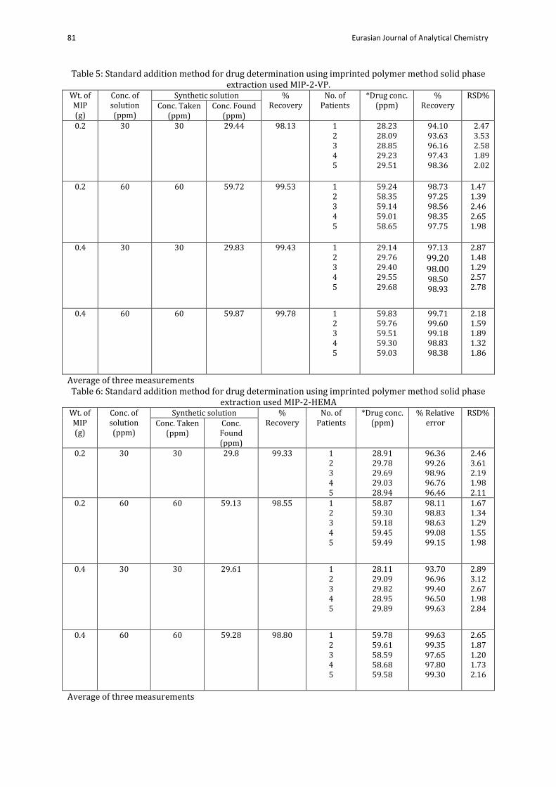
81 Eurasian Journal of Analytical Chemistry
Table 5: Standard addition method for drug determination using imprinted polymer method solid phase extraction used MIP-2-VP.
Wt. of MIP (g)
Conc. of solution (ppm)
Synthetic solution % Recovery
No. of Patients
*Drug conc. (ppm)
% Recovery
RSD% Conc. Taken
(ppm) Conc. Found
(ppm) 0.2 30 30 29.44 98.13 1
2 3 4 5
28.23 28.09 28.85 29.23 29.51
94.10 93.63 96.16 97.43 98.36
2.47 3.53 2.58 1.89 2.02
0.2 60 60 59.72 99.53 1 2 3 4 5
59.24 58.35 59.14 59.01 58.65
98.73 97.25 98.56 98.35 97.75
1.47 1.39 2.46 2.65 1.98
0.4 30 30 29.83
99.43 1
2 3 4 5
29.14 29.76 29.40 29.55 29.68
97.13 99.20
98.00 98.50 98.93
2.87 1.48 1.29 2.57 2.78
0.4 60 60 59.87 99.78 1 2 3 4 5
59.83 59.76 59.51 59.30 59.03
99.71 99.60 99.18 98.83 98.38
2.18 1.59 1.89 1.32 1.86
Average of three measurements Table 6: Standard addition method for drug determination using imprinted polymer method solid phase
extraction used MIP-2-HEMA
Wt. of MIP (g)
Conc. of solution (ppm)
Synthetic solution % Recovery
No. of Patients
*Drug conc. (ppm)
% Relative error
RSD% Conc. Taken
(ppm) Conc. Found (ppm)
0.2 30 30 29.8
99.33 1 2 3 4 5
28.91 29.78 29.69 29.03 28.94
96.36 99.26 98.96 96.76 96.46
2.46 3.61 2.19 1.98 2.11
0.2 60 60 59.13
98.55 1 2 3 4 5
58.87 59.30 59.18 59.45 59.49
98.11 98.83 98.63 99.08 99.15
1.67 1.34 1.29 1.55 1.98
0.4 30 30 29.61
1 2 3 4 5
28.11 29.09 29.82 28.95 29.89
93.70 96.96 99.40 96.50 99.63
2.89 3.12 2.67 1.98 2.84
0.4 60 60 59.28 98.80 1 2 3 4 5
59.78 59.61 58.59 58.68 59.58
99.63 99.35 97.65 97.80 99.30
2.65 1.87 1.20 1.73 2.16
Average of three measurements
82 Muhamed Farhan Abd et.al
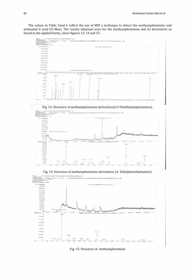
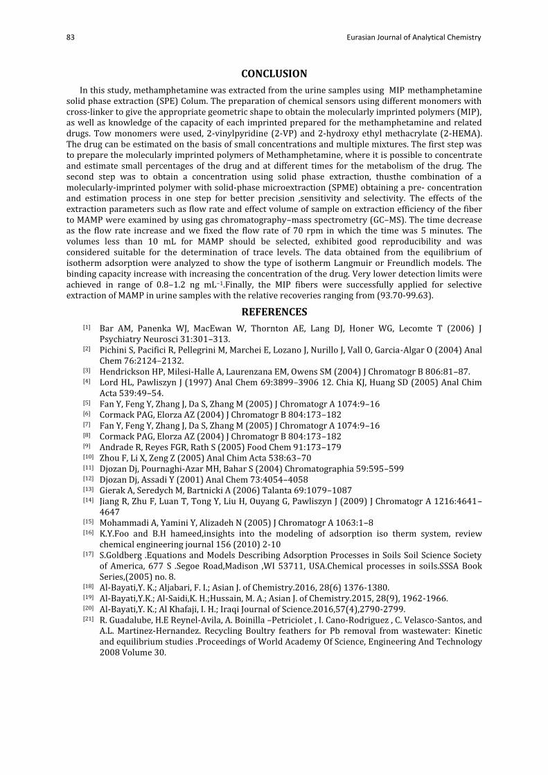
The values in Table 5and 6 reflect the use of MIP a technique to detect the methamphetamine and estimated it used GC-Mass. The results obtained were for the methamphetamine and its derivatives as found in the applied forms, show figures 13, 14 and 15.
Fig. 13: Structure of methamphetamine derivatives(3,5-Dimthylamphetamine).
Fig. 14: Structure of methamphetamine derivatives (4- Ethylphenethylamine).
Fig. 15: Structure of methamphetamine
83 Eurasian Journal of Analytical Chemistry
CONCLUSION
In this study, methamphetamine was extracted from the urine samples using MIP methamphetamine solid phase extraction (SPE) Colum. The preparation of chemical sensors using different monomers with cross-linker to give the appropriate geometric shape to obtain the molecularly imprinted polymers (MIP), as well as knowledge of the capacity of each imprinted prepared for the methamphetamine and related drugs. Tow monomers were used, 2-vinylpyridine (2-VP) and 2-hydroxy ethyl methacrylate (2-HEMA). The drug can be estimated on the basis of small concentrations and multiple mixtures. The first step was to prepare the molecularly imprinted polymers of Methamphetamine, where it is possible to concentrate and estimate small percentages of the drug and at different times for the metabolism of the drug. The second step was to obtain a concentration using solid phase extraction, thusthe combination of a molecularly-imprinted polymer with solid-phase microextraction (SPME) obtaining a pre- concentration and estimation process in one step for better precision ,sensitivity and selectivity. The effects of the extraction parameters such as flow rate and effect volume of sample on extraction efficiency of the fiber to MAMP were examined by using gas chromatography–mass spectrometry (GC–MS). The time decrease as the flow rate increase and we fixed the flow rate of 70 rpm in which the time was 5 minutes. The volumes less than 10 mL for MAMP should be selected, exhibited good reproducibility and was considered suitable for the determination of trace levels. The data obtained from the equilibrium of isotherm adsorption were analyzed to show the type of isotherm Langmuir or Freundlich models. The binding capacity increase with increasing the concentration of the drug. Very lower detection limits were achieved in range of 0.8–1.2 ng mL−1.Finally, the MIP fibers were successfully applied for selective extraction of MAMP in urine samples with the relative recoveries ranging from (93.70-99.63).
REFERENCES [1] Bar AM, Panenka WJ, MacEwan W, Thornton AE, Lang DJ, Honer WG, Lecomte T (2006) J
Psychiatry Neurosci 31:301–313. [2] Pichini S, Pacifici R, Pellegrini M, Marchei E, Lozano J, Nurillo J, Vall O, Garcia-Algar O (2004) Anal
Chem 76:2124–2132. [3] Hendrickson HP, Milesi-Halle A, Laurenzana EM, Owens SM (2004) J Chromatogr B 806:81–87. [4] Lord HL, Pawliszyn J (1997) Anal Chem 69:3899–3906 12. Chia KJ, Huang SD (2005) Anal Chim
Acta 539:49–54. [5] Fan Y, Feng Y, Zhang J, Da S, Zhang M (2005) J Chromatogr A 1074:9–16 [6] Cormack PAG, Elorza AZ (2004) J Chromatogr B 804:173–182 [7] Fan Y, Feng Y, Zhang J, Da S, Zhang M (2005) J Chromatogr A 1074:9–16 [8] Cormack PAG, Elorza AZ (2004) J Chromatogr B 804:173–182 [9] Andrade R, Reyes FGR, Rath S (2005) Food Chem 91:173–179 [10] Zhou F, Li X, Zeng Z (2005) Anal Chim Acta 538:63–70 [11] Djozan Dj, Pournaghi-Azar MH, Bahar S (2004) Chromatographia 59:595–599 [12] Djozan Dj, Assadi Y (2001) Anal Chem 73:4054–4058 [13] Gierak A, Seredych M, Bartnicki A (2006) Talanta 69:1079–1087 [14] Jiang R, Zhu F, Luan T, Tong Y, Liu H, Ouyang G, Pawliszyn J (2009) J Chromatogr A 1216:4641–
4647 [15] Mohammadi A, Yamini Y, Alizadeh N (2005) J Chromatogr A 1063:1–8 [16] K.Y.Foo and B.H hameed,insights into the modeling of adsorption iso therm system, review
chemical engineering journal 156 (2010) 2-10 [17] S.Goldberg .Equations and Models Describing Adsorption Processes in Soils Soil Science Society
of America, 677 S .Segoe Road,Madison ,WI 53711, USA.Chemical processes in soils.SSSA Book Series,(2005) no. 8.
[18] Al-Bayati,Y. K.; Aljabari, F. I.; Asian J. of Chemistry.2016, 28(6) 1376-1380. [19] Al-Bayati,Y.K.; Al-Saidi,K. H.;Hussain, M. A.; Asian J. of Chemistry.2015, 28(9), 1962-1966. [20] Al-Bayati,Y. K.; Al Khafaji, I. H.; Iraqi Journal of Science.2016,57(4),2790-2799. [21] R. Guadalube, H.E Reynel-Avila, A. Boinilla –Petriciolet , I. Cano-Rodriguez , C. Velasco-Santos, and
A.L. Martinez-Hernandez. Recycling Boultry feathers for Pb removal from wastewater: Kinetic and equilibrium studies .Proceedings of World Academy Of Science, Engineering And Technology 2008 Volume 30.