demyelination reduces brain parenchymal stiffness quantified in
TRANSCRIPT

Demyelination reduces brain parenchymal stiffnessquantified in vivo by magnetic resonance elastographyKatharina Schregela,b,1, Eva Wuerfel née Tysiakc,1, Philippe Garteiserb, Ines Gemeinhardtd, Timour Prozorovskie,Orhan Aktase, Hartmut Merzf, Dirk Petersena, Jens Wuerfela,g,2,3, and Ralph Sinkusb,2
aInstitute of Neuroradiology, University Luebeck, 23568 Luebeck, Germany; bUniversité Paris Diderot, Sorbonne Paris Cité, CRB3, UMR 773, Inserm, F-92110Clichy, France; cDepartment of Pediatrics, University Luebeck, 23568 Luebeck, Germany; dInstitute of Radiology, Charité - University Medicine Berlin, 10117Berlin, Germany; eDepartment of Neurology, University Medical Center Duesseldorf, Heinrich-Heine-University, 40225 Duesseldorf, Germany; fDepartment ofPathology, University Luebeck, 23568 Luebeck, Germany; and gNeuroCure Clinical Research Center, Charité - University Medicine Berlin, 10117 Berlin,Germany
Edited by Michael Sela, Weizmann Institute of Science, Rehovot, Israel, and approved March 6, 2012 (received for review January 23, 2012)
The detection of pathological tissue alterations by manual palpa-tion is a simple but essential diagnostic tool, which has beenapplied by physicians since the beginnings of medicine. Recently,the virtual “palpation” of the brain has become feasible usingmagnetic resonance elastography, which quantifies biomechanicalproperties of the brain parenchyma by analyzing the propagationof externally elicited shear waves. However, the precise molecularand cellular patterns underlying changes of viscoelasticity mea-sured by magnetic resonance elastography have not been investi-gated up to date. We assessed changes of viscoelasticity in amurine model of multiple sclerosis, inducing reversible demyelin-ation by feeding the copper chelator cuprizone, and correlated ourresults with detailed histological analyses, comprising myelina-tion, extracellular matrix alterations, immune cell infiltration andaxonal damage. We show firstly that the magnitude of the com-plex shear modulus decreases with progressive demyelination andglobal extracellular matrix degradation, secondly that the lossmodulus decreases faster than the dynamic modulus during thedestruction of the corpus callosum, and finally that those pro-cesses are reversible after remyelination.
magnetic resonance imaging | elasticity imaging | tissue integrity
Palpation of the brain, a hands-on experience long exclusive toneurosurgeons and pathologists detecting brain pathology,
has recently become a domain for physicists and radiologists:Using magnetic resonance elastography (MRE), it is possibletoday to noninvasively assess the biomechanical properties ofbrain parenchyma in vivo. In MRE, viscoelasticity describes thetendency of tissue to resist deformation, thus translating thesubjective tactile information gained from palpation into a quan-tifiable objective measure. These properties can be acquired byanalyzing the propagation of low-frequency shear waves, whichare mechanically elicited in an organ of interest (1, 2).Recent preliminary studies described distinct viscoelastic
characteristics of the brain parenchyma in healthy subjects aswell as changes by aging and brain pathology, underlining theapplicability and relevance of cerebral MRE (3, 4). Duringphysiological aging, there was evidence for a brain parenchymal“liquification” reflected in the decrease of solid-fluid behavior ofthe tissue (5). In patients suffering from multiple sclerosis (MS),a significant decrease of cerebral viscoelasticity was noted al-ready in early disease stages compared with healthy controls (6).However, despite a rising collection of in vivo viscoelasticity
data, no study has yet directly correlated viscoelastic parametersassessed via MRE with histopathological analyses. Thus, thequestion on how in vivo mechanical properties translate intocellular and molecular conditions has remained open.Magnetic resonance imaging (MRI) has emerged as most
important paraclinical tool for the diagnosis and monitoring ofneuroinflammatory diseases like MS, as reflected by current di-agnostic criteria (7). Nevertheless, disease specificity of conven-tional MRI parameters such as T2 lesion load is limited and their
association with clinical course and neurological disability is onlymodest (8). Additionally, these conventional MRI parametersprovide only limited conclusions to be drawn with respect to theunderlying pathology of MS lesions. Novel in vivo parameters arenecessary that are capable of evaluating demyelination and re-pair, and thus may improve diagnostic specificity and predictivevalue in MS.Therefore, we assessed brain parenchymal viscoelasticitiy non-
invasively by MRE in a mouse model of reversible toxic de-myelination, and correlated our findings to detailed histologicalanalyses. The copper chelator cuprizonewas used as demyelinationmodel and fed to susceptible mouse strains (C57BL/6 mice)resulting in progressive demyelination in several brain regions,particularly in the corpus callosum (9–11). We show in this par-ticular model that the degree and the time kinetics of the induceddisruption of extra-axonal tissue integrity (i.e., extensive de-myelination and distinctive extracellular matrix (ECM) degrada-tion) lead to a decrease of viscoelasticity in the corpus callosum.The biomechanical changes of the corpus callosum detected in thisanimalmodel are consistent with previously published human data,showing a global decrease of brain parenchymal viscoelasticity inMS patients (6).
ResultsViscoelasticity Images of Brain Tissue.MRE maps of cerebral tissueviscoelasticity generate an easily interpretable graphic image ofthe underlying brain anatomy, identifying the corpus callosumsignificantly stiffer than all other structures (Fig. 1). As expected,ventricles exhibited low shear properties. A decrease of visco-elasticity within the corpus callosum after 12 wk of cuprizonefeeding was clearly visible.
Physiological Development of MRI and MRE Parameters DuringAdolescence. Initially, we studied physiological changes of MRIand MRE parameters during adolescence in a group of 5- to6-wk-old healthy female C57BL/6 mice. The corpus callosumshowed a significant progressive decrease in T2 signal intensityduring the observation period in this group (Fig. 2A, white bars).Interestingly, the mechanical phase angle y remained constantover time (Fig. 2B, white bars), whereas viscoelasticity expressed
Author contributions: E.W.n.T., J.W., and R.S. designed research; K.S., T.P., J.W., and R.S.performed research; P.G., I.G., D.P., and R.S. contributed new reagents/analytic tools; K.S.,E.W.n.T., P.G., I.G., T.P., H.M., J.W., and R.S. analyzed data; and K.S., E.W.n.T., P.G., O.A., J.W.,and R.S. wrote the paper.
The authors declare no conflict of interest.
This article is a PNAS Direct Submission.1K.S. and E.W.n.T. contributed equally to this work.2J.W. and R.S. contributed equally to this work.3To whom correspondence should be addressed. E-mail: [email protected].
This article contains supporting information online at www.pnas.org/lookup/suppl/doi:10.1073/pnas.1200151109/-/DCSupplemental.
6650–6655 | PNAS | April 24, 2012 | vol. 109 | no. 17 www.pnas.org/cgi/doi/10.1073/pnas.1200151109
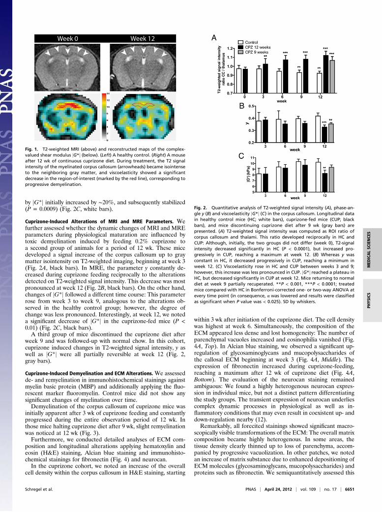
by jG*j initially increased by ∼20%, and subsequently stabilized(P = 0.0009) (Fig. 2C, white bars).
Cuprizone-Induced Alterations of MRI and MRE Parameters. Wefurther assessed whether the dynamic changes of MRI and MREparameters during physiological maturation are influenced bytoxic demyelination induced by feeding 0.2% cuprizone toa second group of animals for a period of 12 wk. These micedeveloped a signal increase of the corpus callosum up to graymatter isointensity on T2-weighted imaging, beginning at week 3(Fig. 2A, black bars). In MRE, the parameter y constantly de-creased during cuprizone feeding reciprocally to the alterationsdetected on T2-weighted signal intensity. This decrease was mostpronounced at week 12 (Fig. 2B, black bars). On the other hand,changes of jG*j followed a different time course: This parameterrose from week 3 to week 9, analogous to the alterations ob-served in the healthy control group; however, the degree ofchange was less pronounced. Interestingly, at week 12, we noteda significant decrease of jG*j in the cuprizone-fed mice (P <0.01) (Fig. 2C, black bars).A third group of mice discontinued the cuprizone diet after
week 9 and was followed-up with normal chow. In this cohort,cuprizone induced changes in T2-weighted signal intensity, y aswell as jG*j were all partially reversible at week 12 (Fig. 2,gray bars).
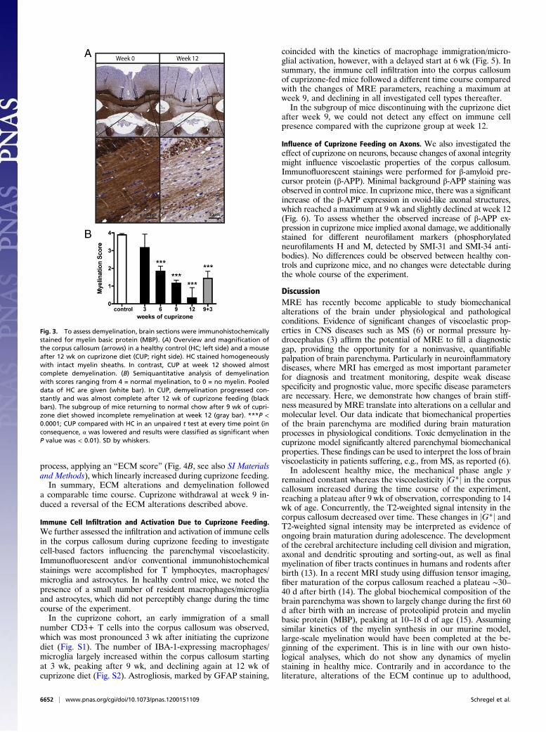
Cuprizone-Induced Demyelination and ECM Alterations. We assessedde- and remyelination in immunohistochemical stainings againstmyelin basic protein (MBP) and additionally applying the fluo-rescent marker fluoromyelin. Control mice did not show anysignificant changes of myelination over time.Demyelination of the corpus callosum of cuprizone mice was
initially apparent after 3 wk of cuprizone feeding and constantlyprogressed during the entire observation period of 12 wk. Inthose mice halting cuprizone diet after 9 wk, slight remyelinationwas noticed at 12 wk (Fig. 3).Furthermore, we conducted detailed analyses of ECM com-
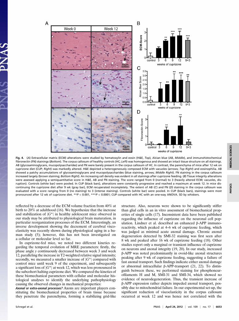
position and longitudinal alterations applying hematoxylin andeosin (H&E) staining, Alcian blue staining and immunohisto-chemical stainings for fibronectin (Fig. 4) and neurocan.In the cuprizone cohort, we noted an increase of the overall
cell density within the corpus callosum in H&E staining, starting
within 3 wk after initiation of the cuprizone diet. The cell densitywas highest at week 6. Simultaneously, the composition of theECM appeared less dense and lost homogeneity: The number ofparenchymal vacuoles increased and eosinophilia vanished (Fig.4A, Top). In Alcian blue staining, we observed a significant up-regulation of glycosaminoglycans and mucopolysaccharides ofthe callosal ECM beginning at week 3 (Fig. 4A, Middle). Theexpression of fibronectin increased during cuprizone-feeding,reaching a maximum after 12 wk of cuprizone diet (Fig. 4A,Bottom). The evaluation of the neurocan staining remainedambiguous: We found a highly heterogenous neurocan expres-sion in individual mice, but not a distinct pattern differentiatingthe study groups. The transient expression of neurocan underliescomplex dynamic processes in physiological as well as in-flammatory conditions that may even result in coexistent up- anddown-regulation nearby (12).Remarkably, all forecited stainings showed significant macro-
scopically visible transformations of the ECM: The overall matrixcomposition became highly heterogenous. In some areas, thetissue density clearly thinned up to loss of parenchyma, accom-panied by progressive vacuolization. In other patches, we notedan increase of matrix substance due to enhanced depositioning ofECM molecules (glycosaminoglycans, mucopolysaccharides) andproteins such as fibronectin. We semiquantitatively assessed this
Fig. 1. T2-weighted MRI (above) and reconstructed maps of the complex-valued shear modulus jG*j (below). (Left) A healthy control. (Right) A mouseafter 12 wk of continuous cuprizone diet. During treatment, the T2 signalintensity of the myelinated corpus callosum (arrowheads) became isointenseto the neighboring gray matter, and viscoelasticity showed a significantdecrease in the region-of-interest (marked by the red line), corresponding toprogressive demyelination.
Fig. 2. Quantitative analysis of T2-weighted signal intensity (A), phase-an-gle y (B) and viscoelasticity jG*j (C) in the corpus callosum. Longitudinal datain healthy control mice (HC; white bars), cuprizone-fed mice (CUP; blackbars), and mice discontinuing cuprizone diet after 9 wk (gray bars) arepresented. (A) T2-weighted signal intensity was computed as ROI ratio ofcorpus callosum and thalami. This ratio developed reciprocally in HC andCUP: Although, initially, the two groups did not differ (week 0), T2-signalintensity decreased significantly in HC (P < 0.0001), but increased pro-gressively in CUP, reaching a maximum at week 12. (B) Whereas y wasconstant in HC, it decreased progressively in CUP, reaching a minimum inweek 12. (C) Viscoelasticity rose in HC and CUP between weeks 3 and 9;however, this increase was less pronounced in CUP. jG*j reached a plateau inHC, but decreased significantly in CUP at week 12. Mice returning to normaldiet at week 9 partially recuperated. **P < 0.001, ***P < 0.0001; treatedmice compared with HC in Bonferroni-corrected one- or two-way ANOVA atevery time point (in consequence, α was lowered and results were classifiedas significant when P value was < 0.025). SD by whiskers.
Schregel et al. PNAS | April 24, 2012 | vol. 109 | no. 17 | 6651
MED
ICALSC
IENCE
SPH
YSICS
process, applying an “ECM score” (Fig. 4B, see also SI Materialsand Methods), which linearly increased during cuprizone feeding.In summary, ECM alterations and demyelination followed
a comparable time course. Cuprizone withdrawal at week 9 in-duced a reversal of the ECM alterations described above.
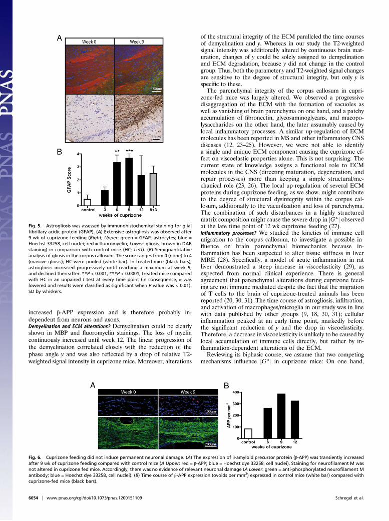
Immune Cell Infiltration and Activation Due to Cuprizone Feeding.We further assessed the infiltration and activation of immune cellsin the corpus callosum during cuprizone feeding to investigatecell-based factors influencing the parenchymal viscoelasticity.Immunofluorescent and/or conventional immunohistochemicalstainings were accomplished for T lymphocytes, macrophages/microglia and astrocytes. In healthy control mice, we noted thepresence of a small number of resident macrophages/microgliaand astrocytes, which did not perceptibly change during the timecourse of the experiment.In the cuprizone cohort, an early immigration of a small
number CD3+ T cells into the corpus callosum was observed,which was most pronounced 3 wk after initiating the cuprizonediet (Fig. S1). The number of IBA-1-expressing macrophages/microglia largely increased within the corpus callosum startingat 3 wk, peaking after 9 wk, and declining again at 12 wk ofcuprizone diet (Fig. S2). Astrogliosis, marked by GFAP staining,
coincided with the kinetics of macrophage immigration/micro-glial activation, however, with a delayed start at 6 wk (Fig. 5). Insummary, the immune cell infiltration into the corpus callosumof cuprizone-fed mice followed a different time course comparedwith the changes of MRE parameters, reaching a maximum atweek 9, and declining in all investigated cell types thereafter.In the subgroup of mice discontinuing with the cuprizone diet
after week 9, we could not detect any effect on immune cellpresence compared with the cuprizone group at week 12.
Influence of Cuprizone Feeding on Axons. We also investigated theeffect of cuprizone on neurons, because changes of axonal integritymight influence viscoelastic properties of the corpus callosum.Immunofluorescent stainings were performed for β-amyloid pre-cursor protein (β-APP). Minimal background β-APP staining wasobserved in control mice. In cuprizone mice, there was a significantincrease of the β-APP expression in ovoid-like axonal structures,which reached a maximum at 9 wk and slightly declined at week 12(Fig. 6). To assess whether the observed increase of β-APP ex-pression in cuprizone mice implied axonal damage, we additionallystained for different neurofilament markers (phosphorylatedneurofilaments H and M, detected by SMI-31 and SMI-34 anti-bodies). No differences could be observed between healthy con-trols and cuprizone mice, and no changes were detectable duringthe whole course of the experiment.
DiscussionMRE has recently become applicable to study biomechanicalalterations of the brain under physiological and pathologicalconditions. Evidence of significant changes of viscoelastic prop-erties in CNS diseases such as MS (6) or normal pressure hy-drocephalus (3) affirm the potential of MRE to fill a diagnosticgap, providing the opportunity for a noninvasive, quantifiablepalpation of brain parenchyma. Particularly in neuroinflammatorydiseases, where MRI has emerged as most important parameterfor diagnosis and treatment monitoring, despite weak diseasespecificity and prognostic value, more specific disease parametersare necessary. Here, we demonstrate how changes of brain stiff-ness measured by MRE translate into alterations on a cellular andmolecular level. Our data indicate that biomechanical propertiesof the brain parenchyma are modified during brain maturationprocesses in physiological conditions. Toxic demyelination in thecuprizone model significantly altered parenchymal biomechanicalproperties. These findings can be used to interpret the loss of brainviscoelasticity in patients suffering, e.g., from MS, as reported (6).In adolescent healthy mice, the mechanical phase angle y
remained constant whereas the viscoelasticity jG*j in the corpuscallosum increased during the time course of the experiment,reaching a plateau after 9 wk of observation, corresponding to 14wk of age. Concurrently, the T2-weighted signal intensity in thecorpus callosum decreased over time. These changes in jG*j andT2-weighted signal intensity may be interpreted as evidence ofongoing brain maturation during adolescence. The developmentof the cerebral architecture including cell division and migration,axonal and dendritic sprouting and sorting-out, as well as finalmyelination of fiber tracts continues in humans and rodents afterbirth (13). In a recent MRI study using diffusion tensor imaging,fiber maturation of the corpus callosum reached a plateau ∼30–40 d after birth (14). The global biochemical composition of thebrain parenchyma was shown to largely change during the first 60d after birth with an increase of proteolipid protein and myelinbasic protein (MBP), peaking at 10–18 d of age (15). Assumingsimilar kinetics of the myelin synthesis in our murine model,large-scale myelination would have been completed at the be-ginning of the experiment. This is in line with our own histo-logical analyses, which do not show any dynamics of myelinstaining in healthy mice. Contrarily and in accordance to theliterature, alterations of the ECM continue up to adulthood,
Fig. 3. To assess demyelination, brain sections were immunohistochemicallystained for myelin basic protein (MBP). (A) Overview and magnification ofthe corpus callosum (arrows) in a healthy control (HC; left side) and a mouseafter 12 wk on cuprizone diet (CUP; right side). HC stained homogeneouslywith intact myelin sheaths. In contrast, CUP at week 12 showed almostcomplete demyelination. (B) Semiquantitative analysis of demyelinationwith scores ranging from 4 = normal myelination, to 0 = no myelin. Pooleddata of HC are given (white bar). In CUP, demyelination progressed con-stantly and was almost complete after 12 wk of cuprizone feeding (blackbars). The subgroup of mice returning to normal chow after 9 wk of cupri-zone diet showed incomplete remyelination at week 12 (gray bar). ***P <0.0001; CUP compared with HC in an unpaired t test at every time point (inconsequence, α was lowered and results were classified as significant whenP value was < 0.01). SD by whiskers.
6652 | www.pnas.org/cgi/doi/10.1073/pnas.1200151109 Schregel et al.
reflected by a decrease of the ECM volume fraction from 40% atbirth to 20% at adulthood (16). We hypothesize that the increaseand stabilization of jG*j in healthy adolescent mice observed inour study may be attributed to physiological brain maturation, inparticular reorganization processes of the ECM. Interestingly, aninverse development showing the decrement of cerebral visco-elasticity was recently shown during physiological aging in a hu-man study (5); however, this has not been investigated ona cellular or molecular level so far.In cuprizone-fed mice, we noted two different kinetics re-
garding the temporal evolution of MRE parameters: firstly, thephase angle y continuously decreased between week 3 and week12, paralleling the increase in T2-weighted relative signal intensity;secondly, we measured a smaller increase of jG*j compared withcontrol mice until week 9 of the experiment, and subsequentlya significant loss of jG*j at week 12, which was less pronounced inthe subcohort halting cuprizone diet. We compared the kinetics ofthese biomechanical parameters with cellular and molecular his-tological analyses to identify the underlying pathophysiologycausing the observed changes in mechanical properties:Axonal or extra-axonal processes? Axons are important players con-stituting the biomechanical properties of brain tissue becausethey penetrate the parenchyma, forming a stabilizing grid-like
structure. Also, neurons were shown to be significantly stifferthan glial cells in an in vitro assessment of biomechanical prop-erties of single cells (17). Inconsistent data have been publishedregarding the influence of cuprizone on the neuronal cell pop-ulation. Lindner et al. described an enhanced β-APP immuno-reactivity, which peaked at 4–6 wk of cuprizone feeding, whichwas judged as minimal acute axonal damage. Chronic axonaldegeneration detected by SMI-32 staining was first seen after8 wk and peaked after 16 wk of cuprizone feeding (18). Otherstudies report only a marginal or transient influence of cuprizoneon neurons and axonal integrity (19, 20). In our study, increasedβ-APP was noted predominantly in ovoid-like axonal structurespeaking after 9 wk of cuprizone feeding, suggesting a failure offast axonal transport. Such findings indicate either axonal damageor abnormal intracellular β-APP-transport (21, 22). To distin-guish between these, we performed staining for phosphoneur-ofilaments H and M, SMI-31 and SMI-34, which showed noevidence of neurodegeneration. Thus, the transient increase ofβ-APP expression rather depicts impeded axonal transport, pos-sibly due to mitochondrial failure. In our experimental set-up, thesignificant reduction of viscoelasticity in the corpus callosumoccurred at week 12 and was hence not correlated with the
Fig. 4. (A) Extracellular matrix (ECM) alterations were studied by hematoxylin and eosin (H&E, Top), Alcian blue (AB, Middle), and immunohistochemicalfibronectin (FN) stainings (Bottom). The corpus callosum of healthy controls (HC; Left) was homogenous and showed an intact tissue structure on all stainings.AB (glycosaminoglycans, mucopolysaccharides) and FN were barely present in the corpus callosum of HC. In contrast, the parenchyma of mice after 12 wk oncuprizone diet (CUP; Right) was markedly altered. H&E depicted a heterogeneously composed ECM with vacuoles (arrows; Top Right) and eosinophilia. ABshowed a patchy accumulations of glycosaminoglycans and mucopolysaccharides (blue staining, arrows; Middle Right). FN staining in the corpus callosumincreased largely (brown staining; Bottom Right). An increasing cell density was evident in all stainings after cuprizone feeding. (B) Tissue integrity alterationswere assessed applying a semiquantitative score in H&E, AB and FN staining. The score ranged from 0 (normal) to 3 (heavily altered ECM; vacuoles, dis-ruption). Controls (white bar) were pooled. In CUP (black bars), alterations were constantly progressive and reached a maximum at week 12. In mice dis-continuing the cuprizone diet after 9 wk (gray bar), ECM recuperated incompletely. The extent of AB (C) and FN (D) staining in the corpus callosum wasevaluated with a score ranging from 0 (no staining) to 3 (intense staining). Controls (white bar) were pooled. In CUP (black bars), stainings were mostpronounced after 12 wk of cuprizone diet. **P < 0.001, ***P < 0.0001; CUP compared with HC with an one-way ANOVA. SD by whiskers.
Schregel et al. PNAS | April 24, 2012 | vol. 109 | no. 17 | 6653
MED
ICALSC
IENCE
SPH
YSICS
increased β-APP expression and is therefore probably in-dependent from neurons and axons.Demyelination and ECM alterations? Demyelination could be clearlyshown in MBP and fluoromyelin stainings. The loss of myelincontinuously increased until week 12. The linear progression ofthe demyelination correlated closely with the reduction of thephase angle y and was also reflected by a drop of relative T2-weighted signal intensity in cuprizone mice. Moreover, alterations
of the structural integrity of the ECM paralleled the time coursesof demyelination and y. Whereas in our study the T2-weightedsignal intensity was additionally altered by continuous brain mat-uration, changes of y could be solely assigned to demyelinationand ECM degradation, because y did not change in the controlgroup. Thus, both the parameter y and T2-weighted signal changesare sensitive to the degree of structural integrity, but only y isspecific to these.The parenchymal integrity of the corpus callosum in cupri-
zone-fed mice was largely altered. We observed a progressivedisaggregation of the ECM with the formation of vacuoles aswell as vanishing of brain parenchyma on one hand, and a patchyaccumulation of fibronectin, glycosaminoglycans, and mucopo-lysaccharides on the other hand, the later assumably caused bylocal inflammatory processes. A similar up-regulation of ECMmolecules has been reported in MS and other inflammatory CNSdiseases (12, 23–25). However, we were not able to identifya single and unique ECM component causing the cuprizone ef-fect on viscoelastic properties alone. This is not surprising: Thecurrent state of knowledge assigns a functional role to ECMmolecules in the CNS (directing maturation, degeneration, andrepair processes) more than keeping a simple structural/me-chanical role (23, 26). The local up-regulation of several ECMproteins during cuprizone feeding, as we show, might contributeto the degree of structural dysintegrity within the corpus cal-losum, additionally to the vacuolization and loss of parenchyma.The combination of such disturbances in a highly structuredmatrix composition might cause the severe drop in jG*j observedat the late time point of 12 wk cuprizone feeding (27).Inflammatory processes? We studied the kinetics of immune cellmigration to the corpus callosum, to investigate a possible in-fluence on brain parenchymal biomechanics because in-flammation has been suspected to alter tissue stiffness in liverMRE (28). Specifically, a model of acute inflammation in ratliver demonstrated a steep increase in viscoelasticity (29), asexpected from normal clinical experience. There is generalagreement that parenchymal alterations during cuprizone feed-ing are not immune mediated despite the fact that the migrationof T cells to the brain of cuprizone-treated animals has beenreported (20, 30, 31). The time course of astrogliosis, infiltration,and activation of macrophages/microglia in our study was in linewith data published by other groups (9, 18, 30, 31); cellularinflammation peaked at an early time point, markedly beforethe significant reduction of y and the drop in viscoelasticity.Therefore, a decrease in viscoelasticity is unlikely to be caused bylocal accumulation of immune cells directly, but rather by in-flammation-dependent alterations of the ECM.Reviewing its biphasic course, we assume that two competing
mechanisms influence jG*j in cuprizone mice: On one hand,
Fig. 5. Astrogliosis was assessed by immunohistochemical staining for glialfibrillary acidic protein (GFAP). (A) Extensive astrogliosis was observed after9 wk of cuprizone feeding (Right; Upper: green = GFAP, astrocytes; blue =Hoechst 33258, cell nuclei; red = fluoromyelin; Lower: gliosis, brown in DABstaining) in comparison with control mice (HC; Left). (B) Semiquantitativeanalysis of gliosis in the corpus callosum. The score ranges from 0 (none) to 4(massive gliosis); HC were pooled (white bar). In treated mice (black bars),astrogliosis increased progressively until reaching a maximum at week 9,and declined thereafter. **P < 0.001, ***P < 0.0001; treated mice comparedwith HC in an unpaired t test at every time point (in consequence, α waslowered and results were classified as significant when P value was < 0.01).SD by whiskers.
Fig. 6. Cuprizone feeding did not induce permanent neuronal damage. (A) The expression of β-amyloid precursor protein (β-APP) was transiently increasedafter 9 wk of cuprizone feeding compared with control mice (A Upper: red = β-APP; blue = Hoechst dye 33258, cell nuclei). Staining for neurofilament M wasnot altered in cuprizone fed mice. Accordingly, there was no evidence of relevant neuronal damage (A Lower: green = anti-phosphorylated neurofilament Mantibody; blue = Hoechst dye 33258, cell nuclei). (B) Time course of β-APP expression (ovoids per mm2) expressed in control mice (white bar) compared withcuprizone-fed mice (black bars).
6654 | www.pnas.org/cgi/doi/10.1073/pnas.1200151109 Schregel et al.
a viscoelasticity-increasing factor supposedly generated by com-bined kinetics of brain maturation such as reorganization of theECM; on the other hand, a viscoelasticity-reducing factor clearlydominated longitudinally. The latter was cuprizone-dependent:Its influence was partially reversible after discontinuing thecuprizone diet. The drop in viscoelasticity could be associatedwith extra-axonal reorganization, i.e., the demyelination and de-struction of the ECM, but not with axonal damage. Our currentdata do not allow the identification of one single molecular orcellular process responsible for the observed biomechanicalalterations. Nevertheless, the temporal evolution of the mechan-ical parameters during physiological maturation in comparisonwith the cuprizone-induced demyelination process reveals oneimportant difference: During physiological maturation, the phaseangle y remains constant, whereas it continuously decreases incuprizone-treated mice. Thus, we hypothesize that the reductionof the phase angle y is exclusively assigned to demyelination.Nevertheless, there are ongoing remodeling processes of theECM during this stage (13, 16). Unfortunately, the cohort ofhealthy control mice investigated in this study was too small todetect significant ECM alterations during adolescence in theconducted histological analyses. A larger trial investigating phys-iological ECM changes during adolescence, and the relevance ofspecific ECM molecules, e.g., in tenascin-receptor deficient (32,33) as well as brevican-deficient (34) murine knock-out models,is warranted and necessary to support this hypothesis.In a previous cross-sectional human study of 45 MS patients
with recent diagnosis and mild relapsing-remitting disease course,we found a reduction of global brain viscoelasticity comparedwith healthy controls measured with MRE. The decrease of
viscoelasticity in the corpus callosum of cuprizone mice is con-sistent with the data acquired in MS patients. The cuprizonemodel mimics particular aspects of the pathology of MS (11, 35,36): The composition of cuprizone-induced pathology corre-sponds to so-called “pattern III lesions” in MS, which are char-acterized by oligodendroglial depletion, demyelination, and aninflammatory infiltration of activated macrophages/microglia andT lymphocytes (37). Conferring our murine cuprizone data toMS, we can assume that globally reduced cerebral viscoelasticityin patients is caused by demyelination and ECM alterations.In conclusion, we identified structural and molecular mecha-
nisms underlying the changes of brain parenchymal bio-mechanics assessed by MRE. Previously acquired human datacould be reproduced in an animal model, supporting the con-sistency of MRE and its applicability in different species.
Materials and MethodsDetailed methods are provided in SI Materials and Methods. These describemouse strains, animal handling including cuprizone diet, in vivo MRI as well asMRE procedures, and post-processing and data analysis algorithms applied.Conventional histological analyses on paraffin-embedded slices included he-matoxylin and eosin (H&E) staining, Alcian blue staining, and the character-ization of microglia/macrophages (anti-IBA-1), astrogliosis (anti-GFAP), T-cells(anti-CD3), myelination (anti-MBP) and the ECM proteins fibronectin (anti-fibronectin) and neurocan (anti-neurocan). Additional immunofluorescentstaining on cryosections was performed for the characterization of microglia/macrophages, astroglia and axonal damage (anti-β-APP, neurofilamentsH and M) as well as myelination (fluoromyelin).
ACKNOWLEDGMENTS.We thank S. Pezet and N. Nadkarni for assistance andvaluable discussions.
1. Green MA, Bilston LE, Sinkus R (2008) In vivo brain viscoelastic properties measured bymagnetic resonance elastography. NMR Biomed 21:755–764.
2. Muthupillai R, Ehman RL (1996) Magnetic resonance elastography. Nat Med 2:601–603.
3. Streitberger KJ, et al. (2010) In vivo viscoelastic properties of the brain in normalpressure hydrocephalus. NMR Biomed 24:385–392.
4. Xu L, et al. (2007) Magnetic resonance elastography of brain tumors: preliminaryresults. Acta Radiol 48:327–330.
5. Sack I, et al. (2009) The impact of aging and gender on brain viscoelasticity. Neuro-image 46:652–657.
6. Wuerfel J, et al. (2010) MR-elastography reveals degradation of tissue integrity inmultiple sclerosis. Neuroimage 49:2520–2525.
7. McDonald WI, et al. (2001) Recommended diagnostic criteria for multiple sclerosis:guidelines from the International Panel on the diagnosis of multiple sclerosis. AnnNeurol 50:121–127.
8. Charil A, et al. (2006) MRI and the diagnosis of multiple sclerosis: expanding theconcept of “no better explanation”. Lancet Neurol 5:841–852.
9. Matsushima GK, Morell P (2001) The neurotoxicant, cuprizone, as a model to studydemyelination and remyelination in the central nervous system. Brain Pathol 11:107–116.
10. Silvestroff L, et al. (2010) Cuprizone-induced demyelination in CNP:GFP transgenicmice. J Comp Neurol 518:2261–2283.
11. Kipp M, Clarner T, Dang J, Copray S, Beyer C (2009) The cuprizone animal model: newinsights into an old story. Acta Neuropathol 118:723–736.
12. Rauch U (2004) Extracellular matrix components associated with remodeling pro-cesses in brain. Cell Mol Life Sci 61:2031–2045.
13. Squire LR (2008) Fundamental neuroscience (Elsevier/Academic Press, Amsterdam,Boston), 3rd Ed, pp 336–576, pp 1167–1200.
14. Baloch S, et al. (2009) Quantification of brain maturation and growth patterns inC57BL/6J mice via computational neuroanatomy of diffusion tensor images. CerebCortex 19:675–687.
15. Matthieu JM, Widmer S, Herschkowitz N (1973) Biochemical changes in mouse braincomposition during myelination. Brain Res 55:391–402.
16. Syková E, Mazel T, Simonová Z (1998) Diffusion constraints and neuron-glia in-teraction during aging. Exp Gerontol 33:837–851.
17. Lu YB, et al. (2006) Viscoelastic properties of individual glial cells and neurons in theCNS. Proc Natl Acad Sci USA 103:17759–17764.
18. Lindner M, Fokuhl J, Linsmeier F, Trebst C, Stangel M (2009) Chronic toxic de-myelination in the central nervous system leads to axonal damage despite remyeli-nation. Neurosci Lett 453:120–125.
19. Sun SW, et al. (2006) Noninvasive detection of cuprizone induced axonal damage anddemyelination in the mouse corpus callosum. Magn Reson Med 55:302–308.
20. Moharregh-Khiabani D, et al. (2010) Effects of fumaric acids on cuprizone inducedcentral nervous system de- and remyelination in the mouse. PLoS ONE 5:e11769.
21. Koo EH, et al. (1990) Precursor of amyloid protein in Alzheimer disease undergoes fastanterograde axonal transport. Proc Natl Acad Sci USA 87:1561–1565.
22. Sisodia SS, Koo EH, Hoffman PN, Perry G, Price DL (1993) Identification and transportof full-length amyloid precursor proteins in rat peripheral nervous system. J Neurosci13:3136–3142.
23. van Horssen J, Dijkstra CD, de Vries HE (2007) The extracellular matrix in multiplesclerosis pathology. J Neurochem 103:1293–1301.
24. Kwok JC, Dick G, Wang D, Fawcett JW (2011) Extracellular matrix and perineuronalnets in CNS repair. Dev Neurobiol 71:1073–1089.
25. Sobel RA, Mitchell ME (1989) Fibronectin in multiple sclerosis lesions. Am J Pathol 135:161–168.
26. Bonneh-Barkay D, Wiley CA (2009) Brain extracellular matrix in neurodegeneration.Brain Pathol 19:573–585.
27. Stephens EH, Grande-Allen KJ (2007) Age-related changes in collagen synthesis andturnover in porcine heart valves. J Heart Valve Dis 16:672–682.
28. Huwart L, van Beers BE (2008) MR elastography. Gastroenterol Clin Biol 32(6, Suppl 1):68–72.
29. Salameh N, et al. (2009) Early detection of steatohepatitis in fatty rat liver by usingMR elastography. Radiology 253:90–97.
30. Remington LT, Babcock AA, Zehntner SP, Owens T (2007) Microglial recruitment,activation, and proliferation in response to primary demyelination. Am J Pathol 170:1713–1724.
31. McMahon EJ, Suzuki K, Matsushima GK (2002) Peripheral macrophage recruitment incuprizone-induced CNS demyelination despite an intact blood-brain barrier. J Neu-roimmunol 130:32–45.
32. Garcion E, Faissner A, ffrench-Constant C (2001) Knockout mice reveal a contributionof the extracellular matrix molecule tenascin-C to neural precursor proliferation andmigration. Development 128:2485–2496.
33. Brückner G, et al. (2000) Postnatal development of perineuronal nets in wild-typemice and in a mutant deficient in tenascin-R. J Comp Neurol 428:616–629.
34. Brakebusch C, et al. (2002) Brevican-deficient mice display impaired hippocampal CA1long-term potentiation but show no obvious deficits in learning and memory. MolCell Biol 22:7417–7427.
35. Torkildsen O, Brunborg LA, Myhr KM, Bø L (2008) The cuprizone model for de-myelination. Acta Neurol Scand Suppl 188:72–76.
36. Denic A, et al. (2011) The relevance of animal models in multiple sclerosis research.Pathophysiology 18:21–29.
37. Lucchinetti C, et al. (2000) Heterogeneity of multiple sclerosis lesions: implications forthe pathogenesis of demyelination. Ann Neurol 47:707–717.
Schregel et al. PNAS | April 24, 2012 | vol. 109 | no. 17 | 6655
MED
ICALSC
IENCE
SPH
YSICS