delta-opioid agonist snc-121 protects retinal ganglion cell function in a chronic ocular...
TRANSCRIPT
Physiology and Pharmacology
Delta-Opioid Agonist SNC-121 Protects Retinal GanglionCell Function in a Chronic Ocular Hypertensive Rat Model
Yasir Abdul, Naseem Akhter, and Shahid Husain
PURPOSE. This study examined if the delta-opioid (d-opioid)receptor agonist, SNC-121, can improve retinal function andretinal ganglion cell (RGC) survival during glaucomatous injuryin a chronic ocular hypertensive rat model.
METHODS. IOP was raised in brown Norway rats by injectinghypertonic saline into the limbal venous system. Rats weretreated with 1 mg/kg SNC-121 (intraperitoneally [IP]) oncedaily for 7 days. Pattern-electroretinograms (PERGs) wereobtained in response to contrast reversal of patterned visualstimuli. RGCs were visualized by fluorogold retrogradelabeling. Expression of TNF-a and p38 mitogen-activatedprotein (MAP) kinase was measured by immunohistochemistryand Western blotting.
RESULTS. PERG amplitudes in ocular hypertensive eyes weresignificantly reduced (14.3 6 0.60 lvolts) when comparedwith healthy eyes (18.0 6 0.62 lvolts). PERG loss inhypertensive eyes was inhibited by SNC-121 treatment (17.206 0.1.3 lvolts; P < 0.05). There was a 29% loss of RGCs inthe ocular hypertensive eye, which was inhibited in thepresence of SNC-121. TNF-a production and activation of p38MAP kinase in retinal sections and optic nerve samples wereupregulated in ocular hypertensive eyes and inhibited in thepresence of SNC-121. Furthermore, TNF-a induced increase inp38 MAP kinase activation in astrocytes was inhibited in thepresence of SNC-121.
CONCLUSIONS. These data provide evidence that activation of d-opioid receptors inhibited the loss of PERG amplitudes andrate of RGC loss during glaucomatous injury. Mechanistic dataprovided clues that TNF-a is mainly produced from glial cellsand activates p38 MAP kinase, which was significantlyinhibited by SNC-121 treatment. Overall, data indicate thatenhancement of d-opioidergic activity in the eye may provideretina neuroprotection against glaucoma. (Invest Ophthalmol
Vis Sci. 2013;54:1816–1828) DOI:10.1167/iovs.12-10741
Glaucoma is a chronic optic neuropathy characterized byretinal ganglion cell (RGC) death, an excavated appear-
ance of the optic nerve, and vision loss.1 Studies have shown
that several factors including elevated IOP,2 oxidative stress,3
mitochondrial dysfunction,4 glutamate neurotoxicity,5 and pro-inflammatory cytokines6 work concomitantly in the pathogen-esis of the disease and in regulating RGC susceptibility toglaucomatous damage.7 Current therapeutic management ofglaucoma aims to halt or slow disease progression by reducingelevated IOP. Although IOP-lowering treatment can retard thedisease progression in many glaucoma patients, it is not alwayssufficient to prevent disease progression. As a result, efforts inour laboratory are focused on developing neuroprotectivetreatment strategies for glaucoma.
TNF-a is a pro-inflammatory cytokine that is rapidlyupregulated in several neurodegenerative disorders, such asmultiple sclerosis, Parkinson’s disease, Alzheimer disease, andglaucoma.6,8 The levels of TNF-a and its receptor, TNF-R1, arealso upregulated significantly in glaucoma.6,9 Growing evi-dence supports that TNF-a through the binding of TNF-R1, adeath receptor, is involved in mediating RGC death duringglaucomatous neurodegeneration.6,9 In addition, the expres-sion of TNF-a receptors on astrocytes and axons of theglaucomatous optic nerve head (ONH) would suggest thatTNF-a is stimulating cytodestructive processes in both theastrocytes and axons.10 Considering a dire need to developneuroprotective agents that can provide neuroprotection andcounterbalance the actions of TNF-a–mediated cytodestructivesignaling pathways, we set out to determine if a selective delta-opioid (d-opioid) receptor agonist, SNC-121, possessed thesenovel properties against glaucomatous injury.
The effects of opioids are mediated through activation ofthree opioid receptor subtypes: d-opioid, j-opioid, and l-opioid.11 Studies have shown that opioid receptor activation byexogenous agonists can facilitate a protective effect againsthypoxia, ischemia, cold, or an acidic environment.12–14 Morerecently, we published that morphine pretreatment canprovide significant retinal neuroprotection against acuteischemic15 and glaucomatous injury,16 and this neuroprotec-tion is mediated, in part, via inhibition of TNF-a produc-tion.16,17 However, the neuroprotective roles of d-opioidreceptors against glaucomatous injury have remained fullyunexplored.
The data presented herein describes the potential partici-pation of d-opioid receptors in a neuroprotective paradigmagainst glaucomatous injury in a chronic ocular hypertensiverat model. The data provide evidence that d-opioid receptoractivation by the exogenous ligand, SNC-121, protects RGCfunction and integrity against glaucomatous injury. Mechanisticdata provide clues that TNF-a is produced mainly from glialcells during the early phase of glaucoma development, whichsubsequently phosphorylate and activate p38 mitogen-activat-ed protein (MAP). These studies provide clues that TNF-a–mediated signaling pathways could be a potential target fordevelopment of antiglaucoma therapies and enhancement of d-opioidergic activity by exogenous means may be a vitalneuroprotective strategy for glaucoma therapy.
From the Hewitt Laboratory of the Ola B. Williams GlaucomaCenter, Department of Ophthalmology, Storm Eye Institute, MedicalUniversity of South Carolina, Charleston, South Carolina.
Supported in part by National Institutes of Health (NIH)/National Eye Institute Grant EY019081 (SH), NIH Grant C06RR015455 from the Extramural Research Facility Program of theNational Center for Research Resources, and an unrestricted grant toMUSC-SEI from Research to Prevent Blindness, New York, New York.
Submitted for publication August 9, 2012; revised December 3,2012 and January 23, 2013; accepted January 31, 2013.
Disclosure: Y. Abdul, None; N. Akhter, None; S. Husain, NoneCorresponding author: Shahid Husain, Storm Eye Institute,
Room 511, Medical University of South Carolina, 167 Ashley Avenue,Charleston, SC 29425; [email protected].
Investigative Ophthalmology & Visual Science, March 2013, Vol. 54, No. 3
1816 Copyright 2013 The Association for Research in Vision and Ophthalmology, Inc.
MATERIALS AND METHODS
Animals
Adult male or female brown Norway rats (3–5 months of age; 150–200
g; Harlan Laboratories, Inc., Indianapolis, IN) were used in this study.
Rats were kept under a cycle of 12 hours light and 12 hours dark for all
the studies. Animal handling was performed in accordance with the
ARVO Statement for the Use of Animals in Ophthalmic and Vision
Research; and the study protocol was approved by the Animal Care and
Use Committee at the Medical University of South Carolina. Stock SNC-
121 solutions (10 mg/mL) were made and diluted in normal saline
(0.9%). SNC-121 (1 mg/kg) was injected intraperitoneally (IP) in brown
Norway rats once daily for 7 days. Drug administration (100–150 lL)
was performed daily at the same time between 9 AM to 11 AM. The
control group was handled in a similar fashion, except that normal
saline was injected without SNC-121.
Development of Glaucoma Model by HypertonicSaline Injection
Brown Norway rats (150–200 g body weight) were housed under a
standard 12:12 (light:dark) cycle. A stable baseline IOP was document-
ed prior to hypertonic saline injection. Rats were anesthetized with
ketamine (75 mg/kg) and xylazine (8 mg/kg) and body temperature
was maintained at 378C with a heating pad. Topical proparacaine
(0.5%) was applied to the cornea. A pulled-glass micropipette attached
to a syringe by PE-50 tubing (Becton Dickinson & Co., Sparks, MD),
was inserted into a circumferential limbal vein near the cornea and
approximately 50 lL of 2 M hypertonic saline was injected into the
limbal venous system as described.16,18 After surgery, an antibacterial
ointment (neomycin) was applied at the injection site of each animal to
prevent infections. IOP was recorded as the average of six to eight
consecutive measurements prior to surgery (baseline IOP; 18–20 mm
Hg) followed by IOP measurement on a weekly basis after treatment,
using a calibrated Tonolab tonometer (Colonial Medical Supply Co.,
Inc., Franconia, NH), as described earlier.19 IOP exposure for each
animal was calculated by performing separate integration of the IOP
over days of exposure for the treated and control eyes as
described.16,20 The control eye integral values were then subtracted
from the treated eye integral, yielding the ‘‘IOP-integral difference’’;
values are expressed as millimeters of mercury over the days. As
inclusion criteria following saline injection, only animals with an
elevated IOP that was at least 25% over baseline were included in the
study.
Pattern-Electroretinogram (PERG) Recordings
Rats were anesthetized with ketamine (75 mg/kg) and xylazine (8 mg/
kg) and body temperature was maintained at 378C with a heating pad.
PERG recordings (without dark adaptation) were conducted in both
eyes (sequentially) 3 days prior to IOP elevation by hypertonic saline
injection, and then biweekly post surgery as described earlier.16 Fifteen
rats were used in this experiment. Rats were divided into two groups:
an untreated hypertensive group and the SNC-121–treated hyperten-
sive group. The PERG electrode was placed on the corneal surface by
means of a micromanipulator and positioned in such a way as to
encircle the undilated pupil without limiting the field of view. A small
drop of saline was applied to keep the cornea and lens moist during
each recording. A visual stimulus generated by black and white
alternating contrast reversing bars (mean luminance, 50 cd/m2; spatial
frequency, 0.033 cycle/deg; contrast, 99%; and temporal frequency, 1
Hz) was aligned with the projection of the undilated pupil at an 11-cm
distance using the UTAS-2000 (LKC Technologies, Gaithersburg, MD).
Each PERG was an average of 300 sweeps at an interval of 1 second.
For the PERG amplitudes, measurements were made between a peak
and adjacent trough of the waveform.
Retrograde Labeling of Retinal Ganglion Cells
Rats were deeply anesthetized with ketamine (75 mg/kg), xylazine (8
mg/kg), and body temperature was maintained at 378C with a heating
pad. Retrograde labeling of RGCs was performed as described earlier.16
Briefly, 3 lL of a 5% solution of fluorogold (Fluorochrome, LLC; Denver,
CO) in PBS was injected into the superior colliculus of twelve
anesthetized ocular hypertensive animals immobilized in a stereotaxic
apparatus. Using a small drill, a 1/8-inch hole was made in the skull 6
mm from bregma and 2 mm from lambda. After making this hole in the
skull, a Hamilton syringe (Hamilton, Reno, NV) was filled with
fluorogold and the syringe needle gently inserted at the hole and
FIGURE 1. IOP measurements of healthy eyes, and eyes from a chronic glaucoma model with and without SNC-121 (1 mg/kg) treatment for 7 days,once daily. Rats were divided into two groups: ocular hypertensive group (n¼ 8) and SNC-121–treated ocular hypertensive group (n¼ 8). IOP waselevated in one eye of brown Norway rats by injecting approximately 50 lL of 2.0 M hypertonic saline, while the contralateral eye served as thecontrol. Rats were maintained for up to 6 weeks post surgery. A total of 16 rats were used in this experiment. Data are mean 6 SE. *P < 0.05; n¼ 8for each group.
IOVS, March 2013, Vol. 54, No. 3 Retina Neuroprotection by SNC-121 against Glaucoma 1817

going down 4 mm, whereupon the fluorogold was injected. The needle
was left in the brain for 30 to 60 seconds, then slowly removed. The
skull hole was filled with bone wax 903 (Lukens Cat # 2007–05; World
Precision Instruments, Inc., Sarasota, FL). Seven days post injection,
animals were euthanized and their eyes were enucleated and fixed in
4% paraformaldehyde (PFA) for 24 hour at 48C. After rinsing with PBS,
each retina was detached from the eyecup and prepared as a flat mount
by mounting the vitreous side up. Fluorescent RGCs were visualized
under Zeiss microscopy (Jena, Germany). Each retina was divided into
four quadrants, and each quadrant was further divided in two regions
(inner and peripheral retina). RGCs in the inner and peripheral retina
were counted at 1.5 to 2.00 mm and 3.5 to 4.00 mm from the optic
disc, respectively. RGCs were counted in exactly same fashion in
normal, hypertensive, and SNC-121–treated hypertensive retinas. RGCs
were counted and averaged per eight microscopic fields of identical
size (150 lm2; 320 magnification) per retina by using ImageJ software
(NIH, Bethesda, MD). The automated RGC numbers generated by
ImageJ software were comparable when RGCs were counted manually
by two operators in a masked fashion.
Immunohistochemistry
Eyes and optic nerve were removed at 3, 7, and 42 days, respectively,
post hypertonic saline injections and immunohistochemistry was
FIGURE 2. (A) PERG recording in normal and ocular hypertensive rat eyes. Each waveform is a mean of 300 individual waveforms taken at aninterval of 1 second for each data point. (B) Example of PERG recorded in SNC-121–treated (1 mg/kg) healthy and ocular hypertensive rat eyes. Eachwaveform is also a mean of 300 individual waveforms. (C) Changes observed in PERG of untreated and SNC-121–treated ocular hypertensive rateyes. In these experiments, brown Norway rats were treated with 1 mg/kg SNC-121 (IP) immediately after hypertonic saline injections, andsubsequently once daily for 7 days. PERG data shown in this figure were collected at weeks 2, 4, and 6, post saline injection. Prior to the surgery,PERG values in both hypertensive and SNC-121–treated hypertensive eyes (e.g., ranging from 16–20 lvolts) were considered as 100% at day 0. Thechanges in PERG of hypertensive eyes or SNC-121–treated eyes at 2, 4, and 6 weeks were calculated using their respective values at day 0. A total of15 rats (e.g., ocular hypertensive group, n¼ 8 and SNC-121–treated ocular hypertensive group, n¼ 7) were used in this experiment. IOP data forthese rats is shown in Figure 1. Data are mean 6 SE; *P < 0.05; n¼ 7 to 8.
1818 Abdul et al. IOVS, March 2013, Vol. 54, No. 3

performed as described previously.15 After removing the anterior
segment of the eye and the lens, eyes, and isolated optic nerves were
fixed in 4% PFA for 4 hours, then cryoprotected in 25% sucrose
solution overnight at 48C. The eyecups and optic nerves were washed
in ice cold PBS and frozen in optimal cutting temperature compound
embedding medium over dry ice. Eyes and optic nerves were either
stored at �208C or cryosections were cut. Cryosections were cut at
�208C, fixed in cold methanol for 10 minutes and rinsed in 10 mM Tris-
buffered saline (TBS), pH 7.5. Tissues were permeabilized with 0.2%
Triton-X-100 in TBS and washed again with TBS. Tissues were then
blocked with 5% BSA in TBS for 1 hour at room temperature, followed
by incubation with primary antibodies (e.g., anti–TNF-a antibody,
1:100 dilution; anti-cellular retinaldehyde binding protein (CRALBP),
1:500 dilution; anti–glial fibrillary-acidic protein [GFAP], 1:150 dilution;
anti–phospho-p38 MAP kinase, 1:500 dilution; or 0.5% BSA) for
overnight at 48C. Cryosections were then washed with TBS and
incubated with fluorescein-conjugated secondary antibody (antimouse
Immunoglobulin [Ig] G, 1:400; DyLight 488 and anti-Rabbit IgG, 1:600
Rhodamine; Jackson Immuno Research Laboratories, Inc., West Grove,
PA) at room temperature for 1 hour. Negative control slides were
incubated with 0.5% BSA in place of the primary antibody. The sections
were observed under a bright-field microscope equipped with
epifluorescence, and digitized images were captured by a digital
camera (Zeiss).
Optic Nerve Head Astrocyte Cultures
Ten human eyes from five donors were obtained from National Disease
Research Interchange (NDRI, Philadelphia, PA) to isolate ONH
astrocytes as described previously.17 Briefly, primary ONH astrocytes
FIGURE 3. (A) Fluorescence micrographs of flat-mounted retinas depicting Fluorogold-labeled RGCs in normal (a), ocular hypertensive (b), SNC-121–treated normal (c), and SNC-121–treated ocular hypertensive (d), eyes. Briefly, 3 lL of a 5% solution of fluorogold was injected into thesuperior colliculus of anesthetized animals. Seven days post injection, animals were euthanized and retinas were prepared as flat-mounts, vitreousside facing up. Fluorescent RGCs were visualized under Zeiss microscopy. Bar: 20 lm. (B) Quantification of RGCs. Rats were divided into twogroups: ocular hypertensive group (n¼ 6) and SNC-121–treated ocular hypertensive group (n¼ 6). A total of 12 rats were used in this experiment.IOP and PERG data for these rats are shown in Figures 1 and 2, respectively. RGCs were counted in eight microscopic fields of identical size (150lm2 area) for each retina using ImageJ software. *P < 0.05; n¼ 6 for each group.
IOVS, March 2013, Vol. 54, No. 3 Retina Neuroprotection by SNC-121 against Glaucoma 1819

were isolated from ONH tissue of human eyes. Optic nerve freed of
sclera tissue and central retinal vessels was cut into four pieces. These
pieces were then plated onto a collagen-I–coated cell culture plate and
allowed to grow 2 to 3 weeks in Dulbecco’s minimum essential
medium F12 (DMEM F12; Cellgro, Pittsburg, PA) containing 10% fetal
bovine serum (FBS) and antibiotics. ONH astrocytes from the mixed
cell culture were purified by immunopanning as described earlier.17
Briefly, culture plates were coated with the C5-neuroepithelial
monoclonal antibody, which was obtained from the Developmental
Studies Hybridoma Bank at the University of Iowa (Iowa City, IA). The
mixed population of cells derived from tissue explants was trypsinized
and resuspended in DMEM and placed on the C5-antibody–coated
plates for 40 minutes. Nonadherent cells were removed and adherent
cells were washed gently and cultured in astrocyte growth medium
(Cambrex; Lonza, Walkersville, MD) containing 3% FBS. The purity of
the astrocyte culture was determined by positive immunostaining for
the astrocyte markers GFAP and neural cell adhesion molecule (a cell
surface adhesion-molecule) in each batch. In this study, ONH astrocytes
of passages two to six were used. ONH astrocytes were pretreated with
the d-opioid agonist, SNC-121 (1 lmol/L), for 15 minutes followed by
treatment with 25 ng/mL TNF-a for 6 hours for the measurement of
phospho- and total-p38 MAP kinase expression.
Western Blotting
Equivalent amounts of optic nerve extracts or cell lysates of human
ONH astrocytes (15 lg protein/lane) were loaded onto 10% SDS-PAGE,
proteins separated, and proteins transferred to nitrocellulose mem-
branes as described earlier.17 The membranes were blocked with 5%
nonfat dry milk followed by incubation for 12 hours at 48C with
appropriate primary antibodies (e.g., TNF-a, total-p38 MAP kinase,
phospho-p38 MAP kinase, at 1:1000 dilutions or b-actin at 1:3000
dilutions). After washing, membranes were incubated for 1 hour at
208C with appropriate secondary antibodies (horseradish peroxidase
[HRP]-conjugated; dilution 1:3000). Prestained molecular weight
markers were run in parallel to identify the molecular weight of
proteins of interest. For chemiluminescent detection, the membranes
were treated with enhanced chemiluminescent reagent, and the signal
was monitored using a Biorad Versadoc imaging system (Biorad,
Hercules, CA).
Statistical Analysis
Statistical comparisons were made using the Student’s t-test for paired
data or ANOVA using the Bonferroni posttest for multiple comparisons
(GraphPad Software, Inc., San Diego, CA). P less than or equal to 0.05
was considered significant.
RESULTS
A significant elevation in IOP was seen as early as 7 days, whichwas maintained for up to 6 weeks post surgery in an ocularhypertensive rat model of glaucoma. To determine the effectsof a selective d-opioid receptor agonist, SNC-121, animals weretreated with 1 mg/Kg SNC-121 for 7 days, once daily. We havenot seen any significant difference in retina neuroprotectionbetween the groups that were either treated 7 or 28 days with1 mg/kg SNC-121 (data not shown). Since there was no addedbenefit for a prolonged SNC-121 treatment, we chose to treatanimals with SNC-121 (1 mg/kg) for 7 days in all subsequentstudies. As shown in Figure 1, treatment of SNC-121 (1 mg/kg;
FIGURE 4. Eyes of brown Norway rats were enucleated 7 days post hypertonic saline injections. Contralateral eyes were used as the normal control.Cryosections were immunostained by anti–TNF-a and anti-GFAP antibodies as indicated horizontally. Ocular treatments are indicated vertically.Green color indicates staining for TNF-a, red for GFAP, and blue nuclei for DAPI. Far right panels represent double-labeling of TNF-a and GFAP.There was no positive staining when primary antibodies were omitted (not shown). Fluorescence microscopy; bar is 20 lm. Data shown in thisfigure are a representation of at least four independent experiments. A total of 10 animals were used in this experiment. Comparable staining forTNF-a and GFAP was seen in at least four animals in each treatment group.
1820 Abdul et al. IOVS, March 2013, Vol. 54, No. 3
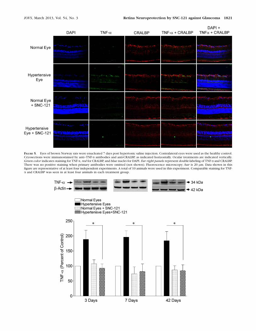
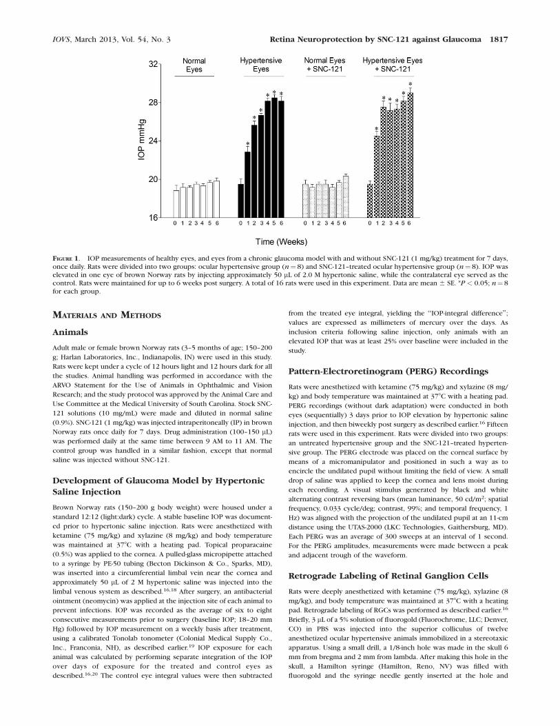
FIGURE 5. Eyes of brown Norway rats were enucleated 7 days post hypertonic saline injection. Contralateral eyes were used as the healthy control.Cryosections were immunostained by anti–TNF-a antibodies and anti-CRALBP, as indicated horizontally. Ocular treatments are indicated vertically.Green color indicates staining for TNF-a, red for CRALBP, and blue nuclei for DAPI. Far right panels represent double-labeling of TNF-a and CRALBP.There was no positive staining when primary antibodies were omitted (not shown). Fluorescence microscopy; bar is 20 lm. Data shown in thisfigure are representative of at least four independent experiments. A total of 10 animals were used in this experiment. Comparable staining for TNF-a and CRALBP was seen in at least four animals in each treatment group.
IOVS, March 2013, Vol. 54, No. 3 Retina Neuroprotection by SNC-121 against Glaucoma 1821
IP) had no significant effect on IOP when measured once aweek. Additionally, IOP was measured at 2, 4, 6, 24, and 72hours, post SNC-121 treatment and we have not seen anysignificant changes in the IOP (data not shown). Furthermore,the IOP-integral difference of IOP elevation over the days ofexposure for both the control and hypertensive eyes wascalculated using the area under the curve (e.g., elevated IOPminus baseline IOP), as described earlier.16 IOP-integraldifferences were averaged for each group and compared. Thecumulative average group IOP-integral differences rose steadilyover the 6 week duration. At week 6, cumulative IOP of theuntreated ocular hypertensive group (319 6 5 mm Hg; n¼ 8)was not significantly different from the SNC-121–treated ocularhypertensive group (331 6 8 mm Hg; n¼8). However, the IOPremained significantly elevated in both the untreated and SNC-121–treated ocular hypertensive groups, when compared withhealthy eyes (Fig. 1).
To determine the functional response of RGCs in healthyand ocular hypertensive rats, PERGs were performed. Figure2A presents changes in PERG amplitudes in normal and ocularhypertensive eyes, whereas Figure 2B shows changes in PERGamplitudes of SNC-121–treated healthy and hypertensive eyes.Figure 2C presents the change in the PERG of ocularhypertensive eyes and SNC-121–treated ocular hypertensive
eyes at different time points, post injury. PERG amplitudeswere significantly reduced in ocular hypertensive eyes whencompared with healthy eyes (P < 0.05; Fig. 2A). To determineif d-opioid receptor activation during glaucomatous injurypreserved the function of RGCs, animals were treated withSNC-121 (1 mg/kg, IP) each day for 7 days. SNC-121 treatmentpreserved RGC function significantly (P < 0.05; Fig. 2B), asdetermined by PERG. Overall, PERGs were reduced to 80%,76%, and 76% in hypertensive eyes at 2, 4, and 6 weeks postinjury, respectively, which were increased to 101%, 96%, and101% when animals were treated with SNC-121 (Fig. 2C).
To confirm that declines in PERG amplitudes are due to theloss of RGCs, they were visualized by retrograde labeling withbilateral injections of fluorogold into the superior colliculus.Representative micrographs of fluorogold-labeled cells indicatea clear loss of RGCs in the ocular hypertensive eye ascompared with the contralateral healthy eye (i.e., Fig. 3A-bcompared with Fig. 3A-a, respectively). The loss of RGCs wasreduced when the ocular hypertensive animals were treatedwith SNC-121 (1 mg/kg; IP) each day for 7 days (Fig. 3A-d; Fig.3A-c serving as the treatment control). Quantification of RGCsin healthy eyes, ocular hypertensive eyes, SNC-121–treatedhealthy eyes, and SNC-121–treated ocular hypertensive eyesare shown in Figure 3B. The mean number (6 SE) of
FIGURE 6. TNF-a production in response to glaucomatous injury and its suppression by SNC-121 treatment in the optic nerve extracts. Optic nervesof untreated and SNC-121–treated healthy and hypertensive eyes were collected at 3, 7, or 42 days post injury and analyzed for TNF-a expression byWestern blotting using anti–TNF-a antibodies. Rats were divided into two groups: ocular hypertensive group (n¼ 4) and SNC-121–treated ocularhypertensive group (n¼ 4). Rats were treated with SNC-121 (1 mg/kg) once daily for 7 days. IOP was elevated in one eye of brown Norway rats byinjecting approximately 50 lL of 2.0 M hypertonic saline into limbal veins, while the contralateral eye served as the control. Band intensities ofWestern blots were quantitated by densitometry. A total of 24 animals were used in this experiment. Data are expressed as mean 6 SE. *P < 0.05; n
¼ 4 for each group.
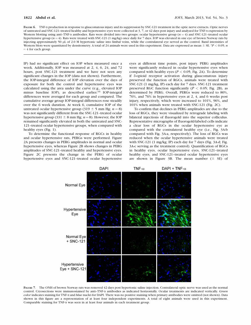
FIGURE 7. The ONH of brown Norway rats was removed 42 days post hypertonic saline injection. Contralateral optic nerve was used as the normalcontrol. Cryosections were immunostained by anti–TNF-a antibodies as indicated horizontally. Ocular treatments are indicated vertically. Green
color indicates staining for TNF-a and blue nuclei for DAPI. There was no positive staining when primary antibodies were omitted (not shown). Datashown in this figure are a representation of at least four independent experiments. A total of eight animals were used in this experiment.Comparable staining for TNF-a was seen in at least four animals in each treatment group.
1822 Abdul et al. IOVS, March 2013, Vol. 54, No. 3
fluorogold-positive RGCs were: healthy eyes, 1572 6 81;ocular hypertensive eyes, 1122 6 38 (28.6% less than healthyeyes); and SNC-121–treated ocular hypertensive eyes, 1533 6
92 (36.6% greater than the ocular hypertensive eyes).To determine if TNF-a plays a detrimental role in the early
stages of glaucoma development and d-opioid receptor agonist,SNC-121, counterbalances TNF-a production, retinal sectionsof chronic ocular hypertensive rat eyes were analyzed for TNF-a expression by immunohistochemistry. As shown in Figure 4,no staining for TNF-a was seen in normal eyes (contralateraleye); however, the staining for TNF-a was increased in ocularhypertensive eyes, which was inhibited in SNC-121–treatedanimals. Strong immunostaining was observed for TNF-a in thenerve fiber layer and RGC layer. In addition, an occasionalpunctate staining was observed in the inner plexiform andinner nuclear layers of ocular hypertensive eyes (Fig. 5). Toidentify the cell type responsible for TNF-a production, a dualimmunolabeling using GFAP (a glial cell marker) and CRALBP(an RPE and Muller cell marker) was performed. As shown inFigure 4, TNF-a and GFAP immunostaining were colocalized(far right panels). Additionally, we have noticed a mild stainingfor GFAP in healthy eyes and it was increased in the nerve fiberlayer of ocular hypertensive eyes, which was also inhibited inSNC-121–treated ocular hypertensive eyes.
As shown in Figure 5, strong CRALBP staining was observedin the RPE and glial Muller cells. However, CRALBP stainingwas not colocalized with TNF-a staining. To further confirmthat TNF-a seen in immunohistochemistry data are mainlyproduced from glial cells, we have analyzed the ONH (non-myelinated) samples of normal and ocular hypertensiveanimals at 3, 7, and 42 days, post hypertonic saline injections.
We chose 3 days as the earliest time point for TNF-a detectionin optic nerve samples because we have not seen anappreciable increase in the IOP prior to 3 days, in thehypertonic saline injected eyes. As shown in Figure 6, TNF-aproduction was significantly increased in the optic nerve (1896 30% over the control) as early as 3 days post injury, andremained significantly elevated up to 42 days (184 6 31% overthe control). The increase in TNF-a production was completelyinhibited when animals were treated with SNC-121 at eachtime point. To further confirm our Western blot data, weanalyzed optic nerve sections by immunohistochemistry forTNF-a production at 42 days, post glaucomatous injury. In thisexperiment, IOP was measured at 3, 7, 14, 28, and 42 days, andpattern ERG was measured at 14, 28, and 42 days (data notshown). Changes seen in IOP and PERG were comparable tothe data shown in Figures 1 and 2. As shown in Figure 7, wesaw mild TNF-a staining in normal optic nerve, which wasfurther increased in the hypertensive optic nerve. Theincreased TNF-a staining in the ocular hypertensive opticnerve was inhibited in SNC-121–treated animals.
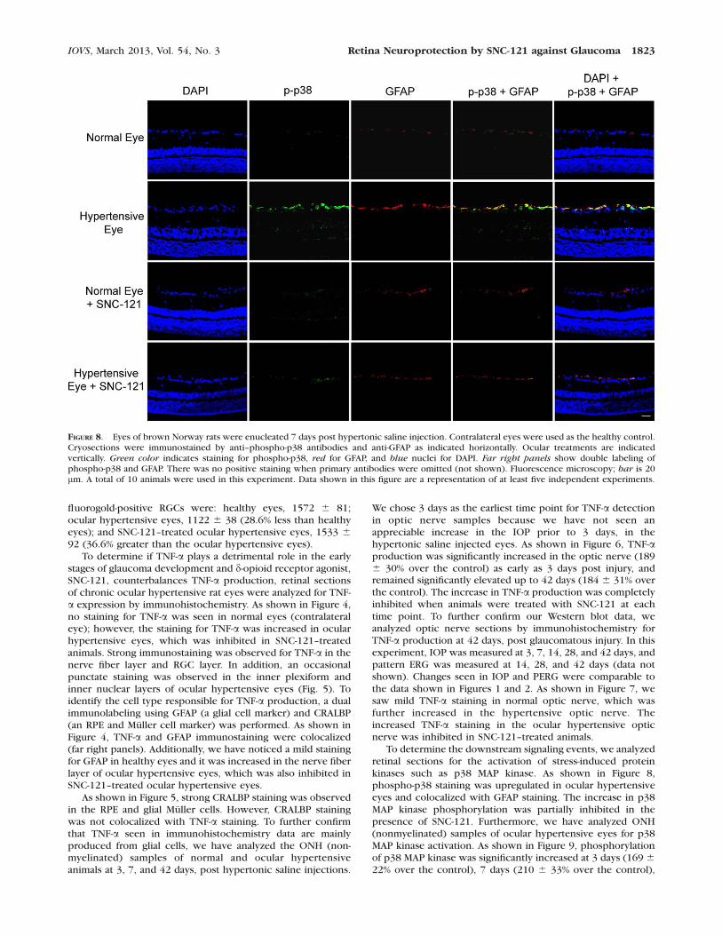
To determine the downstream signaling events, we analyzedretinal sections for the activation of stress-induced proteinkinases such as p38 MAP kinase. As shown in Figure 8,phospho-p38 staining was upregulated in ocular hypertensiveeyes and colocalized with GFAP staining. The increase in p38MAP kinase phosphorylation was partially inhibited in thepresence of SNC-121. Furthermore, we have analyzed ONH(nonmyelinated) samples of ocular hypertensive eyes for p38MAP kinase activation. As shown in Figure 9, phosphorylationof p38 MAP kinase was significantly increased at 3 days (169 6
22% over the control), 7 days (210 6 33% over the control),
FIGURE 8. Eyes of brown Norway rats were enucleated 7 days post hypertonic saline injection. Contralateral eyes were used as the healthy control.Cryosections were immunostained by anti–phospho-p38 antibodies and anti-GFAP as indicated horizontally. Ocular treatments are indicatedvertically. Green color indicates staining for phospho-p38, red for GFAP, and blue nuclei for DAPI. Far right panels show double labeling ofphospho-p38 and GFAP. There was no positive staining when primary antibodies were omitted (not shown). Fluorescence microscopy; bar is 20lm. A total of 10 animals were used in this experiment. Data shown in this figure are a representation of at least five independent experiments.
IOVS, March 2013, Vol. 54, No. 3 Retina Neuroprotection by SNC-121 against Glaucoma 1823

and 42 days (161 6 17% over the control), post injury. In opticnerve, the increase in p38 MAP kinase phosphorylation wascompletely inhibited when animals were treated with SNC-121at all time points. In contrast, we have not seen any changes inthe expression pattern of total p38 MAP kinase in theuntreated and SNC-121–treated ocular hypertensive eyes (datanot shown).
To establish a causal link between TNF-a and p38 MAPkinase activation in glial cells, we used primary cultures ofoptic nerve head (ONH) astrocytes. Cells were pretreated withSNC-121 followed by TNF-a (25 ng/mL) treatment for 6 hoursand phosphorylation of p38 MAP kinase was determined byWestern blotting using selective anti–phospho-p38 MAP kinaseantibodies. A concentration response study was performed inONH astrocytes using TNF-a for p38 MAP kinase activation,where we found that 25 ng/mL TNF-a provided maximumphosphorylation and activation of p38 MAP kinase (data notshown). As shown in Figure 10, TNF-a increased thephosphorylation of p38 MAP kinase over 2-fold at 6 hours(control 100 6 00 versus TNF-a 231 6 31; n ¼ 4; P ¼ 0.024).TNF-a–induced increase in p38 MAP kinase phosphorylation inONH astrocytes was significantly inhibited in the presence ofSNC-121 (TNF-a 231 6 31 versus SNC-121þ TNF-a 128 6 25;n¼ 4; P ¼ 0.041).
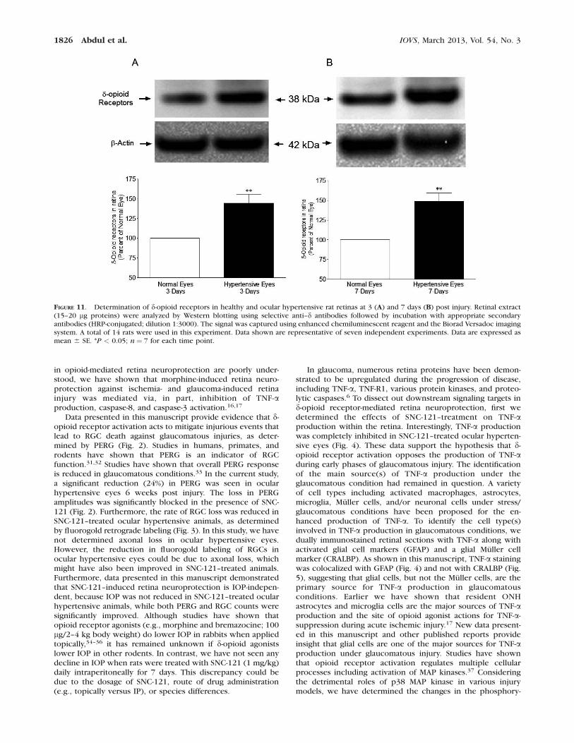
To determine if d-opioid receptors were downregulated orupregulated in response to glaucomatous injury, expression ofd-opioid receptors was measured in ocular hypertensive eyes at3, 7, and 42 days post injury. IOP was increased whenmeasured at 3 and 7 days. As shown in Figures 11A and 11B, d-opioid receptors were moderately upregulated in ocular
hypertensive eyes at 3 days (44.6% over the healthy eye; n ¼7; P¼ 0.0078) and 7 days (49% over the healthy eye; n¼ 7; P¼0.0028).
DISCUSSION
Opioids have been used clinically for centuries as analgesics.However, other biological effects induced by opioids includecytoprotection, immunomodulation, neuroendocrine regula-tion, and behavioral modification. Most of these biologicalresponses are presumed to be manifested through theactivation of G-protein–coupled d-, j-, and l-opioid receptors.In the eye, activation of opioid receptors has been implicatedin the regulation of iris function, accommodative power,regulation of aqueous humor dynamics (e.g., IOP), cornealwound healing, retinal development, and retina neuroprotec-tion.15,17,21–23
d-opioid receptors (also known as DOR or DOP receptors inthe International Union of Basic and Clinical Pharmacology[IUPHAR] nomenclature) are increasingly attractive due totheir therapeutic potential. Along with the development ofhighly selective d-opioid receptor agonists and rapid progressin mouse mutagenesis approaches targeting the d-opioidreceptor gene (Oprd1), novel functions of d-opioid receptorshave emerged. For example, activation of d-opioid receptorsreduced infarct size in stroke and myocardial ischemiamodels.24,25 Activation of d-opioid receptors is protectiveagainst hypoxia/excitotoxic injury in cortical neurons.26
DADLE, a d-opioid receptor agonist, has been shown toimprove hippocampal neuronal survival against ischemia in a
FIGURE 9. Activation of p38 MAP kinase in response to glaucomatous injury and its suppression by SNC-121 treatment in the optic nerve extracts.Optic nerves of untreated and SNC-121 treated healthy and ocular hypertensive eyes were collected at 3, 7, or 42 days post injury. Changes inphosphorylation of p38 MAP kinase were analyzed by Western blotting using selective anti–phospho-p38 MAP kinase antibodies. Rats were dividedinto two groups: ocular hypertensive group (n¼ 6) and SNC-121–treated ocular hypertensive group (n¼ 6). IOP was elevated in one eye of brownNorway rats by injecting approximately 50 lL of 2.0 M hypertonic saline, while the contralateral eye served as the control. A total of 36 animalswere used in this experiment. Band intensities of Western blots were quantitated by densitometry. Data are expressed as mean 6 SE. *P < 0.05; n¼6 for each group at each time point.
1824 Abdul et al. IOVS, March 2013, Vol. 54, No. 3

rat model.27 Studies have also shown that d-opioid receptoractivation attenuates oxidative injuries in the brain exposed toischemia/reperfusion by enhancing antioxidant ability andinhibition of caspase activity.28 Moreover, d-opioid receptorshave been involved in neuroprotective mechanisms ofAlzheimer29 and Parkinson’s30 diseases. In this manuscriptwe have shown an upregulation of d-opioid receptors inresponse to the glaucomatous injury, which may have beeninvolved in the endogenous neuroprotection. Although d-opioid receptors are upregulated in response to glaucomatousinjury, there may not be enough endogenous ligand to initiatedownstream neuroprotective signaling cascades to protect theretina against glaucomatous injury. When animals were treatedwith an exogenous ligand, such as SNC-121, d-opioid receptorswere activated and initiated a neuroprotective downstreamsignaling cascade to protect the retina. d-opioid receptoractivation curtailed the detrimental cellular events at an earlystage of the injury. Once the initial cellular events werecounteracted by SNC-121 in the initial phase of glaucomatousinjury, RGCs can be preserved, as seen here in Figures 2 and 3.Although beneficial effects of d-opioid receptor agonists innonocular systems have been clearly established in the areas of
chronic pain, emotional disorders, and neuroprotection againstvarious forms of injuries, the usefulness of d-opioid receptoragonists against glaucomatous injury remains to be clarified.
Glaucoma is a long term optic neuropathy characterized byoptic disc cupping, RGC death, and vision loss. POAG is themost common type of glaucoma. Glaucoma pathogenesis ismultifactorial; however, a major risk factor for the develop-ment of glaucoma is elevated IOP. The pathophysiologicmechanisms by which elevated IOP leads to optic nerveatrophy and retinal degeneration are unknown. This manu-script provides novel findings that d-opioid receptor agonisttreatment, such as with SNC-121, provides significant retinalneuroprotection against glaucomatous injury, thus, suggestingthat d-opioid receptor agonists have a potential to be used astherapeutic agents against glaucomatous injury. The possibleinvolvement of other opioid receptors (e.g., j and l) in retinaneuroprotection has not been ruled and it will be tested in ourfuture studies. Recently, we have demonstrated that opioidreceptor activation is required for the development of ischemicpreconditioning within the retina, and that the administrationof morphine can reduce retinal ischemic and glaucomatousinjury.15,16 Although the cellular mechanisms that are involved
FIGURE 10. TNF-a–induced activation of p38 MAP kinase in ONH astrocytes. ONH astrocytes were starved in serum-free medium for overnight.Cells were then pretreated with SNC-121 (1 lmol/L) for 15 minutes followed by TNF-a (25 ng/mL) treatment for 6 hours. Cell lysate (15 lg protein)was analyzed by Western blotting using selective anti–phospho-p38 MAP kinase antibodies followed by incubation with appropriate secondaryantibodies (HRP-conjugated; dilution 1:3000). The signal was captured using enhanced chemiluminescent reagent and the Biorad Versadoc imagingsystem. Data shown is a representative of four independent experiments. Data are expressed as mean 6 SE. *P < 0.05; n¼ 4.
IOVS, March 2013, Vol. 54, No. 3 Retina Neuroprotection by SNC-121 against Glaucoma 1825
in opioid-mediated retina neuroprotection are poorly under-stood, we have shown that morphine-induced retina neuro-protection against ischemia- and glaucoma-induced retinainjury was mediated via, in part, inhibition of TNF-aproduction, caspase-8, and caspase-3 activation.16,17
Data presented in this manuscript provide evidence that d-opioid receptor activation acts to mitigate injurious events thatlead to RGC death against glaucomatous injuries, as deter-mined by PERG (Fig. 2). Studies in humans, primates, androdents have shown that PERG is an indicator of RGCfunction.31,32 Studies have shown that overall PERG responseis reduced in glaucomatous conditions.33 In the current study,a significant reduction (24%) in PERG was seen in ocularhypertensive eyes 6 weeks post injury. The loss in PERGamplitudes was significantly blocked in the presence of SNC-121 (Fig. 2). Furthermore, the rate of RGC loss was reduced inSNC-121–treated ocular hypertensive animals, as determinedby fluorogold retrograde labeling (Fig. 3). In this study, we havenot determined axonal loss in ocular hypertensive eyes.However, the reduction in fluorogold labeling of RGCs inocular hypertensive eyes could be due to axonal loss, whichmight have also been improved in SNC-121–treated animals.Furthermore, data presented in this manuscript demonstratedthat SNC-121–induced retina neuroprotection is IOP-indepen-dent, because IOP was not reduced in SNC-121–treated ocularhypertensive animals, while both PERG and RGC counts weresignificantly improved. Although studies have shown thatopioid receptor agonists (e.g., morphine and bremazocine; 100lg/2–4 kg body weight) do lower IOP in rabbits when appliedtopically,34–36 it has remained unknown if d-opioid agonistslower IOP in other rodents. In contrast, we have not seen anydecline in IOP when rats were treated with SNC-121 (1 mg/kg)daily intraperitoneally for 7 days. This discrepancy could bedue to the dosage of SNC-121, route of drug administration(e.g., topically versus IP), or species differences.
In glaucoma, numerous retina proteins have been demon-strated to be upregulated during the progression of disease,including TNF-a, TNF-R1, various protein kinases, and proteo-lytic caspases.6 To dissect out downstream signaling targets ind-opioid receptor-mediated retina neuroprotection, first wedetermined the effects of SNC-121–treatment on TNF-aproduction within the retina. Interestingly, TNF-a productionwas completely inhibited in SNC-121–treated ocular hyperten-sive eyes (Fig. 4). These data support the hypothesis that d-opioid receptor activation opposes the production of TNF-aduring early phases of glaucomatous injury. The identificationof the main source(s) of TNF-a production under theglaucomatous condition had remained in question. A varietyof cell types including activated macrophages, astrocytes,microglia, Muller cells, and/or neuronal cells under stress/glaucomatous conditions have been proposed for the en-hanced production of TNF-a. To identify the cell type(s)involved in TNF-a production in glaucomatous conditions, wedually immunostained retinal sections with TNF-a along withactivated glial cell markers (GFAP) and a glial Muller cellmarker (CRALBP). As shown in this manuscript, TNF-a stainingwas colocalized with GFAP (Fig. 4) and not with CRALBP (Fig.5), suggesting that glial cells, but not the Muller cells, are theprimary source for TNF-a production in glaucomatousconditions. Earlier we have shown that resident ONHastrocytes and microglia cells are the major sources of TNF-aproduction and the site of opioid agonist actions for TNF-a-suppression during acute ischemic injury.17 New data present-ed in this manuscript and other published reports provideinsight that glial cells are one of the major sources for TNF-aproduction under glaucomatous injury. Studies have shownthat opioid receptor activation regulates multiple cellularprocesses including activation of MAP kinases.37 Consideringthe detrimental roles of p38 MAP kinase in various injurymodels, we have determined the changes in the phosphory-
FIGURE 11. Determination of d-opioid receptors in healthy and ocular hypertensive rat retinas at 3 (A) and 7 days (B) post injury. Retinal extract(15–20 lg proteins) were analyzed by Western blotting using selective anti–d antibodies followed by incubation with appropriate secondaryantibodies (HRP-conjugated; dilution 1:3000). The signal was captured using enhanced chemiluminescent reagent and the Biorad Versadoc imagingsystem. A total of 14 rats were used in this experiment. Data shown are representative of seven independent experiments. Data are expressed asmean 6 SE. *P < 0.05; n¼ 7 for each time point.
1826 Abdul et al. IOVS, March 2013, Vol. 54, No. 3

lation and activation of p38 MAP kinase in response to TNF-a–and/or glaucoma-induced injury. Immunostaining data shownin this manuscript clearly demonstrated a sustained increase inthe phosphorylation of p38 MAP kinase in ocular-hypertensiveeyes, which was inhibited in SNC-121–treated hypertensiveeyes. Dual immunostaining data in retinal sections and Westernblot data in optic nerve samples further support the idea thatactivation of p38 MAP kinase is taking place within the opticnerve at an early stage of glaucomatous injury. Studies haveshown that p38 MAP kinase and c-Jun N-terminal kinases (JNK)are activated by stress and contribute in the neurodegenerativesignaling pathways, whereas ERK1/2 have been shown to playneuroprotective roles against various forms of injuries.38–41
Studies also have shown that increased activity of p38 MAPkinase plays a critical role in cell death in neurons under stressconditions.41 Inhibition of p38 MAP kinase confers neuropro-tection in vitro against excitotoxic exposure42 and reducesacute ischemic injury in vivo.43 Studies also have shown that d-opioid receptor agonist, DADLE, provides neuroprotectionagainst oxygen-glucose deprivation-induced neuronal injury viainhibition of p38 MAP kinase phosphorylation and activation.40
Additionally, p38 MAP kinase plays a key role in the glutamate-induced apoptosis of RGCs,44 and the increased phosphoryla-tion of p38 and JNK have been noted in an experimentalglaucoma model.45
To establish a direct link between TNF-a and p38 MAPkinase, we determined the effect of TNF-a on p38 MAP kinaseactivation in isolated ONH astrocytes. TNF-a–induced increasein p38 MAP kinase phosphorylation was significantly inhibitedin the presence of SNC-121. These data provide concretesupport that p38 MAP kinase is a downstream target of TNF-awithin the optic nerve.
Based on the data shown in this manuscript, and previouslypublished work from our laboratory in acute ischemia andchronic glaucoma models,15–17 it is evident that TNF-aproduction in response to retinal injuries is an early event.The current manuscript provides novel information thatsustained IOP elevation and glial cell activation caused asustained TNF-a production within the optic nerve, andactivated downstream signaling targets such as p38 MAPkinase. Activation of these signaling pathways may subse-quently enhance the production of extracellular matrixdegrading enzymes, matrix metalloproteinases (MMPs), whichwill further destabilize the optic nerve by excessive remodel-ing. These detrimental signaling molecules directly and/orindirectly weaken axons and lead to the RGC death. d-opioidreceptor agonists represent a novel class of drugs/agents thatcounteract detrimental events (e.g., TNF-a production and p38MAP kinase activation) within the optic nerve at an early stageof disease development, which delays and/or prevents the lossof RGCs. Thus, therapeutic approaches that primarily inhibitexcessive astrocyte reactivity, proinflammatory cytokine activ-ity, and downstream activation and/or expression of signalingtargets (e.g., stress-activated protein kinase; p38 kinase),should impede deleterious changes in the optic nerve andRGCs in eyes predisposed to glaucoma.
In summary, our study provides evidence that d-opioidreceptors play key roles in retina neuroprotection againstglaucomatous injury because: (1) RGC function was preservedby exogenous SNC-121 treatment in chronic ocular hyperten-sive rat eyes, as determined by PERG; (2) loss of RGCs wasreduced in SNC-121–treated hypertensive eyes, as determinedby retrograde labeling of RGCs; (3) TNF-a is mainly producedfrom glial cells in chronic ocular hypertensive eyes, asdetermined by immunohistochemistry; (4) TNF-a productionin response to glaucomatous injury is inhibited in the presenceof SNC-121; and (5) p38 MAP kinase, a downstream target ofTNF-a, activation is suppressed by SNC-121 treatment. Overall,
TNF-a and p38 MAP kinase appear to be potential therapeutictargets to achieve neuroprotection against glaucomatousinjury. Additionally, enhancement of d-opioidergic activity inthe eye may present a viable neuroprotective strategy for thetreatment of glaucoma.
Acknowledgments
The authors thank Luanna Bartholomew (Medical University ofSouth Carolina-Storm Eye Institute) for critical review of themanuscript.
References
1. Quigley HA, Green WR. The histology of human glaucomacupping and optic nerve damage: clinicopathologic correla-tion in 21 eyes. Ophthalmology. 1979;86:1803–1830.
2. Boland MV, Quigley HA. Risk factors and open-angle glaucoma:classification and application. J Glaucoma. 2007;16:406–418.
3. Liu Q, Ju WK, Crowston JG, et al. Oxidative stress is an earlyevent in hydrostatic pressure induced retinal ganglion celldamage. Invest Ophthalmol Vis Sci. 2007;48:4580–4589.
4. Ju WK, Kim KY, Angert M, et al. Memantine blocksmitochondrial OPA1 and cytochrome c release and subse-quent apoptotic cell death in glaucomatous retina. Invest
Ophthalmol Vis Sci. 2009;50:707–716.
5. Nucci C, Tartaglione R, Rombola L, Morrone LA, Fazzi E,Bagetta G. Neurochemical evidence to implicate elevatedglutamate in the mechanisms of high intraocular pressure(IOP)-induced retinal ganglion cell death in rat. Neurotoxicol-
ogy. 2005;26:935–941.
6. Yang X, Luo C, Cai J, et al. Neurodegenerative and inflamma-tory pathway components linked to TNF-alpha/TNFR1 signal-ing in the glaucomatous human retina. Invest Ophthalmol Vis
Sci. 2011;52:8442–8454.
7. Tezel G. Oxidative stress in glaucomatous neurodegeneration:mechanisms and consequences. Prog Retinal Eye Res. 2006;25:490–513.
8. Shohami E, Ginis I, Hallenbeck JM. Dual role of tumor necrosisfactor alpha in brain injury. Cytokine Growth Factor Rev.1999;10:119–130.
9. Tezel G, Li LY, Patil RV, Wax MB. TNF-alpha and TNF-alphareceptor-1 in the retina of normal and glaucomatous eyes.Invest Ophthalmol Vis Sci. 2001;42:1787–1794.
10. Yuan L, Neufeld AH. Tumor necrosis factor-alpha: a potentiallyneurodestructive cytokine produced by glia in the humanglaucomatous optic nerve head. Glia. 2000;32:42–50.
11. Husain S, Potter DE. The opioidergic system: potential rolesand therapeutic indications in the eye. J Ocul Pharmacol
Ther. 2008;24:117–140.
12. Schultz JE, Gross GJ. Opioids and cardioprotection. Pharma-
col Ther. 2001;89:123–137.
13. Ferri S, Speroni E, Candeletti S, et al. Protection by opioidsagainst gastric lesions caused by necrotizing agents. Pharma-
cology. 1988;36:140–144.
14. Mayfield KP, D’Alecy LG. Delta-1 opioid agonist acutelyincreases hypoxic tolerance. J Pharmacol Exp Ther. 1994;268:683–688.
15. Husain S, Potter DE, Crosson CE. Opioid receptor-activation:retina protected from ischemic injury. Invest Ophthalmol Vis
Sci. 2009;50:3853–3859.
16. Husain S, Abdul Y, Crosson CE. Preservation of retina ganglioncell function by morphine in a chronic ocular-hypertensive ratmodel. Invest Ophthalmol Vis Sci. 2012;53:4289–4298.
17. Husain S, Liou GI, Crosson CE. Opioid-receptor-activation:suppression of ischemia/reperfusion-induced production of
IOVS, March 2013, Vol. 54, No. 3 Retina Neuroprotection by SNC-121 against Glaucoma 1827

TNF-{alpha} in the retina. Invest Ophthalmol Vis Sci. 2011;52:2577–2583.
18. Morrison JC, Moore CG, Deppmeier LM, Gold BG, Meshul CK,Johnson EC. A rat model of chronic pressure-induced opticnerve damage. Exp Eye Res. 1997;64:85–96.
19. Husain S, Whitlock NA, Rice DS, Crosson CE. Effects oflatanoprost on rodent intraocular pressure. Exp Eye Res. 2006;83:1453–1458.
20. McKinnon SJ, Lehman DM, Tahzib NG, et al. Baculoviral IAPrepeat-containing-4 protects optic nerve axons in a ratglaucoma model. Mol Ther. 2002;5:780–787.
21. Isayama T, McLaughlin PJ, Zagon IS. Endogenous opioidsregulate cell proliferation in the retina of developing rat. Brain
Res. 1991;544:79–85.
22. Husain S, Abdul Y, Potter DE. Non-analgesic effects of opioids:neuroprotection in the retina. Curr Pharm Des. 2012;18:5919–5926.
23. Husain S, Potter DE. The opioidergic system: potential rolesand therapeutic indications in the eye. J Ocul Pharmacol
Ther. 2008;24:117–140.
24. Chen TY, Goyagi T, Toung TJ, et al. Prolonged opportunity forischemic neuroprotection with selective kappa-opioid recep-tor agonist in rats. Stroke. 2004;35:1180–1185.
25. Gross ER, Hsu AK, Gross GJ. Opioid-induced cardioprotectionoccurs via glycogen synthase kinase beta inhibition duringreperfusion in intact rat hearts. Circ Res. 2004;94:960–966.
26. Zhang J, Haddad GG, Xia Y. Delta-, but not mu- and kappa-,opioid receptor activation protects neocortical neurons fromglutamate-induced excitotoxic injury. Brain Res. 2000;885:143–153.
27. Iwata M, Inoue S, Kawaguchi M, Nakamura M, Konishi N,Furuya H. Effects of delta-opioid receptor stimulation andinhibition on hippocampal survival in a rat model of forebrainischaemia. British J Anaesth. 2007;99:538–546.
28. Yang Y, Xia X, Zhang Y, et al. delta-Opioid receptor activationattenuates oxidative injury in the ischemic rat brain. BMC
Biol. 2009;7:1–9.
29. Thathiah A, De Strooper B. The role of G protein-coupledreceptors in the pathology of Alzheimer’s disease. Nat Rev
Neurosci. 2011;12:73–87.
30. Mabrouk OS, Marti M, Salvadori S, Morari M. The novel deltaopioid receptor agonist UFP-512 dually modulates motoractivity in hemiparkinsonian rats via control of the nigro-thalamic pathway. Neuroscience. 2009;164:360–369.
31. Holder GE. Pattern electroretinography (PERG) and anintegrated approach to visual pathway diagnosis. Prog Retinal
Eye Res. 2001;20:531–561.
32. Porciatti V. The mouse pattern electroretinogram. Doc
Ophthalmol. 2007;115:145–153.
33. Wanger P, Persson HE. Pattern-reversal electroretinograms inunilateral glaucoma. Invest Ophthalmol Vis Sci. 1983;24:749–753.
34. Dortch-Carnes J, Potter DE. Effect of bremazocine, a kappa-opioid receptor agonist, on inositol phosphate formation inisolated iris-ciliary bodies. Pharmacology. 2002;66:100–106.
35. Dortch-Carnes J, Potter DE. Bremazocine: a kappa-opioidagonist with potent analgesic and other pharmacologicproperties. CNS Drug Rev. 2005;11:195–212.
36. Stagni E, Bucolo C, Motterlini R, Drago F. Morphine-inducedocular hypotension is modulated by nitric oxide and carbonmonoxide: role of mu3 receptors. J Ocul Pharmacol Ther.2010;26:31–35.
37. Burt AR, Carr IC, Mullaney I, Anderson NG, Milligan G. Agonistactivation of p42 and p44 mitogen-activated protein kinasesfollowing expression of the mouse delta opioid receptor inRat-1 fibroblasts: effects of receptor expression levels andcomparisons with G-protein activation. Biochem J. 1996;320:227–235.
38. Park EM, Joh TH, Volpe BT, Chu CK, Song G, Cho S. Aneuroprotective role of extracellular signal-regulated kinase inN-acetyl-O-methyldopamine–treated hippocampal neurons af-ter exposure to in vitro and in vivo ischemia. Neuroscience.2004;123:147–154.
39. Hetman M, Kanning K, Cavanaugh JE, Xia Z. Neuroprotectionby brain-derived neurotrophic factor is mediated by extracel-lular signal-regulated kinase and phosphatidylinositol 3-kinase.J Biol Chem. 1999;274:22569–22580.
40. Ke S, Dian-san S, Xiang-rui W. Delta opioid agonist [D-Ala2, D-Leu5] enkephalin (DADLE) reduced oxygen-glucose depriva-tion caused neuronal injury through the MAPK pathway.Brain Res. 2009;1292:100–106.
41. Takeda K, Ichijo H. Neuronal p38 MAPK signalling: anemerging regulator of cell fate and function in the nervoussystem. Genes Cells. 2002;7:1099–1111.
42. Legos JJ, McLaughlin B, Skaper SD, et al. The selective p38inhibitor SB-239063 protects primary neurons from mild tomoderate excitotoxic injury. Eur J Pharmacol. 2002;447:37–42.
43. Legos JJ, Erhardt JA, White RF, et al. SB 239063, a novel p38inhibitor, attenuates early neuronal injury following ischemia.Brain Res. 2001;892:70–77.
44. Kikuchi M, Tenneti L, Lipton SA. Role of p38 mitogen-activatedprotein kinase in axotomy-induced apoptosis of rat retinalganglion cells. J Neurosci. 2000;20:5037–5044.
45. Levkovitch-Verbin H, Harizman N, Dardik R, Nisgav Y, VanderS, Melamed S. Regulation of cell death and survival pathwaysin experimental glaucoma. Exp Eye Res. 2007;85:250–258.
1828 Abdul et al. IOVS, March 2013, Vol. 54, No. 3