delivery of an enzyme-igfii fusion protein to the mouse ... · with no man6-p have been made for...
TRANSCRIPT

Delivery of an enzyme-IGFII fusion protein to themouse brain is therapeutic for mucopolysaccharidosistype IIIBShih-hsin Kana, Mika Aoyagi-Scharberb,1, Steven Q. Lea, Jon Vinceletteb, Kazuhiro Ohmic, Sherry Bullensb,Daniel J. Wendtd, Terri M. Christiansonb, Pascale M. N. Tigerb, Jillian R. Brownb, Roger Lawrenceb, Bryan K. Yipb,John Holtzingerb, Anil Bagrib, Danielle Crippen-Harmonb, Kristen N. Vondraka, Zhi Chend, Chuck M. Hagued,Josh C. Woloszynekb, Diana S. Cheungb, Katherine A. Websterb, Evan G. Adintorib, Melanie J. Lob, Wesley Wongb,Paul A. Fitzpatrickb, Jonathan H. LeBowitzb, Brett E. Crawfordb, Stuart Buntingb, Patricia I. Dicksona,1,and Elizabeth F. Neufeldc,1
aDepartment of Pediatrics, Los Angeles Biomedical Research Institute at Harbor–UCLA Medical Center, Torrance, CA 90502; bResearch and DrugDiscovery and dAnalytical Chemistry, BioMarin Pharmaceutical, Inc., Novato, CA 94949; and cDepartment of Biological Chemistry, David Geffen School ofMedicine, University of California, Los Angeles, CA 90095
Contributed by Elizabeth F. Neufeld, August 29, 2014 (sent for review August 3, 2014; reviewed by Roscoe O. Brady and William S. Sly)
Mucopolysaccharidosis type IIIB (MPS IIIB, Sanfilippo syndrometype B) is a lysosomal storage disease characterized by profound in-tellectual disability, dementia, and a lifespan of about two decades.The cause ismutation in thegeneencodingα–N-acetylglucosaminidase(NAGLU), deficiency of NAGLU, and accumulation of heparan sulfate.Impediments toenzymereplacement therapyare theabsenceofman-nose 6-phosphate on recombinant human NAGLU and the blood–brain barrier. To overcome the first impediment, a fusion protein ofrecombinant NAGLU and a fragment of insulin-like growth factor II(IGFII) was prepared for endocytosis by the mannose 6-phosphate/IGFII receptor. To bypass the blood–brain barrier, the fusion protein(“enzyme”) in artificial cerebrospinal fluid (“vehicle”) was adminis-tered intracerebroventricularly to the brain of adult MPS IIIB mice,four times over 2 wk. The brains were analyzed 1–28 d later andcompared with brains of MPS IIIB mice that received vehicle aloneor control (heterozygous) mice that received vehicle. There wasmarked uptake of the administered enzyme in many parts of thebrain, where it persisted with a half-life of approximately 10 d. Hep-aran sulfate, and especially disease-specific heparan sulfate, wasreduced to control level. A number of secondary accumulationsin neurons [β-hexosaminidase, LAMP1(lysosome-associatedmem-brane protein 1), SCMAS (subunit c of mitochondrial ATP synthase),glypican 5, β-amyloid, P-tau] were reduced almost to control level.CD68, a microglial protein, was reduced halfway. A large amount ofenzymealso appeared in liver cells, where it reduced heparan sulfateand β-hexosaminidase accumulation to control levels. These resultssuggest the feasibility of enzyme replacement therapy for MPS IIIB.
Mucopolysaccharidosis type III (MPS III, Sanfilippo syn-drome) is a heritable lysosomal disorder of heparan sulfate
degradation, divided into four types (A–D), depending on theenzyme deficiency (1, 2). All four MPS III types are characterizedby severe neurologic problems and relatively mild somatic ones.Profound intellectual disability that progresses to dementia, be-havioral disturbances, and death in the second or third decadebring untold suffering to the MPS III patients and their families.Despite the dire need, treatment for the MPS III disorders haslagged behind other MPS diseases. Hematopoietic stem cell trans-plantation, an effective procedure for MPS I patients with CNS in-volvement (3), is not effective forMPS III (4). Enzyme replacementtherapy has been available for some years for several MPS withextensive somatic involvement [MPS I (5, 6), II (7), andVI (8)], or isnewly approved (MPS IVA), or in clinical trial (MPSVII).However,development of enzyme replacement for MPS III did not seempromising because access to therapeutic enzyme to brain paren-chyma would be limited by the blood–brain barrier. With respect toMPS IIIB, a deficiency of α–N-acetylglucosaminidase, EC 3.2.1.50)
(NAGLU), there is an additional difficulty in that, in contrast tomost other soluble lysosomal enzymes, recombinant NAGLUproduced in Chinese hamster ovary (CHO) cells contains little ornomannose 6-phosphate (Man6-P) (9–11), the signal for receptor-mediated endocytosis and targeting to lysosomes (12, 13). Thereason for the lack of the Man6-P modification is not understoodand appears to apply only to the recombinant enzyme, as humanurinary NAGLU (14) and endogenous NAGLU made by CHOcells (9) contain Man6-P.The lack of Man6-P on the enzyme can be overcome by taking
advantage of the ability of the cation-independent Man6-P re-ceptor to bind insulin-like growth factor II (IGFII) at a sitedistinct from the Man6-P binding sites (15–19). Fusion proteinsconsisting of a fragment of IGFII linked to a lysosomal enzyme
Significance
Mucopolysaccharidosis type IIIB (MPS IIIB) is a devastating andcurrently untreatable disease affecting mainly the brain. Thecause is lack of the lysosomal enzyme, α–N-acetylglucosami-nidase (NAGLU), and storage of heparan sulfate. Using a mousemodel of MPS IIIB, we administered a modified NAGLU by in-jection into the left ventricle of the brain, bypassing the blood–brain barrier. The modification consisted of a fragment of IGFII,which allows receptor-mediated uptake and delivery to lyso-somes. The modified enzyme was taken up avidly by cells inboth brain and liver, where it reduced pathological accumula-tion of heparan sulfate and other metabolites to normal ornear-normal levels. The results suggest the possibility of treat-ment for MPS IIIB.
Author contributions: M.A.-S., J.V., S. Bullens, D.J.W., T.M.C., P.A.F., J.H.L., B.E.C., S. Bunting,P.I.D., and E.F.N. designed research; S.-h.K., S.Q.L., J.V., K.O., D.J.W., T.M.C., P.M.N.T., J.R.B.,R.L., B.K.Y., J.H., A.B., D.C.-H., K.N.V., Z.C., C.M.H., J.C.W., D.S.C., K.A.W., E.G.A., M.J.L., andW.W. performed research; S.-h.K., M.A.-S., S.Q.L., K.O., S. Bullens, D.J.W., T.M.C., J.R.B., R.L.,B.K.Y., J.H., A.B., D.C.-H., Z.C., C.M.H., J.C.W., K.A.W., E.G.A., B.E.C., P.I.D., and E.F.N. ana-lyzed data; and S.-h.K., M.A.-S., and E.F.N. wrote the paper.
Reviewers: R.O.B., National Institute of Neurological Disorders and Stroke; and W.S.S.,Saint Louis University School of Medicine.
Conflict of interest statement: M.A.-S., J.V., S. Bullens., D.J.W., T.M.C., P.M.N.T., J.R.B., R.L.,B.K.Y., J.H., A.B., D.C.-H., Z.C., C.M.H., J.C.W., D.S.C., K.A.W., E.G.A., M.J.L., W.W., P.A.F.,J.H.L., B.E.C., and S. Bunting are employees of BioMarin Pharmaceutical, Inc., which isdeveloping a commercial treatment for mucopolysaccharidosis type IIIB. E.F.N. is principalinvestigator of a sponsored research agreement with BioMarin Pharmaceutical, Inc.
Freely available online through the PNAS open access option.1To whom correspondence may be addressed. Email: [email protected],[email protected], or [email protected].
This article contains supporting information online at www.pnas.org/lookup/suppl/doi:10.1073/pnas.1416660111/-/DCSupplemental.
14870–14875 | PNAS | October 14, 2014 | vol. 111 | no. 41 www.pnas.org/cgi/doi/10.1073/pnas.1416660111
with no Man6-P have been made for β-glucuronidase (20), α-glu-cosidase (21), and NAGLU (11), and shown to have enzymaticactivity similar to that of the original lysosomal enzyme and to betaken up by cultured cells in a manner that is dependent on IGFIIand independent of Man6-P. In addition, the β-glucuronidase–and α-glucosidase–IGFII fusion proteins, administered i.v. to de-ficient mice, were found to be taken up by major somatic organsand muscles, respectively, in which they functioned to reducestorage and pathology (20, 21). On the basis of these promisingearlier studies, we treated the brain of the MPS IIIB mouse byadministering a NAGLU–IGFII fusion protein directly into theleft cerebral ventricle, bypassing the blood–brain barrier. Themodified enzyme was endocytosed mainly into neurons, where itfunctioned to reduce the level of stored heparan sulfate and ofother accumulated substances to a normal or near-normal level. Italso spread to the liver, where it was endocytosed into vascular cellsand hepatocytes and eliminated storage of heparan sulfate. Theresults suggest that the combined use of the IGFII signal for en-docytosis with administration directly into the brainmay overcomethe major obstacles to enzyme replacement therapy for MPS IIIB.A preliminary account of this work has been presented in
abstract form (22, 23).
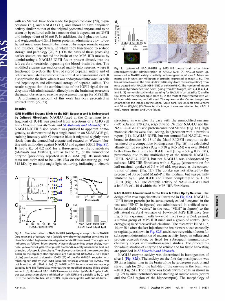
ResultsIGFII-Modified Enzyme Binds to the IGFII Receptor and Is Endocytosedby Cultured Fibroblasts. NAGLU fused at the C terminus to afragment of IGFII was purified from secretions of a CHO cellline (Materials and Methods and SI Materials and Methods). TheNAGLU–IGFII fusion protein was purified to apparent homo-geneity, as demonstrated by a single band on an SDS/PAGE gel,staining intensely with Coomassie blue; it migrated slightly moreslowly than the unmodified enzyme and reacted on Western blot-ting with antibodies against NAGLU and against IGFII (Fig. S1).It had a Km of 0.2 mM for a fluorogenic synthetic substrate(Materials and Methods), similar to the Km of the unmodifiedenzyme [previously reported as 0.22 mM (9)]. Its molecularmass was estimated to be ∼106 kDa on the denaturing gel and315 kDa by multiple angle light scattering, indicating a trimeric
structure, as was also the case with the unmodified enzyme(∼95 kDa and 278 kDa, respectively). Neither NAGLU nor theNAGLU–IGFII fusion protein containedMan6-P (Fig. 1A). Highmannose chains were also lacking, in agreement with a previousreport (11). NAGLU–IGFII, but not unmodified NAGLU, wasbound to domains 10–13 of the Man6-P/IGFII receptor, as de-termined by a competitive binding assay (Fig. 1B); its calculatedaffinity for the receptor (IC50 = 0.28 ± 0.05 nM) was over 10-foldbetter than the affinity for IGFII itself (IC50 = 5.4 ± 0.97 nM),presumably due to the multivalency of the trimeric NAGLU–
IGFII. NAGLU–IGFII, but not NAGLU, was endocytosed bycultured MPS IIIB fibroblasts with a Kuptake (concentration forhalf-maximal uptake) of 5.4 ± 0.9 nM, expressed as the concen-tration of trimer (Fig. 1C). The uptake was not affected by thepresence of 0.5 or 5 mMMan6-P in the medium, but was partiallyinhibited by 0.1 μM IGFII and almost completely so by 1 μMIGFII (Fig. 1D). The catalytic activity of NAGLU–IGFII hada half-life of ∼10 d within the MPS IIIB fibroblasts.
NAGLU–IGFII Administered to the Brain Is Taken Up by Neurons. Thedesign of in vivo experiments is illustrated in Fig. S2A. NAGLU–
IGFII fusion protein (to be subsequently called “enzyme” in thetext and “ENZ” in figures) was administered in artificial cere-brospinal fluid (“vehicle” in the text, “VEH” in figures) to theleft lateral cerebral ventricle of 16-wk-old MPS IIIB mice (seeFig. 5 for experiment with 8-wk-old mice) over a 2-wk period;a similar group of MPS IIIB mice and a group of control (het-erozygous) mice received vehicle alone. The mice were killed 1, 7,14, or 28 d after the last injection; the brains were sliced coronallyor sagittally, as shown in Fig. S2B, and slices were either frozen forsubsequent determination of enzyme activity, heparan sulfate andprotein concentration, or fixed for subsequent immunohisto-chemistry and/or immunofluorescence studies. The proceduresfor administration of enzyme and vehicle and for tissue harvestingare provided in SI Materials and Methods.NAGLU enzyme activity was determined in homogenates of
slice 1 (Fig. S2B). The activity on the first day postinjection was30 times higher than in the brain of the heterozygous control andstayed high for 28 d; the half-life of the enzyme in the brain was∼10 d (Fig. 2A). The enzyme was located within cells, as shown inFig. 2B by immunohistochemical staining of sample areas (cortexand the CA3 region of the hippocampus). The morphology of
Fig. 1. Characterization of NAGLU–IGFII. (A) Glycosylation profiles of NAGLU(Top row) and of NAGLU–IGFII (Middle row) show that neither contained bis-phosphorylated high mannose oligosaccharide (Bottom row). The sugars areindicated as follows: blue squares, N-acetylglucosamine; green circles, man-nose; yellow circles, galactose; purple diamonds,N-acetylneuraminic acid; redtriangles, L-fucose; P, phosphate. The oligosaccharides and the correspondingpeaks on the capillary electrophoresis are numbered. (B) NAGLU–IGFII (opencircles) was bound to domains 10–13 (21) of the Man6-P/IGFII receptor withmuch higher affinity than IGFII (squares), whereas unmodified NAGLU wasnot bound at all (filled circles). (C) NAGLU–IGFII (open circles) was endocy-tosed by MPS IIIB fibroblasts, whereas the unmodified enzyme (filled circles)was not. (D) Uptake of NAGLU–IGFII was not inhibited byMan6-P up to 5mM,but was almost completely inhibited by 1 μM IGFII and partially so by 0.1 μMIGFII; the horizontal bar, set at 100%, represents uptake without inhibitor.
Fig. 2. Uptake of NAGLU–IGFII by MPS IIIB mouse brain after intra-cerebroventricular administration of NAGLU–IGFII. (A) NAGLU taken up,measured as NAGLU catalytic activity in homogenates of slice 1. Measure-ments are in units per milligram of protein, expressed as mean ± SD. Thebrains were taken at the times indicated (in days from the last injection) frommice treated with NAGLU–IGFII (ENZ) or vehicle (VEH). The number of mousebrains analyzed at each time point, going from left to right, was 7, 4, 8, 4, 4, 4,and 8. (B) Immunohistochemical staining for NAGLU in cortex (slice 2) and inCA3 layer of the hippocampus (slice 4), in the mutant mice treated with ve-hicle or with enzyme, as indicated. The squares in the Center images areenlarged for the images on the Right. [Scale bars, 100 μm (Left and Center)and 50 μm (Right).] (C) Characteristic image of a neuron stained for NAGLU(red), NeuN (green), and DAPI (blue).
Kan et al. PNAS | October 14, 2014 | vol. 111 | no. 41 | 14871
MED
ICALSC
IENCE
S
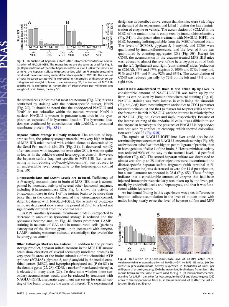
the stained cells indicates that most are neurons (Fig. 2B); this wasconfirmed by staining with the neuron-specific marker, NeuN(Fig. 2C). It should be noted that the endocytosed NAGLU andNeuN do not colocalize within the neuron; whereas NeuN isnuclear, NAGLU is present in punctate structures in the cyto-plasm, as expected of its lysosomal location. The lysosomal loca-tion was confirmed by colocalization with LAMP2, a lysosomalmembrane protein (Fig. S3A).
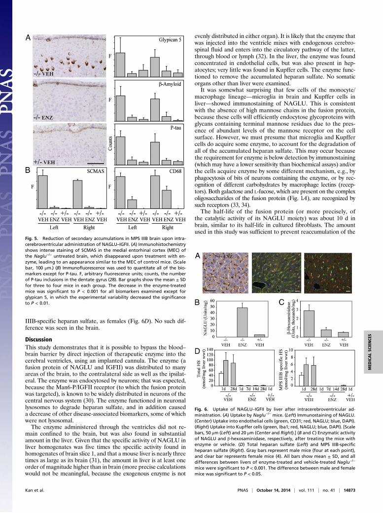
Heparan Sulfate Storage Is Greatly Reduced. The amount of hep-aran sulfate, the primary storage material, was very high in brainof MPS IIIB mice treated with vehicle alone, as determined bythe Sensi-Pro method (24, 25) (Fig. 3A). It decreased rapidlyafter treatment with enzyme, but even after 28 d, it was not quiteas low as in the vehicle-treated heterozygous control. However,the heparan sulfate fragment specific to MPS IIIB (i.e., termi-nating in nonreducing α–N-acetylglucosamine), was reduced toan undetectable level, comparable to the heterozygous control(Fig. 3B).
β-Hexosaminidase and LAMP1 Levels Are Reduced. Deficiency ofα–N-acetylglucosaminidase in brain of MPS IIIB mice is accom-panied by increased activity of several other lysosomal enzymes,including β-hexosaminidase (26). Fig. 4A shows the activity ofβ-hexosaminidase in slice 1 of the mutant brain to be more thantwice that in the comparable area of the heterozygous control.After treatment with NAGLU–IGFII, the activity of β-hexosa-minidase decreased slowly over the period of 28 d, to a level notsignificantly different from the control brain.LAMP1, another lysosomal membrane protein, is expected to
decrease in amount as lysosomal storage is reduced and thelysosomes become smaller. Fig. 4B shows prominent LAMP1staining in neurons of CA3 and in nonneuronal cells (perhapsastrocytes) of the dentate gyrus; upon treatment with enzyme,LAMP1 staining was much reduced, essentially to the level of theheterozygous control.
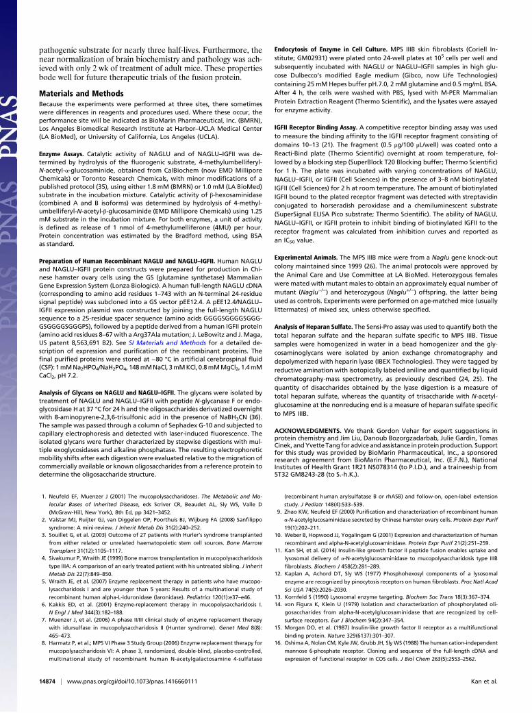
Other Pathologic Markers Are Reduced. In addition to the primarystorage product, heparan sulfate, neurons in theMPS IIIBmousebrain show elevation of several seemingly unrelated proteins invery specific areas of the brain: subunit c of mitochondrial ATPsynthase (SCMAS), glypican 5, and β-amyloid in the medial ento-rhinal cortex (MEC), and hyperphosphorylated tau (P-thr181) inthe dentate gyrus (27, 28). CD68, a marker for activated microglia,is elevated in many areas (29). To determine whether these sec-ondary accumulations would also be reduced by treatment withNAGLU–IGFII, a separate experiment was set up for sagittal cut-ting of the brain to expose the areas of interest. The experimental
designwas as described above, except that themicewere 8wkof ageat the start of the experiment and killed 1 d after the last adminis-tration of enzyme or vehicle. The accumulation of SCMAS in theMEC of the mutant mice is easily seen by immunohistochemistry(Fig. 5A); it disappears after treatment with NAGLU–IGFII, theMEC becoming indistinguishable from the MEC of control brain.The levels of SCMAS, glypican 5, β-amyloid, and CD68 werequantitated by immunofluorescence, and the level of P-tau wasquantitated by counting aggregates (28) (Fig. 5B). Except forCD68, the accumulation in the enzyme-treated MPS IIIB micewas reduced to almost the level of the heterozygote control, bothon the left (ipsilateral) and right (contralateral) sides (reductionin SCMAS, 97% and 97%; glypican 5, 100% and 87%; β-amyloid,91% and 81%; and P-tau, 92% and 93%). The accumulation ofCD68 was reduced partially, by 72% on the left and 44% on theright side.
NAGLU–IGFII Administered to Brain Is also Taken Up by Liver. Aconsiderable amount of NAGLU–IGFII was taken up by theliver, as can be seen by immunohistochemical staining (Fig. 6).NAGLU staining was most intense in cells lining the sinusoids(Fig. 6A,Left); immunostaining with antibodies to CD31 (amarkerfor endothelial cells) and Iba1 (amarker forKupffer cells), showedthe former to be rich in NAGLU and the latter to be relatively freeof NAGLU (Fig. 6A, Center and Right, respectively). Because ofthe intense staining of the endothelial cells, it was difficult to seethe enzyme in hepatocytes; the presence of NAGLU in hepatocyteswas best seen by confocal microscopy, which showed colocaliza-tion with LAMP2 (Fig. S3B).The uptake of NAGLU–IGFII into liver could also be de-
termined bymeasurement of NAGLUenzymatic activity (Fig. 6B)and was seen to be five times higher, permilligram of protein, thanin homogenates of slice 1 of the brain. β-Hexosaminidase activitywas reduced 90% of the way to the normal level, 1 d postfinalinjection (Fig. 6C). The stored heparan sulfate was decreased toalmost zero for up to 28 d after injections were discontinued; thedisease-specific heparan sulfate fragment (with terminal α–N-acetylglucosamine) was decreased to zero for 14 d postinjection,but a small amount reappeared in 28 d (Fig. 6D). These findingsindicate that a considerable amount of enzyme that had beeninjected intracerebroventricularly was taken up by the liver, pri-marily by endothelial cells and hepatocytes, and that it was func-tional within lysosomes.An incidental finding in this experiment was a sex difference in
heparan sulfate accumulation in the liver of mutant mice, withmales having nearly twice the level of heparan sulfate and MPS
Fig. 3. Reduction of heparan sulfate after intracerebroventricular admin-istration of NAGLU–IGFII. The mouse brains are the same as used for Fig. 2.(A) Representation of the total heparan sulfate in slice 3. (B) In the same sliceas in A, the heparan sulfate oligosaccharides with an N-acetylglucosamineresidue at the nonreducing end and therefore specific toMPS IIIB. The amountof total heparan sulfate (HS) is expressed in nanomoles of disaccharide permilligram wet weight of brain tissue, as mean ± SD; the amount of MPS IIIB-specific HS is expressed as nanomoles of trisaccharide per milligram wetweight of brain tissue, mean ± SD.
Fig. 4. Reduction of β-hexosaminidase and of LAMP1 after intra-cerebroventricular administration of NAGLU–IGFII to MPS IIIB mice. (A) De-crease in β-hexosaminidase activity (expressed in thousands of units permilligram of protein, mean± SD) in homogenized brain tissue from slice 1; themouse brains are the same as were used for Fig. 2. (B) Immunohistochemicalstaining for LAMP1, amarker for lysosomes, in the CA3 and dentate gyrus (DG)areas of the hippocampus (slice 4), in brains removed 28 d after the last in-jection. (Scale bar, 50 μm.)
14872 | www.pnas.org/cgi/doi/10.1073/pnas.1416660111 Kan et al.
IIIB-specific heparan sulfate, as females (Fig. 6D). No such dif-ference was seen in the brain.
DiscussionThis study demonstrates that it is possible to bypass the blood–brain barrier by direct injection of therapeutic enzyme into thecerebral ventricles, using an implanted cannula. The enzyme (afusion protein of NAGLU and IGFII) was distributed to manyareas of the brain, to the contralateral side as well as the ipsilat-eral. The enzyme was endocytosed by neurons; that was expected,because the Man6-P/IGFII receptor (to which the fusion proteinwas targeted), is known to be widely distributed in neurons of thecentral nervous system (30). The enzyme functioned in neuronallysosomes to degrade heparan sulfate, and in addition causeda decrease of other disease-associated biomarkers, some of whichwere not lysosomal.The enzyme administered through the ventricles did not re-
main confined to the brain, but was also found in substantialamount in the liver. Given that the specific activity of NAGLU inliver homogenates was five times the specific activity found inhomogenates of brain slice 1, and that a mouse liver is nearly threetimes as large as its brain (31), the amount in liver is at least oneorder of magnitude higher than in brain (more precise calculationswould not be meaningful, because the exogenous enzyme is not
evenly distributed in either organ). It is likely that the enzyme thatwas injected into the ventricle mixes with endogenous cerebro-spinal fluid and enters into the circulatory pathway of the latter,through blood or lymph (32). In the liver, the enzyme was foundconcentrated in endothelial cells, but was also present in hep-atocytes; very little was found in Kupffer cells. The enzyme func-tioned to remove the accumulated heparan sulfate. No somaticorgans other than liver were examined.It was somewhat surprising that few cells of the monocyte/
macrophage lineage—microglia in brain and Kupffer cells inliver—showed immunostaining of NAGLU. This is consistentwith the absence of high mannose chains in the fusion protein,because these cells will efficiently endocytose glycoproteins withglycans containing terminal mannose residues due to the pres-ence of abundant levels of the mannose receptor on the cellsurface. However, we must presume that microglia and Kupffercells do acquire some enzyme, to account for the degradation ofall of the accumulated heparan sulfate. This may occur becausethe requirement for enzyme is below detection by immunostaining(which may have a lower sensitivity than biochemical assays) and/orthe cells acquire enzyme by some different mechanism, e.g., byphagocytosis of bits of neurons containing the enzyme, or by rec-ognition of different carbohydrates by macrophage lectins (recep-tors). Both galactose and L-fucose, which are present on the complexoligosaccharides of the fusion protein (Fig. 1A), are recognized bysuch receptors (33, 34).The half-life of the fusion protein (or more precisely, of
the catalytic activity of its NAGLU moiety) was about 10 d inbrain, similar to its half-life in cultured fibroblasts. The amountused in this study was sufficient to prevent reaccumulation of the
Fig. 5. Reduction of secondary accumulations in MPS IIIB brain upon intra-cerebroventricular administration of NAGLU–IGFII. (A) Immunohistochemistryshows intense staining of SCMAS in the medial entorhinal cortex (MEC) ofthe Naglu−/− untreated brain, which disappeared upon treatment with en-zyme, leading to an appearance similar to the MEC of control mice. (Scalebar, 100 μm.) (B) Immunofluorescence was used to quantitate all of the bio-markers except for P-tau. F, arbitrary fluorescence units; counts, the numberof P-tau inclusions in the dentate gyrus (28). Bar graphs show the mean ± SDfor three to four mice in each group. The decrease in the enzyme-treatedmice was significant to P < 0.001 for all biomarkers examined except forglypican 5, in which the experimental variability decreased the significanceto P < 0.01.
Fig. 6. Uptake of NAGLU–IGFII by liver after intracerebroventricular ad-ministration. (A) Uptake by Naglu−/− mice. (Left) Immunostaining of NAGLU.(Center) Uptake into endothelial cells (green, CD31; red, NAGLU; blue, DAPI).(Right) Uptake into Kupffer cells (green, Iba1; red, NAGLU; blue, DAPI). [Scalebars, 50 μm (Left) and 20 μm (Center and Right).] (B and C) Enzymatic activityof NAGLU and β-hexosaminidase, respectively, after treating the mice withenzyme or vehicle. (D) Total heparan sulfate (Left) and MPS IIIB-specificheparan sulfate (Right). Gray bars represent male mice (four at each point),and clear bar represents female mice (4). All bars show mean ± SD, and alldifferences between livers of enzyme-treated and vehicle-treated Naglu−/−
mice were significant to P < 0.001. The difference between male and femalemice was significant to P < 0.05.
Kan et al. PNAS | October 14, 2014 | vol. 111 | no. 41 | 14873
MED
ICALSC
IENCE
S
pathogenic substrate for nearly three half-lives. Furthermore, thenear normalization of brain biochemistry and pathology was ach-ieved with only 2 wk of treatment of adult mice. These propertiesbode well for future therapeutic trials of the fusion protein.
Materials and MethodsBecause the experiments were performed at three sites, there sometimeswere differences in reagents and procedures used. Where these occur, theperformance site will be indicated as BioMarin Pharmaceutical, Inc. (BMRN),Los Angeles Biomedical Research Institute at Harbor–UCLA Medical Center(LA BioMed), or University of California, Los Angeles (UCLA).
Enzyme Assays. Catalytic activity of NAGLU and of NAGLU–IGFII was de-termined by hydrolysis of the fluorogenic substrate, 4-methylumbelliferyl-N-acetyl-α-glucosaminide, obtained from CalBiochem (now EMD MilliporeChemicals) or Toronto Research Chemicals, with minor modifications of apublished protocol (35), using either 1.8 mM (BMRN) or 1.0 mM (LA BioMed)substrate in the incubation mixture. Catalytic activity of β-hexosaminidase(combined A and B isoforms) was determined by hydrolysis of 4-methyl-umbelliferyl-N-acetyl-β-glucosaminide (EMD Millipore Chemicals) using 1.25mM substrate in the incubation mixture. For both enzymes, a unit of activityis defined as release of 1 nmol of 4-methylumelliferone (4MU) per hour.Protein concentration was estimated by the Bradford method, using BSAas standard.
Preparation of Human Recombinant NAGLU and NAGLU–IGFII. Human NAGLUand NAGLU–IGFII protein constructs were prepared for production in Chi-nese hamster ovary cells using the GS (glutamine synthetase) MammalianGene Expression System (Lonza Biologics). A human full-length NAGLU cDNA(corresponding to amino acid residues 1–743 with an N-terminal 24-residuesignal peptide) was subcloned into a GS vector pEE12.4. A pEE12.4/NAGLU–IGFII expression plasmid was constructed by joining the full-length NAGLUsequence to a 25-residue spacer sequence (amino acids GGGGSGGGGSGGG-GSGGGGSGGGPS), followed by a peptide derived from a human IGFII protein(amino acid residues 8–67with a Arg37Alamutation; J. LeBowitz and J.Maga,US patent 8,563,691 B2). See SI Materials and Methods for a detailed de-scription of expression and purification of the recombinant proteins. Thefinal purified proteins were stored at −80 °C in artificial cerebrospinal fluid(CSF): 1mMNa2HPO4/NaH2PO4, 148mMNaCl, 3mMKCl, 0.8mMMgCl2, 1.4mMCaCl2, pH 7.2.
Analysis of Glycans on NAGLU and NAGLU–IGFII. The glycans were isolated bytreatment of NAGLU and NAGLU–IGFII with peptide N-glycanase F or endo-glycosidase H at 37 °C for 24 h and the oligosaccharides derivatized overnightwith 8-aminopyrene-2,3,6-trisulfonic acid in the presence of NaBH3CN (36).The sample was passed through a column of Sephadex G-10 and subjected tocapillary electrophoresis and detected with laser-induced fluorescence. Theisolated glycans were further characterized by stepwise digestions with mul-tiple exoglycosidases and alkaline phosphatase. The resulting electrophoreticmobility shifts after each digestionwere evaluated relative to themigration ofcommercially available or known oligosaccharides from a reference protein todetermine the oligosaccharide structure.
Endocytosis of Enzyme in Cell Culture. MPS IIIB skin fibroblasts (Coriell In-stitute; GM02931) were plated onto 24-well plates at 105 cells per well andsubsequently incubated with NAGLU or NAGLU–IGFII samples in high glu-cose Dulbecco’s modified Eagle medium (Gibco, now Life Technologies)containing 25 mM Hepes buffer pH.7.0, 2 mM glutamine and 0.5 mg/mL BSA.After 4 h, the cells were washed with PBS, lysed with M-PER MammalianProtein Extraction Reagent (Thermo Scientific), and the lysates were assayedfor enzyme activity.
IGFII Receptor Binding Assay. A competitive receptor binding assay was usedto measure the binding affinity to the IGFII receptor fragment consisting ofdomains 10–13 (21). The fragment (0.5 μg/100 μL/well) was coated onto aReacti-Bind plate (Thermo Scientific) overnight at room temperature, fol-lowed by a blocking step (SuperBlock T20 Blocking buffer; Thermo Scientific)for 1 h. The plate was incubated with varying concentrations of NAGLU,NAGLU–IGFII, or IGFII (Cell Sciences) in the presence of 3–8 nM biotinylatedIGFII (Cell Sciences) for 2 h at room temperature. The amount of biotinylatedIGFII bound to the plated receptor fragment was detected with streptavidinconjugated to horseradish peroxidase and a chemiluminescent substrate(SuperSignal ELISA Pico substrate; Thermo Scientific). The ability of NAGLU,NAGLU–IGFII, or IGFII protein to inhibit binding of biotinylated IGFII to thereceptor fragment was calculated from inhibition curves and reported asan IC50 value.
Experimental Animals. The MPS IIIB mice were from a Naglu gene knock-outcolony maintained since 1999 (26). The animal protocols were approved bythe Animal Care and Use Committee at LA BioMed. Heterozygous femaleswere mated with mutant males to obtain an approximately equal number ofmutant (Naglu−/−) and heterozygous (Naglu+/−) offspring, the latter beingused as controls. Experiments were performed on age-matched mice (usuallylittermates) of mixed sex, unless otherwise specified.
Analysis of Heparan Sulfate. The Sensi-Pro assay was used to quantify both thetotal heparan sulfate and the heparan sulfate specific to MPS IIIB. Tissuesamples were homogenized in water in a bead homogenizer and the gly-cosaminoglycans were isolated by anion exchange chromatography anddepolymerized with heparin lyase (IBEX Technologies). They were tagged byreductive amination with isotopically labeled aniline and quantified by liquidchromatography-mass spectrometry, as previously described (24, 25). Thequantity of disaccharides obtained by the lyase digestion is a measure oftotal heparan sulfate, whereas the quantity of trisaccharide with N-acetyl-glucosamine at the nonreducing end is a measure of heparan sulfate specificto MPS IIIB.
ACKNOWLEDGMENTS. We thank Gordon Vehar for expert suggestions inprotein chemistry and Jim Liu, Danoub Bozorgzadarbab, Julie Gardin, TomasCinek, andYvetteTang for advice andassistance inprotein production. Supportfor this study was provided by BioMarin Pharmaceutical, Inc., a sponsoredresearch agreement from BioMarin Pharmaceutical, Inc. (E.F.N.), NationalInstitutes of Health Grant 1R21 NS078314 (to P.I.D.), and a traineeship from5T32 GM8243-28 (to S.-h.K.).
1. Neufeld EF, Muenzer J (2001) The mucopolysaccharidoses. The Metabolic and Mo-
lecular Bases of Inherited Disease, eds Scriver CR, Beaudet AL, Sly WS, Valle D
(McGraw-Hill, New York), 8th Ed, pp 3421–3452.2. Valstar MJ, Ruijter GJ, van Diggelen OP, Poorthuis BJ, Wijburg FA (2008) Sanfilippo
syndrome: A mini-review. J Inherit Metab Dis 31(2):240–252.3. Souillet G, et al. (2003) Outcome of 27 patients with Hurler’s syndrome transplanted
from either related or unrelated haematopoietic stem cell sources. Bone Marrow
Transplant 31(12):1105–1117.4. Sivakumur P, Wraith JE (1999) Bone marrow transplantation in mucopolysaccharidosis
type IIIA: A comparison of an early treated patient with his untreated sibling. J Inherit
Metab Dis 22(7):849–850.5. Wraith JE, et al. (2007) Enzyme replacement therapy in patients who have mucopo-
lysaccharidosis I and are younger than 5 years: Results of a multinational study of
recombinant human alpha-L-iduronidase (laronidase). Pediatrics 120(1):e37–e46.6. Kakkis ED, et al. (2001) Enzyme-replacement therapy in mucopolysaccharidosis I.
N Engl J Med 344(3):182–188.7. Muenzer J, et al. (2006) A phase II/III clinical study of enzyme replacement therapy
with idursulfase in mucopolysaccharidosis II (Hunter syndrome). Genet Med 8(8):
465–473.8. Harmatz P, et al.; MPS VI Phase 3 Study Group (2006) Enzyme replacement therapy for
mucopolysaccharidosis VI: A phase 3, randomized, double-blind, placebo-controlled,
multinational study of recombinant human N-acetylgalactosamine 4-sulfatase
(recombinant human arylsulfatase B or rhASB) and follow-on, open-label extension
study. J Pediatr 148(4):533–539.9. Zhao KW, Neufeld EF (2000) Purification and characterization of recombinant human
α-N-acetylglucosaminidase secreted by Chinese hamster ovary cells. Protein Expr Purif
19(1):202–211.10. Weber B, Hopwood JJ, Yogalingam G (2001) Expression and characterization of human
recombinant and alpha-N-acetylglucosaminidase. Protein Expr Purif 21(2):251–259.11. Kan SH, et al. (2014) Insulin-like growth factor II peptide fusion enables uptake and
lysosomal delivery of α-N-acetylglucosaminidase to mucopolysaccharidosis type IIIB
fibroblasts. Biochem J 458(2):281–289.12. Kaplan A, Achord DT, Sly WS (1977) Phosphohexosyl components of a lysosomal
enzyme are recognized by pinocytosis receptors on human fibroblasts. Proc Natl Acad
Sci USA 74(5):2026–2030.13. Kornfeld S (1990) Lysosomal enzyme targeting. Biochem Soc Trans 18(3):367–374.14. von Figura K, Klein U (1979) Isolation and characterization of phosphorylated oli-
gosaccharides from alpha-N-acetylglucosaminidase that are recognized by cell-
surface receptors. Eur J Biochem 94(2):347–354.15. Morgan DO, et al. (1987) Insulin-like growth factor II receptor as a multifunctional
binding protein. Nature 329(6137):301–307.16. Oshima A, Nolan CM, Kyle JW, Grubb JH, Sly WS (1988) The human cation-independent
mannose 6-phosphate receptor. Cloning and sequence of the full-length cDNA and
expression of functional receptor in COS cells. J Biol Chem 263(5):2553–2562.
14874 | www.pnas.org/cgi/doi/10.1073/pnas.1416660111 Kan et al.
17. Tong PY, Tollefsen SE, Kornfeld S (1988) The cation-independent mannose 6-phos-phate receptor binds insulin-like growth factor II. J Biol Chem 263(6):2585–2588.
18. Kiess W, et al. (1988) Biochemical evidence that the type II insulin-like growth factorreceptor is identical to the cation-independent mannose 6-phosphate receptor. J BiolChem 263(19):9339–9344.
19. Kornfeld S (1992) Structure and function of the mannose 6-phosphate/insulinlikegrowth factor II receptors. Annu Rev Biochem 61:307–330.
20. LeBowitz JH, et al. (2004) Glycosylation-independent targeting enhances enzymedelivery to lysosomes and decreases storage in mucopolysaccharidosis type VII mice.Proc Natl Acad Sci USA 101(9):3083–3088.
21. Maga JA, et al. (2013) Glycosylation-independent lysosomal targeting of acid α-glu-cosidase enhances muscle glycogen clearance in pompe mice. J Biol Chem 288(3):1428–1438.
22. Aoyagi-Scharber M, et al. (2014) Engineering of a recombinant NAGLU fusionprotein with insulin-like growth factor 2 leads to improved cellular uptake viaa glycosuylation-independent lysopsomal targeting pathway. Mol Genet Metab111(2):S20.
23. Kan SH, et al. (2014) Intracerebroventricular enzyme replacement therapy with gly-cosylation-independent lysosomal targeted NAGLU leads to widespread enzymaticativity, reduction of lysosomal storage and of secondary defects in brain of mice withSanfilippo syndrome type B. Mol Genet Metab 111(2):S59.
24. Lawrence R, et al. (2012) Disease-specific non-reducing end carbohydrate biomarkersfor mucopolysaccharidoses. Nat Chem Biol 8(2):197–204.
25. Lawrence R, et al. (2014) Glycan-based biomarkers for mucopolysaccharidoses. MolGenet Metab 111(2):73–83.
26. Li HH, et al. (1999) Mouse model of Sanfilippo syndrome type B produced by targeteddisruption of the gene encoding alpha-N-acetylglucosaminidase. Proc Natl Acad SciUSA 96(25):14505–14510.
27. Ohmi K, et al. (2009) Sanfilippo syndrome type B, a lysosomal storage disease, is alsoa tauopathy. Proc Natl Acad Sci USA 106(20):8332–8337.
28. Ohmi K, Zhao HZ, Neufeld EF (2011) Defects in the medial entorhinal cortex anddentate gyrus in the mouse model of Sanfilippo syndrome type B. PLoS ONE 6(11):e27461.
29. Ohmi K, et al. (2003) Activated microglia in cortex of mouse models of mucopoly-saccharidoses I and IIIB. Proc Natl Acad Sci USA 100(4):1902–1907.
30. Hawkes C, Kar S (2003) Insulin-like growth factor-II/mannose-6-phosphate receptor:Widespread distribution in neurons of the central nervous system including thoseexpressing cholinergic phenotype. J Comp Neurol 458(2):113–127.
31. The Jackson Laboratory (2014) Morphometric (organ weight) survey of 11 inbredstrains of mice. MPD:Jaxpheno2.
32. Brinker T, Stopa E, Morrison J, Klinge P (2014) A new look at cerebrospinal fluidcirculation. Fluids Barriers CNS 11:10.
33. Stahl PD (1992) The mannose receptor and other macrophage lectins. Curr Opin Im-munol 4(1):49–52.
34. Fadden AJ, Holt OJ, Drickamer K (2003) Molecular characterization of the rat Kupffercell glycoprotein receptor. Glycobiology 13(7):529–537.
35. Marsh J, Fensom AH (1985) 4-Methylumbelliferyl α-N-acetylglucosaminidase activityfor diagnosis of Sanfilippo B disease. Clin Genet 27(3):258–262.
36. Szabo Z, Guttman A, Rejtar T, Karger BL (2010) Improved sample preparation methodfor glycan analysis of glycoproteins by CE-LIF and CE-MS. Electrophoresis 31(8):1389–1395.
Kan et al. PNAS | October 14, 2014 | vol. 111 | no. 41 | 14875
MED
ICALSC
IENCE
S