deletion of integrin linked kinase in endothelial cells...
TRANSCRIPT
DEVELOPMENT AND STEM CELLS RESEARCH ARTICLE 987
Development 140, 987-995 (2013) doi:10.1242/dev.091298© 2013. Published by The Company of Biologists Ltd
INTRODUCTIONIntegrin linked kinase (ILK) is a ubiquitously expressed scaffoldprotein that functions as a central component and mediator of cell-extracellular matrix (ECM) interactions (Novak et al., 1998). ILKbinds the cytoplasmic tail of β1 integrin and consists of threestructural and functional domains (Dedhar and Hannigan, 1996):five ankyrin repeats at the N terminus, which allow interaction withPINCH; a Pleckstrin-homology (PH) domain; and a C-terminalkinase-like domain (Wickström et al., 2010b). By targeting of thePINCH-Parvin complex to integrin adhesion sites, ILK regulatesthe engagement and remodeling of the actin cytoskeletondownstream of integrin adhesion (Wickström et al., 2010a;Wickström et al., 2010b). Moreover, there is evidence for directinteractions of ILK with tyrosine kinase receptors (RTKs) viaPINCH and the NCK2 adaptor protein (Tu et al., 1998). Thus, ILKlinks cell-matrix interactions with signals modulating remodelingof the cytoskeleton and is therefore involved in central cellbiological processes such as cell adhesion, migration, proliferation,survival and differentiation (Hannigan et al., 2007).
ILK has been also reported to affect endothelial cell (EC)apoptosis, proliferation and migration as well as vascular
development in vitro (Friedrich et al., 2004; Vouret-Craviari et al.,2004). In addition, defects in vascular development were observedin vivo in ILK-deficient mice (Friedrich et al., 2004). However,despite the EC-specific deletion of ILK, the mice died at an earlyembryonic stage and, therefore, the molecular defects underlying theobserved delay of vascular development could not be determined(Friedrich et al., 2004). A recent publication in keratinocytes hasprovided new mechanistic insight into the cell biological role of ILKby illustrating its direct involvement in trafficking and its integrationinto the plasma membrane (Wickström et al., 2010a; Wickström etal., 2010b). Caveolae are present in most cell types, but areparticularly abundant in ECs and are known to play a crucial role inEC biology (Cho et al., 2004). The principal component of caveolaein ECs is caveolin 1 and this molecule is known to cluster a greatvariety of different signaling molecules, e.g. RAS, nitric oxide, G-proteins and growth factors such as vascular endothelial growthfactor (VEGF) (Krajewska and Maslowska, 2004). Caveolin 1 assistscompartmentalization of signaling pathways by establishing specificlipid microdomains that act as specialized signaling hubs (Balijepalliet al., 2006; Saliez et al., 2008). Integrin-based adhesion has beenshown to regulate multiple signaling cascades and to providecrosstalk with growth factors. Therefore, we hypothesized that theinvolvement of ILK in the formation of lipid rafts and/or caveolin 1microdomains could be important for this crosstalk (Head et al.,2005; Head et al., 2006). In order to understand the potentialrelevance of ILK for EC biology, we have investigated ECdevelopment and function in Ilk–/– embryonic stem (ES) cells. Ourstudy revealed strongly reduced formation of vessel-like structuresin Ilk–/– embryoid bodies (EBs) because of defective EC signaling.This striking alteration is due to perturbed caveolin 1 positioning invicinity of the plasma membrane.
1Institute of Physiology I, Life and Brain Center, University of Bonn, Bonn, NRW,53105, Germany. 2Department of Molecular and Cellular Sport Medicine, GermanSport University, Cologne, NRW, 50933, Germany. 3Paul Gerson Unna Group ‘Skinhomeostasis and ageing’, Max-Planck Institute for Biology of Ageing, Cologne,NRW, 50931, Germany. 4Department of Dermatology, University of Cologne,Cologne, NRW, 50931, Germany.
*Authors for correspondence ([email protected]; [email protected])
Accepted 24 December 2012
SUMMARYIntegrin linked kinase (ILK) connects the ILK-Pinch-Parvin complex with integrin adhesion sites. Because of the functional relevanceof integrin-linked signaling for endothelial cell (EC) biology, we have explored this pathway in Ilk–/– embryonic stem (ES) cellsdifferentiated into ECs and vessel-like structures. We have focused in particular on the mechanistic relevance of ILK-Pinch-Parvincomplex-related signaling for EC development and tube formation. Our analysis revealed that the formation of vessel-like structureswas strongly reduced in Ilk–/– ES cells and that this phenotype could be rescued by re-expression of ILK in ES cells. ECs were MACS sortedfrom wild-type (WT) and Ilk–/– ES cells and functional analysis using intracellular calcium imaging as the read-out yielded a completelack of vascular endothelial growth factor- and epidermal growth factor-dependent responses. The possibility of a caveolin 1-relateddefect was investigated by transfecting WT and Ilk–/– ECs with a caveolin 1-EGFP fusion protein. Time-lapse microscopy showed thatthe prominent phenotype is due to altered dynamics of caveolin 1 and to a lack of positioning of caveolin 1 in the vicinity of the plasmamembrane and that it is rescued by re-expressing ILK in the Ilk–/– ES cells. We also found that the defect is caused by the perturbedorganization of microtubules and cortical actin filaments. Thus, ILK is required as a scaffold to allow actin-microtubule interactionsand correct positioning of caveolin 1 close to the plasma membrane. This is crucial for signaling compartmentalization in ECs andexplains the key role of ILK for EC development and function.
KEY WORDS: Integrin linked kinase (ILK), Endothelial cells, Tyrosine kinase signaling, Mouse
Deletion of integrin linked kinase in endothelial cells results in defective RTK signaling caused by caveolin 1mislocalizationDaniela Malan1, Andrea Elischer2, Michael Hesse1, Sara A. Wickström3,4, Bernd K. Fleischmann1,* and Wilhelm Bloch2,*
DEVELO
PMENT
988
MATERIALS AND METHODSCell cultureThe mouse ES cell lines D3 (wild type) and Ilk–/– were derived andmaintained in culture in hanging drops (Malan et al., 2010). Time afterplating was indicated as (5+7) and (5+14) days.
Immunohistochemistry and detection of endothelial cells andvesselsEBs were fixed with 4% paraformaldehyde (PFA) and MACS-sorted ECs(M-ECs) were stained with the antibody rat anti-mouse PECAM-1 (CD31)(1:800; Pharmingen, San Diego, CA, USA). Other antibodies/markers usedwere: rabbit polyclonal anti-collagen IV (1:500; Acris Antibodies), mousemonoclonal anti-perlecan (1:500; Biotrend, Köln, Germany), mousemonoclonal anti-fibronectin (1:500; Sigma-Aldrich, Munich, Germany),polyclonal rabbit anti-laminin (1:500; Sigma-Aldrich), rabbit polyclonalPLCγ1 (1:50; abcam, Cambridge, UK), rat anti-mouse flk-1 (1:100, BDPharmingen, Erembodegem Belgium), Alexa Fluor 488 Phalloidin (1:40;Molecular Probes Invitrogen, Karlsruhe, Germany), mouse monoclonal β-tubulin (1:500; Sigma-Aldrich), rabbit polyclonal anti-caveolin 1 (1:500;BD Pharmingen), wheat germ agglutinin conjugate Alexa Fluor 488 (1:500;Molecular Probes Invitrogen). Secondary antibodies were: Cy3- (or Cy2-)conjugated goat anti-rabbit (or goat anti-mouse) Ig (1:1500 and 1:500;Dianova, Hamburg, Germany).
Magnet-associated cell sorting (MACS)Differentiated ES cells (5+7 days) were dissociated with Accutase (PAALaboratories, Linz, Austria). The single cells were stained with anendothelial-specific marker, rat anti-mouse-PECAM-1 (also known asCD31), and MACS sorted as previously described (Schmidt et al., 2004).For analysis, the number of cells in 40 defined areas was calculated usinga 40× objective on an Axiophot microscope (Zeiss Microimaging,Goettingen, Germany).
Proliferation assay and apoptosis assayWild-type and Ilk–/– EBs were fixed and stained with rat anti-PECAM-1 asdescribed above. Proliferating cells were detected with the primary antibodyrabbit anti-mouse Ki67 (1:150, pAb; Dianova) and apoptotic cells with arabbit anti-active caspase 3 (1:500, BD Pharmingen). The number of Ki67-or caspase 3-positive-cells within 50 randomly chosen vessel-like tubes wascounted as proliferating or apoptotic M-ECs (Müller-Ehmsen et al., 2006).
Morphological analysis, apotome and confocal microscopyThe distribution pattern of extracellular matrix proteins, caveolin 1 andcytoskeletal components, as well as live images, were analyzed by confocalmicroscopy using the LSM 510 META Zeiss microscope (ZeissMicroimaging) or by an inverted confocal laser scanning microscope(Nikon Eclipse Ti). Alternatively, image stacks were acquired with afluorescence microscope equipped with the ApoTome (Axiovert 200A,Zeiss Microimaging). Live images were acquired with the confocal laserscanning microscope with one image every second for 50 seconds in total.The analysis of velocity was carried out from the videos with Image J 1.37v(Plug-In: Particle Analysis/Manual Tracking). For every picture, the samecaveolin 1-EGFP was marked and the distance was measured. The programassigns to each analyzed particle a random color, which it is not related tothe velocity. The velocity was then analyzed with a Gaussian distribution.For evaluation of vascular development, PECAM-positive structures in EBswere counted (Malan et al., 2010). In parallel to the quantification ofendothelial tubes we also analyzed the distribution of different PECAM-positive structures in four different EBs at early and late stages. We defined‘endothelial precursors’ as single endothelial cell precursors, ‘clusters’ asaggregates of endothelial precursors and ‘vessel-like structures’ as a networkof tube-like structures with at least five branches. The whole EB wasscreened and results are given as percentage of all PECAM-positivestructures (Malan et al., 2010).
Plastic embedding and electron microscopyM-ECs were fixed in 4% PFA then treated with 1% uranyl acetate. Thespecimens were embedded in Araldite (Serva, Heidelberg, Germany). Semi-thin sections (500 µm) were cut and stained with Methylene Blue. Ultrathin
sections (30-60 nm) for electron microscopic observation were processed ona microtome with a diamond knife and placed on copper grids. Transmissionelectron microscopy was performed using a 902A electron microscope fromZeiss (Zeiss Microimaging).
Protein detection and western blot analysisFor western blotting, samples were submitted to SDS-PAGE and proteinswere transferred to PVDF membrane and incubated with specificantibodies. Immunoreactive proteins were detected by the enhancedchemiluminescence detection system (Amersham Biosciences Europe,Freiburg, Germany) and normalized to the actin content. VEGF (PANBiotech, Aidenbach, Germany) stimulation was achieved by treating thecells for 6 hours with low serum medium (1%), then 20 ng/ml VEGF(PAN Biotech) was applied for 4 minutes. We used a concentration thathas been reported to be physiologically relevant in inducing vasculo-angiogenesis (Schmidt et al., 2005; Hagedorn et al., 2004); thisconcentration also induced a clear Ca2+ release from the sarcoplasmicreticulum. Densitometry analysis was carried out using ImageJ software(NIH) and normalized to the actin content. Antibodies used were: rabbitpolyclonal anti-caveolin 1 (1:500; Acris Antibodies), rabbit polyclonalanti-GFP (1:500; Santa Cruz Biotechnology, Heidelberg, Germany),rabbit polyclonal anti-VEGFR2 and anti-phosphoY1054-1059VEGFR2(1:2000; Abcam), mouse monoclonal anti-MAPkinase activated (1:1000;Sigma-Aldrich), rabbit polyclonal anti-MAPkinase (1:1000; Upstate,Merck Millipore, Billerica, MA, USA), rabbit polyclonal anti-phospho-PLCγ1 (Tyr783) antibody (1:500; Cell Signaling Technology, Danvers,MA, USA), rabbit polyclonal anti-PLCγ1 (1:1000; Cell SignalingTechnology), mouse monoclonal anti-actin (1:4000; Chemicon Millipore,Billerica, MA, USA).
Migration assayMigration assay was performed in a modified Boyden chamber. The totalnumber of PECAM-positive migrated cells was counted as well as thenumber of migrated M-ECs (n=6) as described previously (Schmidt et al.,2004).
Generation of transgenic ES cell clonesA pCL-MFG fusion plasmid containing the ILK-EGFP cDNA (provided byR. Fässler, Max Planck Institute of Biochemistry, Department of MolecularMedicine, Martinsried, Germany). By cutting the construct withEcoRI/NotI, the EGFP cDNA was excised and subsequently cloned intoABD 15-24 pEGFP-N3/β-actin (BD Biosciences Clontech, Heidelberg,Germany) with ABD 15-24-EGFP excised. The resulting β-actin-ILK-EGFP fusion construct was used for electroporation of ILK-ES cells.
Generation of the caveolin 1-EGFP fusion proteinThe caveolin 1 cDNA (Homo sapiens, PubMed BC082246) was cloned in-frame upstream of the EGFP cDNA of the pEGFP-1 plasmid (BDBiosciences Clontech), in the BamHI restriction site. The CAG promoterfrom pDRIVE-CAG (InvivoGen, San Diego, CA, USA) was cloned intothe SmaI/SalI restriction site of the caveolin 1 -EGFP fusion plasmid.
Ca2+ imagingM-ECs were loaded with the intracellular calcium ([Ca2+]i) indicator Fura2 am (5 µM; Molecular Probes Invitrogen) for 10 minutes at roomtemperature. The bath solution contained: 140 mM NaCl, 5.4 mM KCl, 1mM MgCl2, 1.8 mM CaCl2, 10 mM HEPES and 10 mM glucose. Theemitted fluorescence was monitored using a charge-coupled device cooledcamera (TILL Photonics, Planegg, Germany) coupled with an invertedmicroscope (Axiovert 200M, Zeiss Microimaging). The emission data wereanalyzed using the Vision software package (TILL Vision 4.0, TILLPhotonics). Results are displayed as 340 nm/380 nm ratios after backgroundsubtraction. n is the number of cells tested. A [Ca2+]i increase of <10% wasconsidered to be ‘no response’. M-ECs were in some experiments treatedwith 2% methyl-beta-cyclodextrin (MbetaCD) for 2 hours at 37°C. Alldrugs used were from Sigma-Aldrich except for epidermal growth factor(EGF) and VEGF (both from PAN-Biotech) and the PLC activator m-3M3FBS (Calbiochem, Merck Millipore, Darmstadt, Germany).
RESEARCH ARTICLE Development 140 (5)
DEVELO
PMENT
Statistical analysisAll data are presented as mean±s.e.m. Data analysis was performed usinganalysis of variance with Bonferroni post-hoc test and/or Student’s t-testfor paired and unpaired data. Significance was considered at a P-value<0.05. Calculations of significance were carried out using GraphPad Prism5 (GraphPad Software, San Diego, CA, USA).
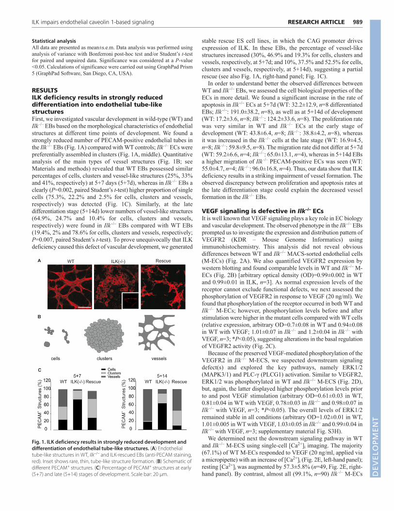
RESULTSILK deficiency results in strongly reduceddifferentiation into endothelial tube-likestructuresFirst, we investigated vascular development in wild-type (WT) andIlk–/– EBs based on the morphological characteristics of endothelialstructures at different time points of development. We found astrongly reduced number of PECAM-positive endothelial tubes inthe Ilk–/– EBs (Fig. 1A) compared with WT controls; Ilk–/– ECs werepreferentially assembled in clusters (Fig. 1A, middle). Quantitativeanalysis of the main types of vessel structures (Fig. 1B; seeMaterials and methods) revealed that WT EBs possessed similarpercentages of cells, clusters and vessel-like structures (25%, 33%and 41%, respectively) at 5+7 days (5+7d), whereas in Ilk–/– EBs aclearly (P=0.002, paired Student’s t-test) higher proportion of singlecells (75.3%, 22.2% and 2.5% for cells, clusters and vessels,respectively) was detected (Fig. 1C). Similarly, at the latedifferentiation stage (5+14d) lower numbers of vessel-like structures(64.9%, 24.7% and 10.4% for cells, clusters and vessels,respectively) were found in Ilk–/– EBs compared with WT EBs(19.4%, 2% and 78.6% for cells, clusters and vessels, respectively;P=0.007, paired Student’s t-test). To prove unequivocally that ILKdeficiency caused this defect of vascular development, we generated
stable rescue ES cell lines, in which the CAG promoter drivesexpression of ILK. In these EBs, the percentage of vessel-likestructures increased (30%, 46.9% and 19.3% for cells, clusters andvessels, respectively, at 5+7d; and 10%, 37.5% and 52.5% for cells,clusters and vessels, respectively, at 5+14d), suggesting a partialrescue (see also Fig. 1A, right-hand panel; Fig. 1C).
In order to understand better the observed differences betweenWT and Ilk–/– EBs, we assessed the cell biological properties of theECs in more detail. We found a significant increase in the rate ofapoptosis in Ilk–/– ECs at 5+7d (WT: 32.2±12.9, n=8 differentiatedEBs; Ilk–/–: 191.0±38.2, n=8), as well as at 5+14d of development(WT: 17.2±3.6, n=8; Ilk–/–: 124.2±33.6, n=8). The proliferation ratewas very similar in WT and Ilk–/– ECs at the early stage ofdevelopment (WT: 43.8±6.4, n=8; Ilk–/–: 38.8±4.2, n=8), whereasit was increased in the Ilk–/– cells at the late stage (WT: 16.9±4.5,n=8; Ilk–/–: 59.8±9.5, n=8). The migration rate did not differ at 5+7d(WT: 59.2±6.6, n=4; Ilk–/–: 65.0±13.1, n=4), whereas in 5+14d EBsa higher migration of Ilk–/– PECAM-positive ECs was seen (WT:55.0±4.7, n=4; Ilk–/–: 96.0±16.8, n=4). Thus, our data show that ILKdeficiency results in a striking impairment of vessel formation. Theobserved discrepancy between proliferation and apoptosis rates atthe late differentiation stage could explain the decreased vesselformation in the Ilk–/– EBs.
VEGF signaling is defective in Ilk–/– ECsIt is well known that VEGF signaling plays a key role in EC biologyand vascular development. The observed phenotype in the Ilk–/– EBsprompted us to investigate the expression and distribution pattern ofVEGFR2 (KDR – Mouse Genome Informatics) usingimmunohistochemistry. This analysis did not reveal obviousdifferences between WT and Ilk–/– MACS-sorted endothelial cells(M-ECs) (Fig. 2A). We also quantified VEGFR2 expression bywestern blotting and found comparable levels in WT and Ilk–/– M-ECs (Fig. 2B) [arbitrary optical density (OD)=0.99±0.002 in WTand 0.99±0.01 in ILK, n=3]. As normal expression levels of thereceptor cannot exclude functional defects, we next assessed thephosphorylation of VEGFR2 in response to VEGF (20 ng/ml). Wefound that phosphorylation of the receptor occurred in both WT andIlk–/– M-ECs; however, phosphorylation levels before and afterstimulation were higher in the mutant cells compared with WT cells(relative expression, arbitrary OD=0.7±0.08 in WT and 0.94±0.08in WT with VEGF; 1.01±0.07 in Ilk–/– and 1.2±0.04 in Ilk–/– withVEGF, n=3; *P<0.05), suggesting alterations in the basal regulationof VEGFR2 activity (Fig. 2C).
Because of the preserved VEGF-mediated phosphorylation of theVEGFR2 in Ilk–/– M-ECS, we suspected downstream signalingdefect(s) and explored the key pathways, namely ERK1/2(MAPK3/1) and PLC-γ (PLCG1) activation. Similar to VEGFR2,ERK1/2 was phosphorylated in WT and Ilk–/– M-ECS (Fig. 2D),but, again, the latter displayed higher phosphorylation levels priorto and post VEGF stimulation (arbitrary OD=0.61±0.03 in WT,0.81±0.04 in WT with VEGF, 0.78±0.03 in Ilk–/– and 0.98±0.07 inIlk–/– with VEGF, n=3; *P<0.05). The overall levels of ERK1/2remained stable in all conditions (arbitrary OD=1.02±0.01 in WT,1.01±0.005 in WT with VEGF, 1.03±0.05 in Ilk–/– and 0.99±0.04 inIlk–/– with VEGF, n=3; supplementary material Fig. S3H).
We determined next the downstream signaling pathway in WTand Ilk–/– M-ECS using single-cell [Ca2+]i imaging. The majority(67.1%) of WT M-ECs responded to VEGF (20 ng/ml, applied viaa micropipette) with an increase of [Ca2+]i (Fig. 2E, left-hand panel);resting [Ca2+]i was augmented by 57.3±5.8% (n=49, Fig. 2E, right-hand panel). By contrast, almost all (99.1%, n=90) Ilk–/– M-ECs
989RESEARCH ARTICLEILK impairs endothelial caveolin 1-based signaling
Fig. 1. ILK deficiency results in strongly reduced development anddifferentiation of endothelial tube-like structures. (A) Endothelialtube-like structures in WT, Ilk–/– and ILK-rescued EBs (anti-PECAM staining,red). Inset shows rare, thin, tube-like structure formation. (B) Schematic ofdifferent PECAM+ structures. (C) Percentage of PECAM+ structures at early(5+7) and late (5+14) stages of development. Scale bar: 20 μm. D
EVELO
PMENT

990
lacked a clear [Ca2+]i response upon application of VEGF (Fig. 2E,middle; the average change in [Ca2+]i was −4.8±1.1% (n=90,Fig. 2E, right-hand panel). These data suggest that VEGFR2-induced signaling is severely impaired in Ilk–/– M-ECs and wetherefore explored in more detail the underlying defect.
ILK deficiency causes defective tyrosine kinasesignalingBecause of the lack of an increase in [Ca2+]i upon VEGF applicationin Ilk–/– M-ECs, we wondered whether similar signaling defects alsooccurred with other RTKs in these cells. Indeed, EGF (20 ng) alsofailed to augment [Ca2+]i in Ilk–/– M-ECs in contrast to WT M-ECs;72.4% of WT cells (n=21) showed an EGF-induced increase of[Ca2+]i by 52.2±10.8%, whereas 58 out of 59 Ilk–/– cells did notrespond to EGF and this is also reflected by the marginal averageincrease of [Ca2+]i by 2.4±0.6% (Fig. 3A). Importantly, we couldalso demonstrate that the defective response to VEGF and EGF wasat least partially restored in rescued Ilk–/– M-ECs (supplementarymaterial Fig. S1A,B) In fact, the majority of rescued cells (73.3%,n=33) responded to EGF and a lower percentage (30.7%, n=11) to
VEGF; the percentage of [Ca2+]i increase amounted to 60.1±3.1%(n=33) and 34.1±5.4% (n=11), respectively (supplementary materialFig. S1C).
Next, we investigated whether other PLC-γ-dependent signalingpathways were also affected by analyzing Gq-coupled agonists.When stimulating M-ECs with bradykinin (100 nM), most of theWT (91.4%, n=64) and Ilk–/– (67.4%, n=58) cells responded with atransient [Ca2+]i response (Fig. 3B) of comparable magnitude.Similar results were also obtained with acetylcholine (ACh; 10 µM),which showed preserved activation of the [Ca2+]i response in Ilk–/–
M-ECs (WT: 73.5% of responders, n=36; Ilk–/–: 69.5% ofresponders, n=16). To pinpoint more precisely the signaling defect,in particular whether it occurred at the receptor level or downstreamof the receptor, we used first the direct PLC activator m-3M3FBS.Application of this compound led to an increase of [Ca2+]i in bothWT and Ilk–/– M-ECs (Fig. 3C), suggesting that PLC-γ and itsrelated downstream signaling components were functional upondirect activation; the percentage of [Ca2+]i increase was 24.7±1.6%in WT cells (n=29) and 29.3±4.8% in Ilk–/– cells (n=19) (Fig. 3C,right-hand panel) (WT: 74.4% of responders; Ilk–/–: 76% of
RESEARCH ARTICLE Development 140 (5)
Fig. 2. VEGF signaling is defective in Ilk–/– M-ECs. (A) VEGFR2distribution in WT and Ilk–/– M-ECs (anti-VEGFR2 staining, red). (B-D) Western blots of VEGFR2 (B), its phosphorylated form (pVEGFR2) (C)and ERK1/2 as well as its phosphorylated form (pERK) (D) in WT and Ilk–/–
M-ECs. The densitometric analysis of these western blots is shown below(n=3, *P<0.05; see also supplementary material Fig. S3H). (E) VEGF (20 nM)evokes an increase of [Ca2+]i in WT (left) but not Ilk–/– (middle) M-ECs,whereas thapsigargin (TH; 1 μM) elevates [Ca2+]i also in M-ECs (middle);representative [Ca2+]i traces are shown, each color labels the 340/380 nmratio in an individual cell over time. Right: statistical analysis of thepercentage increase of [Ca2+]i upon drug application. Error bars represents.e.m. Scale bar: 5 μm.
Fig. 3. Tyrosine kinase signaling is defective in Ilk–/– M-ECs. (A) EGF(20 nM) evokes an increase of [Ca2+]i in WT but not Ilk–/– M-ECs. (B) Bradykinin (Bk, 100 nM) elevates [Ca2+]i in both WT and Ilk–/– M-ECs. (C) Direct activation of PLC with m-3M3FBS (25 μM) also augments [Ca2+]iin WT and Ilk–/– M-ECs; each color labels the 340/380 nm ratio in anindividual cell over time. For A-C, statistical analyses of the percentage[Ca2+]i increase upon drug application in comparison to controlconditions is shown on the right. (D) The cellular distribution of PLC-γ(green) is assessed by immunohistochemistry. (E) Expression analysis ofPLC-γ and its phosphorylated isoform (Tyr783) is performed by westernblotting (left) and quantified by densitometry (right). Error bars represents.e.m. Scale bar: 40 μm. See also supplementary material Fig. S1 and Fig.S3G.
DEVELO
PMENT

responders). We next assessed the PLC-γ distribution pattern in WTand Ilk–/– M-ECs by immunocytochemistry and found that it wasunchanged (Fig. 3D). Moreover, protein expression analysis ofPLC-γ did not reveal significant differences between WT and Ilk–/–
(Fig. 3E; supplementary material Fig. S3G). In addition, ourexperiments revealed that VEGF application lead to thephosphorylation of PLC-γ at the Tyr 783 site in WT and Ilk–/– M-ECs, indicating intact activation (arbitrary OD pPLC=1.02±0.18 inWT and 1.11±0.24 in WT with VEGF; 1.12±0.18 in Ilk–/– and1.20±0.22 in Ilk–/– M-ECs with VEGF; n=4; Fig. 3E); the respectivenumbers are also significantly different (paired t-test WT versusWT with VEGF: P=0.0117; Ilk–/– versus Ilk–/– with VEGF:P=0.0033). Thus, our experiments showed that ILK plays a crucialrole for RTK-dependent signaling in M-ECs.
Caveolin 1 distribution is altered in Ilk–/– M-ECsILK has been recently reported to play a key role in caveolaeformation. As the components of the VEGF/PLC-γ/PtdIns(4,5)P2(PIP2) signaling axis are clustered within caveolae in ECs andearlier experiments underscore the crucial role of the clusteringof signaling components, we explored the subcellular distributionof components within caveolae. For this purpose, we used doubleimmunohistochemical analysis with the plasma membrane markerwheat germ agglutinin and an antibody against caveolin 1.Caveolin 1 is the main protein component of caveolae in ECs andis essential for caveolae formation (Drab et al., 2001). Thestainings illustrated that caveolin 1 is associated with the plasmamembrane in WT (Fig. 4A, left-hand panel, see arrow), but not inIlk–/– M-ECs (Fig. 4A, right-hand panel). This important findingwas corroborated by electron microscopy. Even though theabsolute number of typical caveolar structures is relatively low incultured M-ECs, we could clearly identify these in close vicinityto the plasma membrane in WT cells, whereas this was notobserved at all in any of the Ilk–/– M-ECs (60 cells in eachpreparation were analyzed; Ilk–/–: n=3 preparations; rescued Ilk–/–:n=2 preparations; WT: n=3 preparations) (Fig. 4B). Interestingly,western blotting experiments showed that the overall expressionlevel of caveolin 1 did not significantly differ between WT andIlk–/– M-ECs (WT: 1.01±0.18 arbitrary OD, n=5; Ilk–/–: 0.95±0.16,n=4; P=0.7; Fig. 4D). In addition, even transfection with thecaveolin 1-EGFP fusion protein (see also below) did not changethe overall expression of caveolin 1 in the cells (WT transfected:1.01±0.06 arbitrary OD, n=7; ILK transfected: 1.02±0.08, n=5;P=0.7). Because of the lack of caveolin 1 association with theplasma membrane in Ilk–/– M-ECs, we investigated whetherdisruption of caveolin 1 assembly and caveolin 1 microdomainformation in WT M-ECs could reproduce the functional defectsobserved in the Ilk–/– M-ECs. For this purpose, we used betacyclodextrin (MbetaCD); this compound is a known disruptor oflipid rafts and acts via depletion of cholesterol causingmalpositioning of membrane protein complexes (Barbuti et al.,2004; Jang et al., 2001). In agreement with our hypothesis,MbetaCD (2%) prevented VEGF-induced increase of [Ca2+]i inWT M-ESCs (n=11) (Fig. 4C), whereas, as would be expected (seealso Fig. 3B), the bradykinin response was preserved in theMbetaCD-treated Ilk–/– M-ESCs (Fig. 4C); the percentage of[Ca2+]i increase was 55.5±6.2% in WT cells treated with MbetaCD(n=11) with 42.4% of responders, whereas none of the cells testedresponded to VEGF stimulation. These data suggest that intactcaveolin 1 microdomains in the vicinity of the plasma membraneare required for functional RTK signaling and that deletion of ILKleads to defective caveolin 1 positioning and caveolae formation.
Altered intracellular trafficking and subcellulardistribution of caveolin 1 in Ilk–/– M-ECsBecause of these findings, we investigated the formation andsubcellular localization of caveolae in more detail. To this end, wetransfected M-ECs with a caveolin 1-EGFP fusion protein(Fig. 4E). This colocalized with endogenous caveolin 1 in both WTand Ilk–/– M-ECs (Fig. 4F, red). We could identify a clear differencein the localization of the caveolin 1-EGFP fluorescence betweenWT and Ilk–/– cells; caveolin 1 had membrane localization in WTcells (Fig. 4H, upper panel, arrows), whereas this was lost in Ilk–/–
cells (Fig. 4H, lower panel, arrows). This striking difference in thesubcellular distribution of caveolin 1 was also confirmed when
991RESEARCH ARTICLEILK impairs endothelial caveolin 1-based signaling
Fig. 4. Caveolin 1 positioning is altered in Ilk–/– M-ECs. (A) Caveolin 1insertion into the plasma membrane in Ilk–/– (right) and WT M-ECs (left,arrow) using double staining against wheat germ agglutinin (green) andcaveolin 1 (red). Ext, extracellular side; Int, intracellular side. (B) Electronmicroscopy depicts structures typical for caveolae (asterisks) in WT M-ECs;in Ilk–/– M-ECs, only small invaginations of the membrane, but nocaveolae, can be detected (arrows, right). (C) VEGF does not evoke anincrease of [Ca2+]i in WT M-ECs upon M-β-cyclodextrin (MbetaCD, 2%)treatment, whereas the response to bradykinin remains intact;representative [Ca2+]i traces are shown, each color labels the 340/380 nmratio in an individual cell over time. (D) Statistical analysis of thepercentage of [Ca2+]i increase in WT cells treated with MbetaCD (n=11).(E,F) Assessment of caveolin 1 expression using western blotting (E) andits densitometric analysis (F). (G) Scheme of the caveolin 1-EGFP fusionprotein under control of the CAG promoter. (H) Immunostaining of native(red) and transfected (green) caveolin 1 in WT (upper) and Ilk–/– (lower)cells. Error bars represent s.e.m. Scale bars: 0.5 μm in A; 4 μm in B, left; 5μm in B, right; 20 μm in H.
DEVELO
PMENT
992
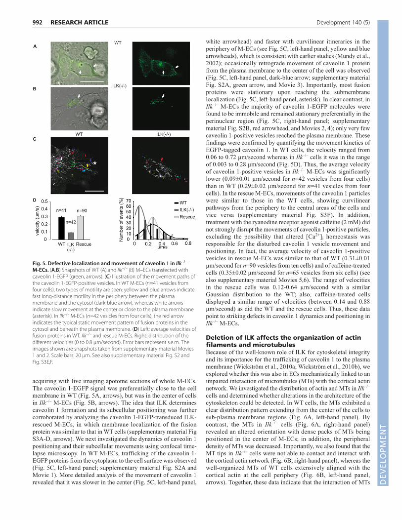
acquiring with live imaging apotome sections of whole M-ECs.The caveolin 1-EGFP signal was preferentially close to the cellmembrane in WT (Fig. 5A, arrows), but was in the center of cellsin Ilk–/– M-ECs (Fig. 5B, arrows). The idea that ILK determinescaveolin 1 formation and its subcellular positioning was furthercorroborated by analyzing the caveolin 1-EGFP-transduced ILK-rescued M-ECs, in which membrane localization of the fusionprotein was similar to that in WT cells (supplementary material FigS3A-D, arrows). We next investigated the dynamics of caveolin 1positioning and their subcellular movements using confocal time-lapse microscopy. In WT M-ECs, trafficking of the caveolin 1-EGFP proteins from the cytoplasm to the cell surface was observed(Fig. 5C, left-hand panel; supplementary material Fig. S2A andMovie 1). More detailed analysis of the movement of caveolin 1revealed that it was slower in the center (Fig. 5C, left-hand panel,
white arrowhead) and faster with curvilinear itineraries in theperiphery of M-ECs (see Fig. 5C, left-hand panel, yellow and bluearrowheads), which is consistent with earlier studies (Mundy et al.,2002); occasionally retrograde movement of caveolin 1 proteinfrom the plasma membrane to the center of the cell was observed(Fig. 5C, left-hand panel, dark-blue arrow; supplementary materialFig. S2A, green arrow, and Movie 3). Importantly, most fusionproteins were stationary upon reaching the submembranelocalization (Fig. 5C, left-hand panel, asterisk). In clear contrast, inIlk–/– M-ECs the majority of caveolin 1-EGFP molecules werefound to be immobile and remained stationary preferentially in theperinuclear region (Fig. 5C, right-hand panel; supplementarymaterial Fig. S2B, red arrowhead, and Movies 2, 4); only very fewcaveolin 1-positive vesicles reached the plasma membrane. Thesefindings were confirmed by quantifying the movement kinetics ofEGFP-tagged caveolin 1. In WT cells, the velocity ranged from0.06 to 0.72 µm/second whereas in Ilk–/– cells it was in the rangeof 0.003 to 0.28 µm/second (Fig. 5D). Thus, the average velocityof caveolin 1-positive vesicles in Ilk–/– M-ECs was significantlylower (0.09±0.01 µm/second for n=42 vesicles from four cells)than in WT (0.29±0.02 µm/second for n=41 vesicles from fourcells). In the rescue M-ECs, movements of the caveolin 1 particleswere similar to those in the WT cells, showing curvilinearpathways from the periphery to the central areas of the cells andvice versa (supplementary material Fig. S3F). In addition,treatment with the ryanodine receptor agonist caffeine (2 mM) didnot strongly disrupt the movements of caveolin 1-positive particles,excluding the possibility that altered [Ca2+]i homeostasis wasresponsible for the disturbed caveolin 1 vesicle movement andpositioning. In fact, the average velocity of caveolin 1-positivevesicles in rescue M-ECs was similar to that of WT (0.31±0.01µm/second for n=90 vesicles from ten cells) and of caffeine-treatedcells (0.35±0.02 µm/second for n=65 vesicles from six cells) (seealso supplementary material Movies 5,6). The range of velocitiesin the rescue cells was 0.12-0.64 µm/second with a similarGaussian distribution to the WT; also, caffeine-treated cellsdisplayed a similar range of velocities (between 0.14 and 0.88µm/second) as did the WT and the rescue cells. Thus, these datapoint to striking defects in caveolin 1 dynamics and positioning inIlk–/– M-ECs.
Deletion of ILK affects the organization of actinfilaments and microtubulesBecause of the well-known role of ILK for cytoskeletal integrityand its importance for the trafficking of caveolin 1 to the plasmamembrane (Wickström et al., 2010a; Wickström et al., 2010b), weexplored whether this was also in ECs mechanistically linked to animpaired interaction of microtubules (MTs) with the cortical actinnetwork. We investigated the distribution of actin and MTs in Ilk–/–
cells and determined whether alterations in the architecture of thecytoskeleton could be detected. In WT cells, the MTs exhibited aclear distribution pattern extending from the center of the cells tosub-plasma membrane regions (Fig. 6A, left-hand panel). Bycontrast, the MTs in Ilk–/– cells (Fig. 6A, right-hand panel)revealed an altered orientation with dense packs of MTs beingpositioned in the center of M-ECs; in addition, the peripheraldensity of MTs was decreased. Importantly, we also found that theMT tips in Ilk–/– cells were not able to contact and interact with the cortical actin network (Fig. 6B, right-hand panel), whereas thewell-organized MTs of WT cells extensively aligned with thecortical actin at the cell periphery (Fig. 6B, left-hand panel,arrows). Together, these data indicate that the interaction of MTs
RESEARCH ARTICLE Development 140 (5)
Fig. 5. Defective localization and movement of caveolin 1 in Ilk–/–
M-ECs. (A,B) Snapshots of WT (A) and Ilk–/– (B) M–ECs transfected withcaveolin 1-EGFP (green, arrows). (C) Illustration of the movement paths ofthe caveolin 1-EGFP-positive vesicles. In WT M-ECs (n=41 vesicles fromfour cells), two types of motility are seen: yellow and blue arrows indicatefast long-distance motility in the periphery between the plasmamembrane and the cytosol (dark-blue arrow), whereas white arrowsindicate slow movement at the center or close to the plasma membrane(asterisk). In Ilk–/– M-ECs (n=42 vesicles from four cells), the red arrowindicates the typical static movement pattern of fusion proteins in thecytosol and beneath the plasma membrane. (D) Left: average velocities offusion proteins in WT, Ilk–/– and rescue M-ECs. Right: distribution of thedifferent velocities (0 to 0.8 μm/second). Error bars represent s.e.m. Theimages shown are snapshots taken from supplementary material Movies1 and 2. Scale bars: 20 μm. See also supplementary material Fig. S2 andFig. S3E,F.
DEVELO
PMENT

with the cortex is impaired in the absence of ILK. In WT M-ECs,a typical regular distribution of stress fibers (Fig. 6C, arrow) wasdetectable throughout the cell, and caveolin 1 (red in Fig. 6C, left-hand panel) colocalized with the cortical actin fibers. By contrast,in Ilk–/– cells (Fig. 6C, right-hand panel) actin was found to bedisorganized and to accumulate within the peripheral areas of thecells, whereas caveolin 1 staining displayed a central distributionsimilar to the native protein (Fig. 4G; Fig. 5B; supplementarymaterial Fig. S2B). Thus, these data reveal that the organization ofboth MT and actin networks are disrupted in Ilk–/– ECs and that theMTs do not interact with the cortical actin. Similar to ourfunctional experiments, we explored next whether disruption ofF-actin in WT cells reiterates the phenotype found in Ilk–/– M-ESCs. For this purpose, we exposed WT cells to latrunculin (1µM) for 30 minutes and thereafter investigated actin and caveolin1 distribution. We observed disrupted actin organization with onlya few stress fibers visible and a re-distribution of caveolin 1molecules to the center of the cells colocalizing with the corticalactin network. These data are reminiscent of those found in Ilk–/–
M-ECs (Fig. 6C, left-hand panel), indicating a cortical actin- andILK-dependent subcellular distribution of caveolin 1.
DISCUSSIONHere, we demonstrate that deletion of ILK results in a strongreduction of endothelial tube formation. This prominent ECphenotype is caused by defective trafficking of caveolin 1 to theplasma membrane resulting in defective VEGF signaling. Thisdisturbance of RTK signaling is due to changes in MTs and corticalactin organization giving rise to the destabilization of caveolin 1microdomains.
Ilk–/– EBs display a striking reduction of endothelial tubeformation with alterations of EC proliferation, apoptosis andmigration. This phenotype was, as demonstrated by rescueexperiments, clearly related to the ILK deficiency. Because keyevents of EC biology are regulated by VEGF signaling, we haveinvestigated expression and function of VEGF-coupled receptor 2(VEGFR2) and downstream signaling pathways (Bhattacharya etal., 2009). Ca2+ imaging was employed to explore VEGF signalingin single M-ECs and revealed that there was no VEGF-induced[Ca2+]i response in mutant cells. This proved to be a more generaldefect of RTK signaling as EGF was also unable to mobilize [Ca2+]iin these cells. Further experiments revealed that VEGFR2 isexpressed and functional and that the signaling defect residesdownstream. Interestingly, VEGFR2 and ERK1/2 displayedincreased basal phosphorylation levels and this is in full agreementwith earlier reports on the effects of MbetaCD treatment (Barbuti etal., 2004) or inhibition of ILK activity (Ruiz-Torres et al., 2006).Because of the intact phosphorylation of VEGFR2 and of PLC-γupon VEGF stimulation and the preserved intracellular Ca2+
handling in Ilk–/– cells, we suspect that defective crosstalk betweensignaling components, most likely between PLC-γ and PIP2,underlies the observed phenotype. In fact, in ECs these signalingcomponents are clustered within caveolin 1-enriched microdomainsand their disruption inhibits PIP2 turnover. (Jang et al., 2001; Pikeand Miller, 1998). Recently, it has been also shown that changes incaveolae density and function, or disruption of the cytoskeletalintegrity perturb the compartmentalization of PIP2 signals (Cui etal., 2010). Future experiments are needed to explore the signalingdefect in more detail; in particular, activation of PLC-γ and itscrosstalk with PIP2 should be assessed using biochemical in vitroactivity assays. Similarly, the distribution and concentration of PIP2in the plasma membrane also needs to be explored with and withoutVEGF activation.
Recently, ILK has been reported to be involved in caveolaeorganization in keratinocytes (Wickström et al., 2010a). Our presentfindings reveal that ILK plays a crucial role for the correctsubcellular positioning of caveolin 1 in close vicinity of the plasmamembrane of M-ECs and illustrate the consequences of its deletionfor cytoskeletal integrity, cellular signaling and EC biology. We alsomimicked the defective VEGF signaling found in Ilk–/– M-ECs bythe treatment of WT cells with MbetaCD and these experimentsunderscore the specific role of lipid rafts in the organization of thissignaling pathway. These findings are in line with earlier reports,in which PLC-γ phosphorylation was found to be preserved despitethe disruption of plasma membrane microdomains, whereas PIP2function was reported to be strongly dependent on their clusteringand on the integrity of the cytoskeleton (Jang et al., 2001). AlthoughG protein-coupled receptors are known to be coupled with caveolae(Razani et al., 2002), these receptors are affected in a differentfashion by caveolar malfunction. In fact, disruption of caveolae byMbetaCD in adult cardiomyocytes affects the β2- but not the β1-adrenoceptor-mediated response (Calaghan et al., 2008), possiblyexplaining why neither acetylcholine nor bradykinin signaling wasaffected in Ilk–/– M-ECs.
993RESEARCH ARTICLEILK impairs endothelial caveolin 1-based signaling
Fig. 6. Loss of ILK affects the subcellular organization of actinfilaments and of MTs. (A) Confocal sections of WT and Ilk–/– M-ECsstained for tubulin to mark MTs (green). Arrows indicate MT pattern. (B) Double immunostaining with actin (red) and tubulin (green) in WT(arrows) and Ilk–/– (arrowheads) M-ECs. (C) Analysis of colocalization ofcaveolin 1 (red) and actin filaments (phalloidin, green) in WT (left) andIlk–/– M-ECs (middle). Latrunculin (1 μM, 30 minutes) treatment of WT M-ECs results in re-positioning of caveolin 1 (arrowhead) to the perinucleararea (right). Scale bars: 20 μm in A; 5 μm in B, left; 2.5 μm in B, right; 40 μmin C, left; 20 μm in C, middle and right.
DEVELO
PMENT

994
As the signaling defects observed in Ilk–/– M-ECs wereassociated with caveolin 1 microdomains, we subsequentlyexplored caveolin 1 positioning and caveolae formation using avariety of cell biological assays. Electron microscopy and high-resolution fluorescence microscopy revealed that Ilk–/– M-ECslacked caveolin 1 at the plasma membrane and sites of caveolaeformation, respectively. This was corroborated by time-lapsemicroscopy of fluorescence-labeled caveolin 1, the central proteincomponent of caveolae (Drab et al., 2001). We observed a cleardifference in motility of caveolin 1-positive vesicles of Ilk–/– M-ECs compared with WT cells. Rapid trafficking of caveolin 1between the cytosol and the plasma membrane was observed inWT M-ECs, which is in accordance with previous studies and isassociated with the high turnover of this protein in the cytosol(Mundy et al., 2002). This was further corroborated in our rescueM-ECs, in which re-expression of ILK restored caveolin 1 particlemovement to that observed in WT cells. By contrast, a strongreduction of caveolin 1 transport to the cell membrane could beobserved in Ilk–/– M-ECs. Also, in keratinocytes caveolin 1distribution was found to be dependent on ILK; in addition, itsimpact on MT stability via mDia1 DIAP1 – Mouse GenomeInformatics) and IQGAP1 has been shown (Wickström et al.,2010a). MTs, in concert with the cortical actin network, areassumed to act as tracks for caveolin 1-positive vesicles, and MT-cortical actin crosstalk is required to enable the transfer of caveolin1 via cortical actin to the plasma membrane, resulting in theformation of caveolae (Mundy et al., 2002; Wickström et al.,2010a). In Ilk–/– M-ECs, we found abnormal organization of theactin cytoskeleton and MT network causing reduced MT-actininteractions. To confirm that the defects in the transport ofcaveolin 1 observed by time-lapse microscopy resulted fromabnormal MT-actin crosstalk, we inhibited actin polymerizationusing latrunculin. Treatment of WT M-ECs with latrunculin ledto disruption of the actin cytoskeleton as well as to re-distributionof caveolin 1 and this was reminiscent of the findings in the Ilk–/–
M-ECs. By contrast, depletion of intracellular Ca2+ stores withcaffeine did not strongly alter subcellular caveolin 1 dynamics.These data support the view that ILK acts in ECs as a directscaffolding protein for actin and MT organization, and that ILK-dependent maintenance of cytoskeletal integrity is crucial for thetransport, positioning and turnover of caveolin 1 in close vicinityof the plasma membrane; these findings also support thoserecently reported in keratinocytes (Wickström et al., 2010a). Thus,our data demonstrate that ILK in ECs, through its regulation ofcaveolin 1 movement and positioning, directly interferes withspecific cellular signaling pathways, in particular RTK-mediatedsignaling, and that intact RTK signaling requires the precise spatialpositioning of downstream signaling components.
AcknowledgementsWe thank Dr P. Sasse (Institute of Physiology 1, Bonn) for experimental advice;and A. Voß (German Sport University, Cologne) for technical help.
FundingThis work was supported by a grant from the German Research Foundation(DFG) [1086 419/1-2BL to W.B. and B.K.F.].
Competing interests statementThe authors declare no competing financial interests.
Supplementary materialSupplementary material available online athttp://dev.biologists.org/lookup/suppl/doi:10.1242/dev.091298/-/DC1
ReferencesBalijepalli, R. C., Foell, J. D., Hall, D. D., Hell, J. W. and Kamp, T. J. (2006).
Localization of cardiac L-type Ca(2+) channels to a caveolar macromolecularsignaling complex is required for beta(2)-adrenergic regulation. Proc. Natl.Acad. Sci. USA 103, 7500-7505.
Barbuti, A., Gravante, B., Riolfo, M., Milanesi, R., Terragni, B. andDiFrancesco, D. (2004). Localization of pacemaker channels in lipid raftsregulates channel kinetics. Circ. Res. 94, 1325-1331.
Bhattacharya, R., Kwon, J., Li, X., Wang, E., Patra, S., Bida, J. P., Bajzer, Z.,Claesson-Welsh, L. and Mukhopadhyay, D. (2009). Distinct role of PLCbeta3in VEGF-mediated directional migration and vascular sprouting. J. Cell Sci. 122,1025-1034.
Calaghan, S., Kozera, L. and White, E. (2008). Compartmentalisation of cAMP-dependent signalling by caveolae in the adult cardiac myocyte. J. Mol. Cell.Cardiol. 45, 88-92.
Cho, C. H., Lee, C. S., Chang, M., Jang, I. H., Kim, S. J., Hwang, I., Ryu, S. H.,Lee, C. O. and Koh, G. Y. (2004). Localization of VEGFR-2 and PLD2 inendothelial caveolae is involved in VEGF-induced phosphorylation of MEK andERK. Am. J. Physiol. 286, H1881-H1888.
Cui, S., Ho, W. K., Kim, S. T. and Cho, H. (2010). Agonist-induced localization ofGq-coupled receptors and G protein-gated inwardly rectifying K+ (GIRK)channels to caveolae determines receptor specificity of phosphatidylinositol4,5-bisphosphate signaling. J. Biol. Chem. 285, 41732-41739.
Dedhar, S. and Hannigan, G. E. (1996). Integrin cytoplasmic interactions andbidirectional transmembrane signalling. Curr. Opin. Cell Biol. 8, 657-669.
Drab, M., Verkade, P., Elger, M., Kasper, M., Lohn, M., Lauterbach, B., Menne,J., Lindschau, C., Mende, F., Luft, F. C. et al. (2001). Loss of caveolae, vasculardysfunction, and pulmonary defects in caveolin-1 gene-disrupted mice.Science 293, 2449-2452.
Friedrich, E. B., Liu, E., Sinha, S., Cook, S., Milstone, D. S., MacRae, C. A.,Mariotti, M., Kuhlencordt, P. J., Force, T., Rosenzweig, A. et al. (2004).Integrin-linked kinase regulates endothelial cell survival and vasculardevelopment. Mol. Cell. Biol. 24, 8134-8144.
Hagedorn, M., Balke, M., Schmidt, A., Bloch, W., Kurz, H., Javerzat, S.,Rousseau, B., Wilting, J. and Bikfalvi, A. (2004). VEGF coordinatesinteraction of pericytes and endothelial cells during vasculogenesis andexperimental angiogenesis. Dev. Dyn. 230, 23-33.
Hannigan, G. E., Coles, J. G. and Dedhar, S. (2007). Integrin-linked kinase at the heart of cardiac contractility, repair, and disease. Circ. Res. 100, 1408-1414.
Head, B. P., Patel, H. H., Roth, D. M., Lai, N. C., Niesman, I. R., Farquhar, M. G.and Insel, P. A. (2005). G-protein-coupled receptor signaling componentslocalize in both sarcolemmal and intracellular caveolin-3-associatedmicrodomains in adult cardiac myocytes. J. Biol. Chem. 280, 31036-31044.
Head, B. P., Patel, H. H., Roth, D. M., Murray, F., Swaney, J. S., Niesman, I. R.,Farquhar, M. G. and Insel, P. A. (2006). Microtubules and actinmicrofilaments regulate lipid raft/caveolae localization of adenylyl cyclasesignaling components. J. Biol. Chem. 281, 26391-26399.
Jang, I. H., Kim, J. H., Lee, B. D., Bae, S. S., Park, M. H., Suh, P. G. and Ryu, S.H. (2001). Localization of phospholipase C-gamma1 signaling in caveolae:importance in EGF-induced phosphoinositide hydrolysis but not in tyrosinephosphorylation. FEBS Lett. 491, 4-8.
Krajewska, W. M. and Masłowska, I. (2004). Caveolins: structure and function insignal transduction. Cell. Mol. Biol. Lett. 9, 195-220.
Malan, D., Wenzel, D., Schmidt, A., Geisen, C., Raible, A., Bolck, B.,Fleischmann, B. K. and Bloch, W. (2010). Endothelial beta1 integrins regulatesprouting and network formation during vascular development. Development137, 993-1002.
Müller-Ehmsen, J., Schmidt, A., Krausgrill, B., Schwinger, R. H. and Bloch,W. (2006). Role of erythropoietin for angiogenesis and vasculogenesis: from embryonic development through adulthood. Am. J. Physiol. 290, H331-H340.
Mundy, D. I., Machleidt, T., Ying, Y. S., Anderson, R. G. and Bloom, G. S.(2002). Dual control of caveolar membrane traffic by microtubules and theactin cytoskeleton. J. Cell Sci. 115, 4327-4339.
Novak, A., Hsu, S. C., Leung-Hagesteijn, C., Radeva, G., Papkoff, J.,Montesano, R., Roskelley, C., Grosschedl, R. and Dedhar, S. (1998). Celladhesion and the integrin-linked kinase regulate the LEF-1 and beta-cateninsignaling pathways. Proc. Natl. Acad. Sci. USA 95, 4374-4379.
Pike, L. J. and Miller, J. M. (1998). Cholesterol depletion delocalizesphosphatidylinositol bisphosphate and inhibits hormone-stimulatedphosphatidylinositol turnover. J. Biol. Chem. 273, 22298-22304.
Razani, B., Woodman, S. E. and Lisanti, M. P. (2002). Caveolae: from cellbiology to animal physiology. Pharmacol. Rev. 54, 431-467.
Ruiz-Torres, M. P., Pérez-Rivero, G., Rodríguez-Puyol, M., Rodríguez-Puyol,D. and Díez-Marqués, M. L. (2006). The leukocyte-endothelial cellinteractions are modulated by extracellular matrix proteins. Cell. Physiol.Biochem. 17, 221-232.
Saliez, J., Bouzin, C., Rath, G., Ghisdal, P., Desjardins, F., Rezzani, R., Rodella,L. F., Vriens, J., Nilius, B., Feron, O. et al. (2008). Role of caveolar
RESEARCH ARTICLE Development 140 (5)
DEVELO
PMENT

compartmentation in endothelium-derived hyperpolarizing factor-mediatedrelaxation: Ca2+ signals and gap junction function are regulated by caveolin inendothelial cells. Circulation 117, 1065-1074.
Schmidt, A., Wenzel, D., Ferring, I., Kazemi, S., Sasaki, T., Hescheler, J.,Timpl, R., Addicks, K., Fleischmann, B. K. and Bloch, W. (2004). Influence of endostatin on embryonic vasculo- and angiogenesis. Dev. Dyn. 230, 468-480.
Schmidt, A., Wenzel, D., Thorey, I., Werner, S., Fleischmann, B. K. and Bloch,W. (2005). Endostatin down-regulates soluble guanylate cyclase (sGC) inendothelial cells in vivo: influence of endostatin on vascular endothelialgrowth factor (VEGF) signaling. Endothelium 12, 251-257.
Tu, Y., Li, F. and Wu, C. (1998). Nck-2, a novel Src homology2/3-containingadaptor protein that interacts with the LIM-only protein PINCH and
components of growth factor receptor kinase-signaling pathways. Mol. Biol.Cell 9, 3367-3382.
Vouret-Craviari, V., Boulter, E., Grall, D., Matthews, C. and Van Obberghen-Schilling, E. (2004). ILK is required for the assembly of matrix-formingadhesions and capillary morphogenesis in endothelial cells. J. Cell Sci. 117,4559-4569.
Wickström, S. A., Lange, A., Hess, M. W., Polleux, J., Spatz, J. P., Krüger, M.,Pfaller, K., Lambacher, A., Bloch, W., Mann, M. et al. (2010a). Integrin-linkedkinase controls microtubule dynamics required for plasma membranetargeting of caveolae. Dev. Cell 19, 574-588.
Wickström, S. A., Lange, A., Montanez, E. and Fässler, R. (2010b). TheILK/PINCH/parvin complex: the kinase is dead, long live the pseudokinase!EMBO J. 29, 281-291.
995RESEARCH ARTICLEILK impairs endothelial caveolin 1-based signaling
DEVELO
PMENT