cytotoxicity in the age of nano: the role of fourth period...
TRANSCRIPT

Chemico-Biological Interactions 206 (2013) 319–326
Contents lists available at ScienceDirect
Chemico-Biological Interactions
journal homepage: www.elsevier .com/locate /chembioint
Cytotoxicity in the age of nano: The role of fourth period transition metaloxide nanoparticle physicochemical properties
0009-2797/$ - see front matter � 2013 Published by Elsevier Ireland Ltd.http://dx.doi.org/10.1016/j.cbi.2013.09.020
⇑ Corresponding authors. Tel.: +1 615 898 2079; fax: +1 615 494 7693 (C.C.Chusuei). Tel.: +1 573 341 6589; fax: +1 573 341 4821 (Y.-W. Huang).
E-mail addresses: [email protected] (C.C. Chusuei), [email protected](Y.-W. Huang).
Charles C. Chusuei a,⇑, Chi-Heng Wu b, Shravan Mallavarapu a, Fang Yao Stephen Hou c, Chen-Ming Hsu d,Jeffrey G. Winiarz e, Robert S. Aronstam b, Yue-Wern Huang b,⇑a Department of Chemistry, Middle Tennessee State University, Box 390, 1301 East Main Street, Murfreesboro, TN 37132, USAb Department of Biological Sciences, Missouri University of Science and Technology, 105 Schrenk Hall, 400 W. 11th Street, Rolla, MO 65409, USAc Department of Clinical Laboratory Science, College of Health Sciences, Marquette University, P.O. Box 1881, Milwaukee, WI 53201, USAd Department of Life Science, National Taiwan Normal University, 88 Ting-Chow Rd, Sec 4, Taipei 116, Taiwan, ROCe Department of Chemistry, Missouri University of Science and Technology, 142 Schrenk Hall, 400 W. 11th Street, Rolla, MO 65409, USA
a r t i c l e i n f o
Article history:Received 22 May 2013Received in revised form 1 September 2013Accepted 30 September 2013Available online 10 October 2013
Keywords:Physicochemical propertiesMetal oxide nanoparticlesCytotoxicitySurface binding sitesPoint-of-zero chargeMetal ion dissolution
a b s t r a c t
A clear understanding of physicochemical factors governing nanoparticle toxicity is still in its infancy. Weused a systematic approach to delineate physicochemical properties of nanoparticles that govern cyto-toxicity. The cytotoxicity of fourth period metal oxide nanoparticles (NPs): TiO2, Cr2O3, Mn2O3, Fe2O3,NiO, CuO, and ZnO increases with the atomic number of the transition metal oxide. This trend was notcell-type specific, as observed in non-transformed human lung cells (BEAS-2B) and human bronchoalve-olar carcinoma-derived cells (A549). Addition of NPs to the cell culture medium did not significantly alterpH. Physiochemical properties were assessed to discover the determinants of cytotoxicity: (1) point-of-zero charge (PZC) (i.e., isoelectric point) described the surface charge of NPs in cytosolic and lysosomalcompartments; (2) relative number of available binding sites on the NP surface quantified by X-ray pho-toelectron spectroscopy was used to estimate the probability of biomolecular interactions on the particlesurface; (3) band-gap energy measurements to predict electron abstraction from NPs which might lead tooxidative stress and subsequent cell death; and (4) ion dissolution. Our results indicate that cytotoxicityis a function of particle surface charge, the relative number of available surface binding sites, and metalion dissolution from NPs. These findings provide a physicochemical basis for both risk assessment and thedesign of safer nanomaterials.
� 2013 Published by Elsevier Ireland Ltd.
1. Introduction
There are currently more than 2800 nanoparticulate-basedapplications commercially available. It is estimated that by 2017,this field will represent a $48.9 billion market [1]. As engineerednanoparticles (NPs) currently occupy a significant portion of themarket and are anticipated to proliferate commercially, there isan urgent need to study their potential impact on human healthand the environment.
To date, there exists no epidemiological or clinical evidencedemonstrating that inhalation of NPs leads to adverse health ef-fects in humans [2]. However, toxicological studies using animalmodels and cell cultures suggest that NPs are more toxic andinflammogenic than larger particles of similar composition and ofequal mass [3]. We have demonstrated intricate relationships
between NPs, production of ROS and changes in intracellular Ca2+
concentrations [Ca2+]in. These studies suggest that NPs can triggercell death by multiple pathways [4]. NPs increase [Ca2+]in. Moder-ation of this increase by nifedipine suggests that a portion of thisincrease reflects the influx of extracellular calcium. Membrane dis-ruption (e.g., as indicated by lipid peroxidation and membranedepolarization) may also play a role in this influx [4,5]. NPs alsodisrupt store-operated calcium entry (SOCE) [6]. The increase inintracellular ROS may also have multiple sources. There exist syn-ergistic relationships between intracellular [Ca2+] and OS as the in-creases in both can be reduced by an antioxidant. Finally, while[Ca2+]in and ROS affect each other, they induce cell death by dis-tinct pathways.
Structural defects on the NPs, which can act as electron-donor/acceptor groups, may alter the electronic configuration andcontribute to the formation of reactive oxygen species (ROS) [7].Particle dissolution has also been considered as a factor in NP-induced toxic responses [8,9]. Particle size and morphology are fac-tors that also contribute to toxicity [10,11]. It remains unclearwhether additional physicochemical properties of metal oxide

320 C.C. Chusuei et al. / Chemico-Biological Interactions 206 (2013) 319–326
NPs dictate the toxic responses. To elucidate these properties, wesystematically examined an array of oxides of transition metalsin the fourth period of the Periodic Table (Ti, Cr, Mn, Fe, Ni, Cu,Zn). These types of nanomaterials have been extensively used incatalysis [12], magnetocooling [13], optical and recording devices[14,15], purification of enzymes and other biological materials[16], water purification devices [17], magnetic field assisted radio-nuclide therapy [18], embolics [19–21], cosmetic and skin careproducts, and targeted drug delivery agents [22–27]. This seriesof NPs offers an opportunity to investigate the determinants of tox-icity, which may lead to the design of safer nanomaterials. Toxicitycan be investigated using in vitro and in vivo systems. Both systemsprovide different information for various scientific purposes and inmany cases are complementary to each other. As there are numer-ous nanomaterials, it is improbable, though not impossible, toinvestigate each nanomaterials with in vivo systems. Therefore,in vitro systems provide an alternative to study nanotoxicity in that(1) it is cost efficient, (2) it provides information to prioritize ani-mal testing, and (3) it informs computational toxicology in the con-text of quantitative structure–activity relationship (QSARS).
We hypothesize that toxicity is a function of multiple physico-chemical properties of nanoparticles. We selected TiO2, Cr2O3,Mn2O3, Fe2O3, NiO, CuO, and ZnO NPs from a single commercialsource to minimize variability. In order to determine whether cyto-toxic responses are cell-type specific, two human lung cells werestudied. Cells were exposed to these NPs and cytotoxicity wasmeasured. Isoelectric points (i.e., point-of-zero charge), numberof available surface binding sites, and band-gap energies of theNPs were measured. The NPs were also subjected to kinetic exper-iments to determine the extent of metal ion dissolution. Our re-sults indicate that certain physicochemical properties of metaloxide NPs strongly correlate with cytotoxicity.
2. Materials and methods
2.1. Nanoparticles, reagents, and instrumentation protocols
The nanoparticles, reagents, and instrumentation protocolsused in the experiments are detailed in the Appendix A. Transmis-sion electron microscopy (TEM), X-ray photoelectron spectroscopy(XPS), and band gap measurements were performed on the NPs.Characterizations of graphite furnace atomic absorption analysis(GFAA) and inductive coupled plasma-mass spectrometry(ICP–MS) of the aqueous solution supernatants exposed to theNPs. Correlations of observed physicochemical properties of thematerials were correlated with cytotoxicity.
2.2. Cell culture and exposure of cells to NPs
Human bronchial epithelial cells (BEAS-2B) and human bron-choalveolar carcinoma-derived cells (A549) are in vitro modelsconsidered as ideal for both studying the prevention of human lungcarcinoma development and nanotoxicity testing [28]. These cellswere maintained using the same procedures described in our pre-vious studies [4,5,29–31].
Cells were grown at 37 �C in a 5% CO2 humidified environment.Upon reaching 85% confluence, the cells were seeded into 24 wellplates and allowed to attach for 24 h. The cell densities used fol-lowed ATCC protocol recommendations, and were well within sen-sitivity and detection limits of the analytical instruments used. Toreduce experimental variation and ensure accuracy, particles weredried in a desiccator before being weighed on an analytical bal-ance. Particles were suspended in cell culture medium, vortexedvigorously, and then sonicated. A series of dilutions in cell culturemedium were performed to achieve desired concentrations. The
suspensions were immediately applied to cells to minimizeagglomeration. Cells without NPs and reagent blanks were usedas controls in each experiment.
2.3. Cytotoxicity assay and apoptosis
At the end of cell exposure to NP suspensions, the medium wasdiscarded and the sulforhodamine B assay was used to determinecell viability relative to the control group [31]. Briefly, the cellswere fixed with cold 10% trichloroacetic acid (TCA) for 1 h at4 �C. The TCA solution was then discarded and the cells werewashed three times with distilled water, followed by completedrying. Sulforhodamine B (0.2% in 1% acetic acid) was added tostain the cells for 30 min at room temperature. The staining solu-tion was discarded and the cells were washed with 1% acetic acidthree times to eliminate excess dye. After complete drying, thedye was dissolved in cold 10 mM Tris buffer (pH = 10.5). Aliquots(100 lL) of dye solution were transferred onto a 96-well plate,and absorbance was measured at 550 nm using a microplate read-er (FLOURstar, BMG Labtechnologies, Durham, NC, USA).
Apoptotic cells were stained with annexin V–FITC and 7-amin-oactinomycin D (7-AAD) followed by quantification using a Beck-man Coulter Cell Lab Quanta SC System. Morphologicalexamination of apoptotic cells was performed using the same dyesand observed with an Olympus IX 51 epifluorescence microscope.
2.4. Statistical analysis
For toxicity studies, three independent experiments were con-ducted, using triplicates for each treatment group. Data are ex-pressed as mean ± standard deviation. The relationship betweencytotoxicity and the physicochemical properties of nanoparticleswere analyzed with Spearman’s Rank Correlation Analysis.
3. Results
3.1. Size, morphology, and specific surface area
The approximate physical sizes (APS) of the seven commerciallyavailable transition metal oxide NPs ranged from 16 ± 5 nm (NiO)to 82 ± 31 nm (Mn2O3) (Table 1). The morphology of NPs observedwith TEM was needle-like (TiO2), spherical (Mn2O3, Fe2O3), ornearly spherical (Cr2O3, NiO, CuO, ZnO) (Fig. A1). The specific sur-face area (SSA) of NPs ranged from 8.71 m2/g (Mn2O3) to178.95 m2/g (TiO2). While TiO2, Fe2O3, and CuO had similar sizes,they possessed distinctly different specific surface areas. This couldbe due to variations in surface porosity and discrepancy inmorphology.
3.2. Influence of pH in cell culture medium on cell viability
Cytotoxicity of NPs may simply reflect changes in pH over time.To evaluate this possibility, NPs were added to the cell culturemedium and pH was measured at 0, 6, 12, 18, and 24 h. Immedi-ately after adding NPs to cells in medium, the pH became slightlyelevated compared to the control cell in medium only. The pH fluc-tuated briefly, and then stabilized, except for NiO, which increasedthe pH between 12 and 24 h. Very little change in pH was observedat low NP concentrations (Table A1). As concentrations of NPs in-crease, pH variations increased with all NPs. However, the extentof pH fluctuations was 0.29 ± 0.14 and 0.31 ± 0.03 units with andwithout NPs, respectively. Cell morphology and size in the groupswith NPs were similar to those in the control groups.

Table 1Measured specific surface area, approximate physical size, and morphology of nanoparticles.
TiO2 Cr2O3 Mn2O3 Fe2O3 NiO CuO ZnO
SSA (m2/g) 178.95 11.41 8.71 31.44 70.86 9.02 44.61APS (nm) 46 ± 20 63 ± 34 82 ± 31 48 ± 13 16 ± 5 47 ± 24 27 ± 13Morphology Needle-like Nearly spherical Spherical Spherical Nearly spherical Nearly spherical Nearly spherical
C.C. Chusuei et al. / Chemico-Biological Interactions 206 (2013) 319–326 321
3.3. Cytotoxicity and apoptosis
Seven nanosized oxides of transition metals (Ti, Cr, Mn, Fe, Ni,Cu, Zn) from the fourth period of the Periodic Table of Elementswere selected to test our hypothesis that certain physicochemicalproperties of NPs contribute to cytotoxicity in human cells. Twohuman lung cell lines, BEAS-2B and A549, were tested to determinewhether cytotoxicity is cell-type specific.
A 24-h study of A549 cells exposed to NPs shows a trend of tox-icity, as revealed by cell viability. As the atomic number of thetransition metal increases within the fourth period, cytotoxicity in-creases (Fig. 1). The toxicity falls into three categories: (1) TiO2,Cr2O3, and Fe2O3 have zero to minimal toxicity (close to 100% cellviability); (2) Mn2O3 and NiO show typical dose-dependent toxic-ity (�40% cell viability); and (3) CuO and ZnO induce potent toxic-ity within a narrow dose range ([20% cell viability). A similartrend of toxicity is observed with BEAS cells (data not shown),
120
100
rol
80ontro
l
80
f Con
60 C O % o
f C
60 CuOTiOlit
y, %
TiO2
Cr Oiabi
lity
40 Cr2O3
NiOll Vi
ab
NiOMn OC
ell V
20Mn2O3
Fe O
C
Fe2O3
ZnO0
ZnO
0 20 40 60 80 1000
Particle Mass/Medium Volume (µ g/mL)
120
100
rol
80ontro
l
80
f Con
60 % o
f C
60
lity,
%ia
bilit
y
40ll Vi
abC
ell V
20
C
00 5 10 15 20
0
Particle Specific Surface AParticle Specific Surface A
A
C
Fig. 1. Response of A549 cells to transition metal oxide nanoparticles based upon variomass/seeding area (lg/cm2), and (C) particle specific surface area/seeding area (cm2/cm2
experiments were conducted, using triplicates for each treatment group. Data are expre
albeit with slight variations. Notably, both CuO and ZnO cause tox-icity with a steep concentration range. There is a good correlationbetween cytotoxicity and atomic number (q = 0.93, Fig. 2). Cyto-toxicity is observed in the form of apoptosis and necrosis(Fig. 3A–B). The combined populations of early apoptotic cellsand late apoptotic/early necrotic cells treated with the highest con-centrations of CuO (20 lg/mL) and ZnO (28 lg/mL) are 71.8 ± 7.6%and 28.4 ± 11.7%, respectively. The combined populations of earlyapoptotic cells and late apoptotic/early necrotic cells of the restof five NPs range from 2.2 ± 0.7% and 6.1 ± 0.7%. The degree ofapoptosis/necrosis corresponds with severity of cytotoxicity.
3.4. Physicochemical properties and toxicity
We hypothesize that cytotoxicity is a function of particle (i) sur-face charge, (ii) available surface binding sites, and (iii) dissolutionof metal ions from metal oxide NPs. The particle surface charge,
120120
100100
80ntro
l
80
f Con
tr o
f C
60CuOty
, % o
CuOTiO2bi
lity,
402
Cr2O3 Via
bi
2 3
NiO
Cel
l Vi
20 Mn2O3
Ce
20 Fe2O3
0 ZnO
0 10 20 30 40 50 600
0 10 20 30 40 50 60
Particle Mass/Seeding Area (µ g/cm2)Particle Mass/Seeding Area (µ g/cm )
CuOTiO TiO2
C O Cr2O3
NiO NiOMn O Mn2O3
Fe O Fe2O3
ZnOZnO
25 30 35 50 100
rea/Seeding Area (cm2/cm2)rea/Seeding Area (cm /cm )
B
us dosimetry expressions: (A) particle mass/medium volume (lg/mL), (B) particle). Cell viability was determined by the sulforhoamine B method. Three independentssed as mean ± standard deviation.

Fig. 2. (A) Spearman’s Rank Correlation between cytotoxicity and atomic number of seven oxides of transition metals. As atomic number increases, cytotoxicity increases. Thesame trend occurs in both A549 and BEAS-2B cells. (B) Correlation between cytotoxicity of point-of-zero charge (PZC) of nanoparticles. Without the outlier Mn2O3, q = 0.94.(C) Correlation between cytotoxicity and available particle surface binding sites. Experiments were conducted at pH 7.4. Available surface binding sites were not estimated atpH 4.5 conditions. Acid etching effects observed in this pH region would skew quantitative measurements of physisorbed-to-metal oxide oxygen ratios.
10010
(%) 100
CuO ZnO10
80CuO ZnO
cel
ls (%
)
80 µg/mLic
cel
ls
rotic
c
6
0 µg/mL5 µg/mL
crot
ic
60
nec
rot
6µg
25 µg/mL50 µg/mL n
ecro
40arly
ne
450 µg/mL100 µg/mL
arly
n
40
is/E
arl4 µg
is/E
ar
20
ptos
is/
2
tosi
s/
0Apo
pto
0popt
o
0 1 5 10 20 0 15 22 24 280A
p
TiO2 Cr2O3 Mn2O3 Fe2O3 NiO0A
po
µg/mLTiO2 Cr2O3 Mn2O3 Fe2O3 NiO
A
B
Fig. 3. Transition metal oxide nanoparticles induced cell death in A549 cells. Cells were treated with nanoparticles for 24 h followed by flow cytometric analysis usingannexin V–FITC (AV, x-axis) and 7-aminoactinomycin D (7-AAD, y-axis): (A) AV positive/7-AAD negative (apoptotic) and AV-positive/7-AAD positive (late apoptosis, earlynecrosis); (B) Percentage of late apoptotic and early necrotic populations. Three independent experiments were conducted, using triplicates for each treatment group.
322 C.C. Chusuei et al. / Chemico-Biological Interactions 206 (2013) 319–326
measured as PZCs, of the metal oxides are summarized in Fig. 4.Horizontal dashed lines within the PZC plot at pH = 4.5 and 7.4 de-note the pH of the lysosomal and cytosolic environments, respec-tively. Noteworthy is the fact that as PZC increases, the cytotoxiceffects of the NPs on the BEAS-2B and A549 cell lines increase, ex-cept for Mn2O3. Most of the PZC values cluster between 8 and 9,above both lysosomal and cytosolic environments, with the excep-tion of TiO2, which had a PZC of 6.9. Fig. 2B shows a good correla-tion between cytotoxicity and PZC calculated with Mn2O3 as anoutlier (q = 0.94), and without Mn2O3 as an outlier (q = 0.78).
The relative number of available particle surface binding siteswas measured by XPS (Table A2). A greater physisorbed-to-metaloxide oxygen ratio denotes more adsorption sites potentially avail-able for cellular molecular binding. Fig. 5 shows a stack plot of XPSspectra of the O 1s orbitals of all NPs following a 16 h CC reaction atpH = 7.4. The chemical oxidation state denoting metal oxide (bluetrace) is clearly defined for each respective nanoparticle. The XPSbinding energies (BE) with full-width-at-half-maxima (fwhm) inparentheses were found to be at 530.0 (1.7), 529.6 (1.1), and529.5 (1.4) eV, matching literature values for the metal oxide

1414
1212
1010
8 7 48
O7.4
6 Mn2O3
6 ZnOFina
l pH
4.5 CuO4 NiO4
Fe2O32
2 3Cr2O32 Cr2O3TiOTiO2
0 2 4 6 8 10 12 140
0 2 4 6 8 10 12 140
Initial pH
Fig. 4. Point-of-zero charge (PZC) initial pH versus final pH plots of Mn2O3, ZnO,CuO, NiO, Fe2O3, Cr2O3, and TiO2 metal oxide nanoparticles. Horizontal lines at pH7.4 and pH 4.5 denote cytosolic and lysosomal environments, respectively.
Fig. 5. XPS of O 1s orbitals of Fe2O3, Mn2O3, and TiO2 metal oxide nanoparticles. XP speShirley background subtractions. Blue envelopes denote the metal oxide chemical oxidatlines denote the BE position for adsorbed H2O. (For interpretation of the references to c
Fig. 6. ICP–MS and GFAA data from supernatants extracted from constant composition (CpH 7.4, showing: (A) metal dissolution kinetics of CuO and ZnO nanoparticles and (B) meanalysis was performed to analyze ions of Cu, Zn, Ti, Cr, Mn, and Ni in solution. GFAA anwere expressed as mean ± standard deviation.
C.C. Chusuei et al. / Chemico-Biological Interactions 206 (2013) 319–326 323
oxidation state for TiO2 [32], Mn2O3 [33,34], and Fe2O3 [35,36],respectively. BEs observed at 531.9 (2.4) eV on TiO2 [37–39],530.9 (1.1) and 531.5 (4.0) eV on Mn2O3, and 529.5 (1.4) and530.6 (2.4) eV on Fe2O3 are consistent with adsorbed hydroxyl spe-cies on these surfaces [40]. BEs of the metal oxide chemical stateobserved at O 1s = 529.0 (0.9), 529.7 (1.0), 529.7 (1.0) and 529.8(1.1) eV, matched their literature values for NiO [41], Cr2O3 [42],CuO [43,44], and ZnO [45], respectively. BE peak centers at 531.0(2.3) eV on NiO, 530.7 (2.1) and 532.5 (2.3) eV on Cr2O3, 531.1(1.4) eV on CuO, and 531.6 (2.2) eV on ZnO are also consistent withthe presence of adsorbed surface hydroxyls. The peak position at�531.5 eV could also emanate from adsorbed carbonyls (fromatmospheric CO2) [40]. The vertical dashed line (Fig. 5) denotesthe BE chemical shift for the H2O oxidation state. The O 1s BE peakcenters at 532.9 (1.7) eV on NiO, 532.5 (2.3) eV on Cr2O3, 532.6(1.3) and 532.9 (3.7) eV on CuO, and 532.9 (2.0) eV on ZnO denoteadsorbed H2O on these surfaces [39,40]. There was a good correla-tion between cytotoxicity and available nanoparticle surface bind-ing sites (q = 0.71, Fig. 2C). According to these data, the relativenumber of available binding sites (Table A3) for each oxide is in
ctra were deconvoluted using a 70:30 Gaussian–Lorenztian lineshape and applyingion state. Red envelopes denote adsorbed non-metal oxide oxygen. Vertical dashed
olour in this figure legend, the reader is referred to the web version of this article.)
C) experiments of solutions in contact with metal oxide nanoparticles at pH 4.5 andtal dissolution kinetics of TiO2, Cr2O3, Mn2O3, Fe2O3, and NiO nanoparticles. ICP–MSalysis was performed to quantify the amount of ions of Fe in solution. N = 3–6. Data
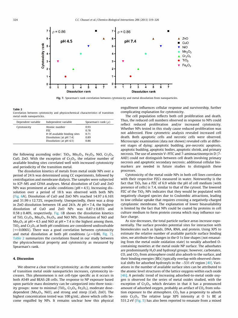
Fig. 7. Spearman’s rank correlation between cytotoxicity and metal dissolution from nanoparticles.
Table 2Correlation between cytotoxicity and physicochemical characteristics of transitionmetal oxide nanoparticles.
Dependent variable Independent variable Spearman’s rank (q)
Cytotoxicity Atomic number 0.93PZC 0.78# Of available binding sites 0.71Dissolution (at pH 7.4) 0.89Dissolution (at pH 4.5) 0.86
324 C.C. Chusuei et al. / Chemico-Biological Interactions 206 (2013) 319–326
the following ascending order: TiO2, Mn2O3, Fe2O3, NiO, Cr2O3,CuO, ZnO. With the exception of Cr2O3, the relative number ofavailable binding sites correlated well with increased cytotoxicityand periodicity of the transition metal.
The dissolution kinetics of metals from metal oxide NPs over aperiod of 24 h was determined using CC experiments, followed bycentrifugation and membrane dialysis. The samples were subjectedto ICP–MS and GFAA analyses. Metal dissolution of CuO and ZnONPs was prominent at acidic conditions (pH = 4.5). Increasing dis-solution over a period of 18 h was observed with both NPs(Fig. 6A). Dissolution of CuO and ZnO NPs reached 14.97 ± 6.16%and 31.99 ± 12.72%, respectively. Unexpectedly, there was a dropin ZnO dissolution between 18 and 24 h. At pH = 7.4, the highestdissolution of CuO and ZnO NPs was 0.87 ± 0.85% and0.58 ± 0.40%, respectively. Fig. 6B shows the dissolution kineticsof TiO, Cr2O3, Mn2O3, Fe2O3, and NiO NPs. Dissolution of NiO andMn2O3 at pH = 4.5 and NiO at pH = 7.4 is the highest among them.TiO2 and Cr2O3 at both pH conditions are considered undissolvable(<< 0.006%). There was a good correlation between cytotoxicityand metal dissolution at both pH conditions (q = 0.86, Fig. 7).Table 2 summarizes the correlations found in our study betweenthe physicochemical property and cytotoxicity as measured bySpearman’s rank.
4. Discussion
We observe a clear trend in cytotoxicity: as the atomic numberof transition metal oxide nanoparticles increases, cytotoxicity in-creases. This phenomenon is not cell-type specific as it occurs inboth A549 and BEAS-2B cells. The response to NP exposure basedupon particle mass dosimetry can be categorized into three toxic-ity groups: none to minimal (TiO2, Cr2O3, Fe2O3), moderate dose-dependent (Mn2O3, NiO), and strong and steep (CuO, ZnO). Thehighest concentration tested was 100 g/mL; above which cells be-come engulfed by NPs. It remains unclear how this physical
engulfment influences cellular response and survivorship, furthercomplicating explanation for cytotoxicity.
The cell population reflects both cell proliferation and death.Thus, the reduced cell numbers observed in response to NPs couldreflect reduced proliferation and/or increased cytotoxicity.Whether NPs tested in this study cause reduced proliferation wasnot addressed. Flow cytometric analysis revealed increased celldeath. Both apoptotic cells and necrotic cells were observed.Microscopic examination (data not shown) revealed cells at differ-ent stages of dying: apoptotic budding, pre-necrotic apoptosis,apoptotic budding, apoptotic bodies, apoptotic shrink, and primarynecrosis. The use of annexin V–FITC and 7-aminoactinomycin D (7-AAD) could not distinguish between cell death involving primarynecrosis and apoptotic secondary necrosis; additional cellular bio-markers are needed in future studies to distinguish theseprocesses.
Cytotoxicity of the metal oxide NPs in both cell lines correlateswith their respective PZCs measured in water. Noteworthy is thefact that TiO2 has a PZC of 6.9 while the pH of cell medium (withpresence of cells) is 7.4, similar to that of the cytosol. The loweredPZC of the TiO2 NPs indicates that they would be populated withnegatively charged species due to Coulombic attractions, leadingto low cellular uptake that requires crossing a negatively-chargedcytoplasmic membrane. The explanation of lower bioavailabilityis limited by the fact that NPs could be coated by proteins in cellculture medium to form protein corona which may influence sur-face charge.
As size decreases, the total particle surface areas increase expo-nentially. The surface provides potential sites for interaction withbiomolecules such as lipids, DNA, RNA, and protein. Using XPS toestimate the relative number of available particle surface bindingsites, we attribute the changes in the O 1s line shapes (not emanat-ing from the metal oxide oxidation state) to weakly adsorbed O-containing moieties at the metal oxide NP surface. The adsorbatesare predominantly H2O and hydroxyl groups; however, carbonates,CO, and CO2 from atmosphere could also adsorb to the surface, andtheir binding energies (BEs) typically overlap with observed chem-ical shifts for adsorbed hydroxyls in the �531 eV region [40]. Vari-ations in the number of available surface sites can be attributed tothe atomic level structures of the lattice oxygens within each oxide[46]. A periodic trend of increasing adsorbed-to-metal oxide oxy-gen is observed for the series of metal oxides studied, with theexception of Cr2O3, which deviates in that it has a pronouncedamount of adsorbed oxygen, probably an artifact of CO2 from solu-tion exposure to the atmosphere. CO2 has a propensity to adsorbonto Cr2O3. The relative large XPS intensity at O 1s BE at531.2 eV (Fig. 5) has also been reported to emanate from a mixed
C.C. Chusuei et al. / Chemico-Biological Interactions 206 (2013) 319–326 325
complex of Cr2O3�nH2O�xCO2 formed from adsorbed atmosphericCO2 into the aqueous solution [47]. The most toxic of nanoparticlesanalyzed in this series also has the highest adsorbed H2O content;the vertical, dashed line denotes the chemical oxidation state foradsorbed H2O, ZnO, and CuO have the highest PZCs and hencegreatest degree of ‘‘protonation’’ via adsorption of hydronium ions(H3O+). Under aqueous solution physiological conditions, the metaloxide surface would be populated by excess H3O+, in accordancewith Gouy–Chapman theory. During adsorption, the adsorbatewould be electrically neutralized resulting in the observed, en-hanced intensity denoting chemisorbed H2O at 532.9 eV on theCuO and ZnO surfaces. Adsorbed H2O is not pronounced on theCr2O3, Mn2O3, Fe2O3, and NiO NP surfaces. Lesser absorption is ob-served with TiO2 (Fig. 5), which has the lowest PZC in the series(below that of physiological pH), appearing at the leading edge ofthe BE envelope indicative of adsorbed hydroxyls at �532 eV.
Dissolution of metals from metal oxides correlate with observedcytotoxicity. Dissolution kinetics of metals from metal oxide NPssuggests a significant release of Cu2+ and Zn2+ from CuO and ZnOoxides in acidic environment (pH = 4.5), but not neutral environ-ment (pH = 7.4). A drop in metal dissolution of ZnO is observed be-tween 18 and 24 h. We postulate that high concentrations of Zn2+
leads to re-absorption of the released ions onto the ZnO NPs. Thisaction could result in an incomplete separation of ions from oxidesduring sample preparation. George et al. [48] also found significantZn2+ dissolution from ZnO NPs in a 1000-min. kinetics study; equi-librium was not reached at the end of the experiment.
Metal dissolution of NiO at both pH = 4.5 and 7.4, and Mn2O3 atpH = 4.5 were lower than 1%, except for NiO at 24 h at pH = 4.5.Though the released Cu2+ and Zn2+ concentrations at the neutralenvironment are small, their potential effects may be significant,as these two ions are very toxic. Elevated levels of Zn2+ are toxicto a variety of cells, including PC-12, HeLa, and HT-29 cell lines,as well as primary cultures of cardiac myocytes and neurons[49]. Cu2+ increases cell death and impairs the colony-forming effi-ciency of human hepatoma cells [50]. Ni2+ released from nickelhydroxide nanoparticles plays a role of pulmonary toxicity in awhole-body inhalation study [51]. Additional studies have shownthat exposure of particulate matter (PM)-associated ions, such asCu2+ and Zn2+, elevate oxidative stress and induce inflammatory re-sponses [52–54]. Many NPs have been shown to use endocytosis asa major route for cellular entry [55,56]. Vesicles formed in endocy-tosis become early endosomes, late endosomes, and eventuallyacidic lysosomes. The time period that NPs remain in the acidicenvironment remains to be elucidated, as this factor would influ-ence the degree of metal dissolution from the metal oxides. Exper-iments with soluble compounds such as ZnSO4 and CuCl2 canfacilitate the understanding of the role of ions in nanotoxicity, withthe limitation that kinetics of ions from these compounds differfrom those released from transition metal oxides.
To summarize, we note the following trends correlating NPphysicochemical properties with the cytotoxicity. As the atomicnumber of the transition metal increases, cytotoxicity increases.Cytotoxicity is not cell-type specific and does not reflect changesin pH or material band gap. Instead, cytotoxicity appears to pre-dominantly be a function of (1) particle surface charge, (2) thenumber of available particle surface sites, and (3) metal ion disso-lution from the NPs. Particle surface charge is pH dependent, andmay thus influence the rate and routes of their cellular uptake aswell as subsequent partitioning between organelles. The correla-tion of available surface binding sites with cytotoxicity increasesthe likelihood of NP interaction with biomolecules such as DNA,RNA, protein, and lipids. Dissolution of metals from oxides is pHdependent. Among the seven oxide NPs, release of Cu2+ and Zn2+
from their respective oxides is most likely to contribute to toxicity.Our observations show interplay of these three variables governing
cytotoxicity, and highlight this important consideration for riskassessment and design of safer nanomaterials.
Conflict of interest
None.
Acknowledgments
We thank Ching Chang, Chun-Jen Hsiao, Jong-Sik Moon, Hsiu-Jen Wang, and Honglan Shi for technical support. SM and CCCgratefully acknowledge support from the Faculty Research andCreative Activity Committee (FRAC) of MTSU. YH acknowledgessupport from Missouri S&T cDNA Resource Center.
Appendix A. Supplementary data
Supplementary data associated with this article can be found, inthe online version, at http://dx.doi.org/10.1016/j.cbi.2013.09.020.
References
[1] A. McWilliams, Nanotechnology: a realistic market assessment, NAN031E, BCCResearch, 2012.
[2] NIOSH Current Intelligence Bulletin: Occupational Exposure to TitaniumDioxide, 2011-160, NIOSH, 2011.
[3] G. Oberdorster, E. Oberdorster, J. Oberdorster, Nanotoxicology: an emergingdiscipline evolving from studies of ultrafine particles, Environ. Health Perspect.113 (2005) 823–839.
[4] C.C. Huang, R.S. Aronstam, D.R. Chen, Y.W. Huang, Oxidative stress, calciumhomeostasis, and altered gene expression in human lung epithelial cellsexposed to ZnO nanoparticles, Toxicol. In Vitro 24 (2010) 45–55.
[5] W. Lin, I. Stayton, Y.-W. Huang, X.-D. Zhou, Y. Ma, Cytotoxicity and cellmembrane depolarization induced by aluminum oxide nanoparticles in humanlung epithelial cells A549, Toxicol. Environ. Chem. 90 (2008) 983–996.
[6] H.J. Wang, A.C. Growcock, T.H. Tang, J. O’Hara, Y.-W. Huang, R.S. Aronstam, Zincoxide nanoparticle disruption of store-operated calcium entry in a muscarinicreceptor signaling pathway, Toxicol. In Vitro 24 (2010) 1953–1961.
[7] J. Jiang, G. Oberdorster, A. Elder, R. Gelein, P. Mercer, P. Biswas, Doesnanoparticle activity depend upon size and crystal phase?, Nanotoxicology 2(2008) 33–42
[8] N.M. Franklin, N.J. Rogers, S.C. Apte, G.E. Batley, G.E. Gadd, P.S. Casey,Comparative toxicity of nanoparticulate ZnO, bulk ZnO, and ZnCl2 to afreshwater microalga (Pseudokirchneriella subcapitata): the importance ofparticle solubility, Environ. Sci. Technol. 41 (2007) 8484–8490.
[9] T. Xia, Y. Zhao, T. Sager, S. George, S. Pokhrel, N. Li, D. Schoenfeld, H. Meng, S.Lin, X. Wang, M. Wang, Z. Ji, J.I. Zink, L. Madler, V. Castranova, S. Lin, A.E. Nel,Decreased dissolution of ZnO by iron doping yields nanoparticles with reducedtoxicity in the rodent lung and zebrafish embryos, ACS Nano 5 (2011) 1223–1235.
[10] Y. Pan, S. Neuss, A. Leifert, M. Fischler, F. Wen, U. Simon, G. Schmid, W.Brandau, W. Jahnen-Dechent, Size-dependent cytotoxicity of goldnanoparticles, Small 3 (2007) 1941–1949.
[11] X. Peng, S. Palma, N.S. Fisher, S.S. Wong, Effect of morphology of ZnOnanostructures on their toxicity to marine algae, Aquat. Toxicol. 102 (2011)186–196.
[12] F.B. Noronha, M. Schmal, C. Nicot, B. Moraweck, R. Frety, Characterization ofgraphite supported palladium–cobalt catalysts by temperature-programmedreduction and magnetic measurements, J. Catal. 168 (1997) 42–50.
[13] S. Roy, D. Das, D. Chakravorty, D.C. Agrawal, Magnetic-properties of glass–metal nanocomposites prepared by the sol–gel route and hot-pressing, J. Appl.Phys. 74 (1993) 4746–4749.
[14] G.A. Prinz, Magnetoelectronics, Science 283 (1999) 330.[15] J.K. Vassiliou, V. Mehrotra, M.W. Russell, E.P. Giannelis, R.D. Mcmichael, R.D.
Shull, R.F. Ziolo, Magnetic and optical-properties of gamma-Fe2O3
nanocrystals, J. Appl. Phys. 73 (1993) 5109–5116.[16] S.S. Airapetyan, G.G. Balayan, A.G. Khachatryan, Synthesis and some
characteristics of magnetic matrices for fixation of biologically activesubstances, Russ. J. Appl. Chem. 74 (2001) 519–521.
[17] S. Kobe, G. Drazic, P.J. McGuiness, J. Strazisar, The influence of the magneticfield on the crystallisation form of calcium carbonate and the testing of amagnetic water-treatment device, J. Magn. Magn. Mater. 236 (2001) 71–76.
[18] C. Gruttner, J. Teller, Preparation and characterization of magneticnanoparticles for in vivo applications, in: U. Hafeli, W. Schutt, J. Teller (Eds.),Scientific and Clinical Applications of Magnetic Carriers, Plenum PublishingCorporation, New York, 1997, p. 53.
[19] M.A. Howard III, M.S. Grady, R.C. Ritter, G.T. Gillies, W.C. Broaddus, R.G. Dacey,Magnetic neurosurgery, in: gildenbert (Ed.), Stereotactic and FunctionalNeurosurgery, Karger Pulbisher, Basel, Switzerland, 1996, pp. 102–107.

326 C.C. Chusuei et al. / Chemico-Biological Interactions 206 (2013) 319–326
[20] M.A. Howard III, B.A. Abkes, M.C. Ollendieck, M.D. Noh, R.C. Ritter, G.T. Gillies,Measurement of the force required to move a neurosurgical probe throughin vivo human brain tissue, IEEE Trans. Biomed. Eng. 46 (1999) 891–894.
[21] M.S. Grady, M.A. Howard, R.G. Dacey, W. Blume, M. Lawson, P. Werp, R.C.Ritter, Experimental study of the magnetic stereotaxis system for cathetermanipulation within the brain, J. Neurosurg. 93 (2000) 282–288.
[22] E. Comini, G. Faglia, G. Sberveglieri, Z. Pan, Z.L. Wang, Stable and highlysensitive gas sensors based on semiconducting oxide nanobelts, Appl. Phys.Lett. 81 (2002) 1869–1871.
[23] X.D. Bai, P.X. Gao, Z.L. Wang, E.G. Wang, Dual-mode mechanical resonance ofindividual ZnO nanobelts, Appl. Phys. Lett. 28 (2003) 4806–4808.
[24] G.G. Ramakrishna, H.N. Ghosh, Effect of particle size on the reactivity ofquantum size ZnO nanoparticles and charge-transfer dynamics with adsorbedcatechols, Langmuir 19 (2003) 3006–3012.
[25] S.Y. Bae, H.W. Seo, Vertically aligned sulfur-doped ZnO nanowires synthesizedvia chemical vapor deposition, J. Phys. Chem. 108 (2004) 5206–5210.
[26] Y. Ding, Z.L. Wang, Structure analysis of nanowires and nanobelts bytransmission electron microscopy, J. Phys. Chem. 108 (2004) 12280–12291.
[27] B.L. Zhu, C.S. Xie, D.W. Zeng, W.L. Song, A.H. Wang, Investigation of gassensitivity of Sb-doped ZnO nanoparticles, Mater. Chem. Phys. 89 (2005) 148–153.
[28] A.K. Prahalad, J. Inmon, L.A. Dailey, M.C. Madden, A.J. Ghio, J.E. Gallagher, Airpollution particles mediated oxidative DNA base damage in a cell free systemand in human airway epithelial cells in relation to particulate metal contentand bioreactivity, Chem. Res. Toxicol. 14 (2001) 879–887.
[29] W. Lin, Y.-W. Huang, X.-D. Zhou, Y. Ma, In vitro toxicity of silica nanoparticlesin human lung cancer cells, Toxicol. Appl. Pharmacol. 217 (2006) 252–259.
[30] W. Lin, Y.-W. Huang, X.D. Zhou, Y. Ma, Toxicity of cerium oxide nanoparticlesin human lung cancer cells, Int. J. Toxicol. 25 (2006) 451–457.
[31] W. Lin, Y. Xu, C.-C. Huang, Y. Ma, K.B. Shannon, D.-R. Chen, Y.-W. Huang,Toxicity of nano- and micro-sized ZnO particles in human lung epithelial cells,J. Nanopart. Res. 11 (2009) 25–39.
[32] C.C. Chusuei, D.W. Goodman, M.J. Van Stipdonk, D.R. Justes, K.H. Loh, Solid–liquid adsorption of calcium phosphate on TiO2, Langmuir 15 (1999) 7355–7360.
[33] M. Oku, K. Hirokawa, X-ray photoelectron spectroscopy of manganese–oxygensystems, J. Electron Spectrosc. Relat. Phenom. 7 (1975) 465–473.
[34] B.R. Strohmeier, D.M. Hercules, Surface spectroscopic characterization of Mn/Al2O3 catalysts, J. Phys. Chem. 88 (1984) 4922–4929.
[35] J. Haber, J. Stoch, L. Ungier, X-ray photoelectron spectra of oxygen in oxides ofcobalt, nickel, iron, and zinc, J. Electron Spectrosc. Relat. Phenom. 9 (1976)459–467.
[36] N.S. McIntyre, D.G. Zetaruk, X-ray photoelectron spectroscopic studies of ironoxides, Anal. Chem. 49 (1977) 1521–1529.
[37] D. Gonbeau, C. Guuimon, G. Pfister-Guillouzo, A. Levasseur, G. Meunier, R.Dormoy, XPS study of thin films of titanium oxysulfides, Surface Sci. 254(1991) 81–89.
[38] W.E. Slinkard, P.B. DeGroot, Vanadium–titanium oxide catalysts for oxidationof butene to acetic acid, J. Catal. 68 (1981) 423–432.
[39] C.D. Wagner, D.A. Zatko, R.H. Raymond, Use of the oxygen KLL Auger lines inidentification of surface chemical states by electron spectroscopy for chemicalanalysis, Anal. Chem. 52 (1980) 1445–1451.
[40] C.D. Wagner, W.M. Riggs, L.E. Davis, J.F. Moulder, G.E. Muilenbuerg, Handbookof X-ray Photoelectron Spectroscopy, Perkin–Elmer Corporation, Minnesota,1979. p. 190.
[41] E.E. Khawaja, M.A. Salim, M.A. Khan, F.F. Al-del, G.D. Khatak, Z. Hussain, XPS,Auger, electrical and optical studies of vanadium phosphate glasses dopedwith nickel oxide, J. Non-Cryst. Solids 110 (1989) 33–43.
[42] W.-Y. Howng, R.J. Thorn, Investigation of the electronic structure ofmagnesium- and strontium-doped lanthanum chromite, chromia, andlanthana by X-ray photoelectron spectroscopy, J. Phys. Chem. Solids 41(1980) 75–81.
[43] G. Ertl, R. Hierl, H. Knozinger, N. Thiele, H.P. Urbach, XPS study of copperaluminate catalysts, Appl. Surf. Sci. 5 (1980) 49–64.
[44] J.C. Otamiri, S.L.T. Andersson, A. Andersson, Ammoxidation of toluene byYBa2Cu3O6+x and copper oxides: activity and XPS studies, Appl. Catal. 65(1990) 159–174.
[45] B.R. Strohmeier, D.M. Hercules, Surface spectroscopic characterization of theinteraction between zinc ions and c-alumina, J. Catal. 86 (1984) 266–279.
[46] V.E. Henrich, P.A. Cox, The Surface Science of Metal Oxides, CambridgeUniversity Press, New York, 1994. p. 464.
[47] D.L. Perry, L. Tsao, J.A. Taylor, The galena/dichromate solution interaction andthe nature of the resulting chromium (III) species, Inorg. Chim. Acta 85 (1984)L57–L60.
[48] S. George, S. Pokhrel, T. Xia, B. Gilbert, Z. Ji, M. Schowalter, A. Rosenauer, R.Damoiseaux, K.A. Bradley, L. Madler, A.E. Nel, Use of a rapid cytotoxicityscreening approach to engineer a safer zinc oxide nanoparticle through irondoping, ACS Nano 4 (2010) 15–29.
[49] R.A. Bozym, F. Chimienti, L.J. Giblin, G.W. Gross, I. Korichneva, Y. Li, S. Libert,W. Maret, M. Parviz, C.J. Frederickson, R.B. Thompson, Free zinc ions outside anarrow concentration range are toxic to a variety of cells in vitro, Exp. Biol.Med. (Maywood) 235 (2010) 741–750.
[50] N.S. Aston, N. Watt, I.E. Morton, M.S. Tanner, G.S. Evans, Copper toxicity affectsproliferation and viability of human hepatoma cells (HepG2 line), Hum. Exp.Toxicol. 19 (2000) 367–376.
[51] G.S. Kang, P.A. Gillespie, A. Gunnison, H. Rengifo, J. Koberstein, L.C. Chen,Comparative pulmonary toxicity of inhaled nickel nanoparticles; role ofdeposited dose and solubility, Inhal. Toxicol. 23 (2011) 95–103.
[52] S. Becker, L.A. Dailey, J.M. Soukup, S.C. Grambow, R.B. Devlin, Y.-C. Huang,Seasonal variations in air pollution particle induced inflammatory mediatorrelease and oxidative stress, Environ. Health Persp. 113 (2005) 1032–1038.
[53] S. Becker, J.M. Soukup, J.E. Gallagher, Differential particulate air pollutioninduced oxidant stress in human granulocytes, monocytes, and alveolarmacrophages, Toxicol. In Vitro 16 (2002) 209–218.
[54] G.E. Hatch, E. Boykin, J.A. Graham, J. Lewtas, F. Pott, K. Loud, J.L. Mumford,Inhalable particles and pulmonary host defense: in vivo and in vitro effects ofambient air and combustion particles, Environ. Res. 36 (1985) 67–80.
[55] S.H. Wang, C.W. Lee, A. Chiou, P.K. Wei, Size-dependent endocytosis of goldnanoparticles studied by three-dimensional mapping of plasmonic scatteringimages, J. Nanobiotechnol. 8 (2010) 33.
[56] S. Zhang, J. Li, G. Lykotrafitis, G. Bao, S. Suresh, Size-dependent endocytosis ofnanoparticles, Adv. Mater. 21 (2009) 419–424.