cytotoxicity evaluation of magnetite (fe3o4) nanoparticles in mouse embryonic stem cells
TRANSCRIPT
Cs
Ca
b
K
a
ARRAA
KMEES
1
1taocfayct
vRt[niiS
0h
Colloids and Surfaces B: Biointerfaces 97 (2012) 221– 225
Contents lists available at SciVerse ScienceDirect
Colloids and Surfaces B: Biointerfaces
jou rn al h om epage: www.elsev ier .com/ locate /co lsur fb
ytotoxicity evaluation of magnetite (Fe3O4) nanoparticles in mouse embryonictem cells
higusa Shundoa, Hong Zhangb, Takuya Nakanishia, Tetsuya Osakaa,∗
Department of Applied Chemistry, Graduate School of Advanced Science and Engineering, Waseda University, 3-4-1 Okubo, Shinjuku-ku, Tokyo 169-8555, JapanCooperative Major in Advanced Health Science, Graduate School of Bio-Applications and Systems Engineering, Tokyo University of Agriculture and Technology, 2-24-16 Naka-cho,oganei, Tokyo 184-8588, Japan
r t i c l e i n f o
rticle history:eceived 30 January 2012eceived in revised form 28 March 2012ccepted 4 April 2012vailable online 13 April 2012
a b s t r a c t
Magnetite nanoparticles are expected to be applied in the medical field because of their biocompatibilityand high saturated magnetization. In this paper, magnetite nanoparticles with a diameter of approxi-mately 40 nm were evaluated for their safety by using mouse embryonic stem (mES) cells. First, variousdoses of magnetite nanoparticles were added to mES cells to find an optimal dose and to evaluate viabil-ity and keeping undifferentiated states of mES. The uptake of nanoparticles by mES cells was confirmed
eywords:agnetite nanoparticles
mbryonic stem cellsxcretionafety evaluation
by using cytospin and transmission electron microscopy. Next, mES cells containing magnetite nanopar-ticles were collected by a magnet column 24 h after the addition of magnetite nanoparticles, and thechange in the ratio of those mES cells to the total mES cells was assayed by FACS 0, 4, 8, 12, 16, 24, 48 and72 h after incubation. The result showed that the ratio decreased with time, indicating that the mES cellsexcreted the nanoparticles, for there was no change in the total number of cells. Based on these results,it was concluded that magnetite nanoparticles were safe to mES cells.
. Introduction
Nanoparticles are defined as materials with sizes in the range of–100 nm [1]. The number of atoms comprising them is smallerhan that of bulk materials, and they have high specific surfacereas and small sizes which make them suitable for the fabricationf catalysts, magnetic recording media, and sensors [2]. Espe-ially, magnetite nanoparticles are being intensively investigatedor applications in the medical field because they are superparam-gnetic and biocompatible. They are also being developed in recentears as contrasting agents in magnetic resonance imaging (MRI),arriers in drug delivery systems and heating elements of hyper-hermia [3–7].
For practical applications of nanoparticles to the therapy inivo, “cellular uptake” and “intracellular behavior” are important.egarding the cellular uptake, the synthesis of magnetite nanopar-icles which easily go into cancer cells leads to an effective therapy8,9]. It should be noted that normal tissues and cells also inter-alize magnetite nanoparticles [10], and thus understanding their
ntracellular behavior is also important for the purpose of safety. Its apparent that stem cells are useful for the evaluation of safety.tem cells have an ability to differentiate between certain kinds of
∗ Corresponding author. Tel.: +81 3 5286 3202; fax: +81 3 3205 2074.E-mail address: [email protected] (T. Osaka).
927-7765/$ – see front matter © 2012 Elsevier B.V. All rights reserved.ttp://dx.doi.org/10.1016/j.colsurfb.2012.04.003
© 2012 Elsevier B.V. All rights reserved.
cells, and they have a self-renewability which leads to unlimitedproliferation, i.e., the role of stem cells is to make new cells [11].Especially, embryonic stem (ES) cells derived from the inner cellmass of preimplantation embryos have an ability to be maintainedas undifferentiated cells in culture and also have high capacity todifferentiate into a broad range of cell types. Therefore, ES cellsprovide a unique tool to study and evaluate the nanoparticles cyto-toxicity on various types of cells in vitro.
In this study, we focus our attention on ES cells. We investigatedthe safety of magnetite nanoparticles with mouse embryonic stem(mES) cells. The final aim of this research is to establish a processof certification for safety and to feedback information to synthesizenanoparticles, which are ingested by targets selectively.
2. Material and methods
2.1. Preparation of magnetite nanoparticles
Magnetite nanoparticles were synthesized from ferrous chlo-ride and 1,6-hexanediamine (Kanto Chemical Co., Japan) asreported in the previous paper [12].
2.2. Cell cultures
Mouse embryonic stem (mES) cell line employed in this studywas CCE mouse ES cell line, purchased from Stemcell Technologies,
2 aces B: Biointerfaces 97 (2012) 221– 225
IpissaldfgI5
2
waeptpHa41wut
2n
cwsrns
2
mstwa1wnBTofl7
2
na2a
22 C. Shundo et al. / Colloids and Surf
nc., USA. We maintained the cells at the low passage, betweenassage 25 and passage 30. They were cultured in Dulbecco’s Mod-
fied Eagle Medium (DMEM, Sigma) containing 15% fetal bovineerum (Hyclone), MEM non-essential amino acid (0.1 mM, Gibco),odium pyruvate (1 mM, Sigma), l-glutamine (2 mM, Invitrogen),ntibiotic–antimycotic (Gibco), 2-mercaptoethanol (100 M) andeukemia inhibitory factor (10 ng/mL, Millipore) in a gelatin-coatedish. The human umbilical vein endothelial cells were purchasedrom Cambrex, Inc., USA (Cell lot number: 1F1804). They wererown in Endothelial Cell Growth Medium-2 Bullet Kit (Cambrex,nc., USA). All cells were incubated at 37 ◦C in an atmosphere with% CO2.
.3. Measurements of cellular uptake of magnetite nanoparticles
Cells were prepared at the density of 1.5 × 10⁴ cells/mL in 6-ell gelatin-coated plates for co-culture with 125, 250, 500, 750,
nd 1000 �g of magnetite nanoparticles. After incubation for 24 h,xcess magnetite nanoparticles were removed by rinsing withhosphate buffered saline (PBS). Next, cells were harvested andreated by 0.25% trypsin–EDTA for measuring the amount of incor-orated magnetite nanoparticles. Cells were dissolved in 500 �L ofCl (12 M). Then, 2.5 mL of trichloroacetic acid solution was addednd the mixture stored at 4 ◦C for 30 min. After centrifugation, a00 �L portion of the supernatant liquid was collected, to which0 mL of H2O2 and 4 mL of potassium thiocyanate solution (1 mol/L)ere added. One milliliter of this mixture was diluted with 2 mL ofltrapure water to measure the absorbance at 480 nm by a spec-rophotometer.
.4. Viability assay of mES cells co-cultured with magnetiteanoparticles
For measurement of the viability of mES cells, those mES cells co-ultured with magnetite nanoparticles were collected and stainedith 10 �L of propidium iodide (PI, BD). The cells were mea-
ured with flow cytometry (FACSCanto II, BD, CA). Dead cells wereecognized by PI positive response, and the uptake of magnetiteanoparticles by mES cells was derived in the height SSC (sidecatter) section.
.5. Selection of cells containing magnetite nanoparticles
To study the internalization of magnetite nanoparticles intoES cells, the cells were inoculated in a 10-cm dish at the den-
ity of 9.0 × 10⁴ cells/mL. The cells were cultured until they grewo a sufficient density in the dish. Next, magnetite nanoparticlesere mixed with the medium at the concentration of 1 mg/mL
dded to the dish with mES cells at the ratio of 500 �g per.5 × 105 cells, which were then incubated for 24 h. After washingith PBS to remove excess nanoparticles, cells containing mag-etite nanoparticles were sorted by a magnet column (Miltenyiiotec, Germany) and a magnet stand (Miltenyi Biotec, Germany).hen the nanoparticle-containing cells were cultured, and the ratiof magnetite nanoparticles to positive cells was evaluated using aow cytometer and a spectrophotometer at 0, 4, 8, 16, 24, 48, and2 h.
.6. Transmission electron microscopic observation
Cells were evaluated with TEM to confirm the uptake of mag-
etite nanoparticles. The mES cells with added nanoparticles in15-mL tube were centrifuged. Next, mES cells were fixed with% of glutaraldehyde, dehydrated through 50%, 60%, 70%, 80%, 90%nd 100% ethanol, and firmed to slice by resin. Then, samples were
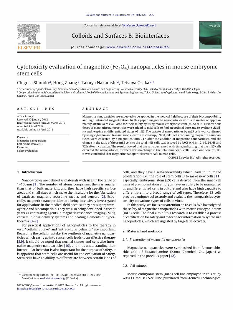
Fig. 1. TEM images of magnetite nanoparticles.
prepared by slicing with a diamond cutter, and they were observedwith TEM (Hitachi, Japan).
2.7. Cytospin observation
Cells were collected with centrifugation after co-culturewith magnetite nanoparticles for 24 h and dissolved in PBS(>107 cells/mL PBS). Next, samples were dispensed to a cuvettewith slide glass and they were centrifuged (860 rpm, 5 min) withCytospin 4 (Thermo, Japan). The cells on the slide glass were dried,and the magnetite nanoparticles were stained with potassium fer-rocyanide (Sigma). Magnetite nanoparticles turned blue 10 minlater, at which time the residues were rinsed away with PBS. Then,the cells were stained red with pararosaniline chloride solution(Sigma), and after 5 min the residues were rinsed away with PBS.The sample on the slide glass was observed with an optical micro-scope.
2.8. FACS analysis of undifferentiated state of mES
For staining, mES cells and mES cells co-cultured with mag-netite nanoparticles were collected by 0.25% trypsin–EDTA solutionand suspended in PBS with 0.1% bovine serum albumin (BSA) at aconcentration of 1 × 10⁵ cells per sample. In all experiments, cellsamples were preincubated in 0.1 mL PBS supplemented with 10 �Lnormal rabbit serum (Funakoshi, Tokyo, Japan) for 30 min to blocknonspecific binding. After a wash with PBS, cells were stained for40 min on ice with monoclonal antibodies rat antimous SSEA-1 con-jugated by phycoerythrin (PE). Stained cells were washed by PBSand analyzed by using FACSCanto II (BD, CA). PE-conjugated ratantimous SSEA-1 were purchased from BD.
3. Results and discussion
3.1. Shape and size of magnetite nanoparticles
After the synthesis of magnetite nanoparticles, their shape andsize were observed by TEM. The TEM images in Fig. 1 reveal
that magnetite nanoparticles were spherical or truncated cubic inshape, measuring 42.7 ± S.D. 16.8 nm from randomly selected 100nanoparticles. Those results were essentially same as reported inthe previous paper [12].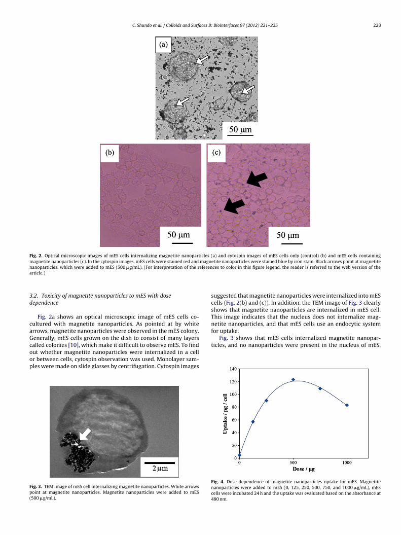
C. Shundo et al. / Colloids and Surfaces B: Biointerfaces 97 (2012) 221– 225 223
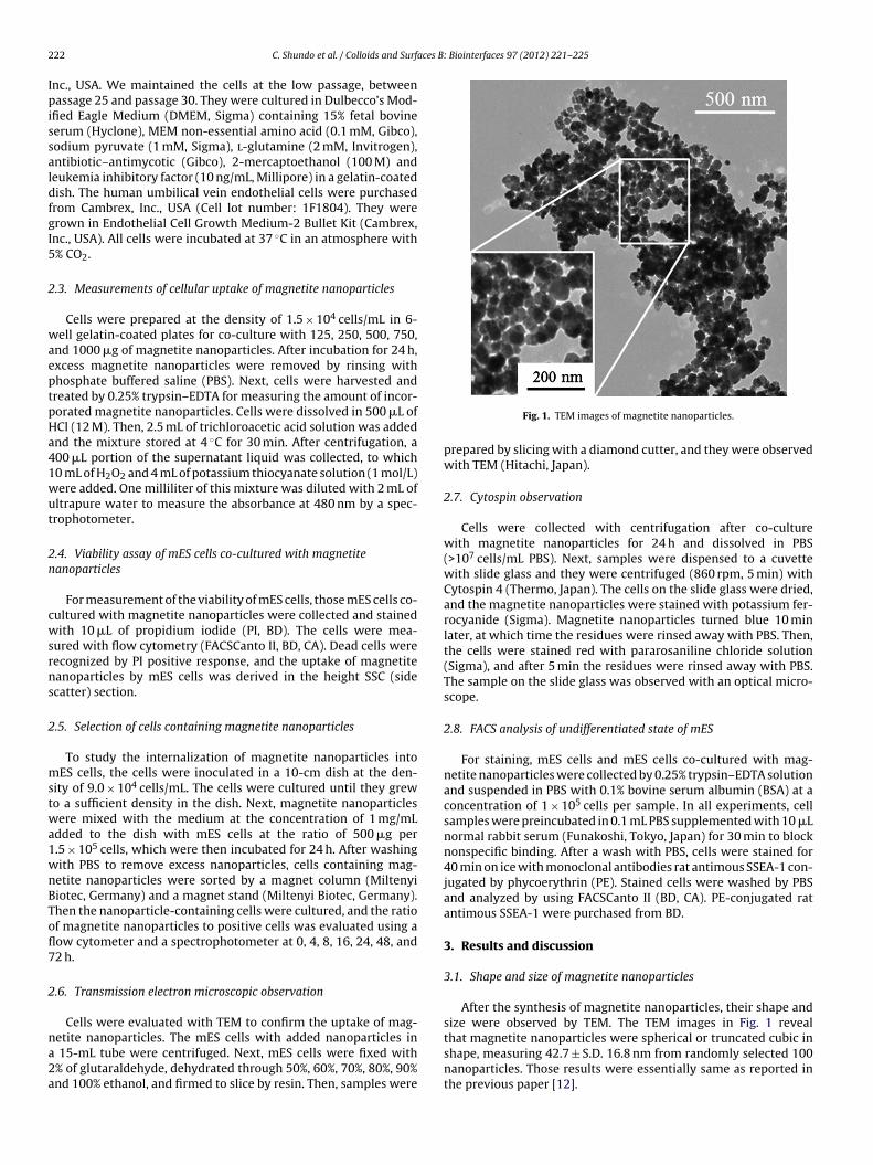
Fig. 2. Optical microscopic images of mES cells internalizing magnetite nanoparticles (a) and cytospin images of mES cells only (control) (b) and mES cells containingmagnetite nanoparticles (c). In the cytospin images, mES cells were stained red and magnetite nanoparticles were stained blue by iron stain. Black arrows point at magnetiten eferena
3d
caGcoop
Fp(
netite nanoparticles, and that mES cells use an endocytic systemfor uptake.
Fig. 3 shows that mES cells internalized magnetite nanopar-ticles, and no nanoparticles were present in the nucleus of mES.
anoparticles, which were added to mES (500 �g/mL). (For interpretation of the rrticle.)
.2. Toxicity of magnetite nanoparticles to mES with doseependence
Fig. 2a shows an optical microscopic image of mES cells co-ultured with magnetite nanoparticles. As pointed at by whiterrows, magnetite nanoparticles were observed in the mES colony.enerally, mES cells grown on the dish to consist of many layersalled colonies [10], which make it difficult to observe mES. To find
ut whether magnetite nanoparticles were internalized in a cellr between cells, cytospin observation was used. Monolayer sam-les were made on slide glasses by centrifugation. Cytospin imagesig. 3. TEM image of mES cell internalizing magnetite nanoparticles. White arrowsoint at magnetite nanoparticles. Magnetite nanoparticles were added to mES500 �g/mL).
ces to color in this figure legend, the reader is referred to the web version of the
suggested that magnetite nanoparticles were internalized into mEScells (Fig. 2(b) and (c)). In addition, the TEM image of Fig. 3 clearlyshows that magnetite nanoparticles are internalized in mES cell.This image indicates that the nucleus does not internalize mag-
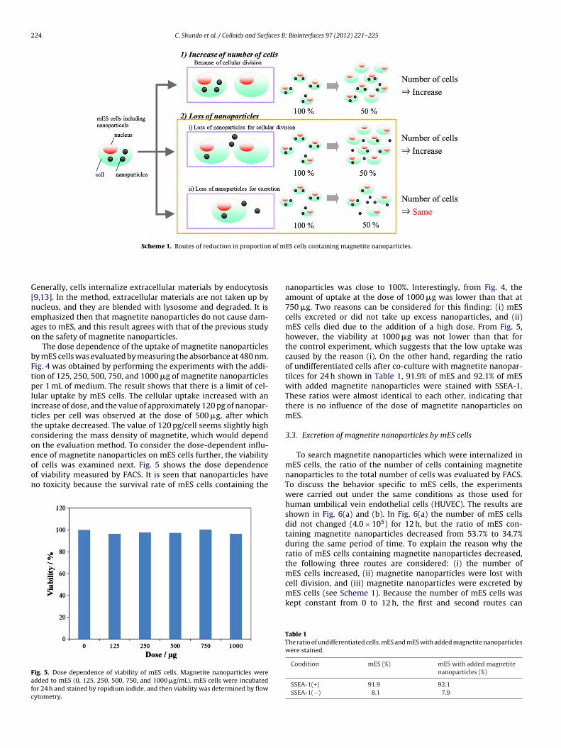
Fig. 4. Dose dependence of magnetite nanoparticles uptake for mES. Magnetitenanoparticles were added to mES (0, 125, 250, 500, 750, and 1000 �g/mL), mEScells were incubated 24 h and the uptake was evaluated based on the absorbance at480 nm.
224 C. Shundo et al. / Colloids and Surfaces B: Biointerfaces 97 (2012) 221– 225
of m
G[neao
bFtplittcoeoon
Fafc
Scheme 1. Routes of reduction in proportion
enerally, cells internalize extracellular materials by endocytosis9,13]. In the method, extracellular materials are not taken up byucleus, and they are blended with lysosome and degraded. It ismphasized then that magnetite nanoparticles do not cause dam-ges to mES, and this result agrees with that of the previous studyn the safety of magnetite nanoparticles.
The dose dependence of the uptake of magnetite nanoparticlesy mES cells was evaluated by measuring the absorbance at 480 nm.ig. 4 was obtained by performing the experiments with the addi-ion of 125, 250, 500, 750, and 1000 �g of magnetite nanoparticleser 1 mL of medium. The result shows that there is a limit of cel-
ular uptake by mES cells. The cellular uptake increased with anncrease of dose, and the value of approximately 120 pg of nanopar-icles per cell was observed at the dose of 500 �g, after whichhe uptake decreased. The value of 120 pg/cell seems slightly highonsidering the mass density of magnetite, which would dependn the evaluation method. To consider the dose-dependent influ-
nce of magnetite nanoparticles on mES cells further, the viabilityf cells was examined next. Fig. 5 shows the dose dependencef viability measured by FACS. It is seen that nanoparticles haveo toxicity because the survival rate of mES cells containing theig. 5. Dose dependence of viability of mES cells. Magnetite nanoparticles weredded to mES (0, 125, 250, 500, 750, and 1000 �g/mL). mES cells were incubatedor 24 h and stained by ropidium iodide, and then viability was determined by flowytometry.
ES cells containing magnetite nanoparticles.
nanoparticles was close to 100%. Interestingly, from Fig. 4, theamount of uptake at the dose of 1000 �g was lower than that at750 �g. Two reasons can be considered for this finding: (i) mEScells excreted or did not take up excess nanoparticles, and (ii)mES cells died due to the addition of a high dose. From Fig. 5,however, the viability at 1000 �g was not lower than that forthe control experiment, which suggests that the low uptake wascaused by the reason (i). On the other hand, regarding the ratioof undifferentiated cells after co-culture with magnetite nanopar-tilces for 24 h shown in Table 1, 91.9% of mES and 92.1% of mESwith added magnetite nanoparticles were stained with SSEA-1.These ratios were almost identical to each other, indicating thatthere is no influence of the dose of magnetite nanoparticles onmES.
3.3. Excretion of magnetite nanoparticles by mES cells
To search magnetite nanoparticles which were internalized inmES cells, the ratio of the number of cells containing magnetitenanoparticles to the total number of cells was evaluated by FACS.To discuss the behavior specific to mES cells, the experimentswere carried out under the same conditions as those used forhuman umbilical vein endothelial cells (HUVEC). The results areshown in Fig. 6(a) and (b). In Fig. 6(a) the number of mES cellsdid not changed (4.0 × 10⁵) for 12 h, but the ratio of mES con-taining magnetite nanoparticles decreased from 53.7% to 34.7%during the same period of time. To explain the reason why theratio of mES cells containing magnetite nanoparticles decreased,the following three routes are considered: (i) the number of
mES cells increased, (ii) magnetite nanoparticles were lost withcell division, and (iii) magnetite nanoparticles were excreted bymES cells (see Scheme 1). Because the number of mES cells waskept constant from 0 to 12 h, the first and second routes canTable 1The ratio of undifferentiated cells. mES and mES with added magnetite nanoparticleswere stained.
Condition mES (%) mES with added magnetitenanoparticles (%)
SSEA-1(+) 91.9 92.1SSEA-1(−) 8.1 7.9
C. Shundo et al. / Colloids and Surfaces B:
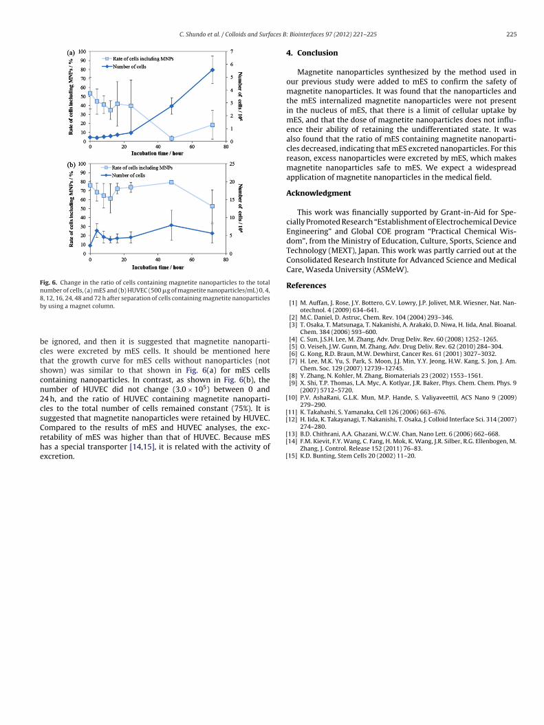
Fig. 6. Change in the ratio of cells containing magnetite nanoparticles to the totalnumber of cells, (a) mES and (b) HUVEC (500 �g of magnetite nanoparticles/mL) 0, 4,8, 12, 16, 24, 48 and 72 h after separation of cells containing magnetite nanoparticlesb
bctscn2csCrhe
[
[[
y using a magnet column.
e ignored, and then it is suggested that magnetite nanoparti-les were excreted by mES cells. It should be mentioned herehat the growth curve for mES cells without nanoparticles (nothown) was similar to that shown in Fig. 6(a) for mES cellsontaining nanoparticles. In contrast, as shown in Fig. 6(b), theumber of HUVEC did not change (3.0 × 10⁵) between 0 and4 h, and the ratio of HUVEC containing magnetite nanoparti-les to the total number of cells remained constant (75%). It isuggested that magnetite nanoparticles were retained by HUVEC.
ompared to the results of mES and HUVEC analyses, the exc-etability of mES was higher than that of HUVEC. Because mESas a special transporter [14,15], it is related with the activity ofxcretion.[[
[
Biointerfaces 97 (2012) 221– 225 225
4. Conclusion
Magnetite nanoparticles synthesized by the method used inour previous study were added to mES to confirm the safety ofmagnetite nanoparticles. It was found that the nanoparticles andthe mES internalized magnetite nanoparticles were not presentin the nucleus of mES, that there is a limit of cellular uptake bymES, and that the dose of magnetite nanoparticles does not influ-ence their ability of retaining the undifferentiated state. It wasalso found that the ratio of mES containing magnetite nanoparti-cles decreased, indicating that mES excreted nanoparticles. For thisreason, excess nanoparticles were excreted by mES, which makesmagnetite nanoparticles safe to mES. We expect a widespreadapplication of magnetite nanoparticles in the medical field.
Acknowledgment
This work was financially supported by Grant-in-Aid for Spe-cially Promoted Research “Establishment of Electrochemical DeviceEngineering” and Global COE program “Practical Chemical Wis-dom”, from the Ministry of Education, Culture, Sports, Science andTechnology (MEXT), Japan. This work was partly carried out at theConsolidated Research Institute for Advanced Science and MedicalCare, Waseda University (ASMeW).
References
[1] M. Auffan, J. Rose, J.Y. Bottero, G.V. Lowry, J.P. Jolivet, M.R. Wiesner, Nat. Nan-otechnol. 4 (2009) 634–641.
[2] M.C. Daniel, D. Astruc, Chem. Rev. 104 (2004) 293–346.[3] T. Osaka, T. Matsunaga, T. Nakanishi, A. Arakaki, D. Niwa, H. Iida, Anal. Bioanal.
Chem. 384 (2006) 593–600.[4] C. Sun, J.S.H. Lee, M. Zhang, Adv. Drug Deliv. Rev. 60 (2008) 1252–1265.[5] O. Veiseh, J.W. Gunn, M. Zhang, Adv. Drug Deliv. Rev. 62 (2010) 284–304.[6] G. Kong, R.D. Braun, M.W. Dewhirst, Cancer Res. 61 (2001) 3027–3032.[7] H. Lee, M.K. Yu, S. Park, S. Moon, J.J. Min, Y.Y. Jeong, H.W. Kang, S. Jon, J. Am.
Chem. Soc. 129 (2007) 12739–12745.[8] Y. Zhang, N. Kohler, M. Zhang, Biomaterials 23 (2002) 1553–1561.[9] X. Shi, T.P. Thomas, L.A. Myc, A. Kotlyar, J.R. Baker, Phys. Chem. Chem. Phys. 9
(2007) 5712–5720.10] P.V. AshaRani, G.L.K. Mun, M.P. Hande, S. Valiyaveettil, ACS Nano 9 (2009)
279–290.11] K. Takahashi, S. Yamanaka, Cell 126 (2006) 663–676.12] H. Iida, K. Takayanagi, T. Nakanishi, T. Osaka, J. Colloid Interface Sci. 314 (2007)
274–280.13] B.D. Chithrani, A.A. Ghazani, W.C.W. Chan, Nano Lett. 6 (2006) 662–668.14] F.M. Kievit, F.Y. Wang, C. Fang, H. Mok, K. Wang, J.R. Silber, R.G. Ellenbogen, M.
Zhang, J. Control. Release 152 (2011) 76–83.15] K.D. Bunting, Stem Cells 20 (2002) 11–20.