cytotoxicity and negligible genotoxicity of borax and borax ores to cultured mammalian cells
TRANSCRIPT

American Journal of Industrial Medicine 7:31-43 (1985)
Cytotoxicity and Negligible Genotoxicity of Borax and Borax Ores to Cultured Mammalian Cells
Joseph R. Landolph, PhD
The cytotoxicity and genotoxicity of refined borax and borax ores were studied in cultured mammalian cells. In V79 Chinese hamster cells, C3H/lOTl/2 mouse embryo fibroblasts, and diploid human foreskin fibroblasts, crude borax ore, kernite ore, and refined borax were all cytotoxic. The lowest concentrations at which cytotoxicity was observed were 0.02 mg/ml and 0.1 mg/ml for borax ore in C3H/lOT1/2 and human fibroblasts, respectively, 0.2 mg/ml for kernite ore in both cell types, and 0.1 mg/ml for refined borax in both C3H/IOT1/2 and human fibroblasts. The cytotoxicity was dose dependent above these concentrations. The concentrations of borax ore, kernite ore, and refined borax that reduced the relative plating efficiency to 50% were approximately 3.2, 1.6, and 0.8 mg/ml, respectively, in human fibroblasts and were 0.8 mglml for all three substances in C3H/lOT1/2 cells. All three borax samples were not significantly mutagenic in assays for mutation to ouabain resistance in human fibroblasts and C3H/ 10T1/2 cells and were at most only weakly mutagenic in an assay for mutation to 8- azaguanine resistance in V79 Chinese hamster cells. Refined borax did not induce neoplastic transformation in C3H/lOT1/2 cells. Crude borax ore and kernite ore induced weak transformation that was not dose-dependent and was not reproducible in another experiment. Therefore, borax and its ores are cytotoxic to mammalian cells at high (mg/ ml) concentrations and are at most weakly mutagenic but not significantly oncogenic as measured in a cell transformation assay.
Key words: borax ore, refined borax, cytotoxicity, mutation, neoplastic transformation, cells
INTRODUCTION
The safety of borax has been a concern owing to its widespread commerical use and because of occupational exposure in the borax mining and processing industries. Boric acid has been widely used in medicine and has been responsible for inadvertent poisonings in both children and adults [Goldbloom and Goldbloom, 1953; Valdes- Dapena and Arey, 1962; Schowing and Cuevas, 1975; Locksley and Farr, 19551. Accidental ingestion of boric acid in large quantities and topical medicinal application of large amounts of boric acid in infants have resulted in deaths [Goldbloom and Goldbloom, 1953; Valdes-Dapena and Arey, 19621, and there are toxic symptoms in
Departments of Microbiology and Pathology, Norris Cancer Hospital and Research Institute, Compre- hensive Cancer Center, University of Southern California School of Medicine, Los Angeles. Address reprint requests to Dr. Joseph R. Landolph, Norris Cancer Hospital and Research Institute, University of Southern California School of Medicine, Los Angeles, CA 90033. Accepted for publication August 15, 1984.
0 1985 Alan R. Liss, Inc.

32 Landolph
adults treated intravenously with large quantities of boric acid during neutron capture therapy [Locksley and Farr, 19551. Boric acid produces skeletal abnormalities and inhibits pigmentation in chicks exposed to 0.1 mg [Schowing and Cuevas, 19751 or 2.5 mg [Landauer, 1953 a,b,c] of this compound. Borax accumulates in the testes of rats fed this compound and causes depletion of germ cells and dose dependent reduction in fertility [Lee et al, 1978; Dixon et al, 19791. Borax and boric acid cause growth suppression, skin desquamation, and testicular degeneration in rats and dogs [Weir and Fisher, 19721 and sterility in rats fed these compounds at sufficiently high concentrations [Lee et al, 1978; Dixon et al, 1979; Weir and Fisher, 19721. Boric acid and borax are therefore poisonous at high concentrations. Boric acid has been reported not to mutate E. coli from streptomycin dependence to streptomycin independence [Szybalslu, 19581.
Workers in the borax industries are often exposed to high levels of particulates of borax, and there has been concern about adverse health effects associated with these exposures.
Previous studies of the toxicological properties of borax have centered on the systemic toxicity of this compound in animal feeding. Little is known of the mecha- nisms of cytotoxicity of borax. One purpose of the present study was to determine the cytotoxicity in mammalian cell culture systems of crude borax ore, intermediately refined kernite ore, and the final product, refined borax, to which workers are exposed in high concentrations.
Since many well-defined mammalian cell assays are now available to determine genotoxic effects, a further purpose of this study was to investigate whether these borax preparations possessed mutagenic or carcinogenic properties. To do this , borax samples were studied in three mammalian cell cultures designed to measure mutation at defined loci and in one mammalian cell assay to measure neoplastic transformation caused by chemical carcinogens. The assays for mutation detect the induction of ouabain resistant colonies in C3H/ 10T 1 /2 mouse embryo fibroblasts [Landolph and Heidelberger, 1979; Landolph et al, 1980al and in diploid human foreskin fibroblasts [Buchwald, 19771, which reflects base substitution mutations. These assays also detect induction of 9-azaguanine-resistant colonies, which reflects point mutations, dele- tions, and additions in V79 Chinese hamster cells [Peterson and Peterson, 1979; Peterson et al, 1979; Caskey and Kruh, 19791. The assay for neoplastic transformation detects the morphological transformation of cells to focus formation in C3H/ 10T 1 /2 mouse embryo fibroblasts [Reznikoff et al, 1973 a,b]. Foci induced in this latter assay have altered growth control properties and a high proportion of them form tumors when injected into immunosuppressed C3H mice [Reznikoff et al, 1973bl.
Abbreviations
The abbreviations used in this study were the following: MNNG, N-methyl- N'-nitro-N-nitrosoguanidine; PBS, phosphate-buffered isotonic saline, pH 7.2; MCA, 3-methylcholanthrene; (Na2B407 - 10H20), borax, sodium tetraborate decahydrate; (Na2B407 .4H20 , kernite ore, sodium tetraborate with impurities; borax ore, borax with impurities; Oua', oubain-resistant; Azg', 8-azaguanine-resistant, LDS0, concen- tration of substance that reduces the plating efficiency of treated cells to 50% of the plating efficiency of nontreated (control) cells. Note that this LDsO definition for cytotoxicity is different from, and should not be confused with, the classical LDsO used in whole-animal toxicology.

Borax Genotoxicity in Mammalian Cells 33
MATERIALS AND METHODS Cells and Cell Culture
C3H/lOT1/2 C1 8 mouse embryo fibroblasts were cultured in Basal Eagle's medium containing 10% fetal calf serum without antibiotics as described in Reznikoff et a1 [1973a] and used from passages five to fifteen. V79 Chinese hamster cells were cultured in Dulbecco's medium without phenol red, fortified with 10% dialyzed fetal calf serum without antibiotics as described in Peterson and Peterson [ 19791. Diploid human foreskin fibroblasts were prepared from freshly obtained circumcised fore- skins, cultured in modified minimal Eagle's medium containing 15% fetal calf serum without antibiotics, as described in Silinskas et a1 [1981], and used up to passage fifteen. All cell lines were routinely verified to be negative for mycoplasma by growth on mycoplasma agar [Hayflick, 19561. Heat-inactivated fetal calf serum, dialyzed fetal calf serum, and powdered tissue cultured medium were purchased from Grand Island Biological Company, Grand Island, NY. Dialyzed fetal calf serum was heat- inactivated at 56°C for 30 min before use. Tissue culture flasks and dishes were purchased from Corning Glass Works, Corning, NY.
Preparation of Borax Samples for Toxicity Testing
Borax samples were covered by a clean cloth, crushed to a fine powder with a hammer, and weighed on a Metler balance. The powered samples were then dissolved in phosphate-buffered saline (PBS) at 37 "C overnight with stirring. A concentration of 3.2 mg/ml of borax ore, kernite ore, or refined borax was approximately the highest concentration that could be obtained in PBS. The pH of the solution was then adjusted to the pH of blood, 7.4, and the solutions were filter-sterilized through 0.2 pM Nalgene filters and used in the assays as described below.
Cytotoxicity Assays
Cytotoxicity assays were conducted by measuring reduction in plating efficiency of treated cells. C3H/lOT1/2 cells were seeded at 200 cells per 60-mm dish; V79 cells, at 100 cells per 60-mm dish; and diploid human foreskin fibroblasts, at 200 cells per 60-mm dish. Twenty-four hr later, 0.05 ml of borax solution was added to each of five 60-mm dishes for 48 hr, and medium was then removed, cells were rinsed with isotonic saline, fixed with methanol, and stained with Giemsa, and colonies containing greater than 20 cells were scored by microscope. Penicillin G at 100 unitdm1 and streptomycin sulfate at 100 pg/ml were added to all cytotoxicity, mutagenesis, and transformation assays upon initiation of experiments and with each medium change.
Mutation Assays
Assays for mutation to ouabain resistance (Oua') in C3H/lOT1/2 cells were performed according to methods we developed earlier [Landolph and Heidelberger, 1979; Landolph et al, 1980al that quantitatively detect base-substitution mutations [Landolph et al, 1980b; Landolph and Jones, 1982; Gehly et al, 1982; Landolph and Fournier, 19821 Twenty 100-mm dishes were each seeded with lo5 cells, and treated 24 hr later for 48 hr with 0.1 ml of sterile solutions of borax samples dissolved in PBS. Medium was then removed and replaced with medium without test samples. After two days' further expression-time, cells were trypsinized; 20 100-rnm dishes
34 Landolph
were each seeded with 2 X lo5 cells to select mutants, five 60-mm dishes were each seeded with 200 cells, and another five dishes were seeded with 2,000 cells to determine plating efficiencies of mutagenized cells. Plating efficiency determinations were fixed, stained, and counted 10 days later. One, 7, and 13 days after reseeding mutagenized cells into 100-mm dishes, medium was removed and replaced with filter- sterilized medium containing 3 mM ouabain. After 16 days of selection in ouabain, Oua' colonies were fixed with methanol, stained with Giemsa, and scored by micro- scope. Assays for mutation to Oua' in diploid foreskin fibroblasts were performed similarly to those in C3H/lOT1/2 cells, except that cells were selected in 1 pM ouabain [Buchwald, 19771.
Assays to detect mutation to 8-azaguanine resistance (Azg') in V79 cells were performed similarly to previously published protocols [Peterson et al, 19791, which eliminate metabolic cooperation and score stringently and quantitatively for Axg' mutants [Peterson and Peterson 19791. Five 100-mm dishes were seeded with lo5 cells each and treated 24 hr later for 48 hr with 0.1 ml of sterile solutions of borax in PBS. Medium was then removed and replaced with medium without test compound. After 1 day of expression-time, cells were trypsinized and reseeded into ten 100-mm dishes at lo5 cells per dish. Following a further five days' expression-time, during which cells were maintained in logarithmic growth phase by passaging, cells were trypsinized and reseeded into 20 100-mm dishes at lo5 cells per dish to select 8-aza- guanine (Azg') resistant mutants and into five 60-mm dishes at 100 cells per dish and into five 60-mm dishes at 1,000 cells per dish to determine the plating efficiency of mutagenized cells. Plating efficiency determinations were fixed and stained and colonies containing greater than 20 cells were counted by microscope 8 to 10 days after seeding. Twenty-four hr after reseeding cells into 100-mm dishes to select mutants, 0.1 ml of a filter-sterilized solution of 4 mg/ml of 8-azaguanine in PBS was added to each dish (final concentration of 40 pg/ml 8-azaguanine in the medium). Five days later, medium was removed and replaced with fresh 8-azaguanine-contain- ing medium, and after a further 5 to 7 days, visible Azg' colonies were fixed, stained, and scored by microscope.
In all mutation assays, 1 pgiml of the known mutagen MNNG was added to each of ten 100-mm dishes as a positive control to ensure that mutation assays were functioning properly. Cytotoxicity assays were conducted to determine the level of cytotoxicity caused by each assayed concentration of test compound.
In all assays, mutation frequencies were calculated as follows:
Mutation frequency =
Total number of mutant colonies/total number of dishes Plating efficiency of reseeded cells x number of cells reseeded/dish
If no actual mutant colonies were observed, then we assumed one mutant colony was present and used the above calculation to determine an upper bound to the possible mutant frequency. In this case, the mutant frequency is preceded by a " < " (less than) sign.
Assay to Detect Neoplastic Transformation These assays were performed as described in Reznikoff et a1 [1973b] with
modifications. For each concentration of sample tested, 20 60-mm dishes containing

Borax Genotoxicity in Mammalian Cells 35
2,000 cells each were plated and treated 24 hr later for 48 hr with 0.05 ml of a filter- sterilized solution of borax sample in PBS [Landolph and Heidelberger, 19791. One p g / d of the known carcinogen 3-methylcholanthrene (MCA) was added separately to each of 20 60-mm dishes as a positive control. The medium was then removed and replaced with fresh medium without test substance and then renewed each week for 6 weeks. Cytotoxicity assays were conducted in parallel with transformation assays to determine the cytotoxicity each concentration of borax caused. At the end of 6 weeks, cells were fixed with methanol and stained with Giemsa. Type I1 foci, which are darkly staining areas of multilayered cells, and type I11 foci, which are darkly staining areas of multilayered cells with cells in crisscrossed assays at the periphery of the focus, were scored as morphologically transformed by microscope as previously described [Reznikoff et al, 1973bl.
Chemicals
Ouabain octahydrate, 8-azaguanine (grade II), and N-methyl-N’-nitro’-N-nitroso- guanidine were purchased from Sigma Chemical Company, St. Louis, MO. 3-Methy- lcholanthrene was purchased from Aldrich Chemical Company, Milkwaukee, WI. Borax ore, kernite ore, and refined borax were obtained through the courtesy of Dr. David Garabrant, USC Division of Occupational Health, who obtained them from the United States Borax and Chemical Corporation. Penicillin G and streptomycin sulfate were purchased from Grand Island Biological Company, Grand Island, NY.
Statistical Analysis
In mutational assays, the significance of the differences between borax-treated and control (PBS-treated) mutation frequencies was determined by use of the Poisson distribution test method [Daniel, 19741 and the extensive Poisson distribution tables [Pearson and Hartley, 19761.
RESULTS Cytotoxicity of Borax Samples to C3H/lOT1/2 and Human Diploid Foreskin Fibroblasts
The cytotoxicity of borax and borax ores to C3H/lOT1/2 cells is shown in Table I. Little or no cytotoxicity occurred with either borax ore, kernite, or refined borax at concentrations up to 0.02 mg/ml. At concentrations of 0.2 mg/ml and above, there were dose dependent cytotoxic effects for all three substances. The LD50 values were approximately 0.8 mg/ml for all three substances. At 3.2 mg/ml, the highest concen- tration tested, the highest cytotoxicity was observed: 71 % cell kill, 67% cell kill, and 65% cell kill for borax ore, kernite ore, and refined borax, respectively.
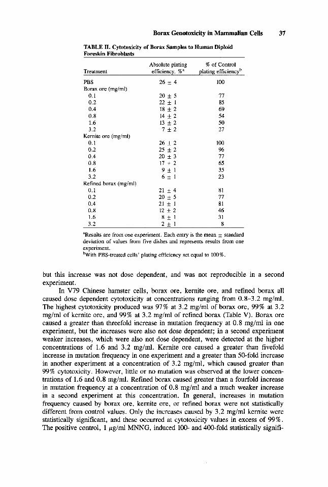
Borax ore, kernite ore, and refined borax also caused dose dependent cytotox- icity to diploid human foreskin fibroblasts in the concentration range 0.1-3.2 mg/ml. At a concentration of 3.2 mg/ml, borax ore caused 73% cytotoxicity; kernite ore, 77 % cytotoxicity; and refined borax, 92 % cytotoxicity to human foreskin fibroblasts (Table 11).
There were difficulties in dissolving the samples at concentrations much higher than 3.2 mg/ml in phosphate-buffered saline (PBS), possibly because of the buffering action of PBS; therefore, cytotoxic concentrations ranging from 0.4 to 3.2 mg/ml of the borax samples were next used to study their mutagenic and transforming potentials

36 Landolph
TABLE I. Cytotoxicity of Borax Samples to C3HllOT112 Cells
Relative plating % of control,b mean + standard efficiency deviation
Concentration, mg/mla Borax ore Kernite ore Refined borax
1 0 - ~ 92 f 2 (2)' 115 f 5 (1) 113 * lO(1) 10-6 93 f 3 (2) 83 f 3 (2) 87 * 7 (3) 10P 106 f 3 (2) 101 * 2 (2) 110 * I0 (3) 10-4 80 5 20 (2) 96 k 6 (2) 97 f 7 (3) 10-3 140 f 20 (2) 80 f 30 (2) loo * 10 (3) 10-2 70 k 10 (2) 117 f 12 (2) 120 f 10 (3) 2 x 10⁻² 91 * 3 (2) 97 f 9 (1) loo f 3 (1) 0.1 80 f 20 (4) 96 If: 6 (3) 82 f 14 (4) 0.2 88 k 0(1) 90 f 7 (1) 85 k 4 (1) 0.3 84 k 9 (1) 71 f 9(1) 57 k 6(1) 0.4 58 5 2 (2) 60 & 2 (2) 75 + 13 (2) 0.8 50 k 10 (3) 59 * 17 (4) 51 f 13 (2) 1 .o 50 * 10 (1) 70 f 20 (1) 31 f 2 (1) 1.6 32 * 8 (3) 39 f 13 (3) 32 f 10 (4) 3.2 29 k 9 (2) 33 f 11 (2) 35 * 20 (2)
'Cells were treated with test compounds for 48 hr. bPlating efficiency of treated cells/plating efficiency of control (PBS-treated) cells X 100%, averaged over the number of experiments conducted. The absolute plating efficiencies of control cells ranged from 28-40% in these experiments. 'Number in parentheses is the number of experiments conducted.
in C3H/lOT1/2 cells, in diploid human foreskin fibroblasts, and in V79 Chinese hamster cells.
Mutagenesis Assays In C3H/10T1/2 cells borax samples at concentrations of 0.4, 0.8, and 1.6 mg/
ml caused from 23-60% cytotoxicity, and the positive control MNNG induced a greater than 243-fold increase in mutation frequency, indicating that the mutation assay was functioning properly (Table 111). Borax ore and kernite ore at either 0.4, 0.8, or 1.6 m g / d did not induce measurable mutation of these cells to Oua'. Two Oua' colonies were observed on one dish of cells treated with 0.8 m g / d but not with 0.4 or 1.6 mg/ml of refined borax, and the induced mutation frequency of 0.8 mg/ml was not statistically significantly different from control values.
In two mutation experiments in diploid human foreskin fibroblasts, borax ore, kernite ore, and refined borax at concentrations of 0.8, 1.6, and 3.2 mg/ml caused dose dependent cytotoxicity (Table IV). The highest concentrations of samples tested, 3.2 mg/ml for borax ore, 3.2 m g / d for kernite ore, and 3.2 mg/ml for refined borax, caused an average of 51, 81, and 91 % cytotoxicity, respectively, in the two experi- ments. The positive control, 1 p g / d of MNNG, caused reproducible 32- and 43-fold increases in the Oua' mutant frequency, indicating that the mutation assays were functioning correctly. Kernite ore and refined borax at concentrations of 0.8, 1.6 and 3.2 m g / d and borax ore at concentrations of 1.6 and 3.2 m g / d did not cause any measurable increases in the Oua' mutant frequency in two experiments. Cells treated with borax ore at a concentration of 0.8 m g / d in one experiment showed an eightfold increase in the Oua' mutation frequency over the spontaneous mutation frequency,
Borax Genotoxicity in Mammalian Cells 37
TABLE 11. Cytotoxicity of Borax Samples to Human Diploid Foreskin Fibroblasts
Treatment efficiency, %" plating efficiencyb
PBS 26 f 4 100 Borax ore (mg/ml)
0.1 20 * 5 77 0.2 22 f 1 85 0.4 18 * 2 69 0.8 14 f 2 54 1.6 13 f 2 50 3.2 7 + 2 27
0.1 26 f 2 100 0.2 25 f 2 96 0.4 20 f 3 77 0.8 17 f 2 65 1.6 9 * 1 35 3.2 6 * 1 23
0.1 21 f 4 81 0.2 20 f 5 77 0.4 21 f 1 81 0.8 12 f 2 46 1.6 8 * 1 31 3.2 2 f 1 8
Absolute plating % of Control
Kernite ore (mg/ml)
Refined borax (mg/ml)
"Results are from one experiment. Each entry is the mean & standard deviation of values from five dishes and represents results from one experiment. bWith PBS-treated cells' plating efficiency set equal to 100%.
but this increase was not dose dependent, and was not reproducible in a second experiment.
In V79 Chinese hamster cells, borax ore, kernite ore, and refined borax all caused dose dependent cytotoxicity at concentrations ranging from 0.8-3.2 mg/ml. The highest cytotoxicity produced was 97% at 3.2 mg/ml of borax ore, 99% at 3.2 mg/ml of kernite ore, and 99% at 3.2 mg/ml of refined borax (Table V). Borax ore caused a greater than threefold increase in mutation frequency at 0.8 mg/ml in one experiment, but the increases were also not dose dependent; in a second experiment weaker increases, which were also not dose dependent, were detected at the higher concentrations of 1.6 and 3.2 mg/ml. Kernite ore caused a greater than fivefold increase in mutation frequency in one experiment and a greater than 50-fold increase in another experiment at a concentration of 3.2 mg/ml, which caused greater than 99% cytotoxicity. However, little or no mutation was observed at the lower concen- trations of 1.6 and 0.8 mg/ml. Refined borax caused greater than a fourfold increase in mutation frequency at a concentration of 0.8 mg/ml and a much weaker increase in a second experiment at this concentration. In general, increases in mutation frequency caused by borax ore, kernite ore, or refined borax were not statistically different from control values. Only the increases caused by 3.2 mg/ml kernite were statistically significant, and these occurred at cytotoxicity values in excess of 99%. The positive control, 1 p g / d MNNG, induced 100- and 400-fold statistically signifi-

38 Landolph
TABLE 111. Assay of Borax Fractions for Cytotoxicity and Mutagenesis in C3HllOT112 Cells
Treatment efficiencv. % mutant freouencv Plating lo6 x Oua'
PBS 26 f 1 < lb 6.7 pM MNNG 7.6 f 0.8 (29) 243' Borax ore (mg/ml)
0.4 16 4 (61) <3 0.8 14 f 4 (54) < 3 1.6 1 1 * 2 (42) <4
0.4 15 f 3 (58) < 1 0.8 13 & 4 (SO) < 1 1.6 14 * 4 (54) < 1
0.4 20 * 4 (77) < 1 0.8 17 4 (65) 2d 1.6 1 1 & S(42) < 1
Kernite ore (mg/ml)
Refined borax (mg/ml)
'Percent of control (PBS-treated) plating efficiency, with control plating efficiency taken as 100 % . bNo colonies were observed on any dishes with " < " signs in front of the numbers; this is purely a statistical calculation in which one assumes a colony is present and therefore calculates an upper bound to the mutation frequency. 'For MNNG- vs PBS-treated samples, the mean mutational frequencies are highly significantly different at p < 0.000001 using the Poisson distribution test. d T ~ o colonies were observed on one dish in this experiment. The mean mutational frequency was not significantly different from the PBS-treated value, p = 0.184.
cant increases in the frequency of Azg' mutants in these two experiments, indicating that the mutation assays functioned properly.
Assays for Neoplastic Transformation in C3H/lOT1/2 Cells We next studied the ability of these borax samples to oncogenically transform
C3H/10T1/2 cells. In both experiments, the positive control, 1 p g / d of the known carcinogen 3-methylcholanthrene, induced significant numbers of morphologically transformed Type I1 and Type I11 foci, and the negative controls had no foci, indicating the assays were functioning properly (Table VI) . The tested concentrations of borax ore, kernite ore, and refined borax caused an average of up to 73, 67, and 65 % cytotoxicity, respectively, in these experiments.
In both experiments, concentrations of 0.8, 1.6, and 3.2 m g / d of refined borax did not induce any Type I1 or Type 111 foci. In the first experiment, one Type I1 focus was induced at a concentration of 3.2 mg/ml of borax ore and one Type 111 focus was induced at a concentration of 0.8 mg/ml of kernite ore. Concentrations of 0.8 m g / d and 1.6 mg/ml of borax ore, and 1.6 m g / d and 3.2 m g / d of kernite ore did not induce foci. In a second experiment, no transformed foci were observed in cells treated with borax ore at 0.8, 1.6, and 3.2 mglml and kernite ore at 0.8, 1.6, and 3.2 mg/ml .

Borax Genotoxicity in Mammalian Cells 39
TABLE N. Assay of Borax Samples for Cytotoxicity and Mutation in Human Diploid Foreskin Fibroblasts
Plating efficiency, % lo6 x Oua' mutant frequency Treatment Experiment 1 Experiment 2 Experiment 1 Experiment 2
1 p g / d MNNG 12 f 2 (36) 4 f l(13) 43b 32b Borax ore (mg/ml)
PBS 33 rf: 4(100)a 31 f 4(100) < 1 < 1
0.8 28 f 2 (85) 28 k 4 (90) 8' < 1 1.6 26 f 2 (79) 27 k 3 (87) < I < 1 3.2 17 f 3 (52) 14 f 3 (45) < I < 3
0.8 27 & 4 (82) 21 + 5 (68) < I < 1 1.6 21 f 3 (64) 13 rf: 2 (42) < 1 < 2 3.2 6 l(18) 6 * 2 (19) < 2 <4
0.8 20 * 4 (61) 22 * 3 (71) < 2 < 1 1.6 14 f 3 (42) I2 k 2 (39) < 3 <2
Kernite ore (mg/ml)
Refined borax (mg/ml)
3.2 3 * 2 (12) 3 * l(10) < 2 < 8
aValue in parentheses is the plating efficiency of control cells/plating efficiency of treated cells x 100%. For each entry, value is the mean rf: standard deviation of values from five dishes. bFor MNNG-treated vs PBS-treated samples, the mean mutation frequencies are highly significantly different at p < 0.000001 using the Poisson distribution test for both experiments 1 and 2. 'For 0.8 m g / d of borax ore-treated vs PBS samples, the mean mutation frequencies are highly significantly different, p = 0.000009; in experiment 1.
A modified transformation assay that is reported to be more sensitive than the standard transformation assay has recently been developed [Nesnow et al, 19821. In this assay, cells are treated 5 days after seeding instead of 1 day after seeding. Using this assay, there was one spontaneous type I11 focus in the control (PBS-treated group), one type I11 focus in the 0.1 m g / d kernite-treated group, and one type I1 focus in the 0.05 mg/mf anhydrous borax-treated group (Table VII).
Hence, these three experiments demonstrate that refined borax at the tested concentrations has no detectable oncogenic potential in this cell transformation assay, and borax ore and kernite ore have marginal, if any, oncogenic activity.
DISCUSSION
Consistent with previous studies in whole animals [Weir and Fisher, 1972; Lee et al, 19781, borax ore, kernite, and pure refined borax were all cytotoxic in cultured C3H/lOT1/2 mouse cells, in cultured diploid human foreskin fibroblasts, and in cultured V79 Chinese hamster cells at high (mg/ml) concentrations. Interestingly, the range of toxic concentrations was similar in whole-animal feeding experiments [Weir and Fisher, 19721 and in these cultured cell systems. The acute LD50 value of 4.5 g/ kg in whole-animal feeding experiments in Sprague-Dawley rats Weir and Fisher, 19721 would be approximately 4 mg/g or 4 mg/ml if a homogeneous distribution of borax in the animal were assumed and if a density equal to that of water was assumed for the animal. This value is within a factor of 5 of the 0.8 m g / d value we found for the borax samples in cultured C3H/lOT1/2 cells and diploid human foreskin fibro- blasts. These samples were slightly more toxic to V79 cells.

40 Landolph
TABLE V. Cytotoxicity to and Mutation of V79 Cells by Borax Samples
Plating efficiency, % lo6 x Azg' mutant frequencya Treatment Experiment 1 Experiment 2 Experiment 1 Experiment 2
PBS 130b f 10 31 f 7 (100) 0.40 (0/20)d <0.7 (0120)
1 p g / d MNNG 59 rt 6 (45) 10 ~f: 3 (32) 159".' (306/20) 75' (123/20) Borax ore (mg/ml)
<0.6 (0.20)
(WC
0.8 100 rt 16 (77) 7 rt 2 (23) 1.3' (3/20) 1.6 34 rt 7 (26) 0.2 (0.6) 0.73 (1120) 0.6 (1120) 3.2 7 rt I(S) 0.2 (0.6) 0.8 (1120)
I .6 0.2 (0.1) 0.2 (0.6) 0.4 (1/20) <0.8 (0/20) 3.2 0.2 (0.1) 0.2 (0.6) 20.8' (1/20) 3. i 4 ( 4 ~ 0 )
0.4 5 f l(16) <o.s (0/20) 0.8 5 f 2 (4) 0.2 (0.6) 1 .62 (3/20) 0.6 (1/20) 1.6 <0.2 (<0.1) 0.2 (0.6) 0.63 (1120) <0.9 (0.20) 3.2 0.2 (0.6) <0.7 (0/20)
"The significance of the differences between mutation frequencies at any concentration of tested sample and the PBS-treated cells was: 1 , p < O.OOOOO4, highly significantly different; 2, p = 0.21, not significantly different; 3 , p = 0.43, not significantly different; 4, p = 0.028, significantly different. bThe medium change upon removal of borax-containing samples after 48 hr of treatment occasionally causes considerable colony splitting in this assay and forms plating efficiencies of greater than 100%, which are artifactually high. In this experiment, therefore, the plating efficiencies of treated relative to PBS-treated samples are the most significant data. "Value in parentheses is the % of PBS-treated plating efficiencies, with PBS-treated values set equal to 100%. dValue in parentheses is the number of Azg' colonies observed per total number of dishes selected in Azg. ' .2 .3 ,4~ee footnote a.
Kernite ore (mgiml) 0.8 20 f 4 (15) 0.2 (0.6) <0.2 (0/20) <0.6 (0/20)
Refined borax (mg/ml)
The assays used here detect a wide spectrum of mutagenic events. Assays for mutation to Oua' in C3H/lOT1/2 cells [Landolph and Heidelberger, 1979; Landolph et al, 1980 a; Landolph and Jones, 1982; Landolph and Fournier, 19831, in other rodent cells [Baker et al, 19741, in human foreskin fibroblasts [Buchwald, 19771, and in human HeLa cells [Robbins and Baker, 19771 detect base substitution mutations at the (Na,K) adenosinetriphosphate locus such that a mutated enzyme is produced that probably lacks a ouabain-binding site. The assay for mutation to Azg' in V79 cells detects base substitution, deletion, addition, and frameshift mutations at the hypoxan- thine-guanine phosphoribosyl-transferase locus such that a mutated enzyme with reduced activity is produced [Peterson and Peterson, 1979; Landolph and Jones, 1982; Peterson et al, 1979; reviewed in Caskey and Kruh, 19791. Therefore, assay of borax samples in these test systems should detect any substantial mutagenic properties possessed by these compounds. The assay for neoplastic transformation in C3H/ 10T112 cells detects production of Type II and Type 111 foci comprising those cells that have lost the property of contact inhibition of cell division. Since 50% of the type I1 foci and 80% of the type I11 foci form tumors when injected into syngeneic immunosuppressed C3H mice, this assay detects oncogenic transformation of C3H/ 1OT1/2 cells [Reznikoff et al, 1973bl.
The experiments conducted here suggest that borax ore, kernite ore, and refined borax and their associated impurities possess at most weak but not serious mutagenic

Borax Genotoxicity in Mammalian Cells 41
TABLE VI. Assay for Cytotoxicity and Transformation of C3H/lOT1/2 Cells by Borax Samples
Transformation Total dishes
Plating efficiency, Type I1 Dishes with Type I11 Dishes with Treatmenta % foci type I1 foci foci type 111 foci
Experiment 1 PBS 20 f 2 (loo) 0120 1 pglml MCA 16 rt 1 (80) 14/20 Borax ore 0.8 (mglml) 1 1 f 2 (55) 0113 1.6 6 k 2 (30) 0120 3.2 4.0 f 0.5 (20) 1/20 Kernite ore 0.8 (mglml) 12 f 2 (60) 0120 1.6 6 k 1 (30) 0120 3.2 5 f 1 (25) 0120 Refined borax 0.8 (mglml) 8 k 2 (40) 0120 1.6 5 1 (25) 0119 3.2 3 f 1 (15) 0119
PBS 23 f 3 (100) 0119 1 pglml MCA 22 f 3 (96) 9/18 Borax ore 0.8 (mglml) 24 5 3 (104) 0115 1.6 20 f 3 (87) 0115 3.2 8.5 f 2 (37) 0119 Kernite ore 0.8 (mglml) 19 If: 3 (83) 0118 1.6 25 f 3 (108) 0115 3.2 9.5 f 1.5 (41) 0111 Refined borax 0.8 (mglml) 24 f 4 (104) 0120 1.6 9 f 3 (39) 0120 3.2 13 f 2 (56) 0119
'In both assays, cells were exposed to compounds for 48 hr.
Experiment 2a
0120 8/20 0113 0120 1 120 0120 0120 0120 0120 0119 0119
0119 7/18 0115 0115 0119 0118 0115 011 1 0120 0120 0119
0120 15/20 0113 0120 0120 1/20 0120 0120 0120 0119 0119
0119 16118 0115 0115 0119 0118 0115 011 1 0120 0120 0119
0120 1 1/20 0113 0120 0120 1/20 0120 0120 0120 0119 0119
0119 8/18 0115 0115 0119 0118 0115 011 1 0120 0120 0119
properties. Refined borax is of particular interest because it is widely used in commercial products and in industrial processes. This material does not cause neo- plastic transformation in C3H/lOT 1/2 cells or base substitution mutations to Oua' in either C3H/lOT1/2 or human diploid foreskin fibroblasts and causes only small levels of mutation to Azg' in V79 Chinese hamster cells that are not statistically significant.
Similarly, kernite ore and borax ore possess negligible or at most no more than weak cell transformation activity and were not mutagenic in C3H/lOT1/2 cells. Kernite ore was not mutagenic in human diploid foreskin fibroblasts, and borax ore only produced mutants at one low concentration in human fibroblasts in one out of two experiments. This effect was not statistically significant. Hence, these studies suggest that borax ore, kernite ore, and refined borax do not possess significant mutagenic or oncogenic properties as measured in the assays used here when com- pared to the strong mutagen MNNG and the strong carcinogen 3-methylcholanthrene, which were used as positive controls in our assays. The most important biological property of the borax samples was their cytotoxicity at high (mg/ml) concentrations.
ACKNOWLEDGMENTS
I would like to thank Miss Leslie Eva Hamilton and Mr. Stephen Johnston for skilled and dedicated technical assistance during the course of this work; Dr. Dave

42 Landolph
TABLE VII. Cytotoxicity of Borax and Borax Ores in a Modified Transformation Assay
Plating efficiency, % , of cells Total dishes treated with On: Type I1 Dishes with Type 111 Dishes with
Treatment Day la Day 5b foci type I1 foci foci type I11 foci
PBS 35 f 2 (100) 24 k l(100) 0/20 0/20 1 /20 1/20 1 Fg/ml MCA 26 f 2 (74) 25 k 3 (104) 29/20 14/20 20/20 12/20 Borax ore (mg/ml)
0.16 32 * 2 (91) 26 k 3 (108) 0/20 0/20 0/20 0120
0.64 30 k 2 (86) 22 2 (92) 0/20 0/20 0120 0/20
0.8 28 f 3 (80) 21 k l(88) 0117 0117 0/17 0/17 1.6 25 & l(71) 18 k l(75) 0/19 0/19 0/19 0/19 3.2 22 k 2 (63) 18 * 2 (75) 0/20 0/20 1 /20 1/20
1.6 27 k 2 (77) 20 rt 4 (89) 1/19 1/19 0/19 0/19 3.2 25 k 2 (71) 18 f 2 (75) 0/20 0120 0/20 0/20
0.32 28 k 3 (80) 25 k 2 (104) 0116 0/16 0/16 0/16
Kernite (mg/ml)
Anhydrous borax (mg/ml) 0.8 28 f l(80) 20 * 3 (80) 0/18 0/18 0/18 0/18
aCells were treated 24 hr after seeding for 48 hr. bCells were treated 5 days after seeding for 48 hr.
Garabrant, Dr. Charles Heidelberger, and Dr. Paul Billings for helpful discussions; and Dr. Garabrant and Dr. Billings for helpful criticisms of the manuscript. I would also like to acknowledge helpful consultation on statistical analysis with Dr. Stan k e n and Dr. Leslie Bernstein. This work was supported by grant SIG-3 from the American Cancer Society and by a grant from the R.J. Reynolds Company.
REFERENCES
Baker RM, Brunette DM, Mankovitz R, Thompson LH, Whitmore GF, Siminovitch L, Till JE (1974):
Buchwald M (1977): Mutagenesis at the ouabain resistance locus in human diploid fibroblasts. Mut Res
Caskey CT, Kruh GD (1979): The HPRT locus. Cell 16: 1-9. Daniel WW (1974): “Biostatistics: A Foundation for Analysis in the Health Sciences.” New York: John
Wiley and Sons, Inc., pp 66-68. Dixon RL, Sherins RJ, Lee IP (1979): Assessment of environmental factors affecting male fertility.
Environ Health Perspec 3053-68. Gehly EB, Landolph JR, Heidelberger C, Nagasawa H, Little JB (1982): Induction of cytotoxicity,
mutation, cytogenetic changes, and neoplastic transformation by benzo(a)pyrene and derivatives in C3H/lOT1/2 clone 8 mouse fibroblasts. Cancer Res 42:1966-1875.
Goldbloom RB, Goldbloom A (1953): Boric acid poisoning. J Pediat 43:631-643. Hayflick L (1956): Tissue cultures and mycoplasma. Tex Rep Biol Med 23:285-303. Landauer W (1953a): Genetic and environmental factors in the teratogenic effects of boric acid on
chicken embryos. Genetics 38:216-228. Landauer W (1953b): Complex formation and chemical specificity of boric acid in production of chicken
embryo malformations. Proc SOC Exp Biol Med 82:633. Landauer W (1953): Abnormality of down pigmentation associated with experimentally produced
skeletal defects of chicks. Proc Natl Acad Sci USA. 39:54-58. Landolph JR, Heidelberger C (1979): Chemical carcinogens produce mutations to ouabain resistance in
transformable C3H/lOT1/2 C1 8 mouse fibroblasts. Proc Natl Acad Sci USA 76:930-934.
Ouabain-resistant mutants of mouse and hamster cells in culture. Cell 1:9-21.
44 :40 1-4 12.

Borax Genotoxicity in Mammalian Cells 43
Landolph JR, Telfer N, Heidelberger C (1980a): Further evidence that ouabain-resistant variants induced by chemical carcinogens in transformable C3H/lOTl/2 mouse fibroblasts are mutants. Muta Res
Landolph JR, Bhatt RS, Telfer N, Heidelberger C (1980b): Comparison of adriamycin- and ouabain- induced cytotoxicity and inhibition of %ubidium transport in wild-type and ouabain-resistant C3H/lOT1/2 mouse fibroblasts. Cancer Res 40:4581-4588.
Landolph JR, Jones PA (1982): Mutagenicity of 5-azacytidine and related nucleosides in C3H/lOT1/2 and clone 8 and V79 cells. Cancer Res 42:817-832.
Landolph JR, Fournier REK (1983): Microcell-mediated transfer of carcinogen-induced ouabain resis- tance from C3H/lOT1/2 C18 mouse fibroblasts to human cells. Mutat Res 107:447-463.
Lee IP, Sherins RJ, Dixon RL (1978): Evidence for induction of germinal aplasia in male rats by environmental exposure to boron. Toxicol Appl Pharmacol45:577-590.
Locksley HB, Farr LE (1955): The tolerance of large doses of sodium borate intravenously by patients receiving neutron capture therapy. J Pharmacol Exp Ther 114:484489.
Nesnow S, Garland H, Curtis G (1982): Improved transformation of C3H/lOT1/2 C18 cells by direct and indirect-acting carcinogens. Carcinogenesis (London) 3:377-380.
Pearson ES, and Hartley HO (eds) (1976): “Biometrika Tables for Statisticians.” London: Biometrika Trust, Vol. 1.
Peterson AR, Peterson H (1979): Facilitation by pyrimidine deoxyribonucleosides and hypoxanthine of mutagenic and cytotoxic effects of monofunctional alkylating agents in Chinese hamster cells. Mutat Res 61:319-331.
Peterson AR, Peterson H, Heidelberger C (1979): Oncogenesis, mutagenesis, DNA damage, and cytotoxicity in cultured mammalian cells treated with alkylating agents. Cancer Res 39: 131-138.
Resnikoff CA, Brankow DW, Heidelberger C (1973a): Establishment and characterization of a cloned line of C3H/lOT1/2 mouse embryo cells sensitive to post-confluence inhibition of cell division. Cancer Res 33:3231-3238.
Reznikoff CA, Bertram JS, Brankow DW, Heidelberger C (1973b): Quantitative and qualitative studies of chemical transformation of cloned C3H mouse embryo cells sensitive to postconfluence inhibition of cell division. Cancer Res 33:3239-3249.
Robbins AR, Baker RM (1977): (Na,K) ATPase activity in membrane preparations of ouabain-resistant HeLa cells. Biochemistry 165163-5168.
Schowing J, Cuevas P (1975): Teratogenic effects of boric acid upon the chick. Teratology 12:334. Silinskas KC, Kateley SA, Tower JE, Maher VM, McCormick JJ (1981): Induction of anchorage
independent growth in human fibroblasts by propane sultone. Cancer Res 41: 1620-1627. Szybalski W (1958): Special microbiological systems 11. Observations on chemical mutagenesis in
microorganisms. Ann NY Acad Sci 76:475-489. Valdes-Dapena MA, Arey JB (1962): Boric acid poisoning. J Pediat 61:531-546. Weir RJ, Fisher RS (1972): Toxicologic studies on borax and boric acid. Toxicol Appl Pharmacol
72~295-310.
23~351-364.