cytology sel
DESCRIPTION
Clinical Case StudyA 46-year-old inebriated male is brought to the emergency room by paramedics after his girlfriendcalled 911 reporting that he was experiencing a seizure. Over the next hour, the patientbecomes increasingly somnolent (sleepy). While the emergency room staff initiates gastriclavage for presumed drug ingestion, you seek more medical history from the man’s girlfriend.She reports that she found him amidst several empty bottles of antifreeze. Upon hearing this,you immediately order a 10% ethanol solution to be given to the man intravenously.How do enzymes promote metabolism of chemicals? What is meant by “competitive inhibition”and how does this relate to therapy for ethylene glycol poisoning? As you read thischapter, pay attention to other enzyme reactions and recognize that these are important targetsfor therapeutic medications.INTRODUCTION TO CYTOLOGYThe cell is the fundamental structural and functional unit of thebody. Although cells vary widely in size and shape, they havebasic structural similarities, and all cells metabolize to stay alive.Objective 1 Define the terms cell, metabolism, and cytology.Objective 2 Using examples, explain how cells differ fromone another and how the structure of a cell determines itsfunction.TRANSCRIPT
Van De Graaff: Human Anatomy, Sixth Edition
III. Microscopic Structure of the Body
3. Cytology © The McGraw−Hill Companies, 2001
Cytology
Clinical Case StudyA 46-year-old inebriated male is brought to the emergency room by paramedics after his girl-friend called 911 reporting that he was experiencing a seizure. Over the next hour, the patientbecomes increasingly somnolent (sleepy). While the emergency room staff initiates gastriclavage for presumed drug ingestion, you seek more medical history from the man’s girlfriend.She reports that she found him amidst several empty bottles of antifreeze. Upon hearing this,you immediately order a 10% ethanol solution to be given to the man intravenously.
How do enzymes promote metabolism of chemicals? What is meant by “competitive in-hibition” and how does this relate to therapy for ethylene glycol poisoning? As you read thischapter, pay attention to other enzyme reactions and recognize that these are important targetsfor therapeutic medications.
Introduction to Cytology 49Cellular Chemistry 50Cellular Structure 52Cell Cycle 65
CLINICAL CONSIDERATIONS 70
Clinical Case Study Answer 74Chapter Summary 74Review Activities 75
3
FIGURE: Drugs work at the cellular levelwhere a delicate chemical balance ismaintained. A thorough knowledge of cellularstructure is imperative to understand cellularphysiology and drug therapy.
Van De Graaff: Human Anatomy, Sixth Edition
III. Microscopic Structure of the Body
3. Cytology © The McGraw−Hill Companies, 2001
are as yet unknown. Scientists are seeking why and how the bodyages. The answers will come only through a better understandingof cellular structure and function.
Advancements in microscopy have revolutionized the sci-ence of cytology. In a new process called microtomogra-
phy, the capabil it ies of electron microscopy are combined with those of CT scanning to produce high-magnification, three-dimensional, microtomographic images of living cells. With thistechnology, living cells can be observed as they move, grow, anddivide. The clinical applications are immense, as scientists canobserve the response of diseased cells (including cancer cells) tovarious drug treatments.
Cellular DiversityIt is amazing that from a single cell, the fertilized egg, hundredsof kinds of cells arise, producing the estimated 60 trillion to 100 trillion cells that make up an adult human. Cells varygreatly in size and shape. The smallest cells are visible onlythrough a high-powered microscope. Even the largest, an eggcell (ovum), is barely visible to the unaided eye. The sizes ofcells are measured in micrometers (µm)—one micrometerequals 1/1,000th of a millimeter. Using this basis of comparison,an ovum is about 140 µm in diameter and a red blood cell isabout 7.5 µm in diameter. The most common type of whiteblood cell varies in size from 10 to 12 µm in diameter. Althoughstill microscopic, some cells can be extremely long. A nerve cell(neuron), for example, may extend the entire length of a limband be over a meter long.
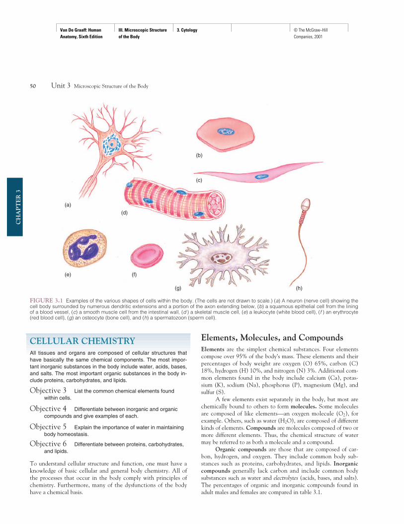
Although a typical diagram of a cell depicts it as roundor cube-shaped, the shapes of cells are actually highly vari-able. They can be flat, oval, elongate, stellate, columnar, andso on (fig. 3.1). The shape of a cell is frequently an indicationof its function. A disc-shaped red blood cell is adapted totransport oxygen. Thin, flattened cells may be bound togetherto form selectively permeable membranes. An irregularlyshaped cell, such as a neuron, has a tremendous ratio of sur-face area to volume, which is ideal for receiving and transmit-ting stimuli.
The surfaces of some cells are smooth, so that substancespass over them easily. Other cells have distinct depressions andelevations on their cell membranes to facilitate absorption. Somecell surfaces support such structures as cilia, flagella, and gelati-nous coats, which assist movement and provide adhesion. Re-gardless of the sizes and shapes of cells, they all have structuralmodifications that serve functional purposes.
Knowledge Check1. Why is the cell considered the basic structural and func-
tional unit of the body?2. What conditions are necessary for metabolism to occur?3. Give some examples of structural modifications that allow
cells to perform specific functions?
Chapter 3 Cytology 49
CH
AP
TE
R 3
INTRODUCTION TO CYTOLOGYThe cell is the fundamental structural and functional unit of thebody. Although cells vary widely in size and shape, they havebasic structural similarities, and all cells metabolize to stay alive.
Objective 1 Define the terms cell, metabolism, and cytology.
Objective 2 Using examples, explain how cells differ fromone another and how the structure of a cell determines itsfunction.
Cells as Functional UnitsHuman anatomy is concerned with the structure of the humanbody and the relationship of its parts. The body is a masterpieceof organization for which the cell provides the basis. For this rea-son, the cell is called the functional unit. As discussed in chapter 2,cellular organization forms tissues, whose organization in turnforms the organs, which in turn form systems. If the organs andsystems are to function properly, cells must function properly.Cellular function is referred to as metabolism. In order for cellsto remain alive and metabolize, certain requirements must bemet. Each cell must have access to nutrients and oxygen and beable to eliminate wastes. In addition, a constant, protective envi-ronment must be maintained. All of these requirements areachieved through organization.
Cells were first observed more than 300 years ago by theEnglish scientist Robert Hooke. Using his crude microscope toexamine a thin slice of cork, he saw a network of cell walls andboxlike cavities. He called them “little boxes or cells,” after thebarren cubicles of a monastery. As better microscopes were devel-oped, the intriguing architectural details of cellular structure weregradually revealed. The improved lenses resulted in a series of de-velopments that culminated in the formulation of the cell theoryin 1838 and 1839 by two German biologists, Matthias Schleidenand Theodor Schwann. This theory states that all living organ-isms are composed of one or more cells and that the cell is thebasic unit of structure for all organisms. The work of Schleidenand Schwann laid the groundwork for a new science called cytol-ogy, which is concerned with the structure and function of cells.
A knowledge of the cellular level of organization is impor-tant for understanding the basic body processes of cellular respi-ration, protein synthesis, mitosis, and meiosis. An understandingof cellular structure gives meaning to the concept of tissue,organ, and system levels of functional body organization. Fur-thermore, many dysfunctions and diseases of the body originatein the cells. Although cellular structure and function have beeninvestigated for many years, we still have much to learn aboutcells. The etiologies, or causes, of a number of complex diseases
metabolism: Gk. metabole, change
cytology: L. cella, small room; Gk. logos, study of
etiology: L. aitia, cause; Gk. logos, study of
Van De Graaff: Human Anatomy, Sixth Edition
III. Microscopic Structure of the Body
3. Cytology © The McGraw−Hill Companies, 2001
CELLULAR CHEMISTRYAll tissues and organs are composed of cellular structures thathave basically the same chemical components. The most impor-tant inorganic substances in the body include water, acids, bases,and salts. The most important organic substances in the body in-clude proteins, carbohydrates, and lipids.
Objective 3 List the common chemical elements foundwithin cells.
Objective 4 Differentiate between inorganic and organiccompounds and give examples of each.
Objective 5 Explain the importance of water in maintainingbody homeostasis.
Objective 6 Differentiate between proteins, carbohydrates,and lipids.
To understand cellular structure and function, one must have aknowledge of basic cellular and general body chemistry. All ofthe processes that occur in the body comply with principles ofchemistry. Furthermore, many of the dysfunctions of the bodyhave a chemical basis.
Elements, Molecules, and CompoundsElements are the simplest chemical substances. Four elementscompose over 95% of the body’s mass. These elements and theirpercentages of body weight are oxygen (O) 65%, carbon (C)18%, hydrogen (H) 10%, and nitrogen (N) 3%. Additional com-mon elements found in the body include calcium (Ca), potas-sium (K), sodium (Na), phosphorus (P), magnesium (Mg), andsulfur (S).
A few elements exist separately in the body, but most arechemically bound to others to form molecules. Some moleculesare composed of like elements—an oxygen molecule (O2), forexample. Others, such as water (H2O), are composed of differentkinds of elements. Compounds are molecules composed of two ormore different elements. Thus, the chemical structure of watermay be referred to as both a molecule and a compound.
Organic compounds are those that are composed of car-bon, hydrogen, and oxygen. They include common body sub-stances such as proteins, carbohydrates, and lipids. Inorganiccompounds generally lack carbon and include common bodysubstances such as water and electrolytes (acids, bases, and salts).The percentages of organic and inorganic compounds found inadult males and females are compared in table 3.1.
50 Unit 3 Microscopic Structure of the Body
CH
AP
TE
R 3
FIGURE 3.1 Examples of the various shapes of cells within the body. (The cells are not drawn to scale.) (a) A neuron (nerve cell) showing thecell body surrounded by numerous dendritic extensions and a portion of the axon extending below, (b) a squamous epithelial cell from the liningof a blood vessel, (c) a smooth muscle cell from the intestinal wall, (d ) a skeletal muscle cell, (e) a leukocyte (white blood cell), (f ) an erythrocyte(red blood cell), (g) an osteocyte (bone cell), and (h) a spermatozoon (sperm cell).
(a)
(g)
(c)
(h)
(e) (f)
(b)
(d)
Van De Graaff: Human Anatomy, Sixth Edition
III. Microscopic Structure of the Body
3. Cytology © The McGraw−Hill Companies, 2001
The disparity of proteins, lipids, and water in adult malesand females can be explained by relative amounts of sex hor-mones. Male sex hormones promote the development of pro-teins, especially in skeletal muscle tissue. Female sex hormonespromote the retention of fats, which are an important food re-source for nursing a child. Because proteins contain more waterthan lipids, there is a disparity between the percent of body fluidsbetween males and females.
WaterWater is by far the most abundant compound found within cellsand in the extracellular environment. Water generally occurswithin the body as a homogeneous mixture of two or more com-pounds called a solution. In this condition, the water is the sol-vent, or the liquid portion of the solution, and the solutes aresubstances dissolved in the solution. Water is an almost universalsolvent, meaning that almost all chemical compounds dissolve init. In addition, it is also used to transport many solutes throughthe cell membrane of a cell or from one part of the cell to an-other. Water is also important in maintaining a constant cellulartemperature, and thus a constant body temperature, because itabsorbs and releases heat slowly. Evaporative cooling (sweating)through the skin also involves water. Another function of wateris as a reactant in the breakdown (hydrolysis) of food material indigestion.
Dehydration is a condition in which fluid loss exceeds fluid in-take, with a resultant decrease in the volume of intracellular
and extracellular fluids. Rapid dehydration through vomiting, diar-rhea, or excessive sweating can lead to serious medical problems byimpairing cellular function. Infants are especially vulnerable becausetheir fluid volume is so small. They can die from dehydration resultingfrom diarrhea within a matter of hours.
ElectrolytesElectrolytes are inorganic compounds that break down into ionswhen dissolved in water, forming a solution capable of conduct-ing electricity. An electrolyte is classified according to the ions ityields when dissolved in water. The three classes of electrolytesare acids, bases, and salts, all of which are important for normalcellular function. The functions of ions include the control ofwater movement through cells and the maintenance of normalacid-base (pH) balance. Ions are also essential for nerve andmuscle function, and some ions serve as cofactors that areneeded for optimal activity of enzymes. Symptoms of electrolyteimbalances range from muscle cramps and brittle bones to comaand cardiac arrest. The three kinds of electrolytes are summa-rized in table 3.2.
ProteinsProteins are nitrogen-containing organic compounds composedof amino acid subunits. An amino acid is an organic compoundthat contains an amino group (—NH2) and a carboxyl group (—COOH). There are 20 different types of amino acids that cancontribute to a given protein. This variety allows each type ofprotein to be constructed to function in very specific ways.
Proteins are the most abundant of the organic compounds.They may exist by themselves or be conjugated (joined) withother compounds; for example, with nucleic acids (RNA orDNA) to form nucleoproteins, with carbohydrates to form glyco-proteins, or with lipids to form lipoproteins.
Proteins may be categorized according to their role in thebody as structural or functional. Structural proteins contributesignificantly to the structure of different tissues. Examples in-clude collagen in connective tissue and keratin in the epidermis of
Chapter 3 Cytology 51
CH
AP
TE
R 3
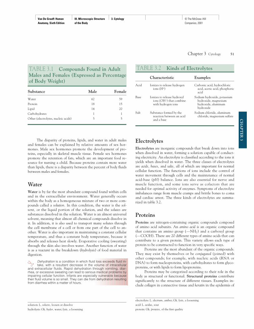
TABLE 3.1 Compounds Found in AdultMales and Females (Expressed as Percentageof Body Weight)
Substance Male Female
Water 62 59
Protein 18 15
Lipid 14 20
Carbohydrates 1 1
Other (electrolytes, nucleic acids) 5 5
solution: L. solvere, loosen or dissolve
hydrolysis: Gk. hydor, water; lysis, a loosening
TABLE 3.2 Kinds of Electrolytes
Characteristic Examples
Acid Ionizes to release hydrogen Carbonic acid, hydrochloric ions (H+) acid, acetic acid, phosphoric
acid
Base Ionizes to release hydroxyl Sodium hydroxide, potassium ions (OH–) that combine hydroxide, magnesium with hydrogen ions hydroxide, aluminum
hydroxide
Salt Substance formed by the Sodium chloride, aluminum reaction between an acid chloride, magnesium sulfateand a base
electrolyte: L. electrum, amber; Gk. lysis, a loosening
acid: L. acidus, sour
protein: Gk. proteios, of the first quality
Van De Graaff: Human Anatomy, Sixth Edition
III. Microscopic Structure of the Body
3. Cytology © The McGraw−Hill Companies, 2001
the skin. Functional proteins assume a more active role in thebody, exerting some form of control of metabolism. Examples in-clude enzymes and antibodies. Many hormones belong to a special-ized group of messenger and regulator proteins produced byendocrine glands. Cellular growth, repair, and division dependon the availability of functional proteins. Proteins, under certainconditions, may even be metabolized to supply cellular energy.
CarbohydratesCarbohydrates are organic compounds that contain carbon, hy-drogen, and oxygen, with a 2:1 ratio of hydrogen to oxygen. Car-bohydrates include monosaccharides, or simple sugars,disaccharides, or double sugars, and polysaccharides, or long-chained sugars. Carbohydrates are the body’s most readily avail-able energy source and also may be used as a fuel reserve.Excessive carbohydrate intake is converted to glycogen (animalstarch) or to fat for storage in adipose tissue.
If a person is deprived of food, the body uses the glycogenand fat reserves first and then metabolizes the protein within
the cells. The gradual destruction of cellular protein accounts for thelethargy, extreme emaciation, and ultimate death of starvation victims.
LipidsLipids are a third group of important organic compounds found incells. They are insoluble in water and include both fats and fat-related substances, such as phospholipids and cholesterol. Fats are im-portant in building cell parts and supplying metabolic energy. Theyalso protect and insulate various parts of the body. Phospholipids andprotein molecules make up the cell membrane and play an impor-tant role in regulating which substances enter or leave a cell.
Lipids, like carbohydrates, are composed of carbon, hydro-gen, and oxygen. Lipids, however, contain a smaller proportionof oxygen than do carbohydrates.
The locations and functions of inorganic and organic sub-stances within cells are summarized in table 3.3.
Knowledge Check4. List the four most abundant elements in the body and state
their relative percentages of body weight.5. Define molecule and compound. What are the two kinds of
compounds that exist in the body? On what basis are theydistinguished?
6. List some of the functions of water relative to cells and de-fine solvent and solute.
7. Discuss the importance of electrolytes in maintaininghomeostasis within cells.
8. Define protein and describe how proteins function within cells.Explain how proteins differ from carbohydrates and lipids.
CELLULAR STRUCTUREThe cell membrane separates the interior of a cell from the extra-cellular environment. The passage of substances into and out ofthe cell is regulated by the cell membrane. Most of the metabolicactivities of a cell occur within the cytoplasmic organelles. The nu-cleus functions in protein synthesis and cell reproduction.
Objective 7 Describe the components of a cell.
Objective 8 Describe the composition and structure of thecell membrane and relate its structure to the functions itperforms.
Objective 9 Distinguish between passive and activetransport and describe the different ways in which each isaccomplished.
52 Unit 3 Microscopic Structure of the Body
CH
AP
TE
R 3
hormone: Gk. hormon, setting in motion
lipid: Gk. lipos, fat
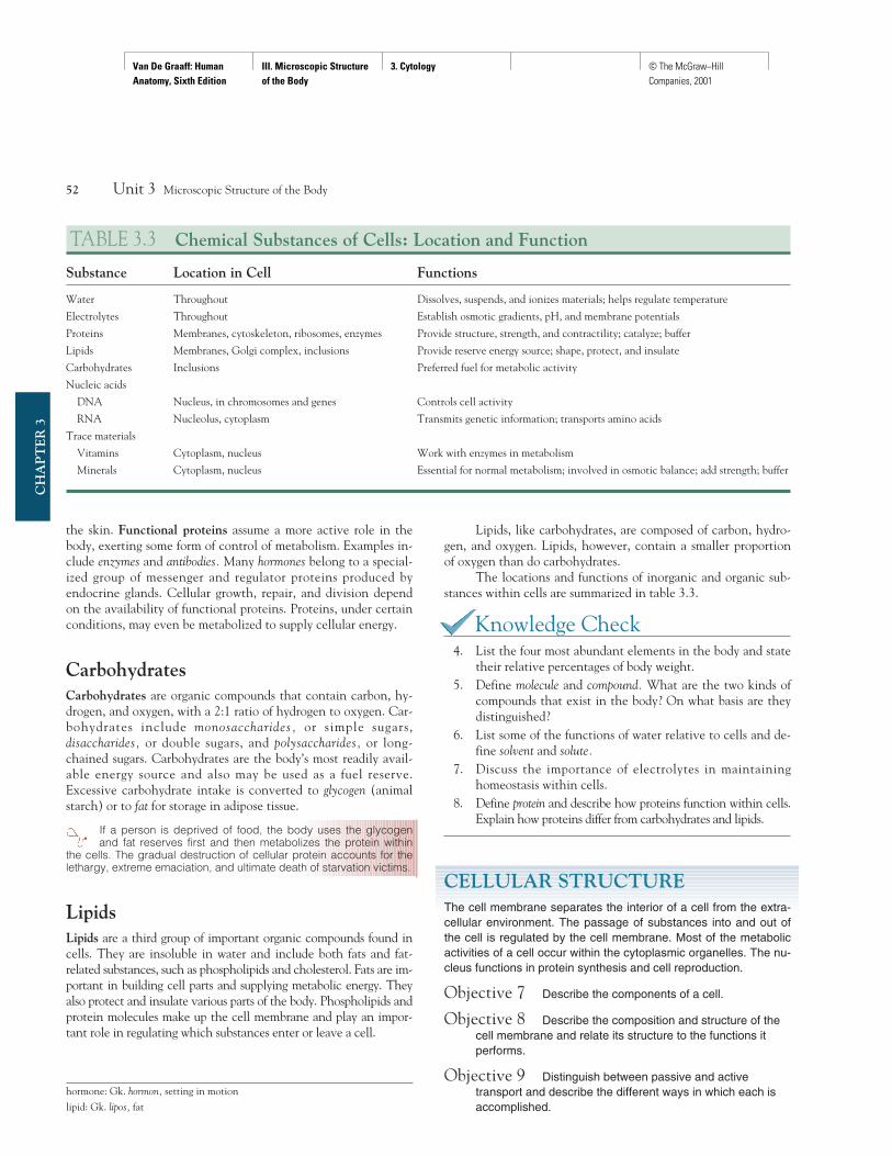
TABLE 3.3 Chemical Substances of Cells: Location and Function
Substance Location in Cell Functions
Water Throughout Dissolves, suspends, and ionizes materials; helps regulate temperature
Electrolytes Throughout Establish osmotic gradients, pH, and membrane potentials
Proteins Membranes, cytoskeleton, ribosomes, enzymes Provide structure, strength, and contractility; catalyze; buffer
Lipids Membranes, Golgi complex, inclusions Provide reserve energy source; shape, protect, and insulate
Carbohydrates Inclusions Preferred fuel for metabolic activity
Nucleic acids
DNA Nucleus, in chromosomes and genes Controls cell activity
RNA Nucleolus, cytoplasm Transmits genetic information; transports amino acids
Trace materials
Vitamins Cytoplasm, nucleus Work with enzymes in metabolism
Minerals Cytoplasm, nucleus Essential for normal metabolism; involved in osmotic balance; add strength; buffer
Van De Graaff: Human Anatomy, Sixth Edition
III. Microscopic Structure of the Body
3. Cytology © The McGraw−Hill Companies, 2001
Objective 10 Describe the structure and function of theendoplasmic reticulum, ribosomes, Golgi complex,lysosomes, and mitochondria.
Objective 11 Describe the structure and function of the nucleus.
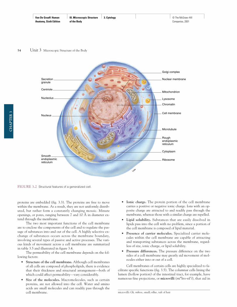
As the basic functional unit of the body, the cell is a highly orga-nized molecular factory. As previously discussed, cells come in agreat variety of shapes and sizes. This variation, which is also ap-parent in subcellular structures (organelles), reflects the diversityof function of different cells in the body. All cells, however, havecertain features in common—a cell membrane, for example, andmost of the other structures listed in table 3.4. Thus, although noone cell can be considered “typical,” the general structure of cellscan be indicated by a single illustration (fig. 3.2).
For descriptive purposes, a cell can be divided into threeprincipal parts:
1. Cell (plasma) membrane. The selectively permeable cellmembrane gives form to the cell. It controls the passage ofmolecules into and out of the cell and separates the cell’sinternal structures from the extracellular environment.
2. Cytoplasm and organelles. The cytoplasm (si′to-plaz″em)is the cellular material between the nucleus and the cellmembrane. Organelles (or″ga-nelz′) are the specializedstructures suspended within the cytoplasm of the cell thatperform specific functions.
3. Nucleus. The nucleus (noo′kle-us) is the large spheroidor oval body usually located near the center of the cell. Itcontains the DNA, or genetic material, that directs the ac-tivities of the cell. Within the nucleus, one or more densebodies called nucleoli (singular, nucleolus) may be seen.The nucleolus contains the subunits for ribosomes, thestructures that serve as sites for protein synthesis.
Cell MembraneThe extremely thin cell (plasma) membrane is composed pri-marily of phospholipid and protein molecules. Its thicknessranges from 65 to 100 angstroms (Å); that is, it is less than a mil-lionth of an inch thick. The structure of the cell membrane isnot fully understood, but most cytologists believe that it consistsof a double layer of phospholipids in which larger globular
Chapter 3 Cytology 53
CH
AP
TE
R 3
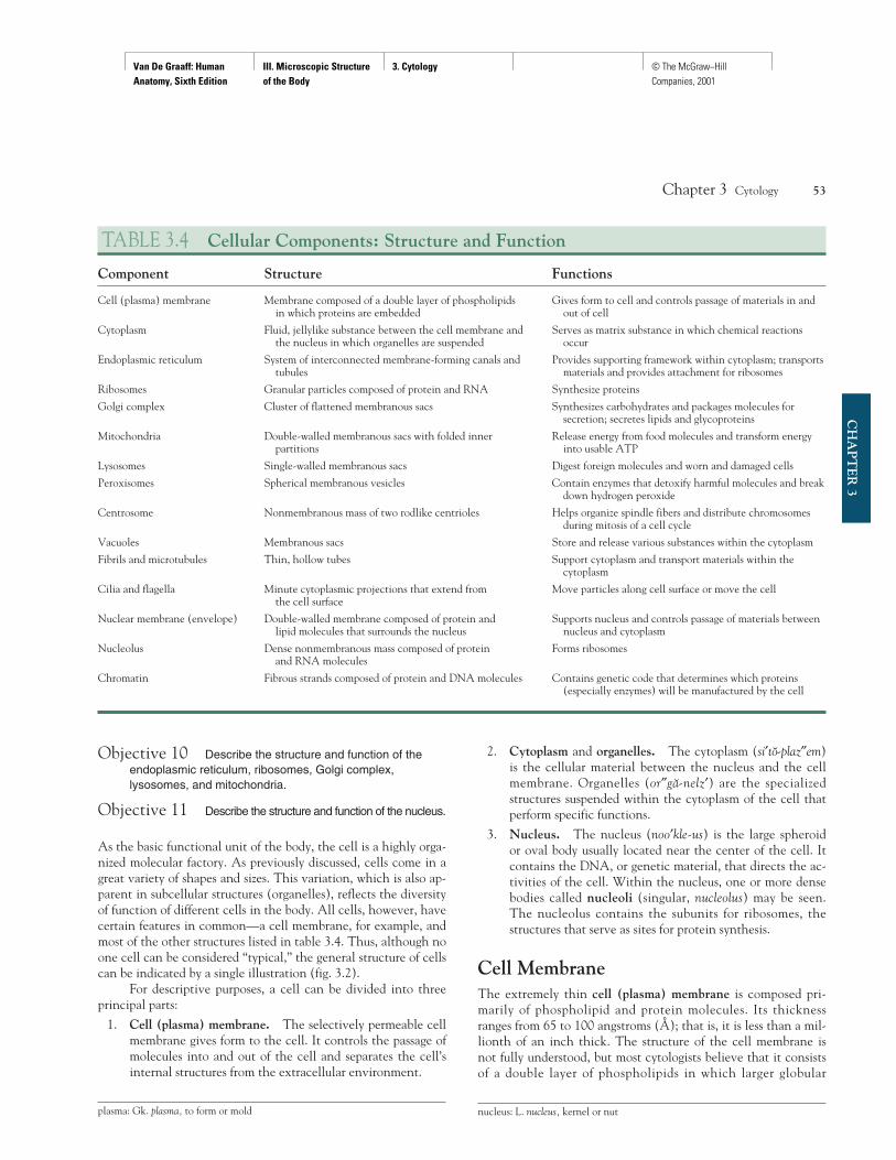
TABLE 3.4 Cellular Components: Structure and Function
Component Structure Functions
Cell (plasma) membrane Membrane composed of a double layer of phospholipids Gives form to cell and controls passage of materials in and in which proteins are embedded out of cell
Cytoplasm Fluid, jellylike substance between the cell membrane and Serves as matrix substance in which chemical reactions the nucleus in which organelles are suspended occur
Endoplasmic reticulum System of interconnected membrane-forming canals and Provides supporting framework within cytoplasm; transports tubules materials and provides attachment for ribosomes
Ribosomes Granular particles composed of protein and RNA Synthesize proteins
Golgi complex Cluster of flattened membranous sacs Synthesizes carbohydrates and packages molecules for secretion; secretes lipids and glycoproteins
Mitochondria Double-walled membranous sacs with folded inner Release energy from food molecules and transform energy partitions into usable ATP
Lysosomes Single-walled membranous sacs Digest foreign molecules and worn and damaged cells
Peroxisomes Spherical membranous vesicles Contain enzymes that detoxify harmful molecules and break down hydrogen peroxide
Centrosome Nonmembranous mass of two rodlike centrioles Helps organize spindle fibers and distribute chromosomes during mitosis of a cell cycle
Vacuoles Membranous sacs Store and release various substances within the cytoplasm
Fibrils and microtubules Thin, hollow tubes Support cytoplasm and transport materials within the cytoplasm
Cilia and flagella Minute cytoplasmic projections that extend from Move particles along cell surface or move the cellthe cell surface
Nuclear membrane (envelope) Double-walled membrane composed of protein and Supports nucleus and controls passage of materials between lipid molecules that surrounds the nucleus nucleus and cytoplasm
Nucleolus Dense nonmembranous mass composed of protein Forms ribosomesand RNA molecules
Chromatin Fibrous strands composed of protein and DNA molecules Contains genetic code that determines which proteins (especially enzymes) will be manufactured by the cell
plasma: Gk. plasma, to form or mold nucleus: L. nucleus, kernel or nut
Van De Graaff: Human Anatomy, Sixth Edition
III. Microscopic Structure of the Body
3. Cytology © The McGraw−Hill Companies, 2001
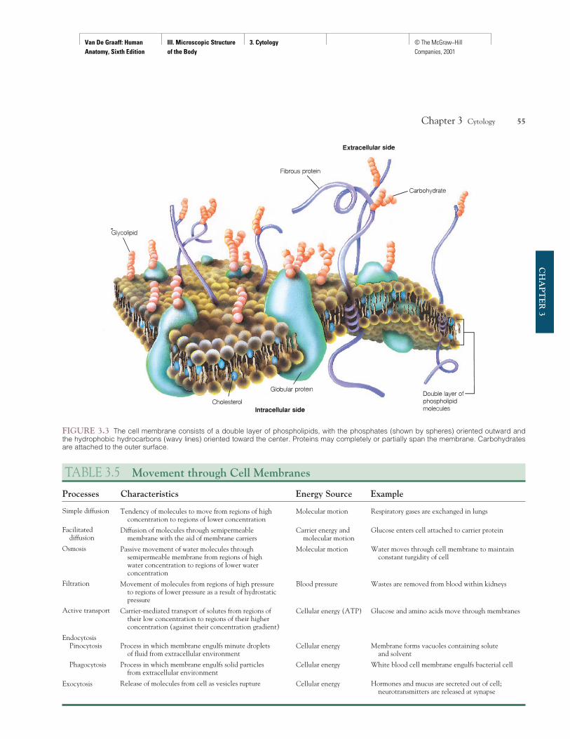
proteins are embedded (fig. 3.3). The proteins are free to movewithin the membrane. As a result, they are not uniformly distrib-uted, but rather form a constantly changing mosaic. Minuteopenings, or pores, ranging between 7 and 10 Å in diameter ex-tend through the membrane.
The two most important functions of the cell membraneare to enclose the components of the cell and to regulate the pas-sage of substances into and out of the cell. A highly selective ex-change of substances occurs across the membrane boundary,involving several types of passive and active processes. The vari-ous kinds of movement across a cell membrane are summarizedin table 3.5 and illustrated in figure 3.4.
The permeability of the cell membrane depends on the fol-lowing factors:
• Structure of the cell membrane. Although cell membranesof all cells are composed of phospholipids, there is evidencethat their thickness and structural arrangement—both ofwhich could affect permeability—vary considerably.
• Size of the molecules. Macromolecules, such as certainproteins, are not allowed into the cell. Water and aminoacids are small molecules and can readily pass through thecell membrane.
• Ionic charge. The protein portion of the cell membranecarries a positive or negative ionic charge. Ions with an op-posite charge are attracted to and readily pass through themembrane, whereas those with a similar charge are repelled.
• Lipid solubility. Substances that are easily dissolved inlipids pass into the cell with no problem, since a portion ofthe cell membrane is composed of lipid material.
• Presence of carrier molecules. Specialized carrier mole-cules within the cell membrane are capable of attractingand transporting substances across the membrane, regard-less of size, ionic charge, or lipid solubility.
• Pressure differences. The pressure difference on the twosides of a cell membrane may greatly aid movement of mol-ecules either into or out of a cell.
Cell membranes of certain cells are highly specialized to fa-cilitate specific functions (fig. 3.5). The columnar cells lining thelumen (hollow portion) of the intestinal tract, for example, havenumerous fine projections, or microvilli (mi″kro-vil ′i), that aid in
54 Unit 3 Microscopic Structure of the Body
CH
AP
TE
R 3
Secretiongranule
Golgi complex
Nuclear membrane
Mitochondrion
Lysosome
Chromatin
Cell membrane
Microtubule
Roughendoplasmicreticulum
Cytoplasm
Ribosome
Lew
Centriole
Nucleolus
Nucleus
Smoothendoplasmicreticulum
FIGURE 3.2 Structural features of a generalized cell.
microvilli: Gk. mikros, small; villus, tuft of hair
Van De Graaff: Human Anatomy, Sixth Edition
III. Microscopic Structure of the Body
3. Cytology © The McGraw−Hill Companies, 2001
Chapter 3 Cytology 55
CH
AP
TE
R 3
FIGURE 3.3 The cell membrane consists of a double layer of phospholipids, with the phosphates (shown by spheres) oriented outward andthe hydrophobic hydrocarbons (wavy lines) oriented toward the center. Proteins may completely or partially span the membrane. Carbohydratesare attached to the outer surface.
TABLE 3.5 Movement through Cell Membranes
Processes Characteristics Energy Source Example
Simple diffusion
Facilitated diffusion
Osmosis
Filtration
Active transport
EndocytosisPinocytosis
Phagocytosis
Exocytosis
Tendency of molecules to move from regions of high concentration to regions of lower concentration
Diffusion of molecules through semipermeable membrane with the aid of membrane carriers
Passive movement of water molecules through semipermeable membrane from regions of high water concentration to regions of lower water concentration
Movement of molecules from regions of high pressure to regions of lower pressure as a result of hydrostatic pressure
Carrier-mediated transport of solutes from regions of their low concentration to regions of their higher concentration (against their concentration gradient)
Process in which membrane engulfs minute droplets of fluid from extracellular environment
Process in which membrane engulfs solid particles from extracellular environment
Release of molecules from cell as vesicles rupture
Molecular motion
Carrier energy and molecular motion
Molecular motion
Blood pressure
Cellular energy (ATP)
Cellular energy
Cellular energy
Cellular energy
Respiratory gases are exchanged in lungs
Glucose enters cell attached to carrier protein
Water moves through cell membrane to maintain constant turgidity of cell
Wastes are removed from blood within kidneys
Glucose and amino acids move through membranes
Membrane forms vacuoles containing solute and solvent
White blood cell membrane engulfs bacterial cell
Hormones and mucus are secreted out of cell; neurotransmitters are released at synapse

Van De Graaff: Human Anatomy, Sixth Edition
III. Microscopic Structure of the Body
3. Cytology © The McGraw−Hill Companies, 2001
56 Unit 3 Microscopic Structure of the Body
CH
AP
TE
R 3 Time
A B
1 2 3
Permeablemembrane
Sugar moleculeWater molecule
A B A B
Time
Selectivelypermeablemembrane
Sugar moleculeWater molecule
AB
A B
1 2
Blood pressure
Capillarymembrane
Tissue fluid
Smallermolecules
Largermolecules
FIGURE 3.4 Examples of various kinds of movements through membranes. (a) Sugar molecules diffuse from compartment A to compart-ment B until equilibrium is achieved in 3. (b) Osmosis occurs as a selectively permeable membrane allows only water to diffuse through themembrane between compartments A and B, causing the level of the liquid to rise in A. (c) Filtration occurs as small molecules are forced througha membrane by blood pressure, leaving the larger molecules behind.
(a) Diffusion (b) Osmosis
(c) Filtration
Mv
(a) (b)
FIGURE 3.5 Microvilli in the small intestine. The microvilli (Mv), are seen here with (a) the transmission and (b) the scanning electron micro-scope. (TW is the terminal web, a protein mesh to which the microvilli are anchored.)Reproduced from R. G. Kessel and R. H. Kardon, Tissues and Organs: A Text Atlas of Scanning Electron Microscopy, W. H. Freeman and Co., 1979.
Van De Graaff: Human Anatomy, Sixth Edition
III. Microscopic Structure of the Body
3. Cytology © The McGraw−Hill Companies, 2001
the absorptive process of digestion. A single columnar cell mayhave as many as 3,000 microvilli on the exposed portion of thecell membrane, and a square millimeter of surface area may con-tain over 200 million microvilli.
Certain sensory organs contain cells that have specializedcell membranes. The photoreceptors, or light-responding rodsand cones of the eye, have double-layered, disc-shaped mem-branes called sacs. These structures contain pigments associatedwith vision. Within the spiral organ (organ of Corti) in the innerear are hair cells. These tactile (touch) receptors are stimulatedthrough mechanical vibration. Hair cells are so named because ofthe fine hairlike processes that extend from their cell membranes.
Cytoplasm and OrganellesCytoplasm refers to the material located within the cell mem-brane but outside the nucleus. The material within the nucleus isfrequently called the nucleoplasm. The term protoplasm is some-times used to refer to the cytoplasm and nucleoplasm collectively.
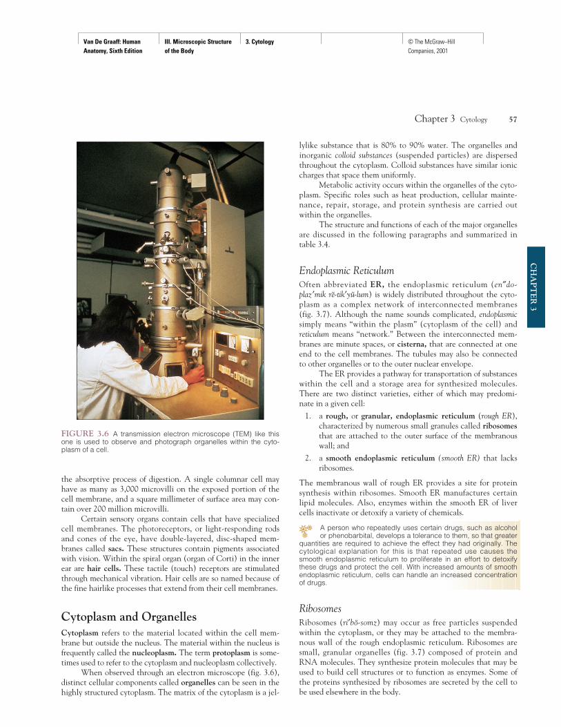
When observed through an electron microscope (fig. 3.6),distinct cellular components called organelles can be seen in thehighly structured cytoplasm. The matrix of the cytoplasm is a jel-
lylike substance that is 80% to 90% water. The organelles andinorganic colloid substances (suspended particles) are dispersedthroughout the cytoplasm. Colloid substances have similar ioniccharges that space them uniformly.
Metabolic activity occurs within the organelles of the cyto-plasm. Specific roles such as heat production, cellular mainte-nance, repair, storage, and protein synthesis are carried outwithin the organelles.
The structure and functions of each of the major organellesare discussed in the following paragraphs and summarized intable 3.4.
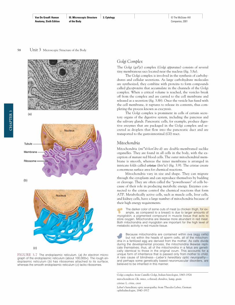
Endoplasmic ReticulumOften abbreviated ER, the endoplasmic reticulum (en″do-plaz ′mik re-tik′yu-lum) is widely distributed throughout the cyto-plasm as a complex network of interconnected membranes (fig. 3.7). Although the name sounds complicated, endoplasmicsimply means “within the plasm” (cytoplasm of the cell) andreticulum means “network.” Between the interconnected mem-branes are minute spaces, or cisterna, that are connected at oneend to the cell membranes. The tubules may also be connectedto other organelles or to the outer nuclear envelope.
The ER provides a pathway for transportation of substanceswithin the cell and a storage area for synthesized molecules.There are two distinct varieties, either of which may predomi-nate in a given cell:
1. a rough, or granular, endoplasmic reticulum (rough ER),characterized by numerous small granules called ribosomesthat are attached to the outer surface of the membranouswall; and
2. a smooth endoplasmic reticulum (smooth ER) that lacksribosomes.
The membranous wall of rough ER provides a site for proteinsynthesis within ribosomes. Smooth ER manufactures certainlipid molecules. Also, enzymes within the smooth ER of livercells inactivate or detoxify a variety of chemicals.
A person who repeatedly uses certain drugs, such as alcoholor phenobarbital, develops a tolerance to them, so that greater
quantities are required to achieve the effect they had originally. Thecytological explanation for this is that repeated use causes thesmooth endoplasmic reticulum to proliferate in an effort to detoxifythese drugs and protect the cell. With increased amounts of smoothendoplasmic reticulum, cells can handle an increased concentrationof drugs.
RibosomesRibosomes (ri′bo-somz) may occur as free particles suspendedwithin the cytoplasm, or they may be attached to the membra-nous wall of the rough endoplasmic reticulum. Ribosomes aresmall, granular organelles (fig. 3.7) composed of protein andRNA molecules. They synthesize protein molecules that may beused to build cell structures or to function as enzymes. Some ofthe proteins synthesized by ribosomes are secreted by the cell tobe used elsewhere in the body.
Chapter 3 Cytology 57
CH
AP
TE
R 3
FIGURE 3.6 A transmission electron microscope (TEM) like thisone is used to observe and photograph organelles within the cyto-plasm of a cell.
Van De Graaff: Human Anatomy, Sixth Edition
III. Microscopic Structure of the Body
3. Cytology © The McGraw−Hill Companies, 2001
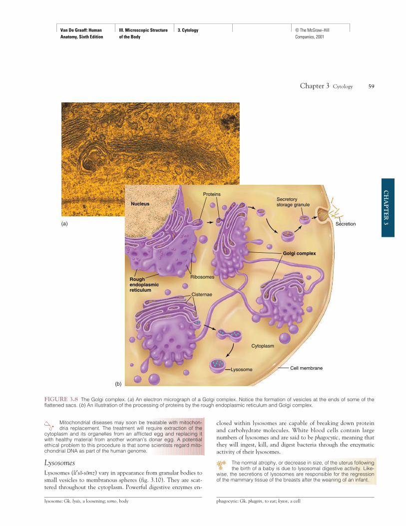
Golgi ComplexThe Golgi (gol′je) complex (Golgi apparatus) consists of severaltiny membranous sacs located near the nucleus (fig. 3.8a).
The Golgi complex is involved in the synthesis of carbohy-drates and cellular secretions. As large carbohydrate moleculesare synthesized, they combine with proteins to form compoundscalled glycoproteins that accumulate in the channels of the Golgicomplex. When a critical volume is reached, the vesicles breakoff from the complex and are carried to the cell membrane andreleased as a secretion (fig. 3.8b). Once the vesicle has fused withthe cell membrane, it ruptures to release its contents, thus com-pleting the process known as exocytosis.
The Golgi complex is prominent in cells of certain secre-tory organs of the digestive system, including the pancreas andthe salivary glands. Pancreatic cells, for example, produce diges-tive enzymes that are packaged in the Golgi complex and se-creted as droplets that flow into the pancreatic duct and aretransported to the gastrointestinal (GI) tract.
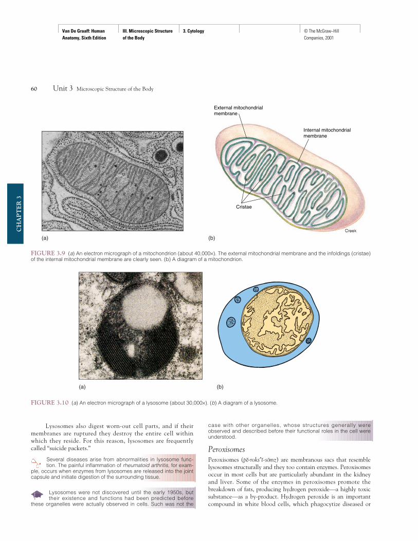
MitochondriaMitochondria (mi″to-kon′dre-a) are double-membraned saclikeorganelles. They are found in all cells in the body, with the ex-ception of mature red blood cells. The outer mitochondrial mem-brane is smooth, whereas the inner membrane is arranged inintricate folds called cristae (kris′te) (fig. 3.9). The cristae createa enormous surface area for chemical reactions.
Mitochondria vary in size and shape. They can migratethrough the cytoplasm and can reproduce themselves by buddingor cleavage. They are often called the “powerhouses” of cells be-cause of their role in producing metabolic energy. Enzymes con-nected to the cristae control the chemical reactions that formATP. Metabolically active cells, such as muscle cells, liver cells,and kidney cells, have a large number of mitochondria because oftheir high energy requirements.
The darker color of some cuts of meat (a chicken thigh, for ex-ample, as compared to a breast) is due to larger amounts of
myoglobin, a pigmented compound in muscle tissue that acts tostore oxygen. Mitochondria are likewise more abundant in red meat.Both mitochondria and myoglobin are important for the high level ofmetabolic activity in red muscle tissue.
Because mitochondria are contained within ova (egg cells)but not within the heads of sperm cells, all of the mitochon-
dria in a fertilized egg are derived from the mother. As cells divideduring the developmental process, the mitochondria likewise repli-cate themselves; thus, all of the mitochondria in a fetus are geneti-cally identical to those in the original ovum. This accounts for aunique form of inheritance that is passed only from mother to child.A rare cause of blindness—Leber’s hereditary optic neuropathy—and perhaps some genetically based neuromuscular disorders, arebelieved to be inherited in this manner.
58 Unit 3 Microscopic Structure of the Body
CH
AP
TE
R 3
Membrane
Ribosome
Nucleus
Tubule
Nucleus
(a)
(b)
(c)
FIGURE 3.7 The endoplasmic reticulum. (a) An electron micro-graph of the endoplasmic reticulum (about 100,000×). The rough en-doplasmic reticulum (b) has ribosomes attached to its surface,whereas the smooth endoplasmic reticulum (c) lacks ribosomes.
Golgi complex: from Camillo Golgi, Italian histologist, 1843–1926
mitochondrion: Gk. mitos, a thread; chondros, lump, grain
cristae: L. crista, crest
Leber’s hereditary optic neuropathy: from Theodor Leber, Germanophthalmologist, 1840–1917
Van De Graaff: Human Anatomy, Sixth Edition
III. Microscopic Structure of the Body
3. Cytology © The McGraw−Hill Companies, 2001
Mitochondrial diseases may soon be treatable with mitochon-dria replacement. The treatment will require extraction of the
cytoplasm and its organelles from an afflicted egg and replacing itwith healthy material from another woman’s donar egg. A potentialethical problem to this procedure is that some scientists regard mito-chondrial DNA as part of the human genome.
LysosomesLysosomes (li′so-somz) vary in appearance from granular bodies tosmall vesicles to membranous spheres (fig. 3.10). They are scat-tered throughout the cytoplasm. Powerful digestive enzymes en-
closed within lysosomes are capable of breaking down proteinand carbohydrate molecules. White blood cells contain largenumbers of lysosomes and are said to be phagocytic, meaning thatthey will ingest, kill, and digest bacteria through the enzymaticactivity of their lysosomes.
The normal atrophy, or decrease in size, of the uterus followingthe birth of a baby is due to lysosomal digestive activity. Like-
wise, the secretions of lysosomes are responsible for the regressionof the mammary tissue of the breasts after the weaning of an infant.
Chapter 3 Cytology 59
CH
AP
TE
R 3
Nucleus
Golgi complex
Rough endoplasmic reticulum
Proteins
Secretion
Ribosomes
Cisternae
Lysosome
Cytoplasm
Secretory storage granule
Cell membrane
FIGURE 3.8 The Golgi complex. (a) An electron micrograph of a Golgi complex. Notice the formation of vesicles at the ends of some of theflattened sacs. (b) An illustration of the processing of proteins by the rough endoplasmic reticulum and Golgi complex.
(a)
(b)
lysosome: Gk. lysis, a loosening; somo, body phagocytic: Gk. phagein, to eat; kytos, a cell
Van De Graaff: Human Anatomy, Sixth Edition
III. Microscopic Structure of the Body
3. Cytology © The McGraw−Hill Companies, 2001
Lysosomes also digest worn-out cell parts, and if theirmembranes are ruptured they destroy the entire cell withinwhich they reside. For this reason, lysosomes are frequentlycalled “suicide packets.”
Several diseases arise from abnormalities in lysosome func-tion. The painful inflammation of rheumatoid arthritis, for exam-
ple, occurs when enzymes from lysosomes are released into the jointcapsule and initiate digestion of the surrounding tissue.
Lysosomes were not discovered until the early 1950s, buttheir existence and functions had been predicted before
these organelles were actually observed in cells. Such was not the
case with other organelles, whose structures generally were observed and described before their functional roles in the cell wereunderstood.
PeroxisomesPeroxisomes (pe-roks′ ı-somz) are membranous sacs that resemblelysosomes structurally and they too contain enzymes. Peroxisomesoccur in most cells but are particularly abundant in the kidneyand liver. Some of the enzymes in peroxisomes promote thebreakdown of fats, producing hydrogen peroxide—a highly toxicsubstance—as a by-product. Hydrogen peroxide is an importantcompound in white blood cells, which phagocytize diseased or
60 Unit 3 Microscopic Structure of the Body
CH
AP
TE
R 3
External mitochondrialmembrane
Internal mitochondrialmembrane
Cristae
FIGURE 3.9 (a) An electron micrograph of a mitochondrion (about 40,000×). The external mitochondrial membrane and the infoldings (cristae)of the internal mitochondrial membrane are clearly seen. (b) A diagram of a mitochondrion.
(a) (b)
(a) (b)
FIGURE 3.10 (a) An electron micrograph of a lysosome (about 30,000×). (b) A diagram of a lysosome.

Van De Graaff: Human Anatomy, Sixth Edition
III. Microscopic Structure of the Body
3. Cytology © The McGraw−Hill Companies, 2001
worn-out cells. Peroxisomes also contain the enzyme catalase,which breaks down excess hydrogen peroxide into water andoxygen so that there is no toxic effect on other organelles withinthe cytoplasm.
Centrosome and CentriolesThe centrosome (central body) is a nonmembranous sphericalmass positioned near the nucleus. Within the centrosome, a pairof rodlike structures called centrioles (sen′tre-o lz) (fig. 3.11) arepositioned at right angles to each other. The wall of each centri-ole is composed of nine evenly spaced bundles, and each bundlecontains three microtubules.
Centrosomes are found only in those cells that can divide.During the mitotic (replication) process, the centrioles moveaway from each other and take positions on either side of the nu-cleus. They are then involved in the distribution of the chromo-somes during cellular reproduction. Mature muscle and nervecells lack centrosomes, and thus cannot divide.
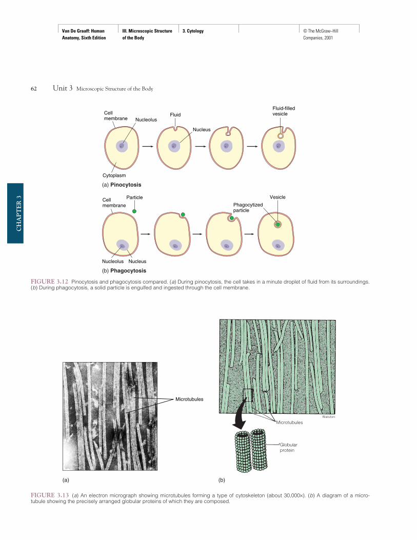
VacuolesVacuoles (vak′yoo-o lz) are membranous sacs of various sizes thatusually function as storage chambers. They are formed when aportion of the cell membrane invaginates and pinches off duringendocytosis. Vacuolation is initiated either by pinocytosis (pin″o-si-to′sis), in which cells take in minute droplets of liquid throughthe cell membrane, or by phagocytosis (fag″o-si-to′sis), in whichthe cell membrane engulfs solid particles (fig. 3.12). Vacuolesmay contain liquid or solid materials that were previously outsidethe cell.
Fibrils and MicrotubulesBoth fibrils and microtubules are found throughout the cyto-plasm. The fibrils are minute rodlike structures, whereas the mi-crotubules are fine, threadlike tubular structures of varyinglengths (fig. 3.13). Both provide the cell with support by forminga type of cytoskeleton. Specialized fibrils called myofilaments areparticularly abundant in muscle cells, where they aid in the con-traction of these cells. Microtubules are also involved in thetransportation of macromolecules throughout the cytoplasm.They are especially abundant in the cells of endocrine organs,where they aid the movement of hormones to be secreted intothe blood. Microtubules in certain cells provide flexible supportfor cilia and flagella.
Cilia and FlagellaAlthough cilia and flagella appear to be extensions of the cellmembrane, they are actually cytoplasmic projections from the in-terior of the cell. These projections contain cytoplasm and sup-portive microtubules bounded by the cell membrane (fig. 3.14).Cilia and flagella should not be confused with microvilli or withstereocilia, both of which are specializations of cell membranes.
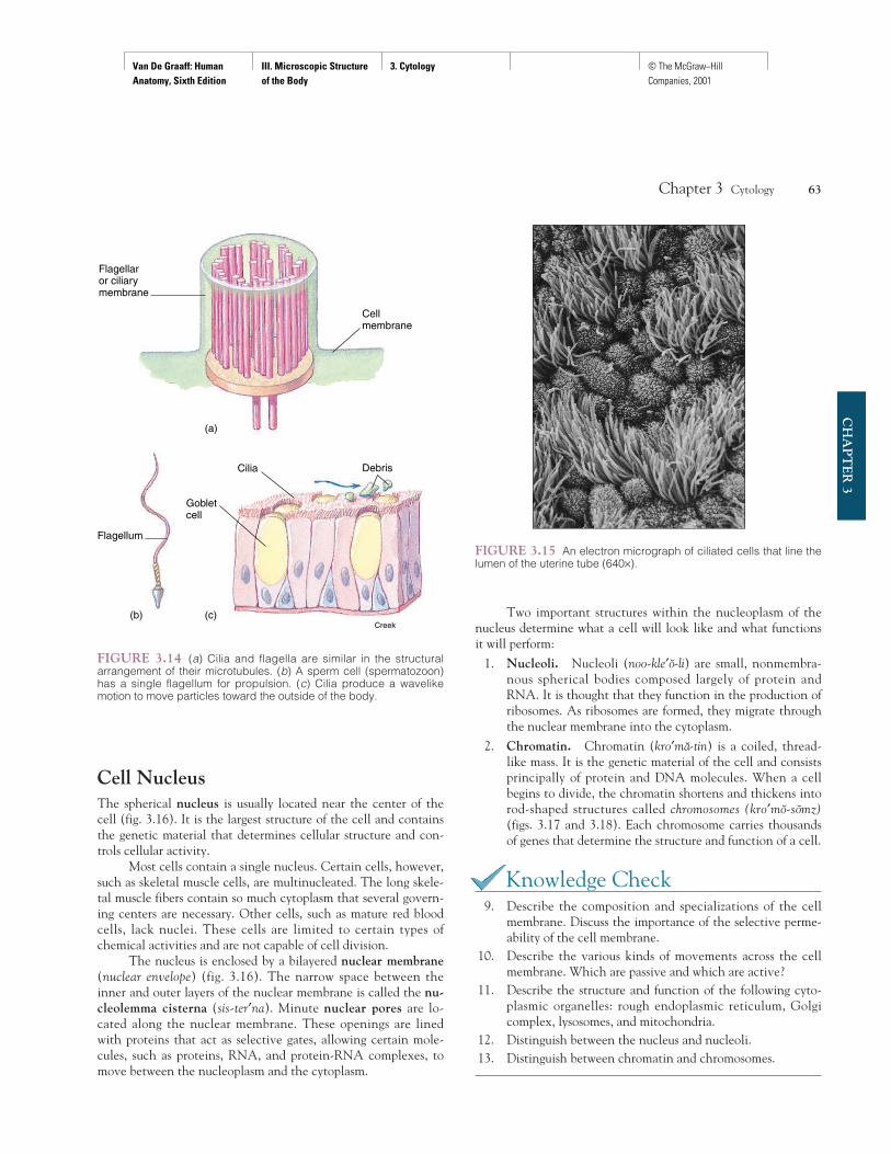
Cilia (sil′e-a) are numerous short projections from the ex-posed border of certain cells (fig. 3.15). Ciliated cells are inter-spersed with mucus-secreting goblet cells. There is always a filmof mucus on the free surface of ciliated cells. Ciliated cells linethe lumina (hollow portions) of sections of the respiratory and re-productive tracts. The function of the cilia is to move the mucusand any adherent material toward the exterior of the body.
Flagella (fla-jel′a) are similar to cilia in basic microtubularstructure (see fig. 3.14), but they are somewhat longer than cilia.The only example of a flagellated cell in humans is the spermcell, which uses the single structure for locomotion.
Chapter 3 Cytology 61
CH
AP
TE
R 3
(a) (b)
FIGURE 3.11 (a) An electron micrograph of centrioles in a centrosome (about 14,200×). (b) A diagram showing that the centrioles are posi-tioned at right angles to each other.
vacuole: L. vacuus, empty
Van De Graaff: Human Anatomy, Sixth Edition
III. Microscopic Structure of the Body
3. Cytology © The McGraw−Hill Companies, 2001
62 Unit 3 Microscopic Structure of the Body
CH
AP
TE
R 3
Nucleus
NucleolusFluid
Fluid-filledvesicle
Cytoplasm
Cellmembrane
Nucleolus Nucleus
Cellmembrane
Particle Vesicle
Phagocytizedparticle
(a) Pinocytosis
(b) Phagocytosis
FIGURE 3.12 Pinocytosis and phagocytosis compared. (a) During pinocytosis, the cell takes in a minute droplet of fluid from its surroundings.(b) During phagocytosis, a solid particle is engulfed and ingested through the cell membrane.
Microtubules
(a) (b)
FIGURE 3.13 (a) An electron micrograph showing microtubules forming a type of cytoskeleton (about 30,000×). (b) A diagram of a micro-tubule showing the precisely arranged globular proteins of which they are composed.
Van De Graaff: Human Anatomy, Sixth Edition
III. Microscopic Structure of the Body
3. Cytology © The McGraw−Hill Companies, 2001
Cell NucleusThe spherical nucleus is usually located near the center of thecell (fig. 3.16). It is the largest structure of the cell and containsthe genetic material that determines cellular structure and con-trols cellular activity.
Most cells contain a single nucleus. Certain cells, however,such as skeletal muscle cells, are multinucleated. The long skele-tal muscle fibers contain so much cytoplasm that several govern-ing centers are necessary. Other cells, such as mature red bloodcells, lack nuclei. These cells are limited to certain types ofchemical activities and are not capable of cell division.
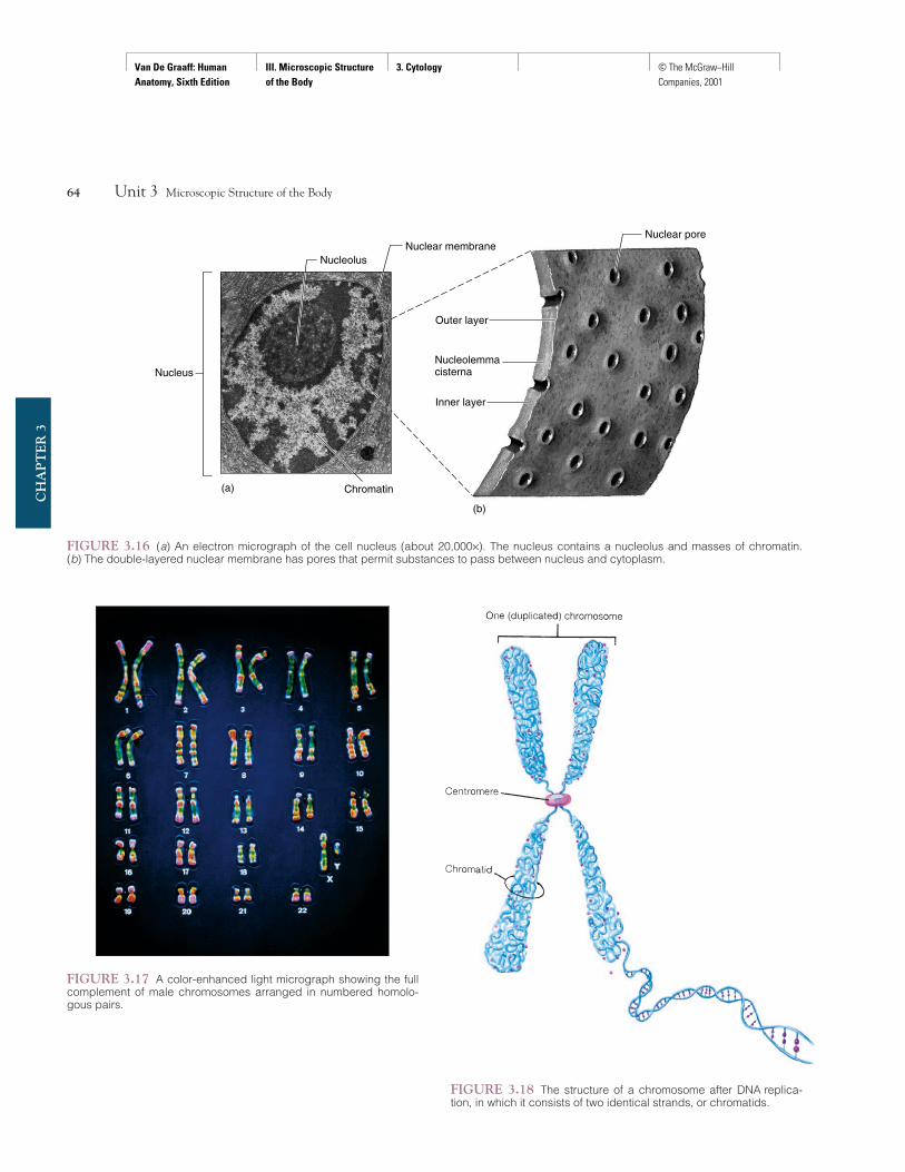
The nucleus is enclosed by a bilayered nuclear membrane(nuclear envelope) (fig. 3.16). The narrow space between theinner and outer layers of the nuclear membrane is called the nu-cleolemma cisterna (sis-ter′na). Minute nuclear pores are lo-cated along the nuclear membrane. These openings are linedwith proteins that act as selective gates, allowing certain mole-cules, such as proteins, RNA, and protein-RNA complexes, tomove between the nucleoplasm and the cytoplasm.
Two important structures within the nucleoplasm of thenucleus determine what a cell will look like and what functionsit will perform:
1. Nucleoli. Nucleoli (noo-kle′o-li) are small, nonmembra-nous spherical bodies composed largely of protein andRNA. It is thought that they function in the production ofribosomes. As ribosomes are formed, they migrate throughthe nuclear membrane into the cytoplasm.
2. Chromatin. Chromatin (kro′ma-tin) is a coiled, thread-like mass. It is the genetic material of the cell and consistsprincipally of protein and DNA molecules. When a cellbegins to divide, the chromatin shortens and thickens intorod-shaped structures called chromosomes (kro′mo-somz)(figs. 3.17 and 3.18). Each chromosome carries thousandsof genes that determine the structure and function of a cell.
Knowledge Check9. Describe the composition and specializations of the cell
membrane. Discuss the importance of the selective perme-ability of the cell membrane.
10. Describe the various kinds of movements across the cellmembrane. Which are passive and which are active?
11. Describe the structure and function of the following cyto-plasmic organelles: rough endoplasmic reticulum, Golgicomplex, lysosomes, and mitochondria.
12. Distinguish between the nucleus and nucleoli.13. Distinguish between chromatin and chromosomes.
Chapter 3 Cytology 63
CH
AP
TE
R 3
Flagellaror ciliarymembrane
(a)
(b) (c)
Flagellum
Cilia
Gobletcell
Debris
Cellmembrane
Creek
FIGURE 3.14 (a) Cilia and flagella are similar in the structuralarrangement of their microtubules. (b) A sperm cell (spermatozoon)has a single flagellum for propulsion. (c) Cilia produce a wavelikemotion to move particles toward the outside of the body.
FIGURE 3.15 An electron micrograph of ciliated cells that line thelumen of the uterine tube (640×).
Van De Graaff: Human Anatomy, Sixth Edition
III. Microscopic Structure of the Body
3. Cytology © The McGraw−Hill Companies, 2001
64 Unit 3 Microscopic Structure of the Body
CH
AP
TE
R 3
Nuclear pore
Nucleolus
Nucleus
(a)
(b)
Nuclear membrane
Outer layer
Nucleolemmacisterna
Chromatin
Inner layer
FIGURE 3.16 (a) An electron micrograph of the cell nucleus (about 20,000×). The nucleus contains a nucleolus and masses of chromatin. (b) The double-layered nuclear membrane has pores that permit substances to pass between nucleus and cytoplasm.
FIGURE 3.17 A color-enhanced light micrograph showing the fullcomplement of male chromosomes arranged in numbered homolo-gous pairs.
FIGURE 3.18 The structure of a chromosome after DNA replica-tion, in which it consists of two identical strands, or chromatids.
Van De Graaff: Human Anatomy, Sixth Edition
III. Microscopic Structure of the Body
3. Cytology © The McGraw−Hill Companies, 2001
CELL CYCLEA cell cycle consists of growth, synthesis, and mitosis. Growth isthe increase in cellular mass resulting from metabolism. Synthesisis the production of DNA and RNA to regulate cellular activity. Mi-tosis is the division of the nucleus and cytoplasm of a cell that re-sults in the formation of two daughter cells.
Objective 12 Describe the structure of DNA and RNA molecules.
Objective 13 Discuss genetic transcription and protein synthesis.
Objective 14 List the stages of mitosis and discuss theevents of each stage.
Objective 15 Discuss the significance of mitosis.
Cellular replication is one of the principal concepts of biology.Through the process of cellular division called mitosis (mi-to′sis),a multicellular organism can develop and be maintained. Mitosisenables body growth and the replacement of damaged, diseased,or worn-out cells. The process ensures that each daughter cellwill have the same number and kind of chromosomes as the orig-inal parent cell.
In an average healthy adult, over 100 billion cells will die andbe mitotically replaced during a 24-hour period. This represents a re-placement of about 2% of the body mass each day. Some of the mostmitotically active sites are the outer layer of skin, the bone marrow,the internal lining of the digestive tract, and the liver.
Before a cell can divide, it must first duplicate its chromo-somes so that the genetic traits can be passed to the succeedinggenerations of cells. A chromosome consists of a coiled deoxyri-bonucleic acid (DNA) molecule that is complexed with protein.As mentioned previously, chromosomes are formed by the short-ening and thickening of the chromatin within the nucleus whenthe cell begins to divide, at which time they are clearly visibleunder the compound microscope. There are 23 pairs of chromo-somes in each human body (somatic) cell and approximately20,000 genes are positioned on each chromosome.
Chromosomes are of varied lengths and shapes—sometwisted, some rodlike. During mitosis, they shorten and con-dense, each pair assuming a characteristic shape (see fig. 3.17).On the chromosome is a small, buttonlike body called a cen-tromere to which are attached the spindle fibers that direct thechromosome toward the pole of the cell during mitosis.
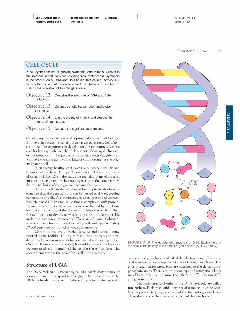
Structure of DNAThe DNA molecule is frequently called a double helix because ofits resemblance to a spiral ladder (fig. 3.19). The sides of theDNA molecule are formed by alternating units of the sugar de-
oxyribose and phosphoric acid called the phosphate group. The rungsof the molecule are composed of pairs of nitrogenous bases. The ends of each nitrogenous base are attached to the deoxyribose-phosphate units. There are only four types of nitrogenous basesin a DNA molecule: adenine (A), thymine (T), cytosine (C),and guanine (G).
The basic structural units of the DNA molecule are callednucleotides. Each nucleotide consists of a molecule of deoxyri-bose, a phosphate group, and one of the four nitrogenous bases.Thus, there is a nucleotide type for each of the four bases.
Chapter 3 Cytology 65
CH
AP
TE
R 3
mitosis: Gk. mitos, thread
FIGURE 3.19 The double-helix structure of DNA. Each strand ofthe helix contains only four kinds of organic bases (A, T, C, and G).
Van De Graaff: Human Anatomy, Sixth Edition
III. Microscopic Structure of the Body
3. Cytology © The McGraw−Hill Companies, 2001
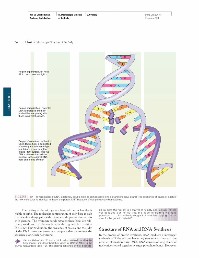
The pairing of the nitrogenous bases of the nucleotides ishighly specific. The molecular configuration of each base is suchthat adenine always pairs with thymine and cytosine always pairswith guanine. The hydrogen bonds between these bases are rela-tively weak and can be easily split during cellular division (fig. 3.20). During division, the sequence of bases along the sidesof the DNA molecule serves as a template that determines thesequence along each new strand.
James Watson and Francis Crick, who devised the double-helix model, first described their vision of DNA in 1953, in the
journal Nature (see table 1.2). The closing sentence of their brief arti-
cle (a mere 900 words) is a marvel of humility and restraint: “It hasnot escaped our notice that the specific pairing we havepostulated . . . immediately suggests a possible copying mecha-nism for the genetic material.”
Structure of RNA and RNA SynthesisIn the process of protein synthesis, DNA produces a messengermolecule of RNA of complementary structure to transport thegenetic information. Like DNA, RNA consists of long chains ofnucleotides joined together by sugar-phosphate bonds. However,
66 Unit 3 Microscopic Structure of the Body
CH
AP
TE
R 3
C G
T
C G
Region of replication. Parental DNA is unzipped and new nucleotides are pairing with those in parental strands.
Region of completed replication. Each double helix is composed of an old parental strand (light purple) and a new daughter strand (dark purple). The twoDNA molecules formed areidentical to the original DNA
Region of parental DNA helix. (Both backbones are light.)
G C
C G
CG
A
A
A
T
T
T
A
C
G
C
GG
T
G
CG
CG
C
T
TA
T
C
G
G
C
G
C
C
TA
T
C G
T
C
helix and to one another.
FIGURE 3.20 The replication of DNA. Each new double helix is composed of one old and one new strand. The sequence of bases of each ofthe new molecules is identical to that of the parent DNA because of complementary base pairing.
Van De Graaff: Human Anatomy, Sixth Edition
III. Microscopic Structure of the Body
3. Cytology © The McGraw−Hill Companies, 2001
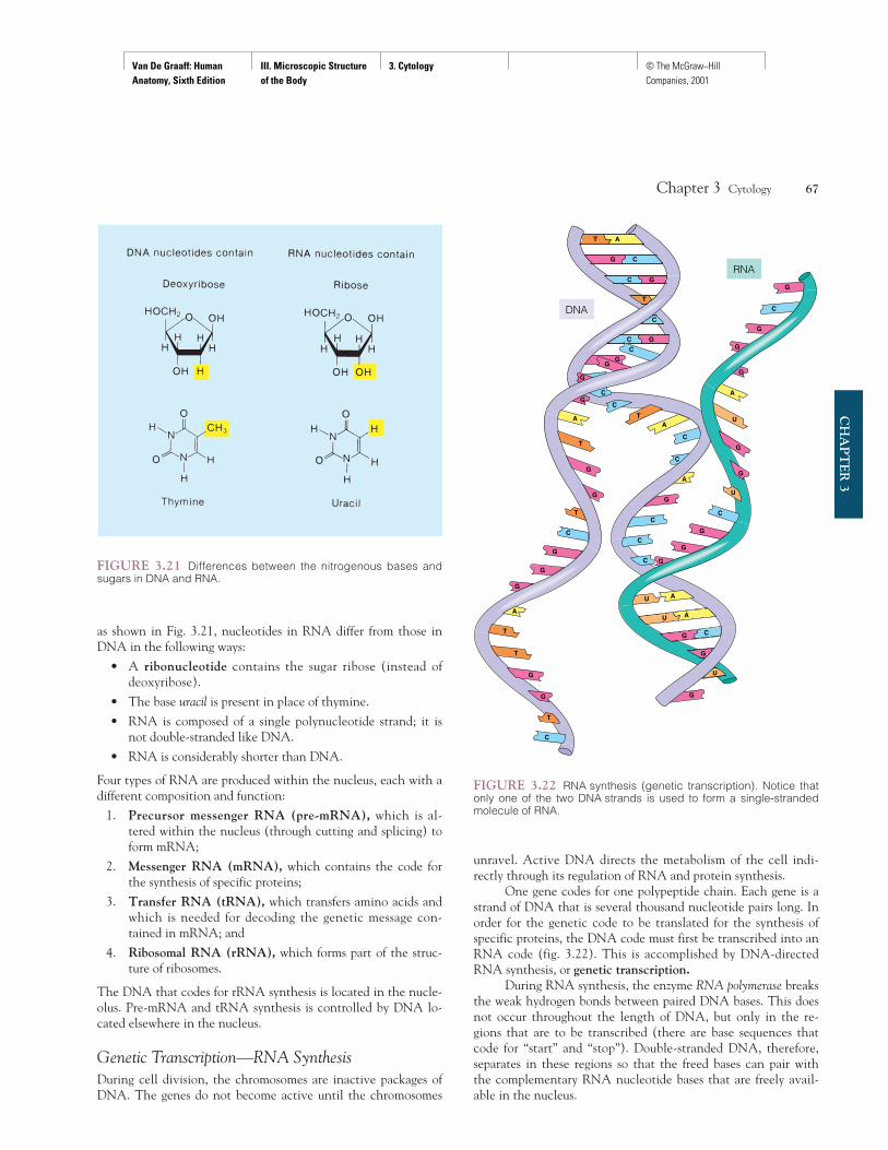
as shown in Fig. 3.21, nucleotides in RNA differ from those inDNA in the following ways:
• A ribonucleotide contains the sugar ribose (instead ofdeoxyribose).
• The base uracil is present in place of thymine.
• RNA is composed of a single polynucleotide strand; it isnot double-stranded like DNA.
• RNA is considerably shorter than DNA.
Four types of RNA are produced within the nucleus, each with adifferent composition and function:
1. Precursor messenger RNA (pre-mRNA), which is al-tered within the nucleus (through cutting and splicing) toform mRNA;
2. Messenger RNA (mRNA), which contains the code forthe synthesis of specific proteins;
3. Transfer RNA (tRNA), which transfers amino acids andwhich is needed for decoding the genetic message con-tained in mRNA; and
4. Ribosomal RNA (rRNA), which forms part of the struc-ture of ribosomes.
The DNA that codes for rRNA synthesis is located in the nucle-olus. Pre-mRNA and tRNA synthesis is controlled by DNA lo-cated elsewhere in the nucleus.
Genetic Transcription—RNA SynthesisDuring cell division, the chromosomes are inactive packages ofDNA. The genes do not become active until the chromosomes
unravel. Active DNA directs the metabolism of the cell indi-rectly through its regulation of RNA and protein synthesis.
One gene codes for one polypeptide chain. Each gene is astrand of DNA that is several thousand nucleotide pairs long. Inorder for the genetic code to be translated for the synthesis ofspecific proteins, the DNA code must first be transcribed into anRNA code (fig. 3.22). This is accomplished by DNA-directedRNA synthesis, or genetic transcription.
During RNA synthesis, the enzyme RNA polymerase breaksthe weak hydrogen bonds between paired DNA bases. This doesnot occur throughout the length of DNA, but only in the re-gions that are to be transcribed (there are base sequences thatcode for “start” and “stop”). Double-stranded DNA, therefore,separates in these regions so that the freed bases can pair withthe complementary RNA nucleotide bases that are freely avail-able in the nucleus.
Chapter 3 Cytology 67
CH
AP
TE
R 3
FIGURE 3.21 Differences between the nitrogenous bases andsugars in DNA and RNA.
G
C G
G C
T A
T
C
C G
C
G
T
G
G
T
G
C
G
G
A
T
T
G
G
T
C
G
G
CG
A
G
U
G
U A
G C
U A
C
C
C
G
A
C
C
AT
C
G
G
C
U
G
G
U
A
G
G
G
C
G
DNA
RNA
FIGURE 3.22 RNA synthesis (genetic transcription). Notice thatonly one of the two DNA strands is used to form a single-strandedmolecule of RNA.
Van De Graaff: Human Anatomy, Sixth Edition
III. Microscopic Structure of the Body
3. Cytology © The McGraw−Hill Companies, 2001
This pairing of bases follows the law of complementary basepairing: guanine bonds with cytosine (and vice versa), and ade-nine bonds with uracil (because uracil in RNA is equivalent tothymine in DNA). In RNA synthesis, only one of the two freedstrands of DNA serves as a guide (see fig. 3.22). Once an RNAmolecule has been produced, it detaches from the DNA strandon which it was formed. This process can continue indefinitely,producing many thousands of RNA copies of the DNA strandbeing transcribed. When the gene is no longer to be transcribed,the separated DNA strands can recoil into their helical form.
In the case of pre-mRNA, the finished molecule is alteredafter synthesis. Within the pre-mRNA are noncoding regionsknown as introns. The introns are removed through the action ofenzymes, and the coding regions are then spliced together so thatthey can direct the synthesis of a specific protein.
Protein SynthesisOnce produced, mRNA leaves the nucleus and enters the cyto-plasm, where it attaches to ribosomes. The mRNA passes througha number of ribosomes to form a polyribosome, or polysome for
short. The association of mRNA with ribosomes is needed forgenetic translation—the production of specific proteins accordingto the code contained in the mRNA base sequences.
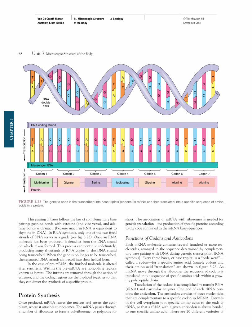
Functions of Codons and AnticodonsEach mRNA molecule contains several hundred or more nu-cleotides, arranged in the sequence determined by complemen-tary base pairing with DNA during genetic transcription (RNAsynthesis). Every three bases, or base triplet, is a “code word”—called a codon—for a specific amino acid. Sample codons andtheir amino acid “translation” are shown in figure 3.23. AsmRNA move through the ribosome, the sequence of codons istranslated into a sequence of specific amino acids within a grow-ing polypeptide chain.
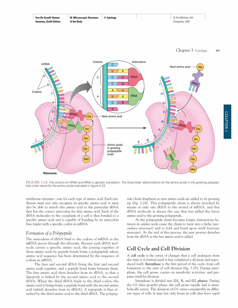
Translation of the codons is accomplished by transfer RNA(tRNA) and particular enzymes. One end of each tRNA con-tains the anticodon. The anticodon consists of three nucleotidesthat are complementary to a specific codon in MRNA. Enzymesin the cell cytoplasm join specific amino acids to the ends oftRNA, so that a tRNA with a given anticodon is always bondedto one specific amino acid. There are 20 different varieties of
68 Unit 3 Microscopic Structure of the Body
CH
AP
TE
R 3
T
A
A
U
G
C
G
C
T
A
A
U
G
C
T
A
G
C
C
G
T
A
G
C
G
T
A
TG
CG
C
AG
C
G
C
T
A
A
T
CC
G
G G
C
G
G
CT
G
CC
G
T
A
Tra
nscr
iptio
n
DNAdoublehelix
Tra
nsla
tion
C
G
C
G
C
G
G
C
A
U
C
G
C
G
G
C
C
G
G
C
DNA coding strand
Messenger RNA
Codon 1 Codon 2 Codon 3 Codon 4 Codon 5 Codon 6 Codon 7
Protein
Methionine Glycine Serine Isoleucine Glycine Alanine Alanine
FIGURE 3.23 The genetic code is first transcribed into base triplets (codons) in mRNA and then translated into a specific sequence of aminoacids in a protein.
Van De Graaff: Human Anatomy, Sixth Edition
III. Microscopic Structure of the Body
3. Cytology © The McGraw−Hill Companies, 2001
synthetase enzymes—one for each type of amino acid. Each syn-thetase must not only recognize its specific amino acid, it mustalso be able to attach this amino acid to the particular tRNAthat has the correct anticodon for that amino acid. Each of thetRNA molecules in the cytoplasm of a cell is thus bonded to aspecific amino acid and is capable of bonding by its anticodonbase triplet with a specific codon in mRNA.
Formation of a PolypeptideThe anticodons of tRNA bind to the codons of mRNA as themRNA moves through the ribosome. Because each tRNA mol-ecule carries a specific amino acid, the joining together ofthese amino acids by peptide bonds forms a polypeptide whoseamino acid sequence has been determined by the sequence ofcodons in mRNA.
The first and second tRNA bring the first and secondamino acids together, and a peptide bond forms between them.The first amino acid then detaches from its tRNA, so that adipeptide is linked by the second amino acid to the secondtRNA. When the third tRNA binds to the third codon, theamino acid it brings forms a peptide bond with the second aminoacid (which detaches from its tRNA). A tripeptide is thus at-tached by the third amino acid to the third tRNA. The polypep-
tide chain lengthens as new amino acids are added to its growingtip (fig. 3.24). This polypeptide chain is always attached bymeans of only one tRNA to the strand of mRNA, and thistRNA molecule is always the one that has added the latestamino acid to the growing polypeptide.
As the polypeptide chain becomes longer, interactions be-tween its amino acids cause the chain to twist into a helix (sec-ondary structure) and to fold and bend upon itself (tertiarystructure). At the end of this process, the new protein detachesfrom the tRNA as the last amino acid is added.
Cell Cycle and Cell DivisionA cell cycle is the series of changes that a cell undergoes fromthe time it is formed until it has completed a division and repro-duced itself. Interphase is the first period of the cycle, from cellformation to the start of cell division (fig. 3.25). During inter-phase, the cell grows, carries on metabolic activities, and pre-pares itself for division.
Interphase is divided into G1, S, and G2 phases. Duringthe G1 (first growth) phase, the cell grows rapidly and is meta-bolically active. The duration of G1 varies considerably in differ-ent types of cells. It may last only hours in cells that have rapid
Chapter 3 Cytology 69
CH
AP
TE
R 3
tRNA
1
2
3
4
5
6
7
8
9
Codons Anticodons
G
G
U A
A U
C G
C G
U A
C
C
Codons
mRNA
tRNA
tRNA
Met
Gly
Ser
Iso
Gly Next amino acid
Amino acidsin growingpolypeptidechain
RibosomeMet
Gly
Ser
Iso
Gly
AlaNext amino acid
tRNA
tRNA
tRNA
4
5
AB
C
D
E
F
G
H
I
tRN
A
FIGURE 3.24 The actions of mRNA and tRNA in genetic translation. The three-letter abbreviations for the amino acids in the growing polypep-tide chain stand for the amino acids indicated in figure 3.23.
Van De Graaff: Human Anatomy, Sixth Edition
III. Microscopic Structure of the Body
3. Cytology © The McGraw−Hill Companies, 2001
division rates, or it may be a matter of days or even years forother cells. At the end of G1, the centrioles replicate in prepara-tion for their role in cell division. During the S (synthetic)phase, the DNA in the nucleus of the cell replicates, so that thetwo future cells will receive identical copies of the genetic mater-ial. During the G2 (second growth) phase, the enzymes andother proteins needed for the division process are synthesized,and the cell continues to grow.
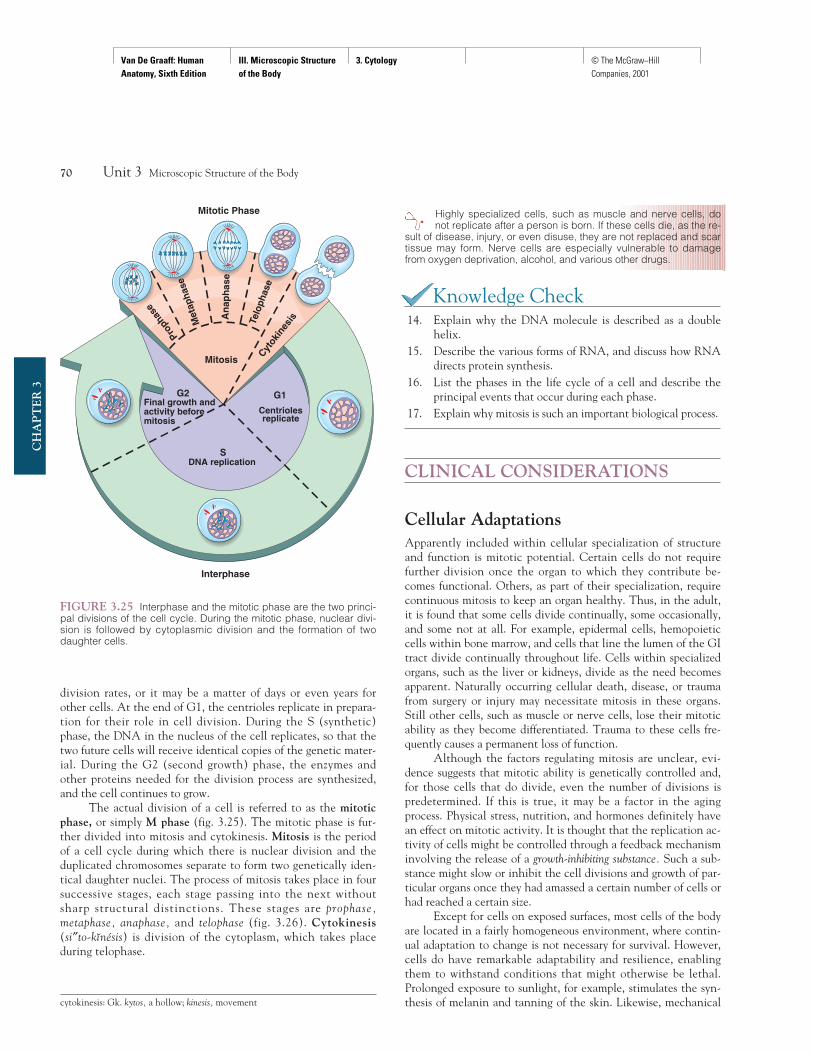
The actual division of a cell is referred to as the mitoticphase, or simply M phase (fig. 3.25). The mitotic phase is fur-ther divided into mitosis and cytokinesis. Mitosis is the periodof a cell cycle during which there is nuclear division and theduplicated chromosomes separate to form two genetically iden-tical daughter nuclei. The process of mitosis takes place in foursuccessive stages, each stage passing into the next withoutsharp structural distinctions. These stages are prophase,metaphase, anaphase, and telophase (fig. 3.26). Cytokinesis(si″to-kınésis) is division of the cytoplasm, which takes placeduring telophase.
Highly specialized cells, such as muscle and nerve cells, donot replicate after a person is born. If these cells die, as the re-
sult of disease, injury, or even disuse, they are not replaced and scartissue may form. Nerve cells are especially vulnerable to damagefrom oxygen deprivation, alcohol, and various other drugs.
Knowledge Check14. Explain why the DNA molecule is described as a double
helix.15. Describe the various forms of RNA, and discuss how RNA
directs protein synthesis.16. List the phases in the life cycle of a cell and describe the
principal events that occur during each phase.17. Explain why mitosis is such an important biological process.
CLINICAL CONSIDERATIONS
Cellular AdaptationsApparently included within cellular specialization of structureand function is mitotic potential. Certain cells do not requirefurther division once the organ to which they contribute be-comes functional. Others, as part of their specialization, requirecontinuous mitosis to keep an organ healthy. Thus, in the adult,it is found that some cells divide continually, some occasionally,and some not at all. For example, epidermal cells, hemopoieticcells within bone marrow, and cells that line the lumen of the GItract divide continually throughout life. Cells within specializedorgans, such as the liver or kidneys, divide as the need becomesapparent. Naturally occurring cellular death, disease, or traumafrom surgery or injury may necessitate mitosis in these organs.Still other cells, such as muscle or nerve cells, lose their mitoticability as they become differentiated. Trauma to these cells fre-quently causes a permanent loss of function.
Although the factors regulating mitosis are unclear, evi-dence suggests that mitotic ability is genetically controlled and,for those cells that do divide, even the number of divisions ispredetermined. If this is true, it may be a factor in the agingprocess. Physical stress, nutrition, and hormones definitely havean effect on mitotic activity. It is thought that the replication ac-tivity of cells might be controlled through a feedback mechanisminvolving the release of a growth-inhibiting substance. Such a sub-stance might slow or inhibit the cell divisions and growth of par-ticular organs once they had amassed a certain number of cells orhad reached a certain size.
Except for cells on exposed surfaces, most cells of the bodyare located in a fairly homogeneous environment, where contin-ual adaptation to change is not necessary for survival. However,cells do have remarkable adaptability and resilience, enablingthem to withstand conditions that might otherwise be lethal.Prolonged exposure to sunlight, for example, stimulates the syn-thesis of melanin and tanning of the skin. Likewise, mechanical
70 Unit 3 Microscopic Structure of the Body
CH
AP
TE
R 3
Final growth andactivity beforemitosis
DNA replication
Centriolesreplicate
Interphase
Mitotic Phase
Proph
ase
Met
apha
se
An
aph
ase
Telo
phas
eCyt
okin
esis
Mitosis
G2 G1
FIGURE 3.25 Interphase and the mitotic phase are the two princi-pal divisions of the cell cycle. During the mitotic phase, nuclear divi-sion is followed by cytoplasmic division and the formation of twodaughter cells.
cytokinesis: Gk. kytos, a hollow; kinesis, movement
Van De Graaff: Human Anatomy, Sixth Edition
III. Microscopic Structure of the Body
3. Cytology © The McGraw−Hill Companies, 2001
Chapter 3 Cytology 71
CH
AP
TE
R 3
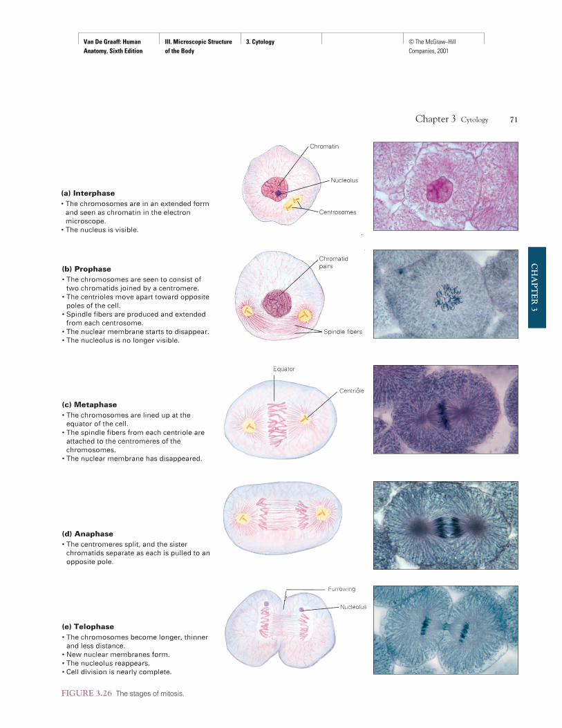
FIGURE 3.26 The stages of mitosis.
Van De Graaff: Human Anatomy, Sixth Edition
III. Microscopic Structure of the Body
3. Cytology © The McGraw−Hill Companies, 2001
friction to the skin stimulates mitotic activity and the synthesisof a fibrous protein, keratin, which results in the formation of aprotective callus.
Cells adapt to potentially injurious stimuli by several spe-cific mechanisms. Hypertrophy (hi″pe′rtro-fe) refers to an in-crease in the size of cells resulting from increased synthesis ofprotein, nucleic acids, and lipids. Cellular hypertrophy can be ei-ther compensatory or hormonal. Compensatory hypertrophy oc-curs when increased metabolic demands on particular cells resultin an increase in cellular mass. Examples of compensatory hyper-trophy include the enlargement of skeletal muscle fibers as a re-sult of exercise and cardiac (heart) muscle fibers or kidney cellsbecause of an increased work demand. Hypertension (high bloodpressure) causes cardiac cells to hypertrophy because they mustpump blood against raised pressures. After the removal of a dis-eased kidney, there is a compensatory increase in the size of thecells of the remaining kidney so that its normal weight is approx-imately doubled. Examples of hormonal hypertrophy are the in-creased size of the breasts and smooth muscles of the uterus in apregnant woman.
Hyperplasia (hi″per-pla′ze-a) refers to an increase in thenumber of cells formed as a result of increased mitotic activity.The removal of a portion of the liver, for example, leads to re-generation, or hyperplasia, of the remaining liver cells to restorethe loss. But the triggering mechanism for hyperplasia is notknown. In women, a type of hormonally induced hyperplasia oc-curs in cells of the endometrium of the uterus after menstruation,which restores this layer to a suitable state for possible implanta-tion of an embryo.
Atrophy (at′ro-fe) refers to a decrease in the size of cellsand a corresponding decrease in the size of the affected organ.Atrophy can occur in the cells of any organ and may be classifiedas disuse atrophy, disease atrophy, or aging (senile) atrophy.
Metaplasia (met′a″-plá ze-a) is a specialized cellular changein which one type of cell transforms into another. Generally, itinvolves the change of highly specialized cells into more general-ized, protective cells. For example, excessive exposure to inhaledsmoke causes the specialized ciliated columnar epithelial cellslining the bronchial airways to change into stratified squamousepithelium, which is more resistant to injury from smoke.
Trauma to CellsAs adaptable as cells are to environmental changes, they are sub-ject to damage from aging and disease. If a trauma causes exten-sive cellular death, the condition may become life threatening.A person dies when a vital organ can no longer perform its meta-bolic role in sustaining the body.
Energy deficit means that more energy is required by a cellthan is available. Cells can tolerate certain mild deficits becauseof various reserves stored within the cytoplasm, but a severe orprolonged deficit will cause cells to die. An energy deficit occurswhen the cells do not have enough glucose or oxygen to allowfor glucose combustion. Examples of energy deficits are low levelsof blood sugar (hypoglycemia) and the impermeability of the cellmembrane to glucose (as in diabetes mellitus). Malnutrition alsomay result in an energy deficit. Few cells can tolerate an inter-ruption in oxygen supply. Cells of the brain and the heart havetremendous oxygen demands, and an interruption of the supplyto these organs can cause death in a matter of minutes.
Physical injury to cells, another type of trauma, occurs ina variety of ways. High temperature (hyperthermia) is generallyless tolerable to cells than low temperature (hypothermia). Res-piratory rate, heart rate, and metabolism accelerate with hyper-thermia. Continued hyperthermia causes protein coagulationwithin cells, and eventually cellular death. In frostbite, rapid orprolonged chilling causes cellular injury. In severe frostbite, icecrystals form and cause the cells to burst.
Burns are particularly significant if they cause damage tothe deeper skin layers, which interferes with the mitotic activityof cells (see fig. 5.20). Of immediate concern with burns, how-ever, is the devastating effect of fluid loss and infection throughtraumatized cell membranes.
Accidental poisoning and suicide through drug overdoseaccount for large numbers of deaths in the United States andelsewhere. Drugs and poisons can cause cellular dysfunction bydisrupting DNA replication, RNA transcription, enzyme sys-tems, or cell membrane activity.
Radiation causes a type of cell trauma that is cumulative ineffect. When X rays are administered for therapeutic purposes(radiotherapy), small doses are focused on a tumorous area over acourse of many days to prevent widespread cellular injury. Somecells are more sensitive to radiation than others. Immature or mi-totically active cells are highly sensitive, whereas cells that areno longer growing, such as neurons and muscle cells, are not asvulnerable to radiation injury.
Infectious agents, or pathogens, also cause cellular dys-function. Viruses and bacteria are the most common pathogens.Viruses usually invade and destroy cells as they reproduce them-selves. Bacteria, on the other hand, do not usually invade cellsbut will frequently poison cells with their toxic metabolic wastes.
Medical GeneticsMedical genetics is a branch of medicine concerned with dis-eases that have a genetic origin. Genetic factors include abnor-malities in chromosome number or structure and mutant genes.Genetic diseases are a diverse group of disorders, including mal-formed blood cells (sickle-cell anemia), defective blood clotting(hemophilia), and mental retardation (Down syndrome).
Chromosomal abnormalities occur in approximately 0.6%of live-birth infants. The majority (70%) are subtle, cause noproblems, and usually go undetected. Structural changes in theDNA that are passed from parent to offspring by means of sex
72 Unit 3 Microscopic Structure of the Body
CH
AP
TE
R 3
hypertrophy: Gk. hyper, over; trophe, nourishment
hyperplasia: Gk. hyper, over; plasis, a molding
atrophy: Gk. a, without; trophe, nourishment
metaplasia: Gk. meta, between; plasis, a molding
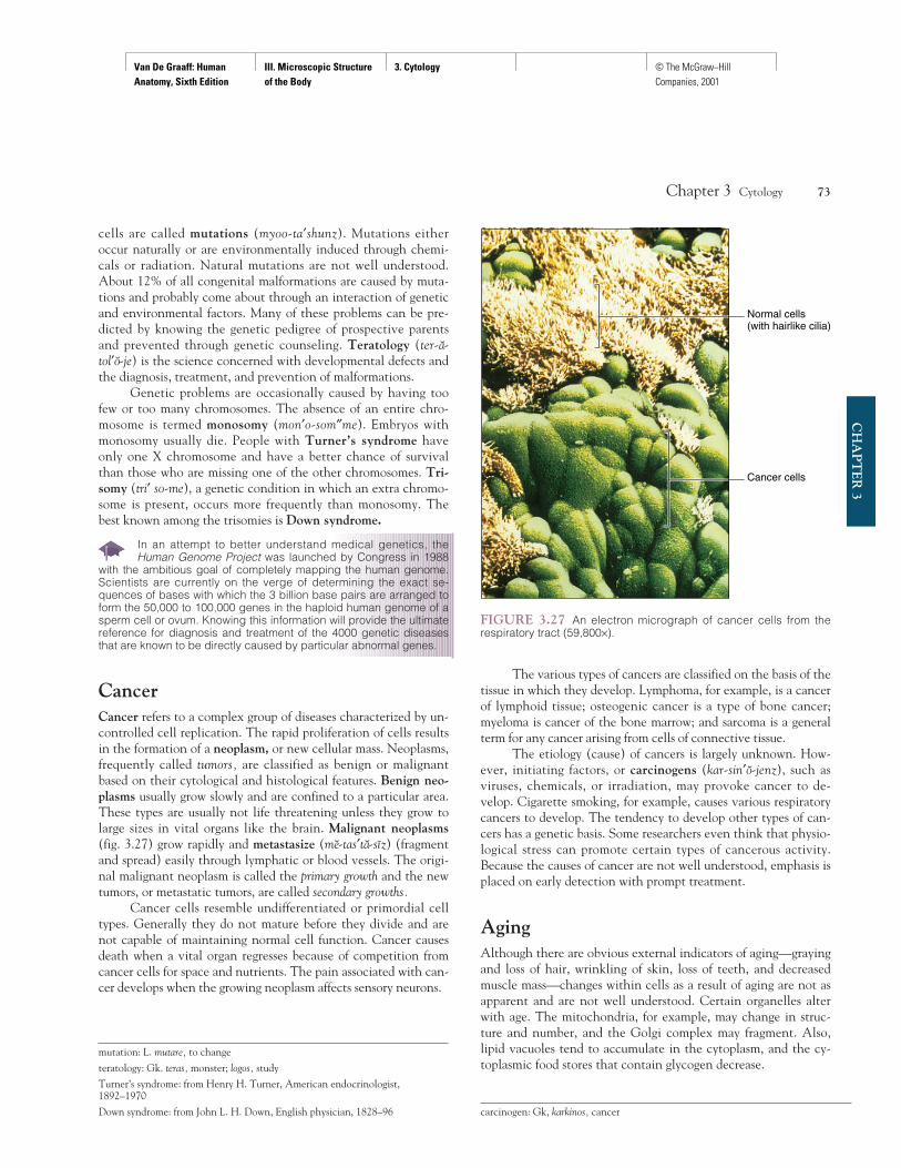
Van De Graaff: Human Anatomy, Sixth Edition
III. Microscopic Structure of the Body
3. Cytology © The McGraw−Hill Companies, 2001
cells are called mutations (myoo-ta′shunz). Mutations eitheroccur naturally or are environmentally induced through chemi-cals or radiation. Natural mutations are not well understood.About 12% of all congenital malformations are caused by muta-tions and probably come about through an interaction of geneticand environmental factors. Many of these problems can be pre-dicted by knowing the genetic pedigree of prospective parentsand prevented through genetic counseling. Teratology (ter-a-tol′o-je) is the science concerned with developmental defects andthe diagnosis, treatment, and prevention of malformations.
Genetic problems are occasionally caused by having toofew or too many chromosomes. The absence of an entire chro-mosome is termed monosomy (mon′o-som″me). Embryos withmonosomy usually die. People with Turner’s syndrome haveonly one X chromosome and have a better chance of survivalthan those who are missing one of the other chromosomes. Tri-somy (tri′ so-me), a genetic condition in which an extra chromo-some is present, occurs more frequently than monosomy. Thebest known among the trisomies is Down syndrome.
In an attempt to better understand medical genetics, theHuman Genome Project was launched by Congress in 1988
with the ambitious goal of completely mapping the human genome.Scientists are currently on the verge of determining the exact se-quences of bases with which the 3 billion base pairs are arranged toform the 50,000 to 100,000 genes in the haploid human genome of asperm cell or ovum. Knowing this information will provide the ultimatereference for diagnosis and treatment of the 4000 genetic diseasesthat are known to be directly caused by particular abnormal genes.
CancerCancer refers to a complex group of diseases characterized by un-controlled cell replication. The rapid proliferation of cells resultsin the formation of a neoplasm, or new cellular mass. Neoplasms,frequently called tumors, are classified as benign or malignantbased on their cytological and histological features. Benign neo-plasms usually grow slowly and are confined to a particular area.These types are usually not life threatening unless they grow tolarge sizes in vital organs like the brain. Malignant neoplasms(fig. 3.27) grow rapidly and metastasize (me-tas′ta-sız) (fragmentand spread) easily through lymphatic or blood vessels. The origi-nal malignant neoplasm is called the primary growth and the newtumors, or metastatic tumors, are called secondary growths.
Cancer cells resemble undifferentiated or primordial celltypes. Generally they do not mature before they divide and arenot capable of maintaining normal cell function. Cancer causesdeath when a vital organ regresses because of competition fromcancer cells for space and nutrients. The pain associated with can-cer develops when the growing neoplasm affects sensory neurons.
The various types of cancers are classified on the basis of thetissue in which they develop. Lymphoma, for example, is a cancerof lymphoid tissue; osteogenic cancer is a type of bone cancer;myeloma is cancer of the bone marrow; and sarcoma is a generalterm for any cancer arising from cells of connective tissue.
The etiology (cause) of cancers is largely unknown. How-ever, initiating factors, or carcinogens (kar-sin′o-jenz), such asviruses, chemicals, or irradiation, may provoke cancer to de-velop. Cigarette smoking, for example, causes various respiratorycancers to develop. The tendency to develop other types of can-cers has a genetic basis. Some researchers even think that physio-logical stress can promote certain types of cancerous activity.Because the causes of cancer are not well understood, emphasis isplaced on early detection with prompt treatment.
AgingAlthough there are obvious external indicators of aging—grayingand loss of hair, wrinkling of skin, loss of teeth, and decreasedmuscle mass—changes within cells as a result of aging are not asapparent and are not well understood. Certain organelles alterwith age. The mitochondria, for example, may change in struc-ture and number, and the Golgi complex may fragment. Also,lipid vacuoles tend to accumulate in the cytoplasm, and the cy-toplasmic food stores that contain glycogen decrease.
Chapter 3 Cytology 73
CH
AP
TE
R 3
mutation: L. mutare, to change
teratology: Gk. teras, monster; logos, study
Turner’s syndrome: from Henry H. Turner, American endocrinologist,1892–1970
Down syndrome: from John L. H. Down, English physician, 1828–96
Normal cells(with hairlike cilia)
Cancer cells
FIGURE 3.27 An electron micrograph of cancer cells from therespiratory tract (59,800×).
carcinogen: Gk, karkinos, cancer
Van De Graaff: Human Anatomy, Sixth Edition
III. Microscopic Structure of the Body
3. Cytology © The McGraw−Hill Companies, 2001
The chromatin and chromosomes within the nucleusshow changes with aging, such as clumping, shrinking, or frag-menting with repeated mitotic divisions. There is strong evi-dence that certain cell types have a predetermined number ofmitotic divisions that are genetically controlled, thus determin-ing the overall vitality and longevity of an organ. If this is true,identifying and genetically manipulating the “aging gene” mightbe possible.
Extracellular substances also change with age. Proteinstrands of collagen and elastin change in quality and number inaged tissues. Elastin plays an important role in the walls of arter-
ies, and its deterioration is thought to be associated with suchvascular diseases as arteriosclerosis in aged persons.
74 Unit 3 Microscopic Structure of the Body
CH
AP
TE
R 3
Clinical Case Study AnswerGiving a substance that competes for the enzyme alcohol dehydrogenasecan inhibit the reaction that forms the toxic metabolite of ethylene gly-col. Thus, infusing a nearly intoxicating dose of alcohol can spare thekidneys from harm. The ethylene glycol is then excreted harmlessly.
Chapter Summary
Introduction to Cytology (p. 49)1. Cells are the structural and functional
units of the body. Cellular function isreferred to as metabolism and the study ofcells is referred to as cytology.
2. Cellular function depends on the specificmembranes and organelles characteristicof each type of cell.
3. All cells have structural modificationsthat serve functional purposes.
Cellular Chemistry (pp. 50–52)1. Four elements (oxygen, carbon, hydrogen,
and nitrogen) compose over 95% of thebody’s mass and are linked together toform inorganic and organic compounds.
2. Water is the most abundant inorganiccompound in cells and is an excellentsolvent.(a) Water is important in temperature
control and hydrolysis.(b) Dehydration, a condition in which
fluid loss exceeds fluid intake, may be aserious problem—especially in infants.
3. Electrolytes are inorganic compounds thatform ions when dissolved in water.(a) The three classes of electrolytes are
acids, bases, and salts.(b) Electrolytes are important in
maintaining pH, in conductingelectrical currents, and in regulatingthe activity of enzymes.
4. Proteins are organic compounds that mayexist by themselves or be conjugated withother compounds.(a) Proteins are important structural
components of the body and arenecessary for cellular growth, repair,and division.
(b) Enzymes and hormones are examplesof specialized proteins.
5. Carbohydrates are organic compoundscontaining carbon, hydrogen, and oxygen,with a 2:1 ratio of hydrogen to oxygen.(a) The carbohydrate group includes the
starches and sugars.(b) Carbohydrates are the most abundant
source of cellular energy.6. Lipids are organic fats and fat-related
substances.(a) Lipids are composed primarily of
carbon, hydrogen, and oxygen.(b) Lipids serve as an important source of
energy, form parts of membranes, andprotect and insulate various parts ofthe body.
Cellular Structure (pp. 52–64)1. A cell is composed of a cell membrane,
cytoplasm and organelles, and a nucleus.2. The cell membrane, composed of
phospholipid and protein molecules,encloses the contents of the cell andregulates the passage of substances intoand out of the cell.(a) The permeability of the cell membrane
depends on its structure, the size of themolecules, ionic charge, lipid solubility,and the presence of carrier molecules.
(b) Cell membranes may be specializedwith such structures as microvilli,sacs, and hair cells.
3. Cytoplasm refers to the materialbetween the cell membrane and thenucleus. Nucleoplasm is the materialwithin the nucleus. Protoplasm is acollective term for both the cytoplasmand nucleoplasm.
4. Organelles are specialized componentswithin the cytoplasm of cells.(a) Endoplasmic reticulum provides a
framework within the cytoplasm andforms a site for the attachment ofribosomes. It functions in thesynthesis of lipids and proteins and incellular transport.
(b) Ribosomes are particles of proteinand RNA that function in proteinsynthesis. The protein particlesmay be used within the cell orsecreted.
(c) The Golgi complex consists ofmembranous vesicles that synthesizeglycoproteins and secrete lipids. TheGolgi complex is extensive insecretory cells, such as those of thepancreas and salivary glands.
(d) Mitochondria are membranoussacs that consist of outer and inner mitochondrial layers andfolded membranous extensions of the inner layer called cristae.The mitochondria produce ATPand are called the “powerhouses”of a cell. Mitochondria are lackingin sperm cells and red blood cells.
(e) Lysosomes are spherical bodies thatcontain digestive enzymes. They areabundant in the phagocytic whiteblood cells.
(f) Peroxisomes are enzyme-containingmembranous sacs that are abundant inthe kidneys and liver. Some of theenzymes in peroxisomes generatehydrogen peroxide, and one of them,catalase, breaks down excess hydrogenperoxide.
Van De Graaff: Human Anatomy, Sixth Edition
III. Microscopic Structure of the Body
3. Cytology © The McGraw−Hill Companies, 2001
(g) The centrosome is the dense area ofcytoplasm near the nucleus thatcontains the centrioles. The pairedcentrioles play an important role incell division.
(h) Vacuoles are membranous sacs thatfunction as storage chambers.
(i) Fibrils and microtubules providesupport in the form of a cytoskeleton.
(j) Cilia and flagella are projections ofthe cell that have the same basicstructure and that function inproducing movement.
5. The cell nucleus is enclosed in a nuclearmembrane that controls the movement ofsubstances between the nucleoplasm andthe cytoplasm.(a) The nucleoli are small bodies of
protein and RNA within the nucleusthat produce ribosomes.
(b) Chromatin is a coiled fiber of proteinand DNA that shortens to formchromosomes during cell reproduction.
6. The nucleotides in DNA consist of the sugardeoxyribose, phosphate, and one of fournitrogenous bases: adenine, guanine,cytosine, or thymine. According to the law ofcomplementary base pairing, bases arespecific in their bonding: adenine bonds withthymine and guanine bonds with cytosine.
7. RNA contains the sugar ribose (instead ofdeoxyribose) and the base uracil (in placeof thymine). The three major forms ofRNA are mRNA, tRNA, and rRNA.
8. The genetic code in mRNA consists ofthree bases called codons. Codons bond toanticodons, which are three bases in tRNA.
9. Each type of tRNA is bonded to a specifictype of amino acid, which the tRNAbrings to the growing polypeptide chain.
Cell Cycle (pp. 65–70)1. The cell cycle consists of growth,
synthesis, and mitosis.(a) Growth is the increase in cellular
mass that results from metabolism.
Chapter 3 Cytology 75
CH
AP
TE
R 3
Synthesis is the production of DNAand RNA to regulate cellular activity.Mitosis is the splitting of the cell’snucleus and cytoplasm that results inthe formation of two diploid cells.
(b) Mitosis permits an increase in the number of cells (body growth)and allows for the replacement of damaged, diseased, or worn-out cells.
2. A DNA molecule is in the shape of adouble helix. The structural unit of themolecule is a nucleotide, which consistsof deoxyribose (sugar), phosphate, and anitrogenous base.
3. Cell division consists of a division of thechromosomes (mitosis) and a division ofthe cytoplasm (cytokinesis). The stages ofmitosis include prophase, metaphase,anaphase, and telophase.
Review Activities
Objective Questions1. Inorganic compounds that form ions
when dissociated in water are(a) hydrolites. (d) ionizers.(b) metabolites. (e) nucleic acids.(c) electrolytes.
2. The four elements that compose over95% of the body are(a) oxygen, potassium, hydrogen, carbon.(b) carbon, sodium, nitrogen, oxygen.(c) potassium, sodium, magnesium,
oxygen.(d) carbon, oxygen, nitrogen, hydrogen.(e) oxygen, carbon, hydrogen, sulfur.
3. Which organelle contains stronghydrolytic enzymes?(a) the lysosome(b) the Golgi complex(c) the ribosome(d) the vacuole(e) the mitochondrion
4. Ciliated cells occur in(a) the trachea. (c) the bronchioles.(b) the ductus (d) the uterine tubes.
deferens. (e) all of the above.5. Osmosis deals with the movement of
(a) gases. (c) oxygen only.(b) water only. (d) both a and c.
6. The phase of mitosis in which thechromosomes line up at the equator(equatorial plane) of the cell is called(a) interphase. (d) anaphase.(b) prophase. (e) telophase.(c) metaphase.
7. The phase of mitosis in which thechromatids separate is called(a) interphase. (d) anaphase.(b) prophase. (e) telophase.(c) metaphase.
8. The organelle that combines protein withcarbohydrates and packages them withinvesicles for secretion is(a) the Golgi complex.(b) the rough endoplasmic reticulum.(c) the smooth endoplasmic reticulum.(d) the ribosome.
9. The enlarged skeletal muscle fibers thatresult from an increased work demandserve to illustrate(a) disuse atrophy.(b) compensatory hypertrophy.(c) metaplasia.(d) inertia.
10. Regeneration of liver cells is an example of(a) compensatory hypertrophy.(b) hyperplasia.
(c) metaplasia.(d) hypertrophy.
11. Which of the following statements aboutDNA is false?(a) It is located in the nucleus.(b) It is double-stranded.(c) The bases adenine and thymine can
bond together.(d) The bases guanine and adenine can
bond together.12. Which of the following statements about
RNA is true?(a) It is made in the nucleus.(b) It contains the sugar deoxyribose.(c) It is a complementary copy of the
entire DNA molecule.(d) It is double-stranded.
Essay Questions1. Explain why a knowledge of cellular
anatomy is necessary for understandingtissue and organ function within thebody. How is the study of cells importantfor the understanding of body dysfunctionand disease?
2. Why is water a good fluid medium of the cell?
Van De Graaff: Human Anatomy, Sixth Edition
III. Microscopic Structure of the Body
3. Cytology © The McGraw−Hill Companies, 2001
3. How are proteins, carbohydrates, andlipids similar? How are they different?What are enzymes and hormones?
4. Describe the cell membrane. List thevarious kinds of movement through the cellmembrane and give an example of each.
5. Describe, diagram, and list the functionsof the following:(a) endoplasmic reticulum,(b) ribosome,(c) mitochondrion,(d) Golgi complex,(e) centrioles, and(f) cilia.
6. Define inorganic compound and organiccompound and give examples of each.
7. Define the terms protoplasm, cytoplasm,and nucleoplasm. Describe the position ofthe membranes associated with each ofthese substances.
8. Describe the structure of the nucleus andthe functions of its parts.
9. What is a nucleotide? How does it relateto the overall structure of a DNAmolecule?
10. Explain the relationship between DNA,chromosomes, chromatids, and genes.
11. Describe how RNA is produced and listthe different forms of RNA.
12. Explain how one DNA strand can serveas a template for the synthesis of anotherDNA strand.
13. Distinguish between mitosis andcytokinesis. Describe the major events ofmitosis and discuss the significance of themitotic process.
14. Give examples of factors that contributecellular hypertrophy, hyperplasia, atrophy,and metaplasia.
15. Explain how cells respond to(a) energy deficit,(b) hyperthermia,(c) burns,(d) radiation, and(e) pathogens.
16. Define the following genetic terms:teratology, monosomy, trisomy, andmutation.
17. In what ways do cells of a neoplasm differfrom normal cells. How may a malignantneoplasm cause death?
18. Discuss the cellular and extracellularchanges that accompany aging.
Critical-Thinking Questions1. How is the structural organization of its
individual cells essential to a multicellularorganism?
2. Construct a table comparing the structureand function of several kinds of cells.Indicate which organelles would be ofparticular importance to each kind of cell.
3. Define medical genetics and give someexamples of genetic diseases. Is spending
76 Unit 3 Microscopic Structure of the Body
CH
AP
TE
R 3
the billions of dollars required tocomplete the Human Genome Projectjustified? Why or why not?
4. The brain is protected to some extent bythe blood-brain barrier—a membranebetween circulating blood and the brainthat keeps certain damaging substancesfrom reaching brain tissue. However, thebrain is still subject to trauma that cancause it to swell, much like an ankleswells with a sprain. Because the craniumis a cavity of fixed size, brain edema(swelling) can rapidly lead to coma anddeath. Knowing what you do aboutmovement of water across a membrane,can you explain why mannitol, a type ofsugar that does not cross the blood-brainbarrier, is commonly used to treat patientswho have suffered head trauma?
5. Your friend knows that you have justreviewed cellular chemistry, and so he asksfor your opinion about his new diet. In anattempt to eliminate the lipid content inadipose tissue and thus lose weight, he hascompletely eliminated fat from his diet.He feels that he is now free to eat as muchfood as he likes, provided it consists onlyof carbohydrates and protein. Is yourfriend’s logic flawed? Would you advisehim to stick with this diet?
Visit our Online Learning Center at http://www.mhhe.com/vdgfor chapter-by-chapter quizzing, additional study resources, and related web links.