cytogenetic automation automated cell distributor on standard microscopic glass side for karyotyping...
TRANSCRIPT

CYTOGENETIC AUTOMATION CYTOGENETIC AUTOMATION Automated cell distributor on standard microscopic glass Automated cell distributor on standard microscopic glass
side for side for karyotyping karyotyping
and and hybridization in situhybridization in situ
Zendropper

Why the Zendropper?
• Automatisation of a very sensitive step
• High reproducibility of distribution from one patient to another and from one slide to another
•No inter-technician variations
•Homogeneisation and reliability of distribution
• Samples sparing
• Time saving and rapid execution

Why the Zendropper?
• Optimization of laboratory tasks
• System using a software allowing traceability of cells dispersion (bar-coded slides + session printing)
• Easier laboratory accreditation
• Better valorization of lab technician work
• Environmental factors under control ( T°, humidity, local aeration) with Water Layer methodology.
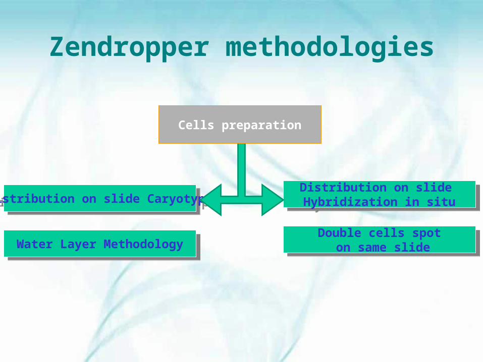
Zendropper methodologies
Distribution on slide CaryotypeDistribution on slide Caryotype
Water Layer MethodologyWater Layer Methodology
Distribution on slide Hybridization in situ
Distribution on slide Hybridization in situ
Double cells spot on same slide
Double cells spot on same slide
Cells preparation

Sampling
Cells culture
Cells preparation
Denaturation/Banding
Microscope Reading
Distribution on slide: ZenDropperDistribution on slide: ZenDropper
1. Karyotype procedure1. Karyotype procedure
• High automatic distribution capacity
• Homogeneous cells distribution
• 8 patients with 1 to 5 slides per patient (40 slides on 1 rack)
• Timing: (dispensing safety 100 %) +/- 25 ‘ for 40 slides Significant time saving
• Protection of technicians not exposed to methanol and acetic acid emanation from Carroy solution.

Karyotype : Water Layer technology
Distribution of a water layer on each slide
Needle containing sample is placed just above the slide
Creation of a central crater in the water layer due to Carroy solution evaporation
Ejection of sample on the slide
Aspiration of residual water on slide
Slide ready for Banding step
1
2
3
4
5
6

Hybridation in situ
Distribution of cell spot I
1
Distribution of cell spot II
2
3Slide ready for next step
Environment preparation :
T° , Hygrometry

Advantages of water layer technology
• Improved mitosis thanks to contact water & acetic acid/methanol mix on slide surface Chromosomes more spread
Banding (R or G) with better quality• Creation of a micro-environnement The
distribution will always be done with same hygrometric conditions independently of climatic conditions
Karyotype : Water Layer technology

Zendropper process
1 Positioning and identification of samples
2 Positioning standard microscopic slide
3 Automatic validation by the Zendropper, of the sample presence and determination of sample volume (from 500µl to 4000µl].
4 Water dispensing on the slide for sample 1
5 Cells mixing following 3 programs of mixing defined automatically in point 3.
6 Aspiration of sample volume needed to dispense on slides
7 Needle positioning above slide , waiting time to create central crater on water due to evaporation of Carroy solution and Sample ejection on slide

Zendropper process
8 Aspiration of residual water on slide
9 Next slide process
10 Needle rinsing and next patient
11 Informatic management of session and possibility to print session steps

Contamination :
Protocol : Cells pellets of 240 patients have been distributed on routine microscopic slide alterning male/female patient
Results : No chromosome Y observed on female samples and confirmed by Quinacrine test (100 mitosis per patient)
Conclusion : No contamination between patients
Comparative study Manual >< ZendropperCERBA Pasteur - Paris
(Genetics departement - More than 32.000 samples with Zendropper since March 2007)
Zendropper process validation

Quality of mitosis :Less crossing of chromosomes with Zendropper
Quantity of mitosis:More reproducible and homogenous dispersion of cells in mitosis with Zendropper
Quality of « Banding » : High level of homogeneity from slide to slide with Zendropper
Reading : Easier with Zendropper due to quality of dispersionVery efficient particularly for metaphasis research
Lab technicians reactions:Easier and fast method than manualLess exposition to toxic vapoursNo more difficult work due to work position on bench or with a glove box.
Zendropper process validation

• Dimensions: 60x60x80 cm• Weight: 62 kg• Rack with slides : 8 patient X max 5
slides• Tube holder for samples: 8• Extra washing needle solution: 2
positions• 2 needles
• Needle 1 :- Mixing, aspiration and distribution of samples + distribution of water
• Needle 2 : Aspiration of residual water
Technical Characteristics
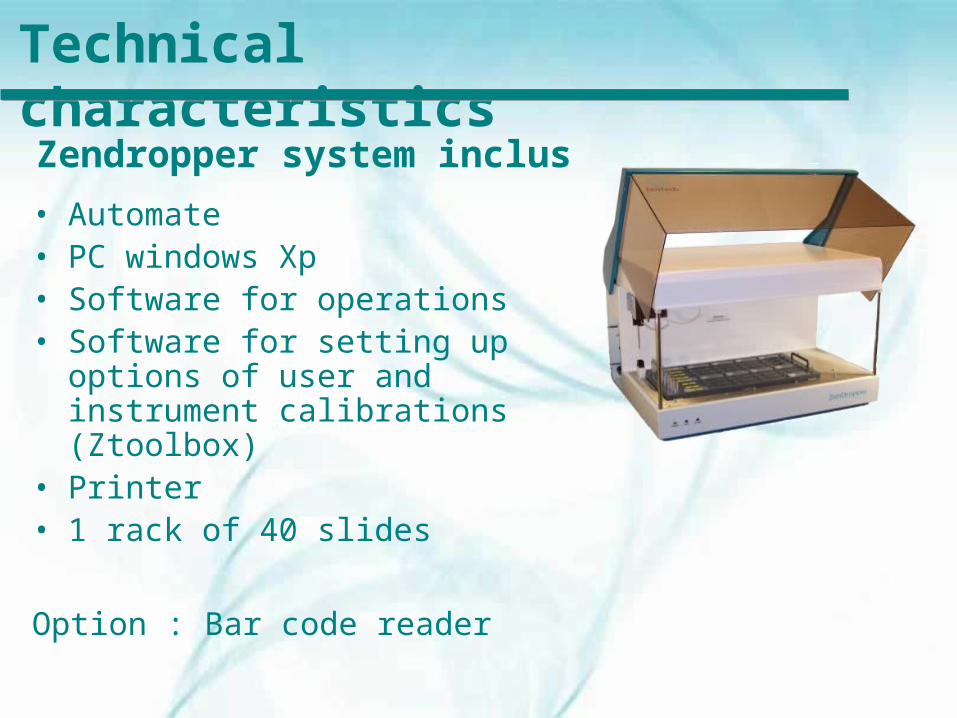
• Automate• PC windows Xp• Software for operations• Software for setting up
options of user and instrument calibrations (Ztoolbox)
• Printer• 1 rack of 40 slides
Option : Bar code reader
Technical characteristicsZendropper system inclus

Zendropper Software• Zendropper software
Session preparation : number of samples, number of slide/patient (codebar available) , print of session with information for tracability
• Ztoolbox Technical tool for maintenance and instrument set up in order to obtain high homogeneous cells dispersion conditions (Humidity and T°)

• Intensive rinsing procedure at start and end of the day
• Regular replacement of needles and tubing due to intense corrosion of solution
Zendropper maintenance
Distribuito in ITALIA da Li StarFish S.r.l.
Via Cavour, 35 - 20063 Cernusco S/N (MI)
Tel 02.92150794 - Fax 02.92157285
[email protected] - www.listarfish.it