cyclic nucleotide permeability through unopposed connexin ... · cyclic nucleotide permeability...
TRANSCRIPT
“fphar-04-00075” — 2013/6/5 — 10:20 — page 1 — #1
ORIGINAL RESEARCH ARTICLEpublished: 06 June 2013
doi: 10.3389/fphar.2013.00075
Cyclic nucleotide permeability through unopposedconnexin hemichannelsVirginijus Valiunas*
Department of Physiology and Biophysics, Stony Brook University, Stony Brook, NY, USA
Edited by:
Stefan Dhein, UniversitätsklinikLeipzig Herzzentrum Leipzig GmbH,Germany
Reviewed by:
John Philip Winpenny, University ofEast Anglia, UKJan Sebastian Schulte, University ofMünster, Germany
*Correspondence:
Virginijus Valiunas, Department ofPhysiology and Biophysics, StonyBrook University, Stony Brook,NY 11794-8661, USAe-mail: [email protected]
Cyclic adenosine monophosphate (cAMP) is a well-known intracellular and intercellularsecond messenger. The membrane permeability of such molecules has potential impor-tance for autocrine-like or paracrine-like delivery. Here experiments have been designedto demonstrate whether gap junction hemichannels, composed of connexins, are apossible entrance pathway for cyclic nucleotides into the interior of cells. HeLa cellsstably expressing connexin43 (Cx43) and connexin26 (Cx26) were used to study the cyclicnucleotide permeability of gap junction hemichannels. For the detection of cAMP uptake,the cells were transfected using the cyclic nucleotide-modulated channel from sea urchinsperm (SpIH) as the cAMP sensor. SpIH derived currents (Im) were recorded in whole-cell/perforated patch clamp configuration. Perfusion of the cells in an external K+ aspartate−(KAsp) solution containing 500 μM cAMP and no extracellular Ca2+, yielded a five tosevenfold increase in the Im current level. The SpIH current increase was associated withdetectable hemichannel current activity. Depolarization of cells in Ca2+-free NaCl perfusatewith 500 μM cAMP also induced a SpIH current increase. Elevating extracellular Ca2+to mM levels inhibited hemichannel activity. Perfusion with a depolarizing KAsp solutioncontaining 500 M cAMP and 2 mM Ca2μ + did not increase SpIH currents. The additionof the gap junction blocker carbenoxolone to the external solution inhibited cAMP uptake.Both cell depolarization and lowered extracellular Ca2+ increase the open probability ofnon-junctional hemichannels. Accordingly, the SpIH current augmentation was induced bythe uptake of extracellular cAMP via open membrane hemichannels in Cx43 and Cx26expressing cells. The data presented here show that hemichannels of Cx43 and Cx26 arepermeable to cAMP, and further the data suggest that hemichannels are, in fact, a potentialpathway for cAMP mediated cell-to-cell signaling.
Keywords: connexin43, connexin26, electrophysiology, gap junction, permeability, cyclic AMP
INTRODUCTIONHemichannels or connexons are composed of connexins, whichare synthesized in the endoplasmic reticulum and assembled intohexameric structures in the Golgi, and subsequently delivered tothe plasma membrane via transport vesicles (Segretain and Falk,2004). Hemichannels are large conductance membrane channelsthat for many years were thought to be silent or nearly silentsubunits. Their function was thought to be only when two suchchannels, between closely apposed cells, formed an intercellularchannel, the gap junction, containing an aqueous pore exclusive ofthe extracellular space. Once formed, gap junction channels even-tually coalesce into aggregates or plaques consisting of hundredsto thousands of channels (Maurer and Weingart, 1987; Bukauskaset al., 2000). However, not all hemichannels are destined to becomecomponent parts of a gap junction channel. Rather, many areapparently randomly distributed within the plasma membrane.The presence of hemichannels within plasma membranes hasbeen well-documented in vitro (De and Schwartz, 1992; Trexleret al., 1996; Valiunas and Weingart, 2000; Valiunas, 2002) andhas prompted speculation about their role in cellular processeslike volume regulation (Quist et al., 2000), the influx/efflux of
metabolically relevant solutes such as ATP (Bruzzone et al., 2001;Dahl and Locovei, 2006), and cell death (Plotkin et al., 2002;Kalvelyte et al., 2003). The electrophysiological data collected invitro have demonstrated that hemichannels open probability isincreased with membrane depolarization in the presence of low-ered extracellular calcium. The open probability can be reducedby acidic pH, calcium, trivalent cations, and quinine derivatives(Trexler et al., 1999; Contreras et al., 2002; Eskandari et al., 2002;Stout et al., 2002; Valiunas, 2002; Srinivas et al., 2005).
Cell to cell communication mediated by gap junction chan-nels represents one of two established intercellular pathwaysfor the movement of signaling molecules, metabolites, andsiRNA/miRNA from one cell to another (Valiunas et al., 2005;Kanaporis et al., 2008). Exocytosis/endocytosis is another inter-cellular delivery pathway that utilizes the extracellular volumefor autocrine and paracrine mediated signaling. The suggestedpossible roles for hemichannels, such as volume regulationand/or cell death would also be examples where delivery is viathe extracellular space and hence, represents an example ofautocrine/paracrine-like delivery. Furthermore, a recent reviewby Wang et al. (2013) also suggests hemichannels are a significant
www.frontiersin.org June 2013 | Volume 4 | Article 75 | 1
“fphar-04-00075” — 2013/6/5 — 10:20 — page 2 — #2
Valiunas cAMP permeability of gap junction hemichannels
source of autocrine and paracrine messengers. When consideringthe role of hemichannels in such a delivery system it is best toconsider the delivery pathway as autocrine-like or paracrine-likebecause delivery of a solute does not necessarily involve vesiculartraffic nor is it necessarily mediated by surface receptors.
Inevitably, an interesting question arises: are hemichannels analternate autocrine/paracrine-like pathway for delivery of relevantsignaling molecules, like adenosine and other related compounds?Two examples focus on cyclic adenosine monophosphate (cAMP)as a signal molecule candidate. The extracellular release of cAMPis known to exert effects such as receptor expression in renalcells (Kuzhikandathil et al., 2011) and inhibition of skeletal muscleinotropism (Duarte et al., 2012). Before addressing further ques-tions, such as if hemichannels are involved in a paracrine-likedelivery of cAMP, it is essential to understand the characteristicsof hemichannel permeability.
As a first step in assessing hemichannels as a potential deliverypathway, HeLa cells were transfected with cyclic nucleotide sensorSpIH in order to investigate the permeability of cAMP of unop-posed connexin43 (Cx43) and connexin26 (Cx26) hemichannels.
MATERIALS AND METHODSCELLS AND CULTURE CONDITIONSExperiments were carried out on human HeLa cells stably trans-fected with wild-type mCx43 and hCx26. HeLa cells were grownin Dulbecco’s Modified Eagle Medium (DMEM; Gibco BRL), sup-plemented with 10% fetal calf serum (FCS; Hyclone), 100 mg/mLstreptomycin (Gibco BRL), and 100 U/mL penicillin (Gibco BRL).The medium also contained 100 mg/mL hygromycin (Sigma) or1 mg/mL puromycin (Sigma). The cells were passaged weekly,diluted 1:10, and kept at 37◦C in a CO2 incubator (5% CO2/95%ambient air). Culture conditions for these cells have been previ-ously published in complete detail (Valiunas et al., 2000, 2001).Electrophysiological experiments were carried out on single cellscultured for 1–3 days.
IMMUNOFLUORESCENT LABELING OF CONNEXINSHeLa cells expressing Cx26 and/or Cx43 were grown on coverslipsand stained as described earlier (Laing and Beyer, 1995). Com-mercially available anti-connexin43 (Sigma) and anti-connexin26(Zymed Labs) antibodies were used for immunostaining. AlexaFluor488 conjugated anti-rabbit IgG (Cell Signaling) was used asa secondary antibody. The protein expression and localization wasmonitored with a 63× oil objective on a Zeiss Axiovert 200 invertedmicroscope and Axiovision (Zeiss) software.
ELECTROPHYSIOLOGICAL MEASUREMENTSExperiments were carried out on single cells using the whole-cell/perforated patch voltage-clamp technique to control themembrane potential and to measure membrane currents of thecell. For electrical recordings, glass coverslips with adherent cellswere transferred to an experimental chamber mounted on thestage of an inverted microscope (Olympus- IX71). During exper-iments, the cells were superfused with depolarizing bath solution(KAsp) at room temperature (∼22◦C) containing (mM): K+aspartate− 120; NaCl 10; CaCl2 2; HEPES 5 (pH 7.4); glucose5; 2 mM CsCl, BaCl2 and TEA+ Cl− were also added. For the
Ca2+-free (0 Ca2+) bath solution CaCl2 was omitted. For the reg-ular modified Tyrode external solution (NaCl), K+ aspartate− inthe superfusate was replaced with an equal molar concentrationof NaCl. The patch pipettes were filled with solution containing(mM): K+ aspartate−, 120; NaCl, 10; MgATP, 3; HEPES, 5 (pH7.2); EGTA, 10 (pCa ∼8); filtered through 0.22 μm pores. In perfo-rated patch experiments, the pipette solution contained 30–50 μMβ-escin (Fan and Palade, 1998). The series resistance with β-escinpatches measured 11–20 M�. Patch pipettes were pulled fromglass capillaries (code GC150F; Harvard Apparatus) with a hor-izontal puller (DMZ-Universal, Zeitz-Instrumente). When filled,the resistance of the pipettes measured 1–4 M�.
cAMP-UPTAKE STUDIESCyclic AMP transfer through gap junction hemichannels wasinvestigated using single HeLaCx43 and/or HeLaCx26 cells. Forthe detection of cAMP uptake, the cells were transfected withthe cAMP sensor, a cyclic nucleotide-modulated channel from seaurchin sperm (SpIH; Gauss et al., 1998; Shin et al., 2001; Kanaporiset al., 2008). Production, characterization, culture conditions,staining, and visualization of these cells have been describedpreviously in Kanaporis et al. (2008).
Wild-type, Cx43 and Cx26 transfected cells were incubatedin either in NaCl or K+ aspartate− (KAsp) bath solution (with2 mM Ca2+ or Ca2+-free). For cAMP uptake experiments cAMP(Sigma-Aldrich) was added to the external bath solution to reacha concentration of 500 μM. The SpIH derived currents (Im) wererecorded from the single cell expressing SpIH and Cx43 and/orCx26. In some experiments 50 μM of cAMP was introducedvia the patch pipette directly in to the cell. In another series ofexperiments 500 μM cAMP was also locally introduced to the cellmembrane via the external pipette. To prevent cAMP degrada-tion a membrane-permeable phosphodiesterase inhibitor IBMX(200 mM, Sigma-Aldrich) was added to the bath solution. Anadenylate cyclase inhibitor, 2′,5′-dideoxyadenosine (5 mM, Cal-biochem) was added to the pipette and bath solutions to inhibitintracellular cAMP production.
SIGNAL RECORDING AND ANALYSISVoltage and current signals were recorded using patch clampamplifiers (Axopatch 200). The current signals were digitized witha 16 bit A/D-converter (Digidata 1322A; Molecular Devices) andstored within a personal computer. Data acquisition and analysiswere performed with pClamp9 software (Molecular Devices). Sta-tistical analysis was performed using SigmaStat (Jandel Scientific).The Mann–Whitney Rank Sum test was used for all cases unlessotherwise noted. The results are presented as means ± SEM.
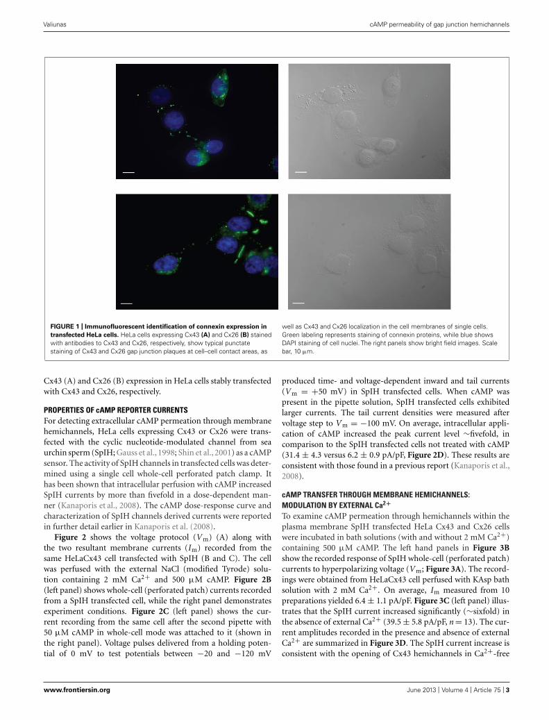
RESULTSLOCALIZATION OF CONNEXINS WITHIN CELLSHeLa cells stably transfected with mCx43 and hCx26 wereimmunostained with anti-Cx43 and anti-Cx26 antibodies, respec-tively. Immunofluorescent staining verified protein expression andlocalization of Cx43 and Cx26 within the cells (Figure 1). As shownin Figure 1, typical punctate staining (in green) at the cell to cellcontact regions and the cell membranes of single cells indicates
Frontiers in Pharmacology | Pharmacology of Ion Channels and Channelopathies June 2013 | Volume 4 | Article 75 | 2
“fphar-04-00075” — 2013/6/5 — 10:20 — page 3 — #3
Valiunas cAMP permeability of gap junction hemichannels
FIGURE 1 | Immunofluorescent identification of connexin expression in
transfected HeLa cells. HeLa cells expressing Cx43 (A) and Cx26 (B) stainedwith antibodies to Cx43 and Cx26, respectively, show typical punctatestaining of Cx43 and Cx26 gap junction plaques at cell–cell contact areas, as
well as Cx43 and Cx26 localization in the cell membranes of single cells.Green labeling represents staining of connexin proteins, while blue showsDAPI staining of cell nuclei. The right panels show bright field images. Scalebar, 10 μm.
Cx43 (A) and Cx26 (B) expression in HeLa cells stably transfectedwith Cx43 and Cx26, respectively.
PROPERTIES OF cAMP REPORTER CURRENTSFor detecting extracellular cAMP permeation through membranehemichannels, HeLa cells expressing Cx43 or Cx26 were trans-fected with the cyclic nucleotide-modulated channel from seaurchin sperm (SpIH; Gauss et al., 1998; Shin et al., 2001) as a cAMPsensor. The activity of SpIH channels in transfected cells was deter-mined using a single cell whole-cell perforated patch clamp. Ithas been shown that intracellular perfusion with cAMP increasedSpIH currents by more than fivefold in a dose-dependent man-ner (Kanaporis et al., 2008). The cAMP dose-response curve andcharacterization of SpIH channels derived currents were reportedin further detail earlier in Kanaporis et al. (2008).
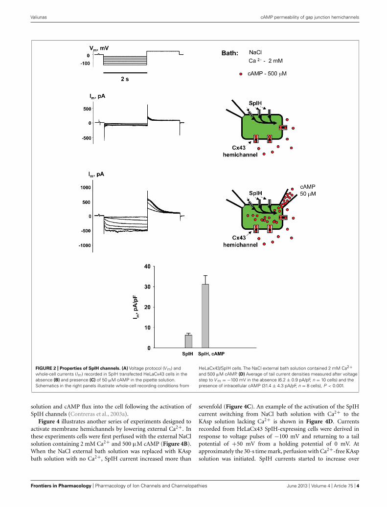
Figure 2 shows the voltage protocol (V m) (A) along withthe two resultant membrane currents (Im) recorded from thesame HeLaCx43 cell transfected with SpIH (B and C). The cellwas perfused with the external NaCl (modified Tyrode) solu-tion containing 2 mM Ca2+ and 500 μM cAMP. Figure 2B(left panel) shows whole-cell (perforated patch) currents recordedfrom a SpIH transfected cell, while the right panel demonstratesexperiment conditions. Figure 2C (left panel) shows the cur-rent recording from the same cell after the second pipette with50 μM cAMP in whole-cell mode was attached to it (shown inthe right panel). Voltage pulses delivered from a holding poten-tial of 0 mV to test potentials between −20 and −120 mV
produced time- and voltage-dependent inward and tail currents(V m = +50 mV) in SpIH transfected cells. When cAMP waspresent in the pipette solution, SpIH transfected cells exhibitedlarger currents. The tail current densities were measured aftervoltage step to V m = −100 mV. On average, intracellular appli-cation of cAMP increased the peak current level ∼fivefold, incomparison to the SpIH transfected cells not treated with cAMP(31.4 ± 4.3 versus 6.2 ± 0.9 pA/pF, Figure 2D). These results areconsistent with those found in a previous report (Kanaporis et al.,2008).
cAMP TRANSFER THROUGH MEMBRANE HEMICHANNELS:MODULATION BY EXTERNAL Ca2+
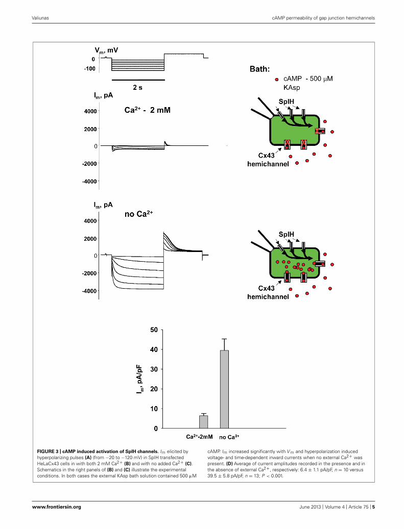
To examine cAMP permeation through hemichannels within theplasma membrane SpIH transfected HeLa Cx43 and Cx26 cellswere incubated in bath solutions (with and without 2 mM Ca2+)containing 500 μM cAMP. The left hand panels in Figure 3Bshow the recorded response of SpIH whole-cell (perforated patch)currents to hyperpolarizing voltage (V m; Figure 3A). The record-ings were obtained from HeLaCx43 cell perfused with KAsp bathsolution with 2 mM Ca2+. On average, Im measured from 10preparations yielded 6.4 ± 1.1 pA/pF. Figure 3C (left panel) illus-trates that the SpIH current increased significantly (∼sixfold) inthe absence of external Ca2+ (39.5 ± 5.8 pA/pF, n = 13). The cur-rent amplitudes recorded in the presence and absence of externalCa2+ are summarized in Figure 3D. The SpIH current increase isconsistent with the opening of Cx43 hemichannels in Ca2+-free
www.frontiersin.org June 2013 | Volume 4 | Article 75 | 3
“fphar-04-00075” — 2013/6/5 — 10:20 — page 4 — #4
Valiunas cAMP permeability of gap junction hemichannels
FIGURE 2 | Properties of SpIH channels. (A) Voltage protocol (V m) andwhole-cell currents (Im) recorded in SpIH transfected HeLaCx43 cells in theabsence (B) and presence (C) of 50 μM cAMP in the pipette solution.Schematics in the right panels illustrate whole-cell recording conditions from
HeLaCx43/SpIH cells. The NaCl external bath solution contained 2 mM Ca2+and 500 μM cAMP. (D) Average of tail current densities measured after voltagestep to V m = −100 mV in the absence (6.2 ± 0.9 pA/pF, n = 10 cells) and thepresence of intracellular cAMP (31.4 ± 4.3 pA/pF, n = 8 cells), P < 0.001.
solution and cAMP flux into the cell following the activation ofSpIH channels (Contreras et al., 2003a).
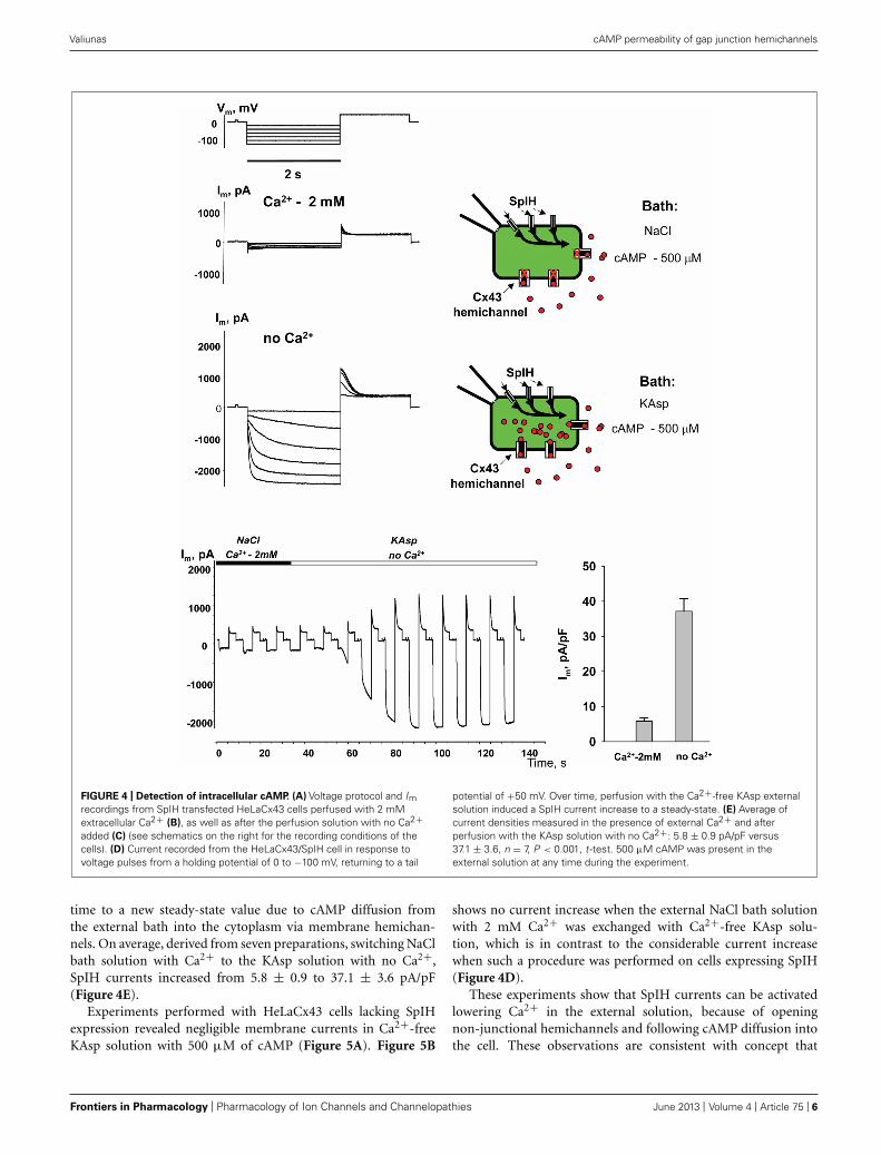
Figure 4 illustrates another series of experiments designed toactivate membrane hemichannels by lowering external Ca2+. Inthese experiments cells were first perfused with the external NaClsolution containing 2 mM Ca2+ and 500 μM cAMP (Figure 4B).When the NaCl external bath solution was replaced with KAspbath solution with no Ca2+, SpIH current increased more than
sevenfold (Figure 4C). An example of the activation of the SpIHcurrent switching from NaCl bath solution with Ca2+ to theKAsp solution lacking Ca2+ is shown in Figure 4D. Currentsrecorded from HeLaCx43 SpIH-expressing cells were derived inresponse to voltage pulses of −100 mV and returning to a tailpotential of +50 mV from a holding potential of 0 mV. Atapproximately the 30-s time mark, perfusion with Ca2+-free KAspsolution was initiated. SpIH currents started to increase over
Frontiers in Pharmacology | Pharmacology of Ion Channels and Channelopathies June 2013 | Volume 4 | Article 75 | 4
“fphar-04-00075” — 2013/6/5 — 10:20 — page 5 — #5
Valiunas cAMP permeability of gap junction hemichannels
FIGURE 3 | cAMP induced activation of SpIH channels. Im elicited byhyperpolarizing pulses (A) (from −20 to −120 mV) in SpIH transfectedHeLaCx43 cells in with both 2 mM Ca2+ (B) and with no added Ca2+ (C).Schematics in the right panels of (B) and (C) illustrate the experimentalconditions. In both cases the external KAsp bath solution contained 500 μM
cAMP. Im increased significantly with V m and hyperpolarization inducedvoltage- and time-dependent inward currents when no external Ca2+ waspresent. (D) Average of current amplitudes recorded in the presence and inthe absence of external Ca2+, respectively: 6.4 ± 1.1 pA/pF, n = 10 versus39.5 ± 5.8 pA/pF, n = 13; P < 0.001.
www.frontiersin.org June 2013 | Volume 4 | Article 75 | 5
“fphar-04-00075” — 2013/6/5 — 10:20 — page 6 — #6
Valiunas cAMP permeability of gap junction hemichannels
FIGURE 4 | Detection of intracellular cAMP. (A) Voltage protocol and Imrecordings from SpIH transfected HeLaCx43 cells perfused with 2 mMextracellular Ca2+ (B), as well as after the perfusion solution with no Ca2+added (C) (see schematics on the right for the recording conditions of thecells). (D) Current recorded from the HeLaCx43/SpIH cell in response tovoltage pulses from a holding potential of 0 to −100 mV, returning to a tail
potential of +50 mV. Over time, perfusion with the Ca2+-free KAsp externalsolution induced a SpIH current increase to a steady-state. (E) Average ofcurrent densities measured in the presence of external Ca2+ and afterperfusion with the KAsp solution with no Ca2+: 5.8 ± 0.9 pA/pF versus37.1 ± 3.6, n = 7, P < 0.001, t -test. 500 μM cAMP was present in theexternal solution at any time during the experiment.
time to a new steady-state value due to cAMP diffusion fromthe external bath into the cytoplasm via membrane hemichan-nels. On average, derived from seven preparations, switching NaClbath solution with Ca2+ to the KAsp solution with no Ca2+,SpIH currents increased from 5.8 ± 0.9 to 37.1 ± 3.6 pA/pF(Figure 4E).
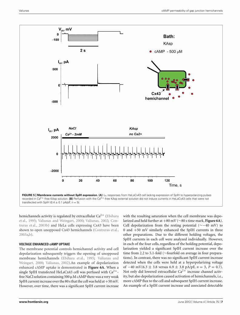
Experiments performed with HeLaCx43 cells lacking SpIHexpression revealed negligible membrane currents in Ca2+-freeKAsp solution with 500 μM of cAMP (Figure 5A). Figure 5B
shows no current increase when the external NaCl bath solutionwith 2 mM Ca2+ was exchanged with Ca2+-free KAsp solu-tion, which is in contrast to the considerable current increasewhen such a procedure was performed on cells expressing SpIH(Figure 4D).
These experiments show that SpIH currents can be activatedlowering Ca2+ in the external solution, because of openingnon-junctional hemichannels and following cAMP diffusion intothe cell. These observations are consistent with concept that
Frontiers in Pharmacology | Pharmacology of Ion Channels and Channelopathies June 2013 | Volume 4 | Article 75 | 6
“fphar-04-00075” — 2013/6/5 — 10:20 — page 7 — #7
Valiunas cAMP permeability of gap junction hemichannels
FIGURE 5 | Membrane currents without SpIH expression. (A) Im responses from HeLaCx43 cell lacking expression of SpIH to hyperpolarizing pulsesrecorded in Ca2+-free KAsp solution. (B) Perfusion with the Ca2+-free KAsp external solution did not induce currents in HeLaCx43 cells that were nottransfected with SpIH (0.4 ± 0.1 pA/pF, n = 5).
hemichannels activity is regulated by extracellular Ca2+ (Ebiharaet al., 1995; Valiunas and Weingart, 2000; Valiunas, 2002; Con-treras et al., 2003b) and HeLa cells expressing Cx43 have beenshown to open unopposed Cx43 hemichannels (Contreras et al.,2003a,b).
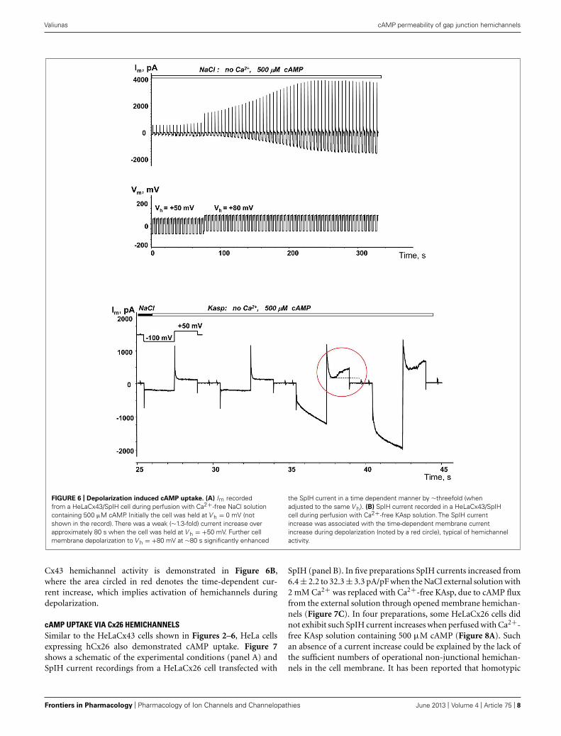
VOLTAGE ENHANCED cAMP UPTAKEThe membrane potential controls hemichannel activity and celldepolarization subsequently triggers the opening of unopposedmembrane hemichannels (Ebihara et al., 1995; Valiunas andWeingart, 2000; Valiunas, 2002).An example of depolarizationenhanced cAMP uptake is demonstrated in Figure 6A. When asingle SpIH transfected HeLaCx43 cell was perfused with Ca2+-free NaCl solution containing 500 μM cAMP there was a very weakSpIH current increase over the 80 s that the cell was held at +50 mV.However, over time, there was a significant SpIH current increase
with the resulting saturation when the cell membrane was depo-larized and held further at +80 mV (∼80 s time mark, Figure 6A).Cell depolarization from the resting potential (∼−40 mV) to0 and +50 mV similarly enhanced the SpIH currents in threeother preparations. Due to the different holding voltages, theSpIH currents in each cell were analyzed individually. However,in each of the four cells, regardless of the holding potential, depo-larization yielded a significant SpIH current increase over thetime from 2.2 to 5.1-fold (∼fourfold on average in four prepara-tions). In contrast, there was no significant SpIH current increasedetected when the cells were held at a hyperpolarizing voltageof −40 mV(6.3 ± 3.8 versus 6.9 ± 3.8 pA/pF, n = 3, P = 0.7).Not only did lowered extracellular Ca2+ increase channel activ-ity, but also depolarization caused activation of hemichannels, i.e.,more cAMP flux to the cell and subsequent SpIH current increase.An example of a SpIH current increase and associated detectable
www.frontiersin.org June 2013 | Volume 4 | Article 75 | 7
“fphar-04-00075” — 2013/6/5 — 10:20 — page 8 — #8
Valiunas cAMP permeability of gap junction hemichannels
FIGURE 6 | Depolarization induced cAMP uptake. (A) Im recordedfrom a HeLaCx43/SpIH cell during perfusion with Ca2+-free NaCl solutioncontaining 500 μM cAMP. Initially the cell was held at V h = 0 mV (notshown in the record). There was a weak (∼1.3-fold) current increase overapproximately 80 s when the cell was held at V h = +50 mV. Further cellmembrane depolarization to V h = +80 mV at ∼80 s significantly enhanced
the SpIH current in a time dependent manner by ∼threefold (whenadjusted to the same V h). (B) SpIH current recorded in a HeLaCx43/SpIHcell during perfusion with Ca2+-free KAsp solution. The SpIH currentincrease was associated with the time-dependent membrane currentincrease during depolarization (noted by a red circle), typical of hemichannelactivity.
Cx43 hemichannel activity is demonstrated in Figure 6B,where the area circled in red denotes the time-dependent cur-rent increase, which implies activation of hemichannels duringdepolarization.
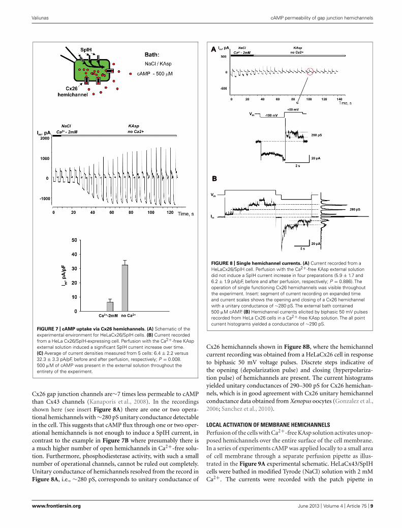
cAMP UPTAKE VIA Cx26 HEMICHANNELSSimilar to the HeLaCx43 cells shown in Figures 2–6, HeLa cellsexpressing hCx26 also demonstrated cAMP uptake. Figure 7shows a schematic of the experimental conditions (panel A) andSpIH current recordings from a HeLaCx26 cell transfected with
SpIH (panel B). In five preparations SpIH currents increased from6.4 ± 2.2 to 32.3 ± 3.3 pA/pF when the NaCl external solution with2 mM Ca2+ was replaced with Ca2+-free KAsp, due to cAMP fluxfrom the external solution through opened membrane hemichan-nels (Figure 7C). In four preparations, some HeLaCx26 cells didnot exhibit such SpIH current increases when perfused with Ca2+-free KAsp solution containing 500 μM cAMP (Figure 8A). Suchan absence of a current increase could be explained by the lack ofthe sufficient numbers of operational non-junctional hemichan-nels in the cell membrane. It has been reported that homotypic
Frontiers in Pharmacology | Pharmacology of Ion Channels and Channelopathies June 2013 | Volume 4 | Article 75 | 8
“fphar-04-00075” — 2013/6/5 — 10:20 — page 9 — #9
Valiunas cAMP permeability of gap junction hemichannels
FIGURE 7 | cAMP uptake via Cx26 hemichannels. (A) Schematic of theexperimental environment for HeLaCx26/SpIH cells. (B) Current recordedfrom a HeLa Cx26/SpIH-expressing cell. Perfusion with the Ca2+-free KAspexternal solution induced a significant SpIH current increase over time.(C) Average of current densities measured from 5 cells: 6.4 ± 2.2 versus32.3 ± 3.3 pA/pF, before and after perfusion, respectively; P = 0.008.500 μM of cAMP was present in the external solution throughout theentirety of the experiment.
Cx26 gap junction channels are∼7 times less permeable to cAMPthan Cx43 channels (Kanaporis et al., 2008). In the recordingsshown here (see insert Figure 8A) there are one or two opera-tional hemichannels with ∼280 pS unitary conductance detectablein the cell. This suggests that cAMP flux through one or two oper-ational hemichannels is not enough to induce a SpIH current, incontrast to the example in Figure 7B where presumably there isa much higher number of open hemichannels in Ca2+-free solu-tion. Furthermore, phosphodiesterase activity, with such a smallnumber of operational channels, cannot be ruled out completely.Unitary conductance of hemichannels resolved from the record inFigure 8A, i.e., ∼280 pS, corresponds to unitary conductance of
FIGURE 8 | Single hemichannel currents. (A) Current recorded from aHeLaCx26/SpIH cell. Perfusion with the Ca2+-free KAsp external solutiondid not induce a SpIH current increase in four preparations (5.9 ± 1.7 and6.2 ± 1.9 pA/pF, before and after perfusion, respectively; P = 0.886). Theoperation of single functioning Cx26 hemichannels was visible throughoutthe experiment. Insert: segment of current recording on expanded timeand current scales shows the opening and closing of a Cx26 hemichannelwith a unitary conductance of ∼280 pS. The external bath contained500 μM cAMP. (B) Hemichannel currents elicited by biphasic 50 mV pulsesrecorded from HeLa Cx26 cells in a Ca2+-free KAsp solution. The all pointcurrent histograms yielded a conductance of ∼290 pS.
Cx26 hemichannels shown in Figure 8B, where the hemichannelcurrent recording was obtained from a HeLaCx26 cell in responseto biphasic 50 mV voltage pulses. Discrete steps indicative ofthe opening (depolarization pulse) and closing (hyperpolariza-tion pulse) of hemichannels are present. The current histogramsyielded unitary conductances of 290–300 pS for Cx26 hemichan-nels, which is in good agreement with Cx26 unitary hemichannelconductance data obtained from Xenopus oocytes (Gonzalez et al.,2006; Sanchez et al., 2010).
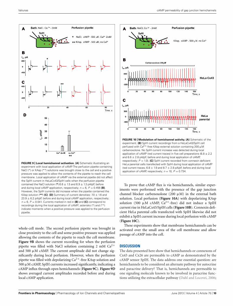
LOCAL ACTIVATION OF MEMBRANE HEMICHANNELSPerfusion of the cells with Ca2+-free KAsp solution activates unop-posed hemichannels over the entire surface of the cell membrane.In a series of experiments cAMP was applied locally to a small areaof cell membrane through a separate perfusion pipette as illus-trated in the Figure 9A experimental schematic. HeLaCx43/SpIHcells were bathed in modified Tyrode (NaCl) solution with 2 mMCa2+. The currents were recorded with the patch pipette in
www.frontiersin.org June 2013 | Volume 4 | Article 75 | 9
“fphar-04-00075” — 2013/6/5 — 10:20 — page 10 — #10
Valiunas cAMP permeability of gap junction hemichannels
FIGURE 9 | Local hemichannel activation. (A) Schematic illustrating anexperiment with local application of cAMP. The perfusion pipette containingNaCl (*) or KAsp (**) solutions was brought close to the cell and a positivepressure was applied to allow the contents of the pipette to reach the cellmembrane. Local application of cAMP via the external pipette did not affectthe SpIH current in HeLaCx43/SpIH cells when the perfusion pipettecontained the NaCl solution (*):6.6 ± 1.5 and 6.9 ± 1.3 pA/pF, beforeand during local cAMP application, respectively; n = 6, P = 0.456 (B).However, the SpIH currents did increase when the pipette contained theKAsp solution (**) (C). (D) Summary of current densities: 7.0 ± 1.6 and23.9 ± 6.2 pA/pF, before and during local cAMP application, respectively;n = 6, P = 0.041. Currents marked in red in (B) and (C) correspond torecordings during the local application of cAMP; asterisks (*) and (**)indicate moments when a positive pressure was applied to the perfusionpipette.
whole-cell mode. The second perfusion pipette was brought inclose proximity to the cell and some positive pressure was appliedallowing the contents of the pipette to reach the cell membrane.Figure 9B shows the current recording for when the perfusionpipette was filled with NaCl solution containing 2 mM Ca2+and 500 μM cAMP. The current amplitude did not change sig-nificantly during local perfusion. However, when the perfusionpipette was filled with depolarizing Ca2+-free KAsp solution and500 μM cAMP, SpIH currents increased significantly, indicating acAMP influx through open hemichannels (Figure 9C). Figure 9Dshows averaged current amplitudes recorded before and duringlocal cAMP application.
FIGURE 10 | Modulation of hemichannel activity. (A) Schematic of theexperiment. (B) SpIH current recordings from a HeLaCx43/SpIH cellperfused with Ca2+-free KAsp external solution containing 200 μMcarbenoxolone. No SpIH current increase was detected during localapplication of cAMP (red current traces) in five cell preparations (6.8 ± 2.2and 6.6 ± 2.8 pA/pF, before and during local application of cAMP,respectively; P = 1.0). (C) SpIH current recorded from connexin deficientHeLa parental cells transfected with SpIH during local application of cAMP(red current traces; 6.8 ± 1.9 and 8.7 ± 2.8 pA/pF, before and during localapplication of cAMP, respectively; n = 10, P = 0.734.
To prove that cAMP flux is via hemichannels, similar exper-iments were performed with the presence of the gap junctionchannel blocker carbenoxolone (200 μM) in the external bathsolution. Local perfusion (Figure 10A) with depolarizing KAspsolution (500 μM cAMP, Ca2+-free) did not induce a SpIHcurrent rise in HeLaCx43/SpIH cells (Figure 10B). Connexin defi-cient HeLa parental cells transfected with SpIH likewise did notexhibit a SpIH current increase during local perfusion with cAMP(Figure 10C).
These experiments show that membrane hemichannels can beactivated over the small area of the cell membrane and allowpassage of cAMP into the cell.
DISCUSSIONThe data presented here show that hemichannels or connexons ofCx43 and Cx26 are permeable to cAMP as demonstrated by thecAMP sensor SpIH. The data address one essential question: arehemichannels to be considered an alternate pathway for autocrineand paracrine delivery? That is, hemichannels are permeable toone signaling molecule known to be involved in paracrine func-tions utilizing the extracellular pathway (Dahl and Locovei, 2006;
Frontiers in Pharmacology | Pharmacology of Ion Channels and Channelopathies June 2013 | Volume 4 | Article 75 | 10
“fphar-04-00075” — 2013/6/5 — 10:20 — page 11 — #11
Valiunas cAMP permeability of gap junction hemichannels
Anselmi et al., 2008; Kanaporis et al., 2008; Kang et al., 2008; Wanget al., 2013).
One issue that has yet to be clearly addressed is the trueoperational limits of hemichannels. Previous works have oftenused lowered extracellular Ca2+ to allow easy demonstrationof hemichannel currents. When extracellular Ca2+ is in theμM range, macroscopic or multichannel data has revealed thathemichannels have a very high open probability with depolariza-tion (Ebihara et al., 1995; Valiunas and Weingart, 2000; Beahm andHall, 2002; Valiunas, 2002; Gonzalez et al., 2006).
Ischemia is known to result in membrane depolarization of car-diac myocytes (Kleber et al., 1978; Kleber, 1983) and cells of otheraerobic tissues (Al-Mehdi et al., 1998; Calabresi et al., 1999; Dreier,2011). As such, hemichannels whether activated by altered Ca2+levels and/or membrane depolarization have the potential to allowboth the influx and efflux of solutes. Consistent with this notion isactivation of hCx26 hemichannels at voltages above −40 mV (Stef-fens et al., 2008). In the case of ischemia it is not yet clear whetherhemichannel activity is the cause of membrane depolarization oris simply part of the effect. One can speculate that if hemichannelsare causal in ischemic depolarization then hemichannels becomereal therapeutic targets.
The large conductance and permissive selectivity of Cx43and Cx26 would also allow significant delivery of solutes likecAMP, possibly with even extremely low open probabilities onthe order of 0.1–1%. In fact, little is known about the activity ofhemichannels when extracellular Ca2+ is between 1 and 2 mM,the expected range for the interstitial space. What remains to bedetermined is the open probability of hemichannels versus extra-cellular Ca2+ while utilizing conditions to silence other channeltypes, i.e., K+ channels. For a cell with 10000 hemichannelson its surface and with an imaged open probability of 1% innormal extracellular Ca2+ the number of functioning channelswould be approximately 100 open channels at any instant in time.Assuming the cAMP/K+ permeability ratio for Cx43 and Cx26hemichannels is similar to their respective gap junction channelsthen the flux per channel is as previously reported (Kanaporiset al., 2008), approximately 6000 molecules/channel/sec for Cx43and approximately 1800 molecules/channel/sec for Cx26. For 100functioning channels the total flux per cell would be 0.6 mil-lion molecules/sec for Cx43 and 0.18 million for Cx26. Whethersuch an efflux is sufficient to function as an autocrine/paracrinesource of signal molecules like cAMP remains tobe seen.
Understanding the potential role of hemichannels, unrelatedto their precursor role in the formation of gap junction chan-nels, as a potential autocrine/paracrine signaling pathway is areal challenge. It is challenging both from a biophysical perspec-tive and the assessment of autocrine and paracrine effects withintissues. Open probability versus calcium is an important biophys-ical parameter to clearly define, but it is also necessary to testautocrine/paracrine delivery mediated by hemichannels withoutan endosomal/vesicular background. The latter can be accom-plished using drugs that inhibit or block endosomal/vesiculartraffic. This study focused on hemichannels composed of con-nexins but an equally plausible hemichannel construct is ahemichannel composed of pannexins (Wang et al., 2013). Thus,it remains to be seen whether connexins or pannexins are trulyable to function as autocrine/paracrine-like delivery systems.
In this study, the SpIH gene, which is a cyclic nucleotide gatedchannel, was used to assess the permeability of hemichannels tocAMP. This is a useful method, which allows accurate estimatesof cyclic nucleotide flux via different membrane hemichannels. Insuch cases, it is the most suitable approach for quick screeningof connexin mutants. Defining the permeability and selectivityproperties of hemichannels are important factors in understand-ing their potential role in normal cell physiology and disease stateswith connexin mutations.
A final question posed is if there is a role for autocrineand paracrine delivery mediated by hemichannels within themyocardium? Presently autocrine and paracrine functions forhemichannels are open to debate, but there is strong evidencethat cAMP influx can act to reduce the sodium current incardiac myocytes (Hofer and Lefkimmiatis, 2007). Autocrineand paracrine-like functions are also a possible explanationfor enhanced endothelin expression in response to ischemia orhypertrophy (Drawnel et al., 2013).
A number of reports reviewed in Wang et al. (2013) demon-strated that hemichannels can function as pathways for paracrinemessengers, including ATP and prostaglandins. This study nowadds an additional potential paracrine-like messenger in the formof cAMP.
ACKNOWLEDGMENTSThis work was supported by the National Institutes of Health grantGM-088181. The author would like to thank Dr. P. Brink and Dr.T. White for their critical comments on the manuscript, C. Gordonand Dr. Kanaporis for the expert technical assistance.
REFERENCESAl-Mehdi, A. B., Zhao, G., and Fisher,
A. B. (1998). ATP-independent mem-brane depolarization with ischemiain the oxygen-ventilated isolated ratlung. Am. J. Respir. Cell Mol.Biol. 18, 653–661. doi: 10.1165/ajr-cmb.18.5.2834
Anselmi, F., Hernandez, V. H., Crispino,G., Seydel, A., Ortolano, S., Roper,S. D., et al. (2008). ATP releasethrough connexin hemichannels andgap junction transfer of secondmessengers propagate Ca2+ signals
across the inner ear. Proc. Natl. Acad.Sci. U.S.A. 105, 18770–18775. doi:10.1073/pnas.0800793105
Beahm, D. L., and Hall, J. E. (2002).Hemichannel and junctional prop-erties of connexin 50. Biophys. J.82, 2016–2031. doi: 10.1016/S0006-3495(02)75550-1
Bruzzone, S., Franco, L., Guida, L.,Zocchi, E., Contini, P., Bisso, A.,et al. (2001). A self-restricted CD38-connexin 43 cross-talk affects NAD+and cyclic ADP-ribose metabolismand regulates intracellular calcium
in 3T3 fibroblasts. J. Biol. Chem.276, 48300–48308. doi: 10.1074/jbc.M107308200
Bukauskas, F. F., Jordan, K.,Bukauskiene, A., Bennett, M. V.,Lampe, P. D., Laird, D. W., et al.(2000). Clustering of connexin 43-enhanced green fluorescent proteingap junction channels and functionalcoupling in living cells. Proc. Natl.Acad. Sci. U.S.A. 97, 2556–2561. doi:10.1073/pnas.050588497
Calabresi, P., Marfia, G. A., Cen-tonze, D., Pisani, A., and Bernardi,
G. (1999). Sodium influx playsa major role in the membranedepolarization induced by oxygenand glucose deprivation in rat stri-atal spiny neurons. Stroke 30,171–179. doi: 10.1161/01.STR.30.1.171
Contreras, J. E., Saez, J. C., Bukauskas,F. F., and Bennett, M. V. (2003a).Functioning of cx43 hemichannelsdemonstrated by single channelproperties. Cell Commun. Adhes.10, 245–249. doi: 10.1080/cac.10.4-6.245.249
www.frontiersin.org June 2013 | Volume 4 | Article 75 | 11
“fphar-04-00075” — 2013/6/5 — 10:20 — page 12 — #12
Valiunas cAMP permeability of gap junction hemichannels
Contreras, J. E., Saez, J. C., Bukauskas,F. F., and Bennett, M. V. (2003b).Gating and regulation of connexin43 (Cx43) hemichannels. Proc. Natl.Acad. Sci. U.S.A. 100, 11388–11393.doi: 10.1073/pnas.1434298100
Contreras, J. E., Sanchez, H. A., Eugenin,E. A., Speidel, D., Theis, M., Wil-lecke, K., et al. (2002). Metabolicinhibition induces opening of unap-posed connexin 43 gap junctionhemichannels and reduces gap junc-tional communication in corticalastrocytes in culture. Proc. Natl.Acad. Sci. U.S.A. 99, 495–500. doi:10.1073/pnas.012589799
Dahl, G., and Locovei, S. (2006). Pan-nexin: to gap or not to gap, is that aquestion? IUBMB Life 58, 409–419.doi: 10.1080/15216540600794526
De, V. S. H., and Schwartz, E. A.(1992). Hemi-gap-junction channelsin solitary horizontal cells of the cat-fish retina. J. Physiol. (Lond.) 445,201–230.
Drawnel, F. M., Archer, C. R., andRoderick, H. L. (2013). The roleof the paracrine/autocrine mediatorendothelin-1 in regulation of car-diac contractility and growth. Br.J. Pharmacol. 168, 296–317. doi:10.1111/j.1476-5381.2012.02195.x
Dreier, J. P. (2011). The role of spreadingdepression, spreading depolarizationand spreading ischemia in neurolog-ical disease. Nat. Med. 17, 439–447.doi: 10.1038/nm.2333
Duarte, T., Menezes-Rodrigues, F. S.,and Godinho, R. O. (2012). Con-tribution of the extracellular cAMP-adenosine pathway to dual couplingof beta2-adrenoceptors to Gs and Giproteins in mouse skeletal muscle. J.Pharmacol. Exp. Ther. 341, 820–828.doi: 10.1124/jpet.112.192997
Ebihara, L., Berthoud, V. M., andBeyer, E. C. (1995). Distinct behaviorof connexin56 and connexin46 gapjunctional channels can be predictedfrom the behavior of their hemi-gap-junctional channels. Biophys. J.68, 1796–1803. doi: 10.1016/S0006-3495(95)80356-5
Eskandari, S., Zampighi, G. A., Leung,D. W., Wright, E. M., and Loo, D.D. (2002). Inhibition of gap junctionhemichannels by chloride channelblockers. J. Membr. Biol. 185, 93–102.doi: 10.1007/s00232-001-0115-0
Fan, J. S., and Palade, P. (1998). Per-forated patch recording with beta-escin. Pflugers Arch. 436, 1021–1023.doi: 10.1007/PL00008086
Gauss, R., Seifert, R., and Kaupp, U.B. (1998). Molecular identification ofa hyperpolarization-activated chan-nel in sea urchin sperm 1. Nature 393,583–587. doi: 10.1038/31248
Gonzalez, D., Gomez-Hernandez,J. M., and Barrio, L. C. (2006).Species specificity of mammalianconnexin-26 to form open voltage-gated hemichannels. FASEB J. 20,2329–2338. doi: 10.1096/fj.06-5828com
Hofer, A. M., and Lefkimmiatis, K.(2007). Extracellular calcium andcAMP: second messengers as “thirdmessengers”? Physiology (Bethesda)22, 320–327. doi: 10.1152/physiol.00019.2007
Kalvelyte, A., Imbrasaite, A.,Bukauskiene, A., Verselis, V. K.,and Bukauskas, F. F. (2003). Connex-ins and apoptotic transformation.Biochem. Pharmacol. 66, 1661–1672. doi: 10.1016/S0006-2952(03)00540-9
Kanaporis, G., Mese, G., Valiuniene, L.,White, T. W., Brink, P. R., and Valiu-nas, V. (2008). Gap junction chan-nels exhibit connexin-specific per-meability to cyclic nucleotides. J.Gen. Physiol. 131, 293–305. doi:10.1085/jgp.200709934
Kang, J., Kang, N., Lovatt, D., Tor-res, A., Zhao, Z., Lin, J., et al.(2008). Connexin 43 hemichan-nels are permeable to ATP. J. Neu-rosci. 28, 4702–4711. doi: 10.1523/JNEUROSCI.5048-07.2008
Kleber, A. G. (1983). Resting membranepotential, extracellular potassiumactivity, and intracellular sodiumactivity during acute global ischemiain isolated perfused guinea pighearts. Circ. Res. 52, 442–450. doi:10.1161/01.RES.52.4.442
Kleber, A. G., Janse, M. J., VanCapelle, F. J., and Durrer, D. (1978).Mechanism and time course of S-T and T-Q segment changes duringacute regional myocardial ischemiain the pig heart determined byextracellular and intracellular record-ings. Circ. Res. 42, 603–613. doi:10.1161/01.RES.42.5.603
Kuzhikandathil, E. V., Clark, L., andLi, Y. (2011). The extracellularcAMP-adenosine pathway regulatesexpression of renal D1 dopaminereceptors in diabetic rats. J. Biol.Chem. 286, 32454–32463. doi:10.1074/jbc.M111.268136
Laing, J. G., and Beyer, E. C.(1995). The gap junction proteinconnexin43 is degraded via theubiquitin proteasome pathway. J.Biol. Chem. 270, 26399–26403. doi:10.1074/jbc.270.44.26399
Maurer, P., and Weingart, R. (1987).Cell pairs isolated from adult guineapig and rat hearts: effects of[Ca2+]i on nexal membrane resis-tance. Pflugers Arch. 409, 394–402.doi: 10.1007/BF00583793
Plotkin, L. I., Manolagas, S. C.,and Bellido, T. (2002). Trans-duction of cell survival signalsby connexin-43 hemichannels. J.Biol. Chem. 277, 8648–8657. doi:10.1074/jbc.M108625200
Quist, A. P., Rhee, S. K., Lin, H., andLal, R. (2000). Physiological roleof gap-junctional hemichannels.Extracellular calcium-dependentisosmotic volume regulation. J.Cell Biol. 148, 1063–1074. doi:10.1083/jcb.148.5.1063
Sanchez, H. A., Mese, G., Srinivas,M., White, T. W., and Verselis,V. K. (2010). Differentially alteredCa2+ regulation and Ca2+ perme-ability in Cx26 hemichannels formedby the A40V and G45E mutationsthat cause keratitis ichthyosis deaf-ness syndrome. J. Gen. Physiol. 136,47–62. doi: 10.1085/jgp.201010433
Segretain, D., and Falk, M. M. (2004).Regulation of connexin biosynthe-sis, assembly, gap junction forma-tion, and removal. Biochim. Bio-phys. Acta 1662, 3–21. doi: 10.1016/j.bbamem.2004.01.007
Shin, K. S., Rothberg, B. S., and Yellen,G. (2001). Blocker state dependenceand trapping in hyperpolarization-activated cation channels: evidencefor an intracellular activation gate 8.J. Gen. Physiol. 117, 91–101. doi:10.1085/jgp.117.2.91
Srinivas, M., Kronengold, J., Bukauskas,F. F., Bargiello, T. A., and Verselis, V.K. (2005). Correlative studies of gat-ing in Cx46 and Cx50 hemichannelsand gap junction channels. Biophys.J. 88, 1725–1739. doi: 10.1529/bio-physj.104.054023
Steffens, M., Gopel, F., Ngezahayo,A., Zeilinger, C., Ernst, A., andKolb, H. A. (2008). Regulation ofconnexons composed of human con-nexin26 (hCx26) by temperature.Biochim. Biophys. Acta 1778, 1206–1212. doi: 10.1016/j.bbamem.2008.01.016
Stout, C. E., Costantin, J. L.,Naus, C. C., and Charles, A. C.(2002). Intercellular calcium signal-ing in astrocytes via ATP releasethrough connexin hemichannels. J.Biol. Chem. 277, 10482–10488. doi:10.1074/jbc.M109902200
Trexler, E. B., Bennett, M. V., Bargiello,T. A., and Verselis, V. K. (1996). Volt-age gating and permeation in a gapjunction hemichannel. Proc. Natl.Acad. Sci. U.S.A. 93, 5836–5841. doi:10.1073/pnas.93.12.5836
Trexler, E. B., Bukauskas, F. F., Bennett,M. V., Bargiello, T. A., and Verselis, V.K. (1999). Rapid and direct effects ofpH on connexins revealed by the con-nexin46 hemichannel preparation. J.
Gen. Physiol. 113, 721–742. doi:10.1085/jgp.113.5.721
Valiunas, V. (2002). Biophysical prop-erties of connexin-45 gap junctionhemichannels studied in vertebratecells. J. Gen. Physiol. 119, 147–164.doi: 10.1085/jgp.119.2.147
Valiunas, V., Gemel, J., Brink, P. R.,and Beyer, E. C. (2001). Gap junc-tion channels formed by coexpressedconnexin40 and connexin43. Am. J.Physiol. Heart Circ. Physiol. 281,H1675–H1689.
Valiunas, V., Polosina, Y. Y., Miller,H., Potapova, I. A., Valiuniene,L., Doronin, S., et al. (2005).Connexin-specific cell-to-cell trans-fer of short interfering RNA by gapjunctions. J. Physiol. 568, 459–468.doi: 10.1113/jphysiol.2005.090985
Valiunas, V., and Weingart, R.(2000). Electrical properties of gapjunction hemichannels identified intransfected HeLa cells. PflugersArch. 440, 366–379. doi: 10.1007/s004240000294
Valiunas, V., Weingart, R., and Brink, P.R. (2000). Formation of heterotypicgap junction channels by connexins40 and 43. Circ. Res. 86, E42–E49.doi: 10.1161/01.RES.86.2.e42
Wang, N., De Bock, M., Decrock, E., Bol,M., Gadicherla, A., Vinken, M., et al.(2013). Paracrine signaling throughplasma membrane hemichannels.Biochim. Biophys. Acta 1828, 35–50. doi: 10.1016/j.bbamem.2012.07.002
Conflict of Interest Statement: Theauthor declares that the research wasconducted in the absence of any com-mercial or financial relationships thatcould be construed as a potential con-flict of interest.
Received: 14 February 2013; accepted:22 May 2013; published online: 06 June2013.Citation: Valiunas V (2013) Cyclicnucleotide permeability through unop-posed connexin hemichannels. Front.Pharmacol. 4:75. doi: 10.3389/fphar.2013.00075This article was submitted to Frontiersin Pharmacology of Ion Channels andChannelopathies, a specialty of Frontiersin Pharmacology.Copyright © 2013 Valiunas. This isan open-access article distributed underthe terms of the Creative CommonsAttribution License, which permits use,distribution and reproduction in otherforums, provided the original authors andsource are credited and subject to anycopyright notices concerning any third-party graphics etc.
Frontiers in Pharmacology | Pharmacology of Ion Channels and Channelopathies June 2013 | Volume 4 | Article 75 | 12