cutting edge research from scotland imaging · edinburgh, glasgow, ... 6222 (science scotland...
TRANSCRIPT
sciencescotland
ISSUE 6 AUTUMN 2007
www.sciencescotland.org
Cutting Edge Research from Scotland
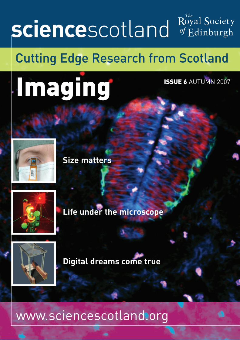
Imaging
Royal Societyof Edinburgh
The
Size matters
Life under the microscope
Digital dreams come true
IntroductionScience Scotland:Imaging
There is nothing more compelling than a visual image and it is no coincidence that the most massive processing system in the human brain is the visual cortex. As a species, we both depend upon and are stimulated by visual signals.Received images help us understand human behaviour andinterpret our surroundings, and our society exploits imaging for a wide variety of purposes, including communication,entertainment, security, remote monitoring, research andmedical diagnosis. In Scotland, we are recognised for ourexcellence in research, innovation and application of imagingtechnologies. In Edinburgh for example, we host the UK’sAstronomy Technology Centre, where many of the instrumentsthat the rest of the world uses to image our galaxy and beyond are developed and manufactured. Meanwhile, throughthe Scottish Funding Council’s research pooling initiative SINAPSE (Scottish Imaging Network: A Platform for ScientificExcellence), complementary expertise from Aberdeen, DundeeEdinburgh, Glasgow, St Andrews and Stirling is being broughttogether to promote brain imaging and interpretation.
In terms of imaging the very small, many regard the EnglishmanRobert Hooke as the father of microscopy and over 300 yearsago, he was the first to reveal some of the secret world of thecells that make up living organisms. He had wide interests inimaging and also made important contributions to astronomy byconstructing one of the world’s first reflecting telescopes basedon the ideas of James Gregory, a professor in mathematics at St.Andrews University. However, many of the limitations on earlymicroscopy imposed by glass optics and the wavelength of lighthave been overcome by applying different approaches to imaging.The extraordinary possibilities of using laser light are described
here by Allister Ferguson of the Centre for Biophotonics inGlasgow, who has developed this technology to allow biologists to observe living tissue without damage. By applying pure physics to biological systems, it has become possible to usepulsed laser light in miniaturised systems to interrogate livingcells for their response to drugs, and to secure a betterunderstanding of normal and abnormal cell function such as in cancers. This is just one example of what can be achieved bynot allowing your mind to be fettered by the artificial boundariesof discrete disciplines.
Some of the simplest questions in biology are often the hardest to study such as “what makes a cell divide”? It’s animportant question as it underlies our understanding of normaldevelopment and also gives us the means to study what goeswrong when cells divide inappropriately, such as during tumourgrowth. Again, imaging is one of our most powerful tools instudying what is going on when cells divide or are exposed to avariety of environmental influences – the challenge though, as in much of modern biology, is to try and make sense of the vastamount of information that is generated. Jason Swedlow atDundee University has confronted the problem by applying aninnovative software solution to accumulate and analyse imagingdata. Another example of thinking across the traditionaldiscipline boundaries to provide fresh solutions.
Building on such creative thinking coupled with advances in technology, we can look forward to an explosion in ourappreciation of basic biology.
But our appetite for more and more sophisticated images is not confined to the study of biology. Go anywhere in the world now and people will be capturing images using mobilephones and sending them round the globe to share with others.At STMicroelectronics’ Imaging Division in Edinburgh, GrahamTownsend predicted this less than ten years ago and thecompany is already working on further miniaturisation ofimaging technology using a different approach to the CCDtechnology that originally dominated the miniaturised worldwideimaging market. Using a new method of integrating the captureand processing of the image, devices can be made smaller,smarter and more cheaply than before, making them ideal forall sorts of applications we never knew we needed, as well asdelivering much needed solutions to emerging problems.
Once we’ve captured the images, we want to see them in avariety of ways. Televisions now look completely different than a few years ago and the large cathode ray tube dinosaurs havebeen replaced by slim panels capable of extraordinarily highdefinition. But, considering the application for miniaturisedsystems led Ian Underwood from MicroEmissive Displays inEdinburgh to develop novel micro display systems which can beembedded in video glasses and imprinted on a variety of flexiblesurfaces. Coupled with digital TV broadcasting, we will now beable to watch TV and videos and other image streams whereverand whenever we wish. There is a huge demand for high qualityimages in the entertainment market, but virtual imaging alsohas a wide variety of other applications such as the planning anddesign of city and country landscapes developed by DouglasPritchard at the Glasgow School of Art. The city of Glasgow is
Professor Anne Glover, FRSE Chief Scientific Advisor for Scotland
ContentsLife under the microscope 4
When cells divide… 6
The future in sight 9
Size matters 12
Digital dreams come true… 15
Mind reading 18
The fast field-cycling revolution 21
Scanning the horizon 23
The no-brainer scanner 25
This magazine is produced by The Royal Society of Edinburgh,with financial support from The Scottish Government, and wasestablished in co-operation with British Council Scotland.
FRONT COVER: A cross section through a 2.5-day-old chick embryo,focusing in on the neural tube, a structure that will eventually make up the spinal cord. DNA (highlighting the nuclei of cells) is labelled in red,Microtubules (a cytoskeletal protein that is involved in cell division) arelabelled in blue, neurons are labelled in green and dividing cells are labelledin pink: by and courtesy of Dr. Arwen Wilcock (the Swedlow Laboratory andthe Storey Laboratory, University of Dundee); Inset Images: MicroEmissiveDisplays’ tiny eyescreen™, courtesy of MED; Laser under construction in thelaboratory, courtesy of Learning Services, University of Strathclyde; Sub-centimetre accurate 3D model of a metal entryway canopy on the Isle ofBute. A digital documentation project for Historic Scotland. Courtesy ofDouglas Pritchard.
Editorial Board:
Chair: Professor John Coggins, FRSE Vice-Principal for the Faculties of Biomedical & Life Sciences,Clinical Medicine and Veterinary Medicine, University of Glasgow
Professor Peter Grant, FRSE, FIEE, FIEEE, FREngRegius Professor of Engineering, Department of Electrical Engineering,University of Edinburgh
Professor Angus Lamond, FRSEProfessor of Biochemistry, Division of Gene Regulation & Expression,School of Life Sciences, University of Dundee
Professor David Milne, OBE, FRSE, FIEE, SMIEEE, FREngExecutive Director, Wolfson Microelectronics Ltd
Editorial Team:
Words: Peter Barr, [email protected]
Design: Emma Quinn
Founding Editor: Michael White, The British Council
Editor: Stuart Brown, The Royal Society of Edinburgh
Print: Henry Ling Ltd
www.royalsoced.org.uk
www.sciencescotland.org
ISSN 1743 – 6222 (Science Scotland Print)ISSN 1743 – 6230 (online)
If you would like more information, please contact:[email protected]
The Royal Society of Edinburgh22 – 26 George StreetEdinburgh EH2 2PQ
ANY VIEWS EXPRESSED IN THIS PUBLICATION DO NOT NECESSARILY REPRESENT THOSEOF THE ROYAL SOCIETY OF EDINBURGH, NOR ALL OF ITS FELLOWS.
sciencescotland ISSUE 6 AUTUMN 2007 / 3
now available as a full 3D model and it is also possible todemonstrate the impact of an elevation in water levels on thecity. Given how receptive we are to “real” images of places weknow, this could be a powerful tool to engage the public withthe likely impacts of climate change and make mitigationmeasures more acceptable. As similar models becomeavailable for locations around the world, virtual tourism mightbe on the horizon, as well as invaluable tools for disasterplanning. These developments underpin new industries andbring economic development to our country, as well as being areal inspiration to young creative people looking for rewardingcareers in science.
It is no surprise that, as we have so much neural activitydevoted to image processing in our brains, we take in vastamounts of information and process it very rapidly to generatea picture of what is happening around us. Professor Schyns inthe Department of Psychology at Glasgow University is trying to unravel how the brain does this, with the aim of producing afamily of new devices from remote response systems allowingdisabled people to control devices by thought, to sophisticatedimage recognition systems to identify the expression of fear,pleasure or particular behaviour patterns. This would challengethe thinking that “our thoughts are our own” and will open upmuch debate on the ethics surrounding this new area of science.
We already know the value of magnetic resonance imaging in medicine and it is widely used as a painless, non-invasivetechnique to provide cross-sectional scans of the whole bodywhich can be used to diagnose disease such as multiplesclerosis, cardiac abnormality and joint problems. David Lurie at Aberdeen University has built upon the history of MRIdevelopment at Aberdeen to develop a new approach that hasthe potential to increase the resolution of the images andtherefore the information that can be gleaned from them. Withbetter information comes earlier diagnoses and more effectivetreatment – good news for an ageing population with ever moredemand on the health service. The SFC Brain Imaging ResearchCentre in Edinburgh, directed by Joanna Wardlaw, is pushing theapplication of MRI scanning to provide better outcomes forpatients with strokes, which are a major killer in Scotland. MRIscanning is undoubtedly leading to enhanced patient care andthis is complemented by the developments from Andy Welch’s team on PET scanning at Aberdeen. PET scanning not onlydelivers diagnoses but also allows the development of newdrugs and more effective treatments. These scanningtechnologies provide both patient care and the tools with whichdrug companies can provide better, more targeted treatmentsfor a variety of neurological diseases, as well as cancers.
The applications for imaging in 21st century life are extensive and range from understanding basic biological processes tomanufacturing new entertainment devices and providing betterdisease treatments. A common theme in this issue of ScienceScotland is the innovative thinking shown by the scientistsinvolved in these developments and their willingness to look for solutions across traditional discipline boundaries.
When you mention lasers, many people still think of death rays, but thenew generation of laser-based multiphoton microscopes are shininglight on all sorts of medical problems, from cancer to toothache…
Life under themicroscope
4 / sciencescotland ISSUE 6 AUTUMN 2007
PROFESSOR ALLISTER I FERGUSON, FRSE
In the early 1990s, biologists used microscopesto study different embryos by staining them withchemicals called fluorophores, then shining avery bright light on the cells. This worked verywell but in the process, the embryos died. Today,the same biologists can track the life of animalslike hamsters from the time they are only a smallbunch of cells until they’re old enough to go for a spin in their wheels, without doing any damageat all.
The key advantage of this new technique – apart from not killing the hamster before it is born – is that biologists can study the sameembryo at every stage of its development, rather than studying different hamster embryos at different stages. If testing the effects ofpharmaceuticals, for example, this means theresults are more consistent throughout theexperiment – and therefore more reliable as well as faster and more economical.
And what has made this possible? The answer is a new kind of technology called multiphotonmicroscopy (MPM) which uses special lasers toilluminate specimens under the lens, withoutdestroying any cells. As well as being minimallyinvasive, MPM can penetrate hundreds ofmicrons under the surface, seeing into thicktissue where other technologies simply can’t see– and even produce three-dimensional images.
The story starts when Professor AllisterFerguson of the physics department at theUniversity of Strathclyde in Glasgow was askedby a biologist called John White, then working in Cambridge, what he knew about somethingcalled ‘confocal microscopy’. After ‘bluffing’ for several minutes, as Ferguson recalls his
reaction, he asked White to explain what‘confocal microscopy’ was, and in the processlearned about the problems of in vivo imaging(studying living organisms under the microscope)– using the available technology.
White had contacted Ferguson after he read ofhis work in developing ultrashort pulse lasers to study atomic hydrogen, thinking this may be the answer to the problem of killing the cells.Another problem for White was that usingfluorophores and other indicators to ‘mark’ living cells sometimes bleached the samples,making them impossible to see. Several otheracademics and commercial developers told White he was wasting his time in his search for a compact laser-based system, but Fergusonimmediately saw that his work with ‘2-photonmicroscopy’ could provide what White needed.
“Too much light cooked the subjects or createdtoxic products, but I knew ultrashort pulse laserscould be the answer,” says Ferguson. “Theobjective was to make a multiphoton systemcompact and easy to use, so that biologists andother scientists without a physics backgroundcould use them just like any other microscope.”
Lasers for multiphoton microscopes at that timewere as big as a room, but Ferguson believed thatit was possible to develop a solid-state version,taking advantage of recent developments in semi-conductors to miniaturise the componentsand pack in more intelligence, using tiny, tightlyfocused lasers with extremely short light pulses(10-13 second) to illuminate the specimens safely,then use raster-scanning (a data mappingmechanism) to build up an image for viewing.
sciencescotland ISSUE 6 AUTUMN 2007 / 5
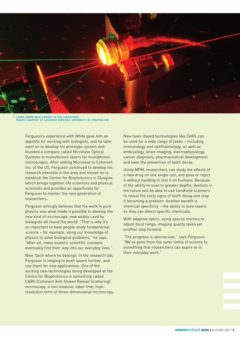
LASER UNDER DEVELOPMENT IN THE LABORATORY.IMAGES COURTESY OF LEARNING SERVICES, UNIVERSITY OF STRATHCLYDE
Ferguson’s experience with White gave him anappetite for working with biologists, and he laterwent on to develop his prototype system andfounded a company called Microlase OpticalSystems to manufacture lasers for multiphotonmicroscopes. After selling Microlase to CoherentInc. of the US, Ferguson continued to develop hisresearch interests in the area and moved on toestablish the Centre for Biophotonics in Glasgow,which brings together life scientists and physicalscientists and provides an opportunity forFerguson to mentor the new generation ofresearchers.
Ferguson strongly believes that his work in purephysics was what made it possible to develop thenew kind of microscope, now widely used bybiologists all round the world. “That is why it’s so important to have people study fundamentalscience – for example, using our knowledge ofphysics to solve biological problems,” he says.“After all, many esoteric scientific conceptseventually find their way into our everyday lives.”
Now ‘back where he belongs’ in the research lab,Ferguson is helping to push lasers further, anduse them for new applications. One of theexciting new technologies being developed at theCentre for Biophotonics is something calledCARS (Coherent Anti-Stokes Raman Scattering)microscopy, a non-invasive, label-free, high-resolution form of three-dimensional microscopy.
New laser-based technologies like CARS can be used for a wide range of tasks – includingimmunology and ophthalmology, as well asembryology, brain imaging, electrophysiology,cancer diagnosis, pharmaceutical developmentand even the prevention of tooth decay.
Using MPM, researchers can study the effects ofa new drug on one single cell, and pass or rejectit without needing to test it on humans. Becauseof the ability to scan to greater depths, dentists inthe future will be able to use handheld scannersto reveal the early signs of tooth decay and stop it becoming a problem. Another benefit ischemical specificity – the ability to tune lasers so they can detect specific chemicals.
With adaptive optics, using special mirrors toadjust focal range, imaging quality takes yetanother step forward.
“The progress is spectacular,” says Ferguson.“We’ve gone from the outer limits of science tosomething that researchers can aspire to in their everyday work.”
The pictures look like psychedelic jellyfishswimming around in a tropical sea, but this is notBlue Planet, it’s a film of mitosis – the momentwhen a cell divides to form a pair of duplicates.
An art gallery in California has exhibited some of the Miro-like images, but despite their aestheticappeal, they could be a significant step forward on the road to new treatments for cancer, braindamage or spinal injuries – plus many othermedical problems.
“If we can illuminate the mechanisms of celldivision,” says Dr Jason Swedlow, “at themechanical and the molecular level, and at thelevel of tissue biology, that would be critical to understanding how cells work – how theyintegrate and make decisions.”
When cells divide, they make a ‘fate choice,’‘deciding’ what to become. The cells in tissues of the sort the Swedlow Laboratory is studying‘choose’ to become neurons or not. Swedlow andhis team are trying to determine which onesbecome neurons, and when. Then they put the filmin reverse, enabling them to trace the cell all theway back to the source – and observe every otherevent that has happened since ‘birth’.
The problem is that in order to observe‘neurogenesis’, you have to film thousands of othercells in action, as they divide and duplicate, and thedata generated in the process adds up to manyterabytes (a terabyte is a million million bytes),which then has to be analysed before you find the ‘needle in a haystack.’ And even though theSwedlow Lab (located in the Wellcome TrustBiocentre at the University of Dundee) has accessto as much computing power as a bank, this is adifficult challenge.
The breakthrough tool that Swedlow and his teamare working on now is a smart piece of software –a relational database management system (RDMS)and an open-source application developed by theOpen Microscopy Environment (OME), an
international consortium which Swedlow helpedto found in 2000. OME builds software tools tomanage and analyse the vast amounts of imagingdata collected by academic and pharmaceuticallabs round the world – for example, the dataproduced by digital fluorescence imaging, whichmarks or bleaches targets under the microscopeso that observers can calculate what’s going on at microscopic level, including processes likemolecular localisation, interaction and mobility.
The OME software can be downloaded free from the OME website (http://openmicroscopy.org), underthe GNU General Public License, which allows re-distribution and resale, as long as the open-source nature of the source code is maintained.What motivates Swedlow himself is the ability toprovide informatics solutions to the biologycommunity at large, so scientists can make morerapid progress with experiments, including drugdiscovery and therapy development.
One of Swedlow’s objectives is to make dataanalysis a more routine procedure, via theconvergence of several disciplines, includinginformatics and biology, and developing newimaging technology to “fine-tune and understandthe role of phospho-specific reporters.”
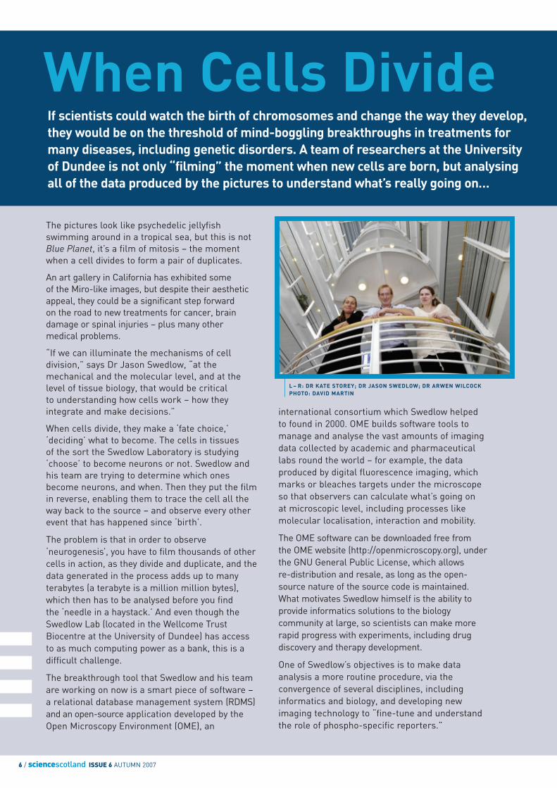
If scientists could watch the birth of chromosomes and change the way they develop,they would be on the threshold of mind-boggling breakthroughs in treatments formany diseases, including genetic disorders. A team of researchers at the Universityof Dundee is not only “filming” the moment when new cells are born, but analysingall of the data produced by the pictures to understand what’s really going on…
When Cells Divide
6 / sciencescotland ISSUE 6 AUTUMN 2007
L – R: DR KATE STOREY; DR JASON SWEDLOW; DR ARWEN WILCOCKPHOTO: DAVID MARTIN
sciencescotland ISSUE 6 AUTUMN 2007 / 7
8 / sciencescotland ISSUE 6 AUTUMN 2007
the eureka momentwas to capture a film of the instant aneuron was bornIn other words, instead of being armchairresearchers who gaze in amazement as cellsreproduce, Swedlow and his team of 13programmers and post-doctoral assistants try to make sense of what they see under themicroscope, translating raw images intomeaningful quantitative data which explains how cells divide.
Another key activity is to ‘perturb’ the wholeprocess, intervening in various ways to test whathappens when we manipulate cells – ultimately todevelop pharmaceuticals and other treatments.
“We are very good at acquiring the images,”Swedlow explains, “but data management anddata analysis turn the images into results whichwill help to solve problems, and that is where OME gives us the edge.”
In partnership with the University of Dundee’sDepartment of Applied Computing, Swedlow hasalso been working to improve the usability of OME,with usability specialists working directly with theuser community and passing on feedback whichSwedlow’s team of developers can use for new features.
So the Swedlow Laboratory combines expertise incell biology, imaging and software to examine howcells progress through mitosis, and in partnershipwith Dr Kate Storey, also based in Dundee, tostudy neurogenesis.
For Swedlow and Storey, supported by their post-doctorate researcher, Arwen Wilcock, the eurekamoment was to capture a film of the instant aneuron was born, by observing cell division in theembryos of chickens. “No-one has ever been ableto see this before,” says Swedlow, explaining thatthe project has so far generated 2.5 terabytes ofdata over the last four years, documenting themagical moment of the birth of a neuron.
In the process, Swedlow’s team has also madesignificant advances in imaging methods, as wellas data management and processing – newimproved tools and techniques which will helpfuture projects, in Scotland and beyond.
The Swedlow Laboratory is funded by theWellcome Trust, Cancer Research UK, theBiotechnology and Biological Sciences ResearchCouncil (BBSRC) and the Engineering and Physical Sciences Research Council (EPSRC). The tools it has developed are already widely used by an increasing number of researchersworldwide, enabling future breakthroughs inbiology and medicine even Swedlow and hissponsors can’t imagine.
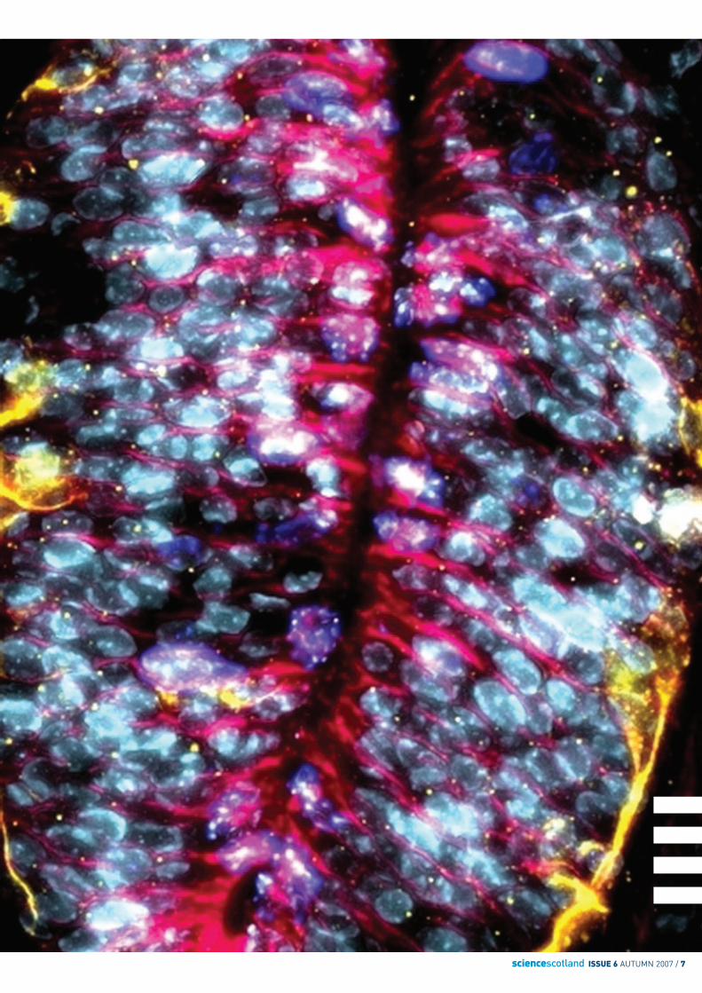
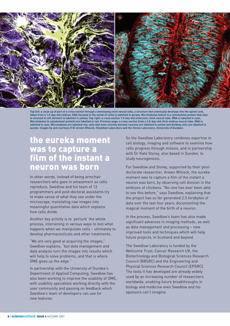
Top left: a close up of part of a cross section through a developing chick neural tube, a structure that eventually develops into the spinal cord, taken from a 1.5-day-old embryo. DNA (located in the nuclei of cells) is labelled in purple, Microtubules (which is a cytoskeletal protein that also is involved in cell division) is labelled in yellow; Top right: a cross section 1.5-day-old embryonic chick neural tube. DNA is labelled in cyan,Microtubules (a cytoskeletal protein) are labelled in red. Previous page: a cross section from a 2.5-day-old chick embryo neural tube. DNA islabelled in cyan, Microtubules are labelled red, cells that have recently become neurons are labelled in yellow and dividing cells are labelled inpurple. Images by and courtesy of Dr Arwen Wilcock, (Swedlow Laboratory and the Storey Laboratory, University of Dundee).
sciencescotland ISSUE 6 AUTUMN 2007 / 9
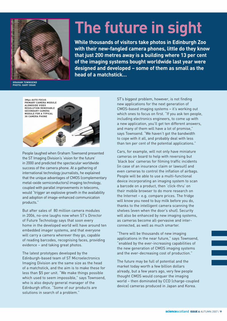
The future in sightWhile thousands of visitors take photos in Edinburgh Zoowith their new-fangled camera phones, little do they knowthat just 200 metres away is a building where 13 per centof the imaging systems bought worldwide last year weredesigned and developed – some of them as small as thehead of a matchstick…
GRAHAM TOWNSENDPHOTO: GARY DOAK
People laughed when Graham Townsend presentedthe ST Imaging Division’s ‘vision for the future’ in 2000 and predicted the spectacular worldwidesuccess of the camera phone. At a gathering ofinternational technology journalists, he explained that the unique advantages of CMOS (complementarymetal-oxide semiconductors) imaging technology,coupled with parallel improvements in telecoms,would “trigger an explosive growth in the availabilityand adoption of image-enhanced communicationproducts.”
But after sales of 80 million camera modules in 2006, no-one laughs now when ST’s Director of Future Technology says that soon every home in the developed world will have around tenembedded imager systems, and that everyone will carry a camera wherever they go, capable of reading barcodes, recognising faces, providingevidence – and taking great photos.
The latest prototypes developed by theEdinburgh-based team of ST MicroelectronicsImaging Division are the same size as the head of a matchstick, and the aim is to make these forless than $5 per unit. “We make things possiblewhich used to seem impossible,” says Townsend,who is also deputy general manager of theEdinburgh office. “Some of our products aresolutions in search of a problem.”
ST’s biggest problem, however, is not finding new applications for the next generation of CMOS-based imaging systems – it’s working outwhich ones to focus on first. “If you ask ten people,including electronics engineers, to come up with a new application, you’ll get ten different answers,and many of them will have a lot of promise,” says Townsend. “We haven’t got the bandwidth to cope with it all, and probably deal with less than ten per cent of the potential applications.”
Cars, for example, will not only have miniaturecameras on board to help with reversing but‘black box’ cameras for filming traffic incidents (in case of an insurance claim or lawsuit) and even cameras to control the inflation of airbags.People will be able to use a multi-functionaldevice incorporating an imaging system to scan a barcode on a product, then ‘click-thru’ on their mobile browser to do more research on the Internet – e.g. compare prices. The fridge will know you need to buy milk before you do,thanks to the intelligent camera scanning theshelves (even when the door’s shut). Security will also be enhanced by new imaging systems, as cameras become all-pervasive and inter-connected, as well as much smarter.
“There will be thousands of new imagingapplications in the near future,” says Townsend,“enabled by the ever-increasing capabilities of the new generation of CMOS imaging systems and the ever-decreasing cost of production.”
The future may be full of potential and the market today worth a few billion dollars already, but a few years ago, very few peoplethought CMOS would conquer the imaging world – then dominated by CCD (charge-coupleddevice) cameras produced in Japan and Korea.
3Mpx AUTO FOCUSPRIMARY CAMERA MODULEALONGSIDE VIDEORESOLUTION REMOVABLESECONDARY CAMERAMODULE FOR A TYPICAL 3G CAMERA PHONE
10 / sciencescotland ISSUE 6 AUTUMN 2007
soon every home in theworld will have ten embeddedimager systems
When Townsend joined Vision, the imagingcompany which had grown out of the University ofEdinburgh during the 1990s and was later boughtout by ST, CMOS-based systems seemed destinedfor lower-end products, not sophisticated productslike digital SLR cameras, and far too expensive tobe built into phones.
Since then, the industry has changed out of allrecognition. Camera phones now account for 70per cent of the worldwide imaging market, andimaging systems are built into 60 per cent of all new models sold – and CMOS has begun tochange the balance of power in the imagingmarket, sweeping aside CCD.
In the 1990s, CMOS imaging systems wereprimarily used in security cameras, producing only black and white pictures. And then there was Barbie…
According to Townsend, when Vision firstapproached Mattel (Barbie’s creator) in 1995, thetoy giant firmly rejected the idea of associatingBarbie with a CMOS-based digital camera, even if it could be sold for less than $50. Young girlswould not want to plug cameras into computers,said the marketing people, but when Townsendshowed them a short animation produced by his11-year-old daughter, using a prototype camera,they quickly changed their mind, and the Barbiecamera soon became an international best-seller.
The latest imaging systems are light years aheadof the earliest models, and are quickly becomingcommodities rather than technical gadgets. ST, for example, supplies its solutions to severalindustry leaders, including auto-focus moduleswhich measure only 1cm x 1cm x 1cm and willsoon be able to deliver up to five megapixels (Mp)i.e. 5000 000 pixels. The 3Mp versions currentlyramping into volume production are alreadybecoming old-fashioned, as consumers begin totake camera phones for granted.
The secret of ST’s success in the imaging market is that it “masters the four pillars of imaging,” says Townsend, including signal processing, optics,mechanics and the sensor itself. This enables ST to keep a foot in both camps of the imaging market– making imaging modules as well as its own imagesensors. This ‘integrated’ approach is reflected inthe design of the product itself – a ‘system on a chip’which integrates the lens with the intelligence thatmakes it all work, reducing signal noise as well asthe production cost. Automated, high-volumeassembly also helps produce quality products ataffordable prices.
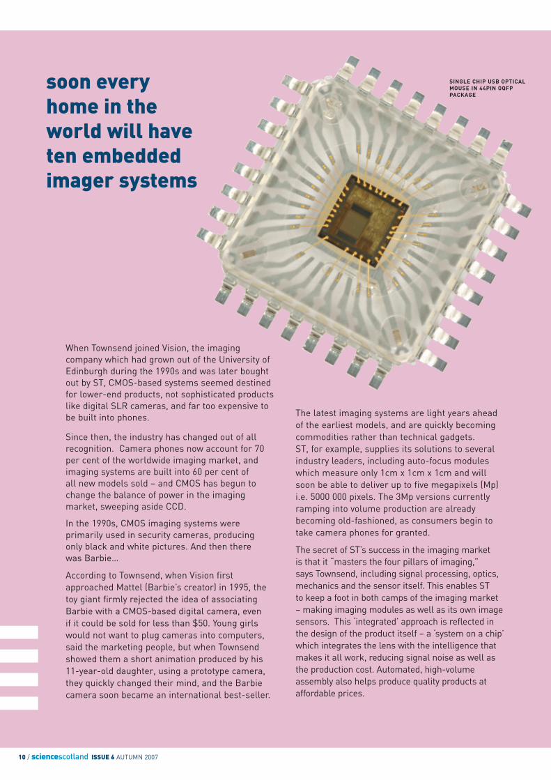
SINGLE CHIP USB OPTICALMOUSE IN 44PIN OQFPPACKAGE
sciencescotland ISSUE 6 AUTUMN 2007 / 11
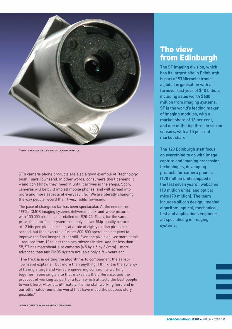
The viewfrom Edinburgh The ST imaging division, whichhas its largest site in Edinburghis part of STMicroelectronics, a global organisation with aturnover last year of $10 billion,including sales worth $600million from imaging systems. ST is the world’s leading makerof imaging modules, with amarket share of 13 per cent, and one of the top three in siliconsensors, with a 15 per centmarket share.
The 120 Edinburgh staff focus on everything to do with imagecapture and imaging processingtechnologies, developingproducts for camera phones (170 million units shipped in the last seven years), webcams(10 million units) and opticalmice (70 million). The teamincludes silicon design, imagingalgorithm, optical, mechanical,test and applications engineers,all specialising in imagingsystems.
ST’s camera-phone products are also a good example of “technologypush,” says Townsend. In other words, consumers don’t demand it – and don’t know they ‘need’ it until it arrives in the shops. Soon,cameras will be built into all mobile phones, and will spread intomore and more aspects of everyday life. “We are literally changingthe way people record their lives,” adds Townsend.
The pace of change so far has been spectacular. At the end of the1990s, CMOS imaging systems delivered black-and-white pictures with 100,000 pixels – and retailed for $20-25. Today, for the same price, the auto-focus systems not only deliver 5Mp-quality pictures at 12 bits per pixel, in colour, at a rate of eighty million pixels persecond, but then execute a further 300-500 operations per pixel toimprove the final image further still. Even the pixels deliver more detail– reduced from 12 to less than two microns in size. And for less than$5, ST has matchhead-size cameras (4.5 by 4.5 by 3.6mm) – moreadvanced than any CMOS system available only a few years ago.
“The trick is in getting the algorithms to complement the sensor,”Townsend explains, “but more than anything, I think it is the synergyof having a large and varied engineering community workingtogether in one single site that makes all the difference, and theprospect of working as part of a team which attracts the best peopleto work here. After all, ultimately, it’s the staff working here and inour other sites round the world that have made the success storypossible.”
“SMIA” STANDARD FIXED FOCUS CAMERA MODULE
IMAGES COURTESY OF GRAHAM TOWNSEND
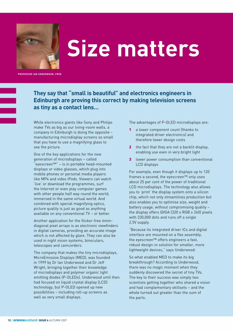
They say that “small is beautiful” and electronics engineers inEdinburgh are proving this correct by making television screensas tiny as a contact lens…
Size matters
12 / sciencescotland ISSUE 6 AUTUMN 2007
PROFESSOR IAN UNDERWOOD, FRSE
While electronics giants like Sony and Philipsmake TVs as big as our living-room walls, acompany in Edinburgh is doing the opposite –manufacturing microdisplay screens so smallthat you have to use a magnifying glass tosee the picture.
One of the key applications for the newgeneration of microdisplays – called“eyescreen™” – is in portable head-mounteddisplays or video glasses, which plug intomobile phones or personal media players like MP4 and video iPods. Viewers can watch‘live’ or download the programmes, surf the Internet or even play computer games with other people half way round the world,immersed in the same virtual world. Andcombined with special magnifying optics, picture quality is just as good as anythingavailable on any conventional TV – or better.
Another application for the flicker-free 6mm-diagonal pixel arrays is as electronic viewfindersin digital cameras, providing an accurate imagewhich is not affected by glare. They can also beused in night vision systems, binoculars,telescopes and camcorders.
The company that makes the tiny microdisplays,MicroEmissive Displays (MED), was founded in 1999 by Dr Ian Underwood and Dr Jeff Wright, bringing together their knowledge of microdisplays and polymer organic lightemitting diodes (P-OLEDs). Underwood until thenhad focused on liquid crystal display (LCD)technology, but P-OLED opened up newpossibilities – including roll-up screens as well as very small displays.
The advantages of P-OLED microdisplays are:
1 a lower component count (thanks to integrated driver electronics) andtherefore lower design costs
2 the fact that they are not a backlit display, enabling use even in very bright light
3 lower power consumption than conventional LCD displays
For example, even though it displays up to 120frames a second, the eyescreen™ only usesabout 25 per cent of the power of traditional LCD microdisplays. The technology also allowsyou to ‘print’ the display system onto a siliconchip, which not only streamlines production butalso enables you to optimise size, weight andbattery usage, without compromising quality –the display offers QVGA (320 x RGB x 240) pixelswith 230,000 dots and runs off a single2.5V supply.
“Because its integrated driver ICs and digitalinterface are mounted on a flex assembly, the eyescreen™ offers engineers a fast, robust design-in solution for smaller, morelightweight devices,” says Underwood.
So what enabled MED to make its bigbreakthrough? According to Underwood,there was no magic moment when theysuddenly discovered the secret of tiny TVs.The key to their success was simply twoscientists getting together who shared a visionand had complementary skillsets – and the whole turned out greater than the sum of the parts.
sciencescotland ISSUE 6 AUTUMN 2007 / 13
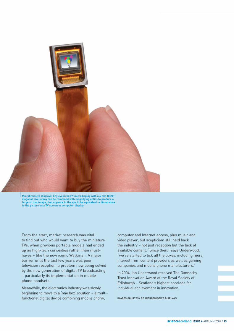
MicroEmissive Displays’ tiny eyescreen™ microdisplay with a 6 mm (0.24”)diagonal pixel array can be combined with magnifying optics to produce alarge virtual image, that appears to the eye to be equivalent in dimensions to the picture on a TV screen or computer display.
From the start, market research was vital,to find out who would want to buy the miniatureTVs, when previous portable models had endedup as high-tech curiosities rather than must-haves – like the now iconic Walkman. A majorbarrier until the last few years was poortelevision reception, a problem now being solvedby the new generation of digital TV broadcasting– particularly its implementation in mobilephone handsets.
Meanwhile, the electronics industry was slowlybeginning to move to a ‘one box’ solution – a multi-functional digital device combining mobile phone,
computer and Internet access, plus music andvideo player, but scepticism still held back the industry – not just reception but the lack ofavailable content. “Since then,” says Underwood,“we’ve started to tick all the boxes, including moreinterest from content providers as well as gamingcompanies and mobile phone manufacturers.”
In 2004, Ian Underwood received The GannochyTrust Innovation Award of the Royal Society ofEdinburgh – Scotland’s highest accolade forindividual achievement in innovation.
IMAGES COURTESY OF MICROEMISSIVE DISPLAYS
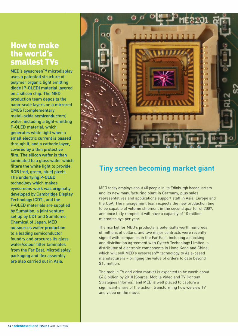
How to makethe world’ssmallest TVsMED’s eyescreen™ microdisplayuses a patented structure ofpolymer organic light emittingdiode (P-OLED) material layeredon a silicon chip. The MEDproduction team deposits thenano-scale layers on a mirroredCMOS (complementary metal-oxide semiconductors)wafer, including a light-emittingP-OLED material, whichgenerates white light when asmall electric current is passedthrough it, and a cathode layer,covered by a thin protective film. The silicon wafer is thenlaminated to a glass wafer whichfilters the white light to provideRGB (red, green, blue) pixels.The underlying P-OLEDtechnology which makeseyescreens work was originallydeveloped by Cambridge DisplayTechnology (CDT), and the P-OLED materials are suppliedby Sumation, a joint venture set up by CDT and SumitomoChemical of Japan. MEDoutsources wafer production to a leading semiconductorfoundry and procures its glasswafer/colour filter laminatesfrom the Far East. Microdisplaypackaging and flex assembly are also carried out in Asia.
14 / sciencescotland ISSUE 6 AUTUMN 2007
Tiny screen becoming market giant
MED today employs about 60 people in its Edinburgh headquartersand its new manufacturing plant in Germany, plus salesrepresentatives and applications support staff in Asia, Europe andthe USA. The management team expects the new production lineto be capable of volume shipment in the second quarter of 2007,and once fully ramped, it will have a capacity of 10 millionmicrodisplays per year.
The market for MED’s products is potentially worth hundreds of millions of dollars, and two major contracts were recentlysigned with companies in the Far East, including a stocking and distribution agreement with Cytech Technology Limited, adistributor of electronic components in Hong Kong and China,which will sell MED's eyescreen™ technology to Asia-basedmanufacturers – bringing the value of orders to date beyond $10 million.
The mobile TV and video market is expected to be worth about £4.8 billion by 2010 (Source: Mobile Video and TV ContentStrategies Informa), and MED is well placed to capture asignificant share of the action, transforming how we view TV and video on the move.
sciencescotland ISSUE 6 AUTUMN 2007 / 15
Digital dreamscome true…
The journey takes you up the River Clyde in thecentre of Glasgow, past the Scottish Exhibition andConference Centre, over the Kingston Bridge, upJamaica Street to Central Station then GeorgeSquare and back again. But even if it’s raining andyou don’t have an umbrella, you will not get theslightest bit wet because this city only existsinside a computer.
Even the computer where the data is stored is in a ‘virtual’ building – the House for an Art Lover,designed by Charles Rennie Mackintosh in 1901and only constructed a century later, in a park inthe middle of Glasgow.
Douglas Pritchard, Head of Visualisation at theDigital Design Studio (part of Glasgow School ofArt) where the virtual city is being developed, is aself-confessed “evangelist” for virtual reality, and he describes the City’s project as a
“ground-breaking” venture which will have ahuge impact on a wide spectrum of activities – some of them hard to imagine.
The project is funded by Glasgow City Council, who realised a virtual city would not only helparchitects and urban planners but also have value to others. This is underlined by the fact that so manyorganisations have since expressed interest – cityplanners, architects, designers, marketers, historicalagencies, educators and the emergency services.
The original concept was to build up a digitaltemplate of the City of Glasgow that architectscould use to place their buildings in an accuratevirtual context, for design as well as planningapproval. Ultimately, this would enable the Cityand its citizens to use the model for design review– comparing height, for example, and showinghow a building fits within the context of its
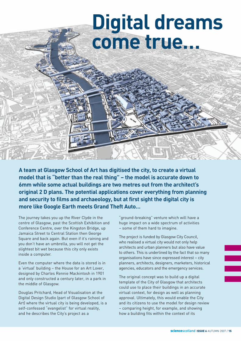
A team at Glasgow School of Art has digitised the city, to create a virtualmodel that is “better than the real thing” – the model is accurate down to6mm while some actual buildings are two metres out from the architect’soriginal 2 D plans. The potential applications cover everything from planningand security to films and archaeology, but at first sight the digital city ismore like Google Earth meets Grand Theft Auto…
16 / sciencescotland ISSUE 6 AUTUMN 2006
neighbourhood. Consider the fact that the Clydewaterfront is currently going through some of the most intense development in the UK, and that Glasgow city centre, with its dense mix ofcontemporary and listed buildings, is constantlyevolving, and you begin to appreciate the need for such an informative tool.
“Two-dimensional drawings or scale models are too abstract and incapable of capturing thecomplexity of a proposed development within theurban context,” says Pritchard. “The architect’s or developer’s ‘artist’s rendering’ is motivated bymarketing and is designed to flatter rather thanproviding accurate information or possiblyunfavourable analysis.”
The virtual city of Glasgow will provide the Counciland the public with a more thorough understandingof proposed architectural projects, to help reducepossible misunderstanding, identify costly designissues before they are built and hopefully encouragegreater participation in the City’s planning process.
How do they do it?Pritchard and his team of ten researchers finishedthe 15-month project in June and are confident ofcutting this to 12 months on their next virtual city.Using a high-definition Leica 3 D laser scannercombined with global satellite positioning, theybuild up a composite scan of the city, then texturethe buildings using digital photos. The initial scanslook like a cross between X-rays and wire-frames,but when they’re put together with aerial photos,and the digital photos, they begin to lookuncannily real.
Early on, the team referred to architect’sdrawings, but soon found that these wereinaccurate, with differences of up to two metresbetween the plan and the actual build. Thescanner, on the other hand, enables them to getwithin sub-centimetre accuracy for building heightand width, and can also identify details on thefacade as small as 5mm – to go beyond thatsimply multiplies the data exponentially.
“The Glasgow model is unique because it reflectsthe actual built city,” says Pritchard. “The 3 Dbuildings are based on the real thing, not idealiseddrawings. If a building’s old or falling down, or if there are undocumented additions, it will be virtually represented that way.”
This degree of accuracy is sufficient to more thanmeet planning requirements. Theoretically, thescanner could be used to image any physicalobject – say, a statue – and create a 3 D modelwhich is so complete that it could then be used to control a manufacturing device capable ofreproducing the statue exactly the same as in real life, to any scale desired.
In the case of Virtual Glasgow, the end result also enables the viewer to ‘fly through’ a model of the city like Flight Simulator and see every detailfrom any perspective, in real time. The researchteam even raised the water level in the River Clydeto demonstrate the impact of a flood – and also justhow realistic the model can be.
Eventually, three real-time versions are planned.A low-resolution version will be available onlinevia the Council web site, primarily for viewingproposals or simply for personal interest.
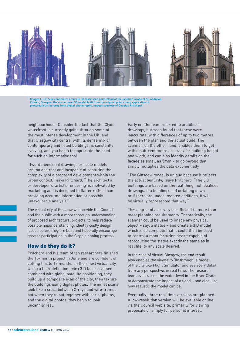
Images L – R: Sub-centimetre accurate 3D laser scan point-cloud of the exterior facade of St. AndrewsChurch, Glasgow; the un-textured 3D model built from the original point cloud; application ofphotorealistic textures from digital photographs. Images courtesy of Douglas Pritchard.
sciencescotland ISSUE 6 AUTUMN 2007 / 17
The DigitalDesign StudioThe DDS was established in 1997 by Paul Anderson and Ian Johnston and focuses onwhat it calls “ReplacementReality: The ability to interactwith highly credible digitalmodels in a natural and intuitivemanner, unencumbered byintrusive equipment, using yoursenses of touch and hearing, as well as vision.”
In pursuit of this wide-rangingmission, its work involvesprojects in real-timevisualisation, gamesdevelopment, human-computerinteraction, voice technologies,prototyping, animation andhaptics (the science of studyingdata obtained by means oftouch), providing services such as digital content creation,architectural modelling, virtualtours, interactive displayapplications and publicinterfaces. Its research partnersinclude Ford, QinetiQ, the BBCand EPSRC, developing projectsfor the automotive, aerospace,ship-building, education, bio-medical and entertainmentindustries.
A medium-resolution version will be for architects and designers, and a super-realistic version will store all the architectural and photorealistic details.
“The aim is to inform and empower,” says Pritchard. “Everyone willhave an interest in the end product, especially the public.”
The Road to GlasgowPritchard is an architect by training who got turned on to visualisation whileworking in Toronto in the early 1990s. He wondered why architects thoughtand designed in 3 D terms and built 3 D buildings, yet developed their ideasin 2 D – an unnatural format that most people find very hard to interpret.Would it not be better if the architect stayed entirely within the 3 D realm?
In the 1990s, the hardware and software he needed to make his idea cometrue were too costly, but as the years went by, technology advanced andPritchard got an opportunity to demonstrate the power of 3 D visualisation,working on Toronto’s bid to host the 2008 Olympics. Not only did the teamshow the Selection Committee what the Games would be like, they also had to prove to local residents that the Games would not ruin the city.
After Toronto, Pritchard’s next project was in Buffalo, New York, where localobjections to a new bridge over the Niagara River, linking the United Stateswith Canada, encouraged the authorities to win greater public support byshowing people what the bridge would look like. The public were eveninvited to vote on a number of options, thus using the 3 D model todemocratise the process as well as to communicate.
Familiar with the 3 D research work done by Tom Maver in the Abacusproject and at Glasgow School of Art (GSA), Pritchard then accepted a postto explore new ideas in visualisation at the Digital Design Studio (DDS).Working with Ian Johnston, an early project which is nearly completed, wasto virtually reconstruct the 1938 British Empire Exhibition held in Glasgow.The project is based on surviving photos and plans, plus interviews withvisitors who went there at the time. Then, in 2005, the DDS won the tenderto create the Virtual Glasgow through the Access Glasgow programme,defeating 15 rivals from the UK and Europe.
Future DDS projects include documenting Scotland’s four WorldHeritage sites. This will be part of the DDS’s contribution to CyArk, aninternational project to digitise the world’s most treasured archaeologicalsites and create a virtual archive for posterity. Both Pritchard and Maver are currently involved in a European-funded research project to documentprominent archeological sites in Germany, Italy and Poland. The project iscentred around the innovative use of visualisation methods within thecontext of archaeology and cultural heritage.
In addition to its international projects, the DDS has also worked withthe Glasgow Housing Association, to show the residents of Gallowgatethe phased regeneration of their neighbourhood and to get more residentsinvolved in the planning process.
“It’s all about communication,” Pritchard comments. “When we build avirtual city, we can all see exactly the same streets and buildings, andnobody needs to imagine it inside their heads. We are fortunate to beworking on the Glasgow city project and are confident that due to itsaccuracy and detail it will be a tremendous benefit to the futuredevelopment of our city.”
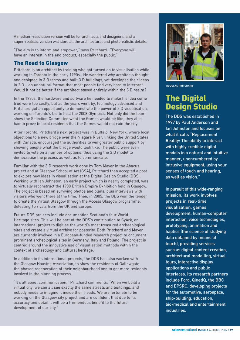
DOUGLAS PRITCHARD
18 / sciencescotland ISSUE 6 AUTUMN 2007
Psychologists at the University of Glasgow haveembarked on a project to map what goes on in themind, using massive computers to analyse how peopleprocess visual information. In the future, this couldlead to “telepathic” and psycho-kinetic computing or superhuman robots flying off to explore outerspace, but researchers today have more modestobjectives – at least for the time being…
Mind Reading
How do we know someone’s happy or frightened?How do we tell the difference between men andwomen? A friend or a foe? What kind of visualsignals do we need to recognise objects? Theanswers may be obvious, but Professor PhilippeSchyns, the head of the department of psychology atGlasgow University, says that answering these basicquestions precisely may eventually enable thedevelopment of tools which control electronicdevices by thinking, or automatons which operate inhostile environments, and see things like humans.
Schyns and his team of researchers are engagedin something called ‘cognitive neuro-imaging’ – ormind reading using computers. And he is happy toadmit that they are in the early stages of a verynew science.
Some researchers focus on the so-called ‘biggerpicture’, but Schyns is much more interested indelving deeper into the mind to establish a fewfundamentals. Instead of claiming that heunderstands what happens inside the brain duringthe process of visual perception, Schyns says thathis work is beginning to highlight how little we doknow: “If we don’t understand the building blocksof visual recognition, we understand nothing,”says Schyns. “Our task is to identify the brainstates and the states of information processing, in order to see the brain as a mechanism.”
The conventional concept of brain scans is imagesshowing a burst of activity or a slice of a region
deep inside the skull. Such images are usefuldiagnostic tools and advance our scientificunderstanding of the processes going on insidethe brain; but even though this is important,it does not help to understand precisely howinformation is processed during even the simplest of visual events. In other words, a static picture is not much good for studying theintricate mechanism of a clock.
Psychologists have known for many years the basic truths of visual recognition. For example, to detect if a person is happy or disgusted, themouth is the critical factor, while to recognise fear or sadness or identify gender, we depend on theeyes. It is easy to reach such conclusions by maskingfacial areas one at a time, then asking people whatthey see and then analysing responses, but this is a“restricted form of knowledge,” says Schyns.
Professor Schyns’s experiments take this muchfurther by trying to identify exactly what ishappening inside the brain when we see aparticular image like somebody’s face, or a featureon somebody’s face, then process visual data inour minds to reach conclusions like “this personlooks happy” or “she is a woman” – a processwhich typically takes about 600 milliseconds.According to Schyns, the critical period is between100 and 350 milliseconds. This is how long it takesto recognise objects – the gap between seeing andmaking your mind up. The rest of the time is takenup with brain ‘administration’ and reaction.
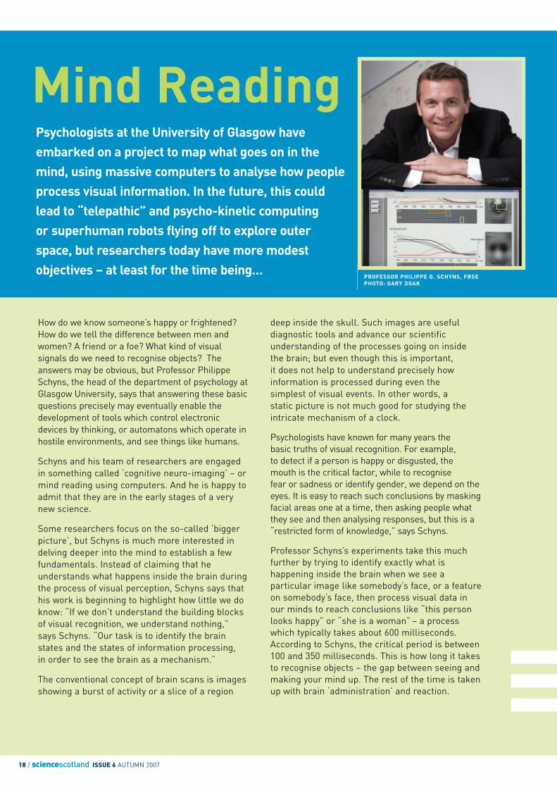
PROFESSOR PHILIPPE G. SCHYNS, FRSEPHOTO: GARY DOAK
sciencescotland ISSUE 6 AUTUMN 2007 / 19
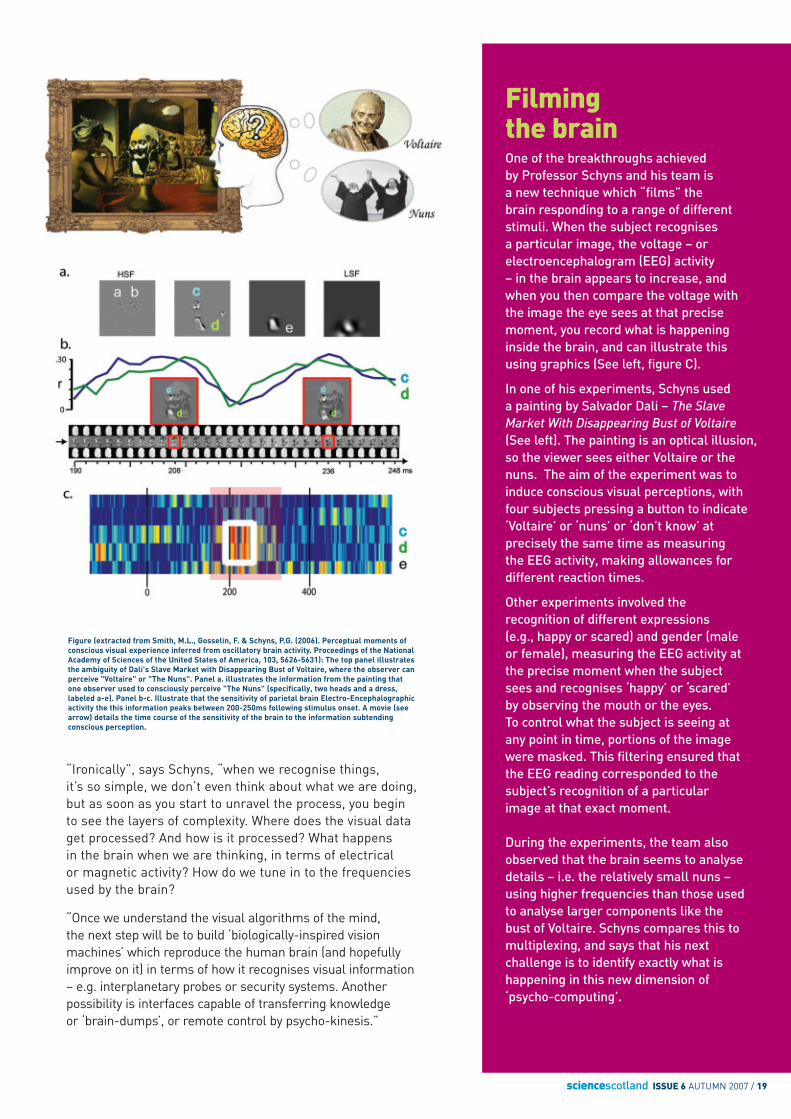
Filmingthe brainOne of the breakthroughs achieved by Professor Schyns and his team is a new technique which “films” the brain responding to a range of differentstimuli. When the subject recognises a particular image, the voltage – orelectroencephalogram (EEG) activity – in the brain appears to increase, andwhen you then compare the voltage withthe image the eye sees at that precisemoment, you record what is happeninginside the brain, and can illustrate thisusing graphics (See left, figure C).
In one of his experiments, Schyns used a painting by Salvador Dali – The SlaveMarket With Disappearing Bust of Voltaire(See left). The painting is an optical illusion,so the viewer sees either Voltaire or thenuns. The aim of the experiment was toinduce conscious visual perceptions, withfour subjects pressing a button to indicate‘Voltaire’ or ‘nuns’ or ‘don’t know’ atprecisely the same time as measuring the EEG activity, making allowances fordifferent reaction times.
Other experiments involved therecognition of different expressions (e.g., happy or scared) and gender (male or female), measuring the EEG activity atthe precise moment when the subject sees and recognises ‘happy’ or ‘scared’ by observing the mouth or the eyes.To control what the subject is seeing at any point in time, portions of the imagewere masked. This filtering ensured thatthe EEG reading corresponded to thesubject’s recognition of a particular image at that exact moment.
During the experiments, the team alsoobserved that the brain seems to analysedetails – i.e. the relatively small nuns –using higher frequencies than those used to analyse larger components like the bust of Voltaire. Schyns compares this tomultiplexing, and says that his nextchallenge is to identify exactly what ishappening in this new dimension of‘psycho-computing’.
“Ironically”, says Schyns, “when we recognise things,it’s so simple, we don’t even think about what we are doing,but as soon as you start to unravel the process, you begin to see the layers of complexity. Where does the visual dataget processed? And how is it processed? What happens in the brain when we are thinking, in terms of electrical or magnetic activity? How do we tune in to the frequenciesused by the brain?
“Once we understand the visual algorithms of the mind, the next step will be to build ‘biologically-inspired visionmachines’ which reproduce the human brain (and hopefullyimprove on it) in terms of how it recognises visual information – e.g. interplanetary probes or security systems. Anotherpossibility is interfaces capable of transferring knowledge or ‘brain-dumps’, or remote control by psycho-kinesis.”
Figure (extracted from Smith, M.L., Gosselin, F. & Schyns, P.G. (2006). Perceptual moments ofconscious visual experience inferred from oscillatory brain activity. Proceedings of the NationalAcademy of Sciences of the United States of America, 103, 5626-5631): The top panel illustratesthe ambiguity of Dali's Slave Market with Disappearing Bust of Voltaire, where the observer canperceive "Voltaire" or "The Nuns". Panel a. illustrates the information from the painting thatone observer used to consciously perceive "The Nuns" (specifically, two heads and a dress,labeled a-e). Panel b-c. Illustrate that the sensitivity of parietal brain Electro-Encephalographicactivity the this information peaks between 200-250ms following stimulus onset. A movie (seearrow) details the time course of the sensitivity of the brain to the information subtendingconscious perception.
20 / sciencescotland ISSUE 6 AUTUMN 2007
Processing PowerHuge computational poweris needed to process the datain Professor Schyns’s project.In each experiment, data aregathered from 64 to a fewhundred sensors on the brain,measuring 5 spatial frequencybands and 30 temporalfrequency bands, at 250different timepoints (every 4 milliseconds), as 4 subjectsrecognise 7 expressions –3,000 times. This generatesabout a terabyte (a terabyte isa million million bytes) of datawhich takes at least two weeksto analyse, using 64 parallelprocessors. “Five years ago,”says Schyns, “we simply didn’thave the computers to do this.”
“The first practical applications will be new enablingdevices,” says Schyns. “For example, tetraplegics couldmanipulate devices and communicate via an implantconnected to the relevant part of the brain. At the moment,we are only beginning to understand the fundamentalprinciples involved in developing these futuristic tools – we haven’t even worked out the alphabet yet, never mind the language.”
Some opportunists will probably want to use ‘visionmachines’ to detect when opponents are bluffing at poker.Others will see the potential for new kinds of securitydevices which ‘recognise’ criminals or terrorists. But nomatter what applications emerge from the currentresearch, Professor Schyns and his colleagues are quietlybuilding the foundations of a serious new kind of sciencewith profound implications for the future of technology and psychology.
Schyns says that his personal ambition is to“understand a few simple tasks” in the brain, but once this understanding is complete, it may lead to majorscientific breakthroughs which will literally change the way we look at things – forever.
New kinds ofsecurity deviceswhich ‘recognise’criminals orterrorists
sciencescotland ISSUE 6 AUTUMN 2007 / 21
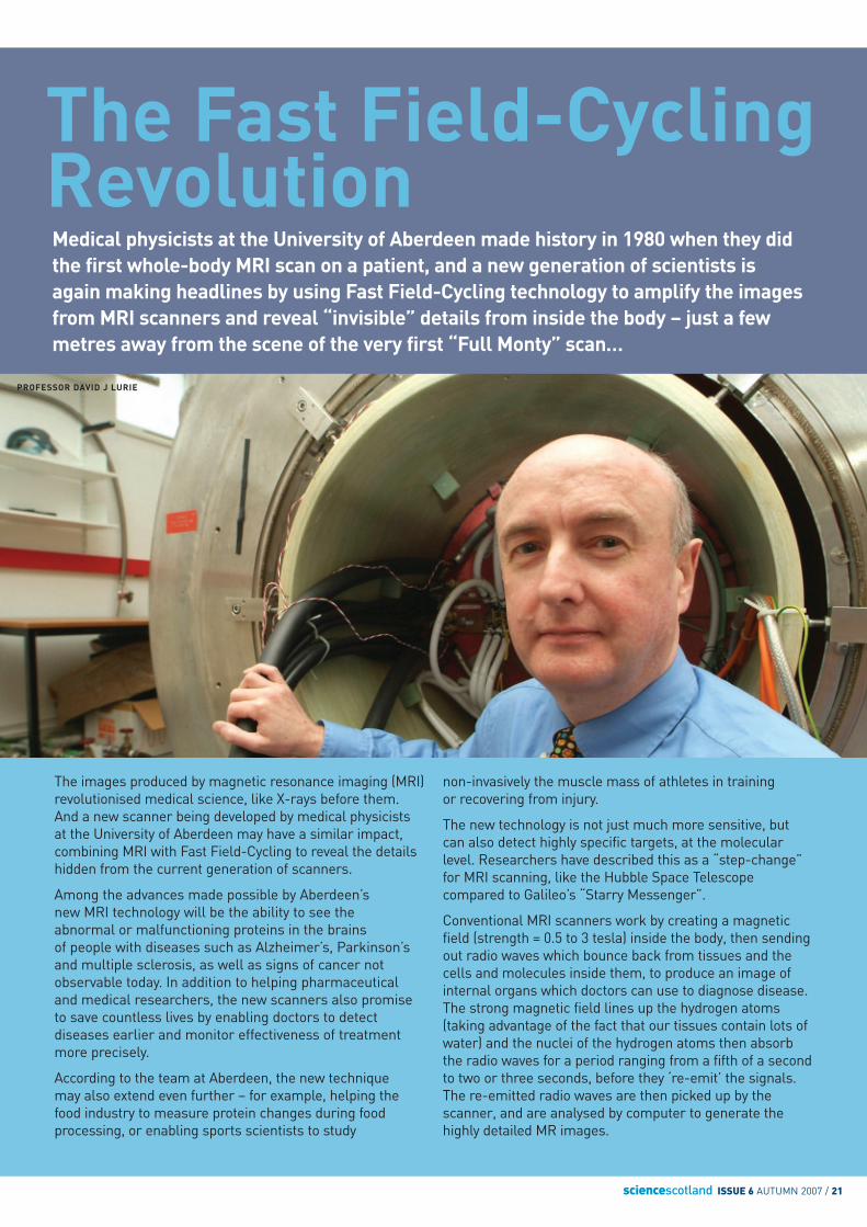
Medical physicists at the University of Aberdeen made history in 1980 when they didthe first whole-body MRI scan on a patient, and a new generation of scientists isagain making headlines by using Fast Field-Cycling technology to amplify the imagesfrom MRI scanners and reveal “invisible” details from inside the body – just a fewmetres away from the scene of the very first “Full Monty” scan…
The Fast Field-CyclingRevolution
The images produced by magnetic resonance imaging (MRI)revolutionised medical science, like X-rays before them.And a new scanner being developed by medical physicistsat the University of Aberdeen may have a similar impact,combining MRI with Fast Field-Cycling to reveal the detailshidden from the current generation of scanners.
Among the advances made possible by Aberdeen’s new MRI technology will be the ability to see the abnormal or malfunctioning proteins in the brains of people with diseases such as Alzheimer’s, Parkinson’sand multiple sclerosis, as well as signs of cancer notobservable today. In addition to helping pharmaceutical and medical researchers, the new scanners also promiseto save countless lives by enabling doctors to detectdiseases earlier and monitor effectiveness of treatmentmore precisely.
According to the team at Aberdeen, the new technique may also extend even further – for example, helping thefood industry to measure protein changes during foodprocessing, or enabling sports scientists to study
non-invasively the muscle mass of athletes in training or recovering from injury.
The new technology is not just much more sensitive, but can also detect highly specific targets, at the molecularlevel. Researchers have described this as a “step-change”for MRI scanning, like the Hubble Space Telescopecompared to Galileo’s “Starry Messenger”.
Conventional MRI scanners work by creating a magneticfield (strength = 0.5 to 3 tesla) inside the body, then sendingout radio waves which bounce back from tissues and thecells and molecules inside them, to produce an image ofinternal organs which doctors can use to diagnose disease.The strong magnetic field lines up the hydrogen atoms(taking advantage of the fact that our tissues contain lots ofwater) and the nuclei of the hydrogen atoms then absorbthe radio waves for a period ranging from a fifth of a secondto two or three seconds, before they ‘re-emit’ the signals.The re-emitted radio waves are then picked up by thescanner, and are analysed by computer to generate thehighly detailed MR images.
PROFESSOR DAVID J LURIE
22 / sciencescotland ISSUE 6 AUTUMN 2007
Earlier diagnosis and treatment monitoringThe delay between the signals going into the tissue andbeing sent back to the scanner is called the relaxation time,and it varies depending on the type of tissue involved andwhether it is healthy or diseased. This is because disease can affect the condition of the tissue at a molecular level –including fluid viscosity, how much protein is present, the size of the proteins and the way they ‘rotate’ or interact witheach other. The relaxation time is also reflected in the rawdata which the scanner translates into pictures – so doctorscan detect ‘molecules behaving badly’ inside the body.In basic terms, healthy tissue typically has a more rapidresponse time than unhealthy tissue.
Every hospital MRI scanner uses a constant magnetic field,and the frequency of the radio waves has to be tuned to thestrength of the field – otherwise it wouldn’t produce anymeaningful pictures of the state of the tissues. The imagesproduced and the way they are modified in diseased tissuesare directly affected by the strength of the magnetic field, but because the field and frequency are constant, what yousee is all you get – even though the image may be extremelydetailed, it is limited to what is revealed by that specificfrequency. In other words, there is a lot of hidden detail which,if revealed, may affect detection, diagnosis and treatment.
MRI would continue to save many lives if no-one ever tried to improve it, but Professor David Lurie, Chair in BiomedicalPhysics at the University of Aberdeen, has different ideas…
According to Lurie, he and his team are breaking “the first law ofMRI” by rapidly changing the magnetic field used by the scanner,at the same time as collecting the radiowave signals for MRimages, and the results could revolutionise medical science byrevealing much more detail than conventional MRI scans.
So how did Lurie arrive at this radical breakthrough?
Lurie has been using MRI for many years, developing methodsto image the distribution of free radicals – “misbehaving”molecules which can indicate problems like inflammatorydiseases, ischaemic heart disease and possibly cancer. In1987, Lurie and his team at Aberdeen pioneered a methodcalled Proton-Electron Double-Resonance Imaging (PEDRI),combining electron spin resonance (ESR) with MRI to generatehigh-resolution images and detect low concentrations of freeradicals. Part of this ongoing project involved designing and building new hardware which incorporates special double magnets (ferrite permanent magnets + copperelectromagnets), so researchers can adjust the strength of the magnetic field very quickly – what is called Fast Field-Cycling (FFC) – to hunt down their targets.
The breakthrough came in 1998, when Lurie attended the first FFC Relaxometry conference in Berlin and learned about the work of a team of researchers at the University ofUlm in Germany who had discovered a very special kind ofrelaxation-time response of protein samples, by observingthem in test-tubes with an FFC ”relaxometer” device that theyhad invented. When the German researchers measured the
nuclear magnetic resonance (NMR) of the protein sample’shydrogen atoms, they noticed that the presence of theprotein’s many hydrogen-nitrogen bonds influenced therelaxation in a particular way, due to the nitrogen nucleihaving a nuclear quadrupole resonance (NQR) interaction with their local environment. In fact, the presence of nuclearmagnetic and nuclear quadrupole resonance in the samesample gives rise to so-called “resonance crossings” – verywell-defined magnetic fields at which the NMR and NQRfrequencies coincide, and this is the reason for the specialrelaxation-time response of proteins. Inspired by thesefindings, which opened a window to a new kind of scanningtechnology, Lurie started thinking about combining FFC withMRI, realising this had the potential to dramatically enhancethe images produced by MRI.
“Resonance crossing is the key to unlocking the images,” Lurieexplains. “By measuring the relaxation times of tissues as afunction of magnetic field, we can also highlight the differencesbetween the behaviour of healthy and diseased tissue.”
Returning to Scotland, Lurie put his new ideas into action,confirming that FFC could be combined with MRI to revealmuch more detail, by scanning his own leg to prove it. He thenwrote special software to calibrate the imaging data, andembarked on the project which is starting to bear fruit today.
The FFC-MRI research is being funded by a grant of £2.4 million from the Engineering and Physical SciencesResearch Council (EPSRC), part of which will support threepost-doctoral staff and two PhD students over the next fouryears. The project also has industrial partners – TeslaEngineering, specialists in electromagnet technology, andOxford Instruments Molecular Biotools, who develop MRIcontrol hardware and software. A third partner is Invento, a spin-off from the University of Turin, which will work withLurie’s team to investigate contrast agents to improve stillfurther the effectiveness of FFC-MRI. The initial target of theresearch project is to optimise the design of the hardware andsoftware on a small-scale scanner, over the next 18 months,before scaling up the scanners to human dimensions. As soonas working scanners are built, they will be put to immediateuse in the laboratories of Aberdeen University’s Institute ofMedical Sciences, where scientists and medical researchers – co-applicants on Lurie’s grant – are researchingfundamental aspects of disease.
Lurie now has the resources at hand to be able to fulfil hisalmost decade-long vision, and is confident that FFC-MRI willhave a significant impact on early diagnosis and “help toelucidate disease mechanisms,” in the very near future. As well as neurological diseases and cancer, the technologywill also be used to investigate thrombosis, adds Lurie,exploiting the fact that proteins become immobile in bloodclots. And if he can make the invisible visible, Lurie and histeam of physicists, biologists and medical researchers will put Aberdeen on the map once again as a pioneer in scanning technology and applications.
sciencescotland ISSUE 6 AUTUMN 2007 / 23
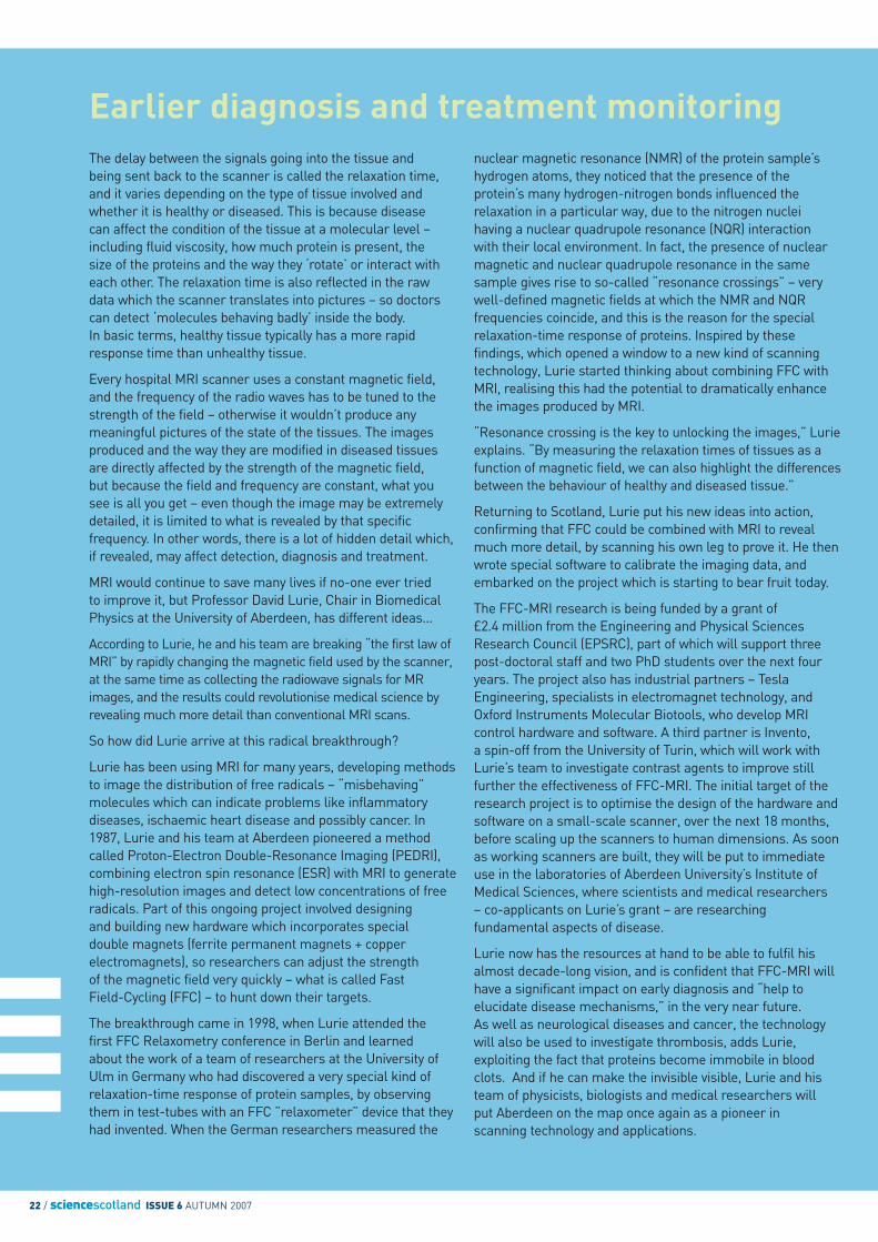
Scanning the horizon
It’s a science with 100,000 targets to aim for, andwhoever gets the next big hit could not only save manylives but also save huge sums of money for health-care providers and pharmaceutical companies.A team of researchers at the University of Aberdeen’sInstitute of Medical Sciences, specialising in PET(Positron Emission Tomography), already have sometargets in their sights.
The John Mallard Scottish PET Centre in Aberdeen – the first in Scotland, was established by imagingpioneer, Professor John Mallard. The physics of PET had been understood for decades but it wasn’t untilrecent years that it started to move from research labs to hospitals, thanks to the development of betterelectronics (including computers and scanners) and newbio-compounds – the biomarkers or ‘tracers’ which thescanners detect and display. Dr Andy Welch, the head ofthe Aberdeen PET team, sees the future of PET goingback to the labs, to develop the new generation oftracers, for drug development as well as diagnosis.
“Until very recently, our focus was on proving that PET is useful in health care,” says Welch, “but now we have established that and started using it fordiagnosing patients, it’s time to move forward andconcentrate more on translational medicine, evolvingfrom an emphasis on clinical uses (e.g. in cancer,heart disease, etc.) to more pre-clinical projects –helping to deliver new drugs to the bedside.”
Despite this shift in focus, the use of PET in healthcare will continue in parallel and be extended to moreand more patients. The PET centre in Aberdeen hasalready helped over 1,000 patients from all overScotland since March 2006 and there are also plans to open a facility in Glasgow in 2007, so as many as 5,000 people a year will have access to PETnationwide, at a cost of about £1,000 per scan.
PET has a wide range of uses in health care.As well as basic diagnosis, it is used to deliver more ‘personalised’ forms of treatment. In a group of women with breast cancer, for example, some
Medical physicists at the University of Aberdeen are looking far beyond the current applications of nuclear medicine, using radioactivity not just to make people better but also to develop better drugs…
DR ANDY WELCH
24 / sciencescotland ISSUE 6 AUTUMN 2007
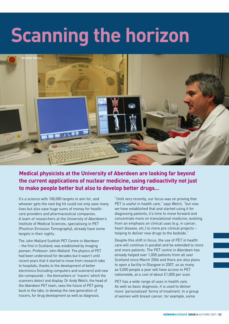
How PET worksPET involves injecting smallamounts of radioactive materialsinto a patient then watching whathappens, using a scanner tocreate a 3 D image, like a crossbetween a thermal photo and anX-ray. Radioactive forms ofimportant biological elements –tagged with atoms like oxygen,nitrogen, carbon and fluorine – are attracted to different“targets” in the body like organsand tumours, and this enablesdoctors to study the target byhighlighting details, revealed bythe radioactive ‘label’ attached tothe compound.
Some tracers and targets aremore ‘photogenic’ than others.For example, radioactive glucoseis attracted to tumours, becauseactive tumours need glucose to live on, so when the ‘hot’glucose arrives at the site of the cancer, it illuminates thetumour as if it has switched on a lightbulb.
As well as tracing where drugsgo and studying their impact,PET scans help to monitor thefunctioning of the brain, forexample, by revealing how many receptors are ‘occupied’before and after treatment.
may respond well to chemotherapy, while others may not benefit at all and suffer side-effects. The problem is how to identify which group iswhich, by measuring the tumour as treatment progresses. The PET scanworks particularly well in this context because it can show changes in thefunction of the tumour, which occur long before any changes in the size or shape (see sidebar left). Similarly, when it comes to lung cancer, somepatients may benefit from surgery while others may not, so PET scans canbe used to decide whether or not to operate – which in some cases may do more damage than good.
In pre-clinical work or translational medicine, PET can also be used tomeasure the effectiveness of new pharmaceuticals, enabling researchersto weed out the failures and focus on more promising candidates. It couldalso be used to evaluate drugs for approval, so they get to market faster.
The ‘magic molecules’ developed by pharmaceutical companies could be the key to the future of PET – or at least give it a boost. Many newmolecules, rejected as the basis of drugs, may have properties useful for PET. By exchanging their discoveries in return for the rights to these patented molecules, the PET Centre in Aberdeen can partner with biotechnology firms to accelerate progress in clinical and pre-clinical projects.
For example, the PET Centre is currently working with a majorpharmaceutical company to apply molecular imaging techniques to the early stages of drug discovery. Ultimately, PET could help developnew therapies for various psychiatric and neurological problems likeschizophrenia, Alzheimer’s disease and Parkinson’s disease, as well as cancer.
With over 100,000 proteins and more than 20,000 different genes, thehuman body has enough targets to keep all the imaging scientists busy for decades. So far, says Welch, most PET researchers have focused on glucose biomarkers, particularly fluoro-deoxy-glucose (FDG), but this is the tip of the iceberg.
“We need to find a smarter way of developing new drugs more quickly, andthis means we have to ask all the right questions,” says Welch. “Whichtracers have good imaging potential? And what are the targets to aim for?”
The PET Centre in Aberdeen has the expertise and infrastructure to buildon, and it’s only a matter of time before Welch and his team find a newbiomarker that changes the future of medical treatment – by finding a new way to see what is happening inside the mind and the body.
sciencescotland ISSUE 6 AUTUMN 2007 / 25
The no-brainerscannerComputed tomographic (CT) scanning has been around for over 30 years.It is widely available, non-invasive and easy to use, and it has revolutionisedthe treatment of many brain diseases. Magnetic resonance imaging (MRI)has been available for routine clinical use for about 20 years and hasadded substantially to the scanning revolution. Scientists in Scotland arenot only busy inventing new medical uses for both MRI and CT but alsohelping health-care providers to focus on the economic benefits of usingscanners for early diagnosis…
PROFESSOR JOANNA WARDLAWPHOTO: GARY DOAK
Weakness down one side, numbness, loss of vision,headache, dizziness, signs of confusion – it looks as if the man has had a stroke, but the wrong diagnosisor treatment may add to the awful statistics, so themedics will have to establish a few basic facts. Is this a haemorrhagic stroke? Ischaemic stroke? Or something which mimics a stroke, like a tumouror an abcess?
Bursting through the doors of the emergency room,the doctor immediately makes a decision: “There’s no time to lose. This man needs scanning – fast!”
The scene above may not appear in next week’s ER,but it’s part of a medical drama that happens for realevery day in hospitals all over Scotland.
Brain imaging now plays a key role in medicine all round the world, and even the abbreviations ‘MRI’ and ‘CT’ have become part of our everydayvocabulary. The advantage of MRI is that, unlike CT scans or X-rays, it uses strong magnetic fieldsrather than ionising radiation, which may damagetissue – e.g. in the eye or a foetus.
MRI is also more versatile than CT. Using ‘contrastagents’ (perfusion) and other techniques, MRI iscapable of distinguishing between different types ofpathologic tissue in the brain and the spinal cord,partly because it “sees” fluids like water andchemicals like lactate so clearly.
As scientists use MRI to investigate more and moreproblems, its applications multiply. According toProfessor Joanna Wardlaw, who directs the SFCBrain Imaging Research Centre in Edinburgh, theknowledge gained from MRI has revolutionised thecare of strokes, and that experience has helped to drive research into other diseases, includingpsychiatric and behavioural disorders like dementiaand schizophrenia.
“MRI has a key role to play in all aspects of stroke,”says Wardlaw. “It improves our understanding of whatcauses the stroke and, more importantly, how thestroke damages the brain. It also gives us insights intohow to treat and prevent strokes, as well as helpingpeople to recover, but it also helps us study manyother conditions affecting the functions of the brain.”
Even though nowadays imaging is taken for grantedin the care of strokes, much as ECG is now routinelyused to help with heart attacks, this was not alwaysthe case. Last year, about 3,000 people died fromstrokes in Scotland – roughly 80 deaths per 100,000people – but without imaging, the figures would beeven worse. In the past, diagnosis and treatmentimmediately after a stroke was a challenge for eventhe most experienced doctor. Despite the fact the vast majority of strokes are ischaemic (caused by ablockage in a blood vessel) rather than haemorrhagic(burst blood vessel), the clinical signs are not alwaysobvious, and many doctors used to make an educatedguess before prescribing the appropriate treatment,
26 / sciencescotland ISSUE 6 AUTUMN 2007
sometimes with unfortunate results. Today, by using CT or MRI, you can tell straight away what hashappened, and act accordingly. Compared to anyother method, MRI is highly sensitive to ischaemicchanges but both CT and MRI can quickly identify a haemorrhage.
Since the early 1990s, brain imaging has alsorevolutionised how strokes are treated by revealingthe effects of different therapies. The text books usedto say that it was best not to unblock a blood vesselafter a stroke, but brain imaging has proved that inmost cases this is the worst thing to do. “MRI isproviding a broader picture of how the brain damageoccurs and allows us to see things in context,” saysWardlaw. “It has made a huge difference to our overallknowledge and is enabling trials of new drugs.”
Before the age of brain imaging, researchers reliedmore on animal studies to observe what happened
during and after a stroke. This is still useful, butWardlaw believes MRI complements this and enables much more rapid progress.
MRI is also used to ‘film’ the progress of recoveringpatients by taking a series of snapshots at variousstages after they suffer a stroke, providing even morerevealing insights into this killer condition. This wouldbe difficult with CT because of the radiation dose frommultiple scans, but with MRI, that’s not a problem.
With several techniques available, strategic use of imaging is also important, says Wardlaw. Forexample, researchers have discovered that about 75per cent of the small haemorrhages which causeminor strokes may be missed by CT scanning. MRI,however, makes these small bleeds visible – thusconfirming its critical role in the treatment of mildstrokes in patients who do not reach hospital quickly.
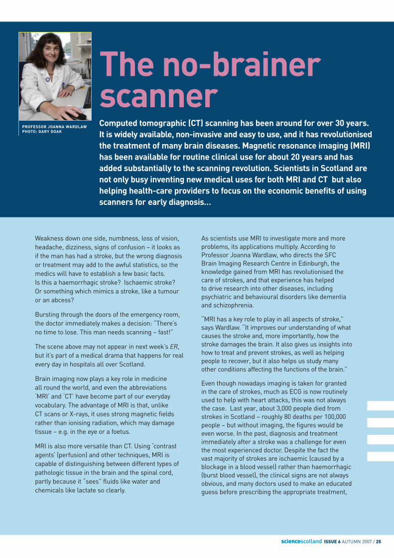
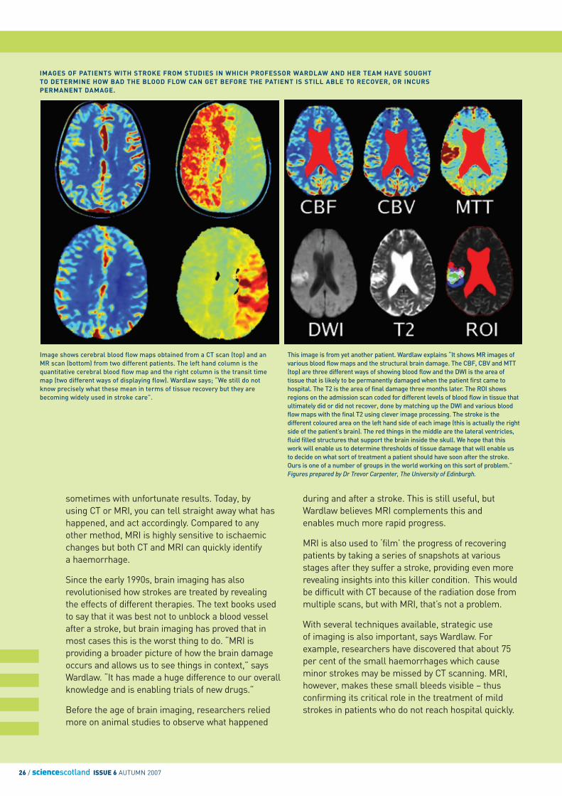
IMAGES OF PATIENTS WITH STROKE FROM STUDIES IN WHICH PROFESSOR WARDLAW AND HER TEAM HAVE SOUGHTTO DETERMINE HOW BAD THE BLOOD FLOW CAN GET BEFORE THE PATIENT IS STILL ABLE TO RECOVER, OR INCURSPERMANENT DAMAGE.
Image shows cerebral blood flow maps obtained from a CT scan (top) and anMR scan (bottom) from two different patients. The left hand column is thequantitative cerebral blood flow map and the right column is the transit timemap (two different ways of displaying flow). Wardlaw says; “We still do notknow precisely what these mean in terms of tissue recovery but they arebecoming widely used in stroke care”.
This image is from yet another patient. Wardlaw explains “It shows MR images ofvarious blood flow maps and the structural brain damage. The CBF, CBV and MTT(top) are three different ways of showing blood flow and the DWI is the area oftissue that is likely to be permanently damaged when the patient first came tohospital. The T2 is the area of final damage three months later. The ROI showsregions on the admission scan coded for different levels of blood flow in tissue thatultimately did or did not recover, done by matching up the DWI and various bloodflow maps with the final T2 using clever image processing. The stroke is thedifferent coloured area on the left hand side of each image (this is actually the rightside of the patient’s brain). The red things in the middle are the lateral ventricles,fluid filled structures that support the brain inside the skull. We hope that thiswork will enable us to determine thresholds of tissue damage that will enable usto decide on what sort of treatment a patient should have soon after the stroke.Ours is one of a number of groups in the world working on this sort of problem.”Figures prepared by Dr Trevor Carpenter, The University of Edinburgh.

sciencescotland ISSUE 6 AUTUMN 2007 / 27
Big boost forneuroscience Six Scottish universities –Aberdeen, Dundee, Edinburgh,Glasgow, St Andrews and Stirling– have been awarded £6 millionby the SFC (Scottish FundingCouncil) to boost brain imagingresearch.
By pooling their resources, the universities aim to recruitinternational researchers to Scotland, purchase newequipment, develop theemerging opportunities foracademic imaging, train the next generation of imaging talent and ensure that newdevelopments in imaging have a long-term impact on the health of the nation.
The programme will primarilyfocus on brain imaging and onthe technologies of magneticresonance imaging (MR),positron emission tomography(PET) and single photon emissioncomputed tomography (SPECT),as well as electrophysiology(EEG).
The universities believe thatneuroscience is a key strategicarea in Scotland, particularlyresearch in strokes, mentalhealth, dementia and psychology,and also hope to encourage newresearch into fields such ascancer, cardiovascular diseaseand diabetes.
The SFC Brain Imaging Research Centre has also helped advance ourunderstanding of imaging economics and as a result encouraged morehealth-care providers to use brain imaging much more strategically. Using data gathered in 2000, the Centre published a report in 2004 whichdemonstrated how CT and MRI scans can actually save money over thelong term by providing a more accurate diagnosis as soon as the patient isadmitted to hospital, thus speeding up decision-making, treatment andrecovery. For example, CT scans cost about £100 and occupying a hospitalbed for a day costs at least £300. If patients who have had a stroke arescanned soon after admission to hospital, that information speeds up thedelivery of more effective treatments which in turn speeds up dischargeand means that patients are less disabled. Thus you only have to reach thepoint of discharge, one-third of a day (eight hours) earlier, to cover the costof the scan. As Wardlaw says, most stroke patients need scanning anyway,so why not do it early when it is most useful?
In addition to its widespread use in hospitals, MRI is also an importantresearch tool, and researchers in Edinburgh have made a number ofsignificant breakthroughs:
• using Magnetic Resonance Spectroscopic Imaging (MRSI) tomeasure temperature changes in the brain (an increase mayindicate problems after a stroke)
• a nationwide study to investigate genetic factors in schizophrenia, using MRI to study the brains of young people at risk
• studying reasons for cognitive decline in the elderly
• using diffusion imaging and three-dimensional DTI tractography to map the white matter (or “wiring”) in brains, to detect changes with ageing, around tumours, in strokes and in schizophrenia
• measuring blood flow in arteries to detect abnormalities which may lead to cardiovascular problems
• new data processing methods
The focus of the SFC Brain Imaging Centre has been different from other research brain imaging centres because it is primarily concernedwith common disorders like strokes and mental illness, which have such a huge impact on public health, with an emphasis on practical,clinically-driven research.
“I now see imaging with CT and MR or other techniques as simply another research tool, like microscopes,” Wardlaw concludes, “but if it has a similar impact on medicine, that will be a major achievement.”

Title
Forename
Surname
Job Title
Organisation
Address
Register online at www.sciencescotland.org for regular updates, or complete and return this form to:Science Scotland, The Royal Society of Edinburgh, 22-26 George Street, Edinburgh, EH2 2PQ, United Kingdom
Science Scotland, bringing you the best of Science and Technology from Scotland, is apublication from the Royal Society of Edinburgh, with support from:
The devolved Government for Scotland is responsible for most of the issues of day-to-day concern to the people of Scotland, including health, education, justice, rural affairs, and transport. The Government was known as the Scottish Executive when it was established in 1999 following the first elections to the Scottish Parliament. The current administration was formed after elections in May 2007.
Seeking your feedback on Science ScotlandWe are committed to making Science Scotland as useful, informative and interesting as possible.
We would greatly appreciate your feedback and invite you please to spend no more than a couple
of minutes to answer a few questions on-line at www.sciencescotland.org
Town/City
Region/State
ZIP/Postcode
Email address
Please tick if you would like to receive a copy of our regular newsletter:
By Email By Post or Both