cutaneous fungal infections odermatophytosis - "ringworm" disease of the nails, hair,...
TRANSCRIPT
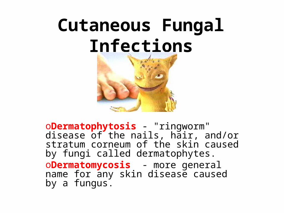
Cutaneous Fungal Infections
oDermatophytosis - "ringworm" disease of the nails, hair, and/or stratum corneum of the skin caused by fungi called dermatophytes. oDermatomycosis - more general name for any skin disease caused by a fungus.
THE SKIN PLANTS
• Etiological agents are called dermatophytes - "skin plants". Three important anamorphic genera, (i.e., Microsporum, Trichophyton, and Epidermophyton), are involved in ringworm.
• Dermatophytes are keratinophilic - "keratin loving". Keratin is a major protein found in horns, hooves, nails, hair, and skin.
• Ringworm - disease called ‘herpes' by the Greeks, and by the Romans ‘tinea' (which means small insect larvae).
Infections by Dermatophytes
• Severity of ringworm disease depends on (1) strains or species of fungus involved and (2) sensitivity of the host to a particular pathogenic fungus.
• More severe reactions occur when a dermatophyte crosses non-host lines (e.g., from an animal species to man). Among dermatophytes there appears to be a evolutionary transition from a saprophytic to a parasitic lifestyle. – Geophilic species - keratin-utilizing soil saprophytes (e.g., M.
gypseum, T. ajelloi). – Zoophilic species - keratin-utilizing on hosts - living animals
(e.g., M. canis, T. verrucosum). – Anthropophilic species - keratin-utilizing on hosts - humans
(e.g., M. audounii, T. tonsurans)
Clinical manifestations of ringworm infections are called different names on
basis of location of infection sites• tinea capitis - ringworm infection of the head, scalp,
eyebrows, eyelashes • tinea favosa - ringworm infection of the scalp (crusty
hair) • tinea corporis - ringworm infection of the body (smooth
skin)• tinea cruris - ringworm infection of the groin (jock itch) • tinea unguium - ringworm infection of the nails • tinea barbae - ringworm infection of the beard • tinea manuum - ringworm infection of the hand • tinea pedis - ringworm infection of the foot (athlete's foot)
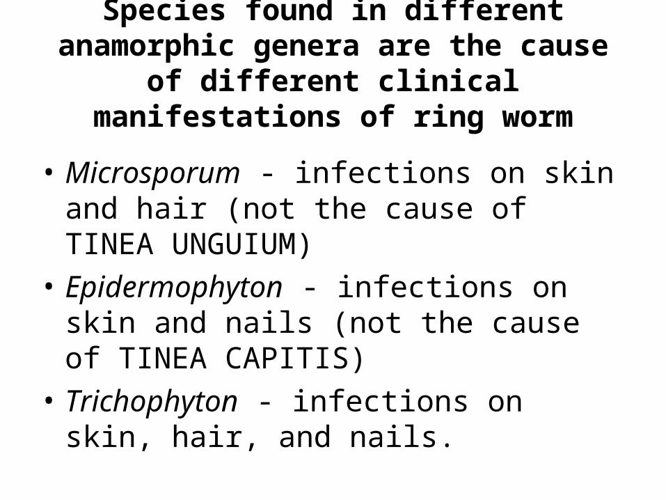
Species found in different anamorphic genera are the cause of different clinical
manifestations of ring worm
• Microsporum - infections on skin and hair (not the cause of TINEA UNGUIUM)
• Epidermophyton - infections on skin and nails (not the cause of TINEA CAPITIS)
• Trichophyton - infections on skin, hair, and nails.
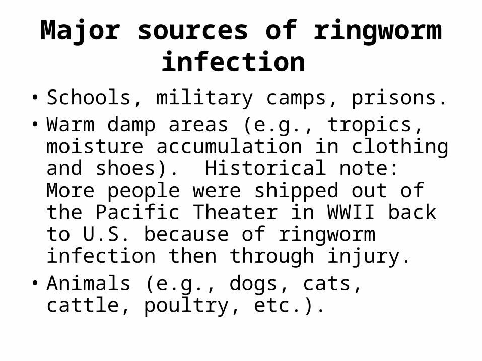
Major sources of ringworm infection
• Schools, military camps, prisons. • Warm damp areas (e.g., tropics, moisture
accumulation in clothing and shoes). Historical note: More people were shipped out of the Pacific Theater in WWII back to U.S. because of ringworm infection then through injury.
• Animals (e.g., dogs, cats, cattle, poultry, etc.).
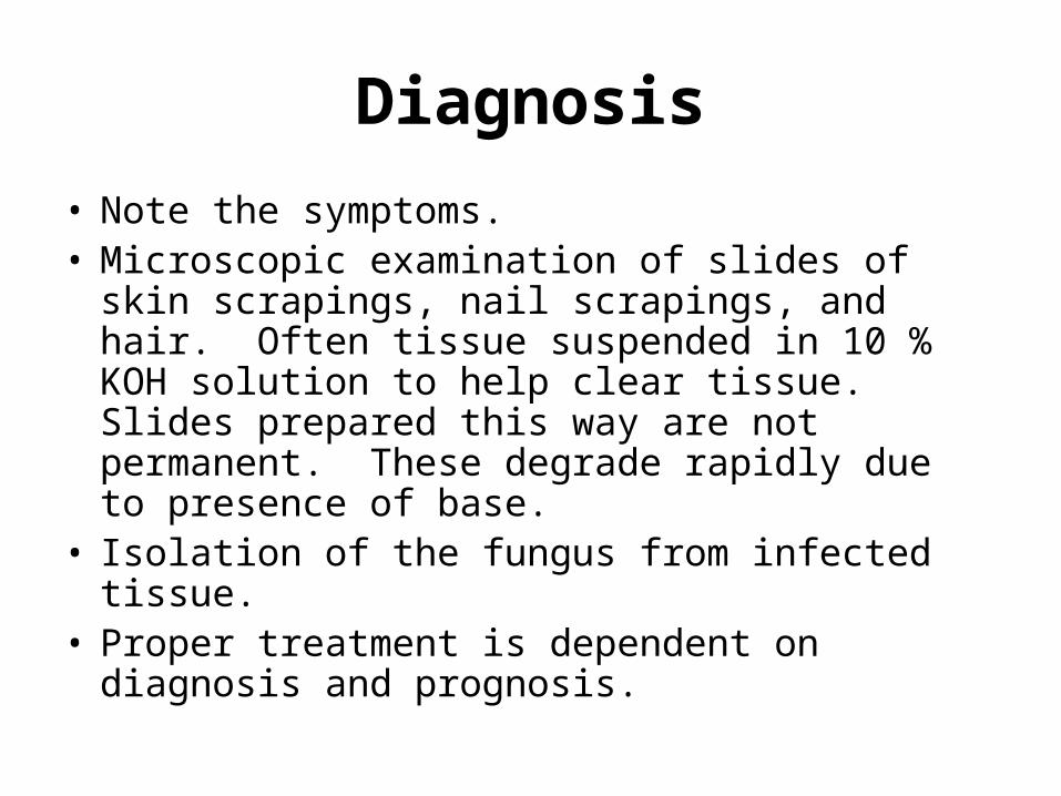
Diagnosis
• Note the symptoms. • Microscopic examination of slides of skin
scrapings, nail scrapings, and hair. Often tissue suspended in 10 % KOH solution to help clear tissue. Slides prepared this way are not permanent. These degrade rapidly due to presence of base.
• Isolation of the fungus from infected tissue. • Proper treatment is dependent on diagnosis and
prognosis.
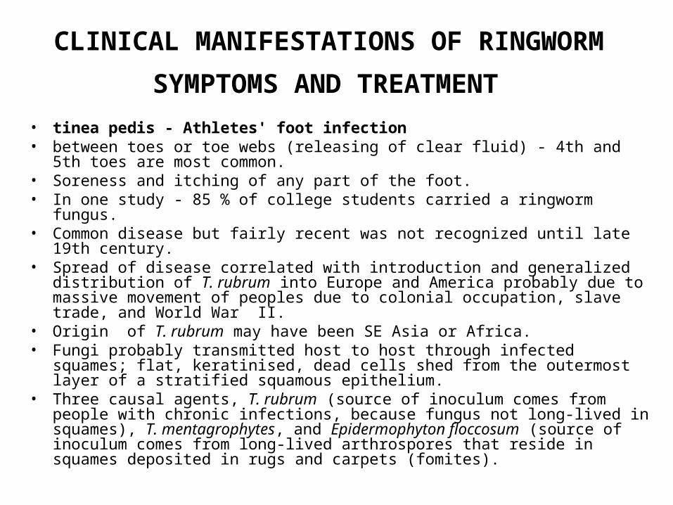
CLINICAL MANIFESTATIONS OF RINGWORM
SYMPTOMS AND TREATMENT • tinea pedis - Athletes' foot infection • between toes or toe webs (releasing of clear fluid) - 4th and 5th toes are most
common. • Soreness and itching of any part of the foot. • In one study - 85 % of college students carried a ringworm fungus. • Common disease but fairly recent was not recognized until late 19th century. • Spread of disease correlated with introduction and generalized distribution of T.
rubrum into Europe and America probably due to massive movement of peoples due to colonial occupation, slave trade, and World War II.
• Origin of T. rubrum may have been SE Asia or Africa. • Fungi probably transmitted host to host through infected squames; flat,
keratinised, dead cells shed from the outermost layer of a stratified squamous epithelium.
• Three causal agents, T. rubrum (source of inoculum comes from people with chronic infections, because fungus not long-lived in squames), T. mentagrophytes, and Epidermophyton floccosum (source of inoculum comes from long-lived arthrospores that reside in squames deposited in rugs and carpets (fomites).
CLINICAL MANIFESTATIONS OF RINGWORM
SYMPTOMS AND TREATMENT • Three Grades of Infection • Grade I - Subclinical
– An itching between toes, skin may be soft and macerated, blistering my occur.
– Treatment - keeping feet dry and clean, drying between the toes lightly each time you bathe to remove some skin. Application of fungicidal powders or ointments containing (1) salicylic acids to promote peeling of the skin and/or (2) tonaftate or other topical fungicides.
• Grade II – Host is conscious of a burning sensation while walking and standing. – Soaks are recommended (paints or liquids) such as 1:4000 KMnO4
(stains the skin purple) or topical fungicides. – Remove clear liquid from blisters by having a doctor puncture near the
base or unroofing the blister. – Dusting powder in morning to help keep feet dry.
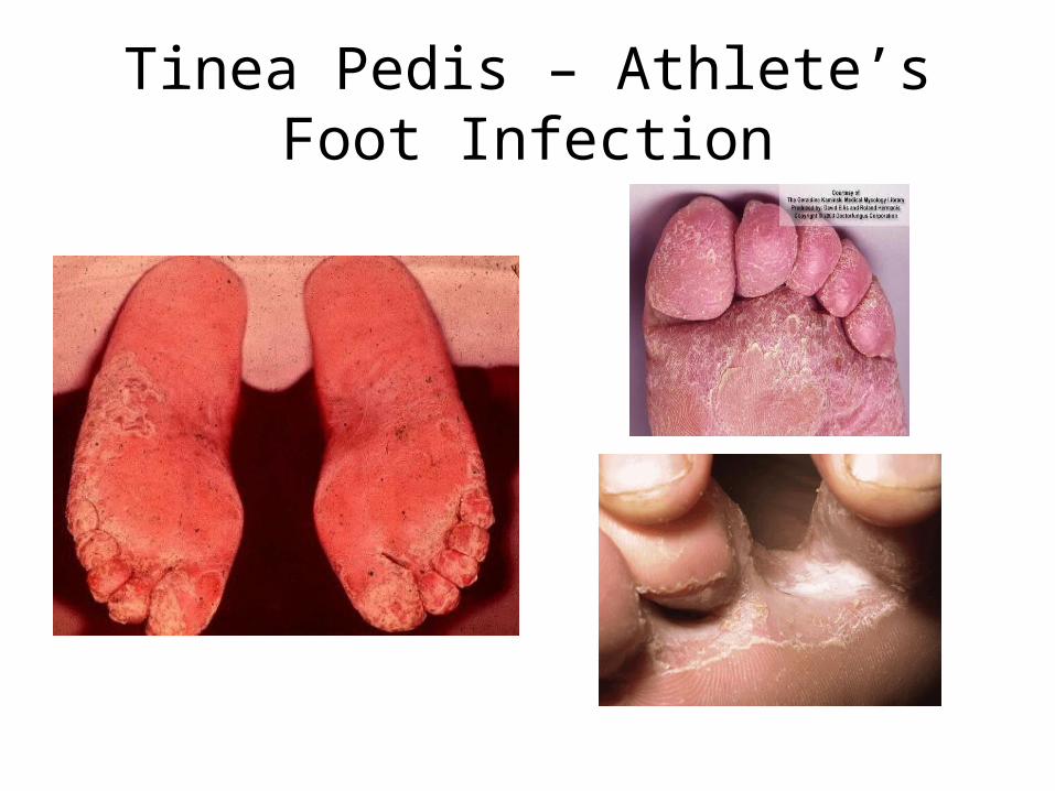
Tinea Pedis – Athlete’s Foot Infection
CLINICAL MANIFESTATIONS OF RINGWORM
SYMPTOMS AND TREATMENT • Grade III
– A secondary bacterial infection sets in. – Patient should go to bed. – Use systemic antibiotics to fight bacterial infection. – Use of soaks and compresses. – After infection subsidies, go to treatments as for Grade I or II
infections. – For persistent cases, (T. rubrum is usually the culprit), resort to
systemic griseofulvin therapy or other antifungal systemic drugs (i.e., Lamisiltrademark or terbinafine HCl)
• Griseofulvin is fungistatic, so it won't kill the fungus, just inhibit its growth.
– Improvement occurs in 2-6 weeks as long as 6 months. – Sometime griseofulvin treatment is ineffective.
• Some patients may not tolerate terbinafine HCL depending on sensitivity to drug.
CLINICAL MANIFESTATIONS OF RINGWORM
SYMPTOMS AND TREATMENT • Allergic reactions are sometimes associated with tinea
pedis and other ringworm infections. • dermatophytid - an "id" allergic reaction. • toxins get into blood stream and reaches a site other
than the site of infection. • blistering occurs on fingers and hands. • in diagnosis, rule out allergic reaction to poison ivy,
detergents or other substances. • during diagnosis, look for tinea (pedis, often) on the
body. • treat the primary site of infection where the antigen is
being produced. • treat secondary site - blisters.
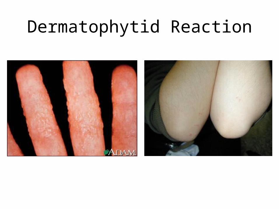
Dermatophytid Reaction
CLINICAL MANIFESTATIONS OF RINGWORM
SYMPTOMS AND TREATMENT • tinea corporis - body ringworm • Generally restricted to stratum corneum of the smooth skin. • Symptoms result form fungi metabolites such as toxin/allergens. • Disease found throughout the world. • Produces concentric or ring-like lesions on skin, and in severe cases these are raised and
may become inflamed. • All forms of tinea corporis caused by T. rubrum, T. mentagrophytes, T. tonsurans, M.
canis, and M. audouinii are treatable with topical agent containing tolnaftate, ketoconazole, miconazole, etc...
• Disease transmitted through infected scales hyphae or arthroconidia on the skin. • Also transmitted through direct contact between infected humans or animals, by fomites
(any agent such a bedding or clothing capable of retaining a pathogen and transmitting to a new host).
• Transfer form on area to the body to another (from tinea pedis to tinea corporis). • Tinea Corporis normally resolves itself in several months. • T. verrucosum and T. violaceum infections require more vigorous treatment including
cleaning of area to remove of scales and older fungicidal topical applications of ammoniated mercury ointment, 3 % salicylic and sulfuric acid, or tincture of iodine for several weeks.
• Widespread tinea corporis and more severe types (lesions) require systemic griseofulvin treatment (about 6 weeks for effective treatment).
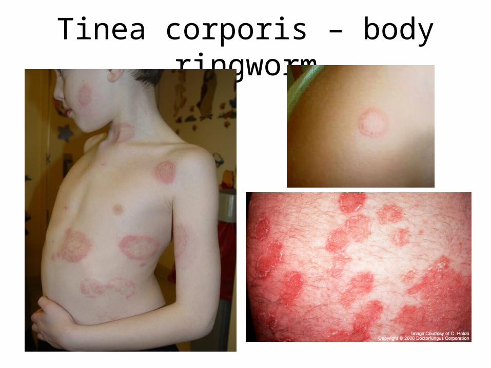
Tinea corporis – body ringworm
CLINICAL MANIFESTATIONS OF RINGWORM
SYMPTOMS AND TREATMENT • tinea cruris - ringworm of the groin and surrounding region • More common in men than women. • Infection seen on scrotum and inner thigh, the penis is usually not infected. • Epidemics associated with grouping of people into tight quarters - athletic teams, troops,
ship crews, inmates of institutions. • Several causes of tinea cruris include T. rubrum (does not normally survive long periods
outside of host), E. flocossum (usually associate with epidemics because resistant arthroconidia in skin scales can survive for years on rugs, shower stalls, locker room floors), T. mentagrophytes (usually of animal origin, such as rodents), and Microsporum gallinae (rarely seen - usually found on gallinaceous birds like turkeys and chickens).
• Predisposing factors include persistent perspiration, high humidity, irritation of skin from clothes, such as tight fitting underwear or athletic supporters, pre-existing disease, such as diabetes and obesity.
• Diagnosis – If lesion "weep", it is likely caused by a yeast, such as, Candida albicans, and not by a
dermatophyte, especially if infections are seen in a woman. – KOH examination of skin scrapings. – Culture of dermatophyte from skin scrapings.
• Treatment – Tolnaftate (Tinactin trademark) treatment protocol for tinea corporis. – Relief of symptoms occur within 3 days and treatment continued until all signs of disease are
gone. – Area is sensitive so the other care needs to be taken into to add to irritation of region.
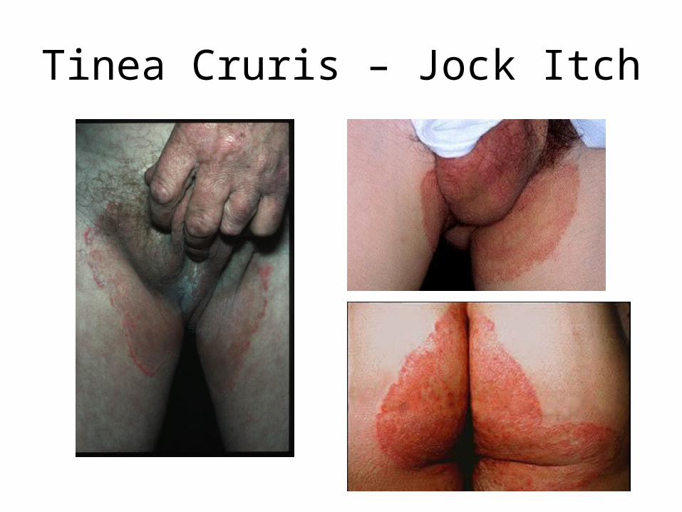
Tinea Cruris – Jock Itch
CLINICAL MANIFESTATIONS OF RINGWORM
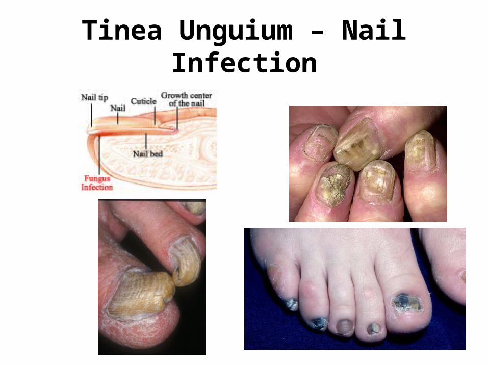
SYMPTOMS AND TREATMENT • tinea unguium - ringworm of the nails • Tinea unguium or onchomycosis can take two forms:
– Leukonychia mycotica - superficial white onychomycosis, invasion of fungus restricted to patches or pits on surface of the toenail.
– Invasive subungual dermatophytosis - lateral or distal edges first involved, followed by establishment of the infection beneath the nail plate. Invasion of nail plates by dermatophytes.
• Onychomycosis (infection of nails caused by non-dermatophytic fungi and yeasts) • Most commonly caused by T. rubrum, then E. floccosum or other Trichophyton
species. • Resistant to treatment, rarely resolves spontaneously. • Topical treatments - poor record of cure. • Ablation - surgical or chemical removal of nail. • Systemic griseofulvin therapy can lead to remission (usually a year or more of
treatment required - results vary about 29 % cure rate). • Use of other systemic antifungal (i.e., Lamisiltrademark or terbinafine HCl). • Filing down the nail to paper thin consistency and soaking or painting with KMnO4
(1:4000), phenol, 10 % salicylic acid, or 1% iodine is useful adjunct to systemic griseofulvin treatment.
Tinea Unguium – Nail Infection
CLINICAL MANIFESTATIONS OF RINGWORM
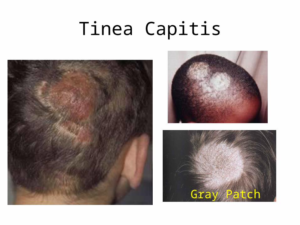
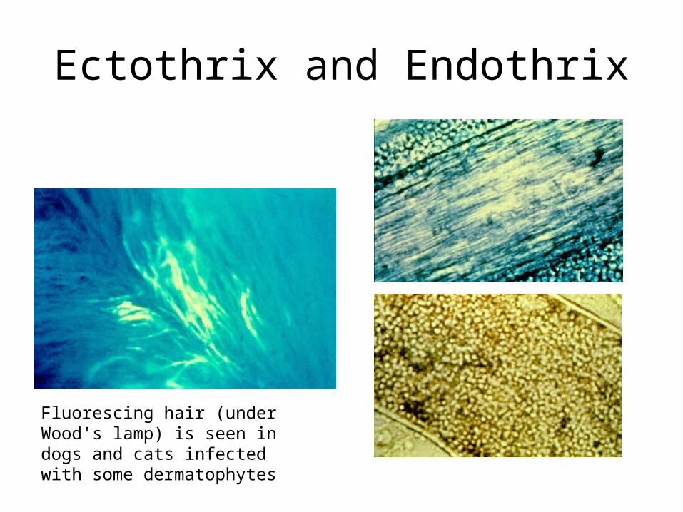
SYMPTOMS AND TREATMENT • tinea capitis - ringworm of the scalp, eyebrows and eyelashes • Caused by species of Microsporum and Trichophyton. • Fungus grows into hair follicle. • Using a Wood's lamp, on hair Microsporum species tend to fluoresce green
while Trichophyton species generally do not fluoresce. • Lack of fluorescence does not mean it isn't Microsporum. • Subculture any strands of hair that fluoresce to help identify the causal agent. • Ectothrix infection - fragmentation of mycelium into conidia (called arthroconidia)
around the hair shaft or just beneath the cuticle with destruction of the cuticle. This type of infection caused by M. audounii, M. canis, M. ferrugineum, T. mentagrophytes, T. verrucosum and T. megninii.
• Endothrix infection - arthroconidia formation occurs by fragmentation of hyphae with the hair shaft with destruction of the cuticle. This type of infection caused T. tonsurans (most common cause), T. violaceum, T. rubrum, and T. gourvillii. All these pathogen species are anthropophilic.
• "Gray patch ringworm" ectothrix common disease in children usually not associated with inflammation.
• Zoophilic and geophilic dermatophytes infections on man associate with inflammation. Microsporum canis, T. verrucosum, and T. mentagrophytes (zoophilic); M. gypseum and M. fulvum (geophilic species).
• "Id" reaction may occur.
Treatment of Tinea Capitis
• Ectothrix infections often resolve on their own. • Endothrix infections my become chronic and may
continue into adulthood. • Topical treatments are ineffective (don't bother using
tonaftate or topical griseofulvin) • Fungistatic agents are somewhat effective (miconazole,
clotrimazole) in combination to systemic administration of griseofulvin.
• Vigorous daily scrubs of scalp help removal of infectious debris. Do not use this treatment on patients with porphyria (an accumulation of blood pigment called porphyrins in blood stream and urine) or is hypersensitive to griseofulvin.
Tinea Capitis
Gray Patch
Ectothrix and Endothrix
Fluorescing hair (under Wood's lamp) is seen in dogs and cats infected with some dermatophytes
DERMATOPHYTES
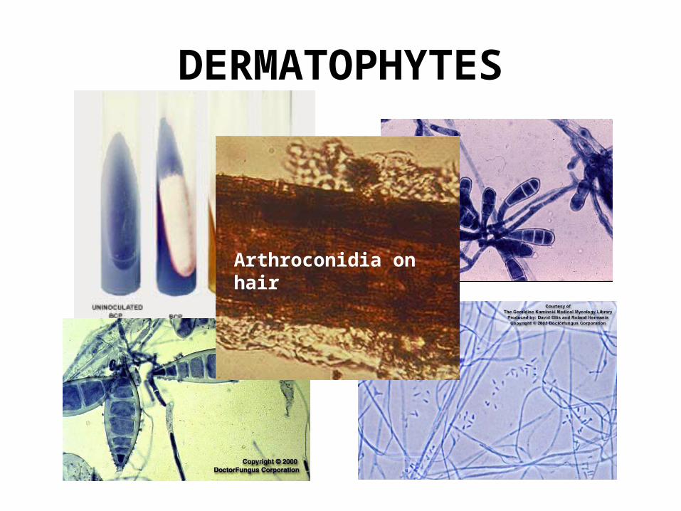
Arthroconidia on hair
DERMATOPHYTES
• Microsporum audouinii • No teleomorph state (sexual reproduction has yet to be
observed in this species). • Usually devoid of conidia (macro- or microconidia). • Septate hyphae with terminal chlamydoconidia, often
pointed at the end. • Macroconidia are often irregular or non-uniform in shape. • Colonies on culture media are flat, silky in appearance. • Growth of colonies on culture media is tight. • On reverse of colony - pigment is reddish-brown in color.
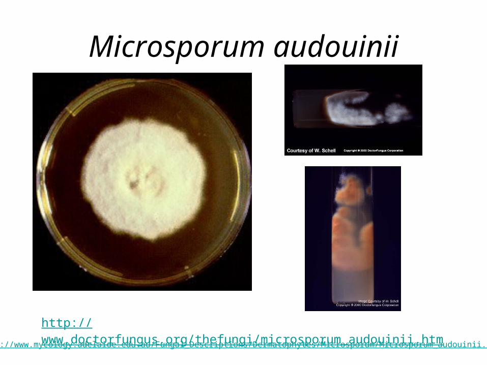
Microsporum audouinii
http://www.doctorfungus.org/thefungi/microsporum_audouinii.htm
http://www.mycology.adelaide.edu.au/Fungal_Descriptions/Dermatophytes/Microsporum/Microsporum_audouinii.html
DERMATOPHYTES
• Microsporum canis complex? • Teleomorph is an ascomycete called
Arthroderma otae. Almost all clinical isolates are minus mating type.
• Macroconidia are abundant, thick-walled with many septa, up to 15. Macroconidia are often hooked or curved at ends.
• Microconidia are small and clavate (club-shaped).
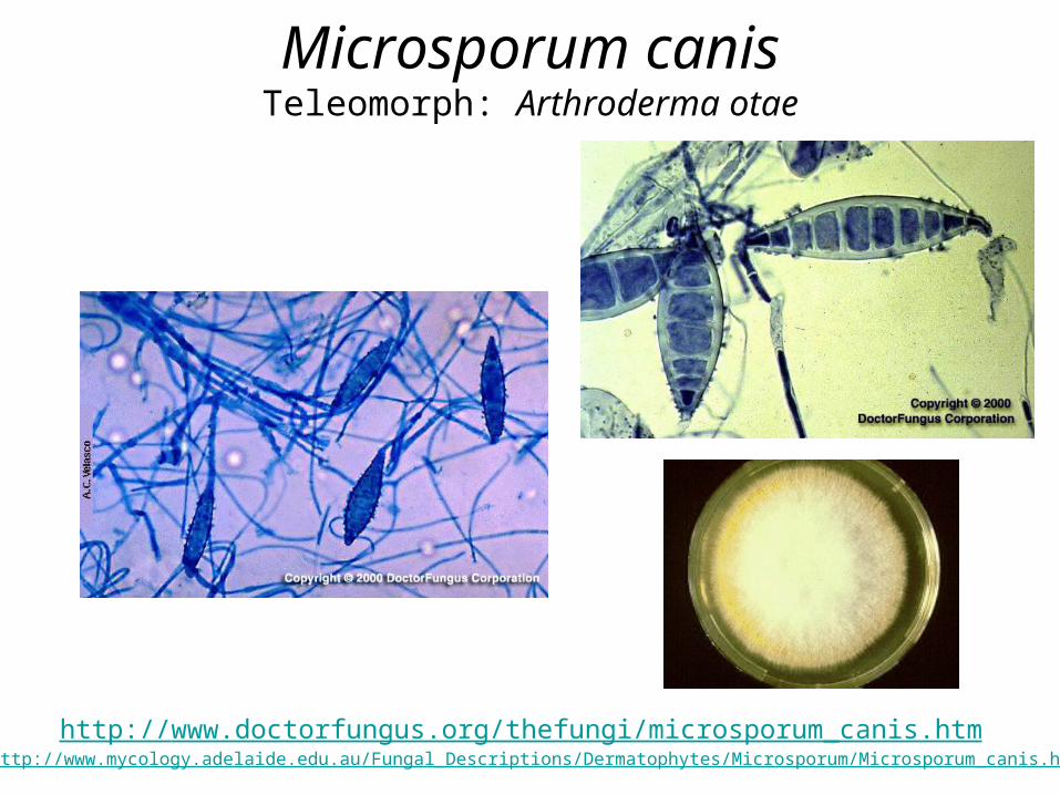
Microsporum canisTeleomorph: Arthroderma otae
http://www.doctorfungus.org/thefungi/microsporum_canis.htmhttp://www.mycology.adelaide.edu.au/Fungal_Descriptions/Dermatophytes/Microsporum/Microsporum_canis.html
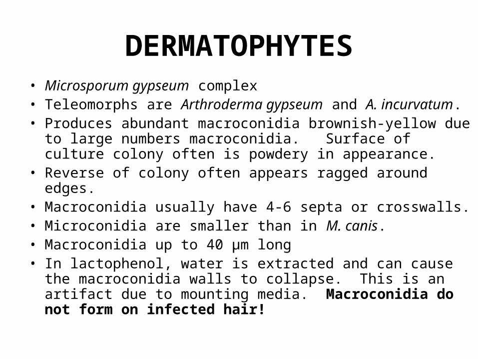
DERMATOPHYTES• Microsporum gypseum complex • Teleomorphs are Arthroderma gypseum and A. incurvatum. • Produces abundant macroconidia brownish-yellow due to
large numbers macroconidia. Surface of culture colony often is powdery in appearance.
• Reverse of colony often appears ragged around edges. • Macroconidia usually have 4-6 septa or crosswalls. • Microconidia are smaller than in M. canis. • Macroconidia up to 40 µm long • In lactophenol, water is extracted and can cause the
macroconidia walls to collapse. This is an artifact due to mounting media. Macroconidia do not form on infected hair!
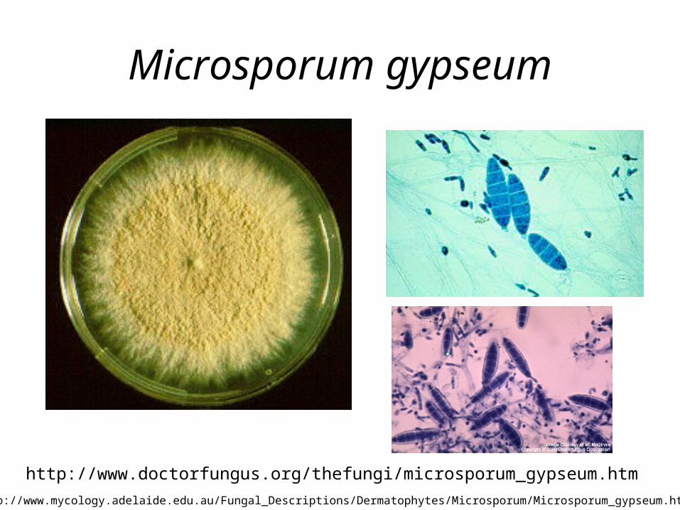
Microsporum gypseum
http://www.mycology.adelaide.edu.au/Fungal_Descriptions/Dermatophytes/Microsporum/Microsporum_gypseum.htm
http://www.doctorfungus.org/thefungi/microsporum_gypseum.htm
DERMATOPHYTES
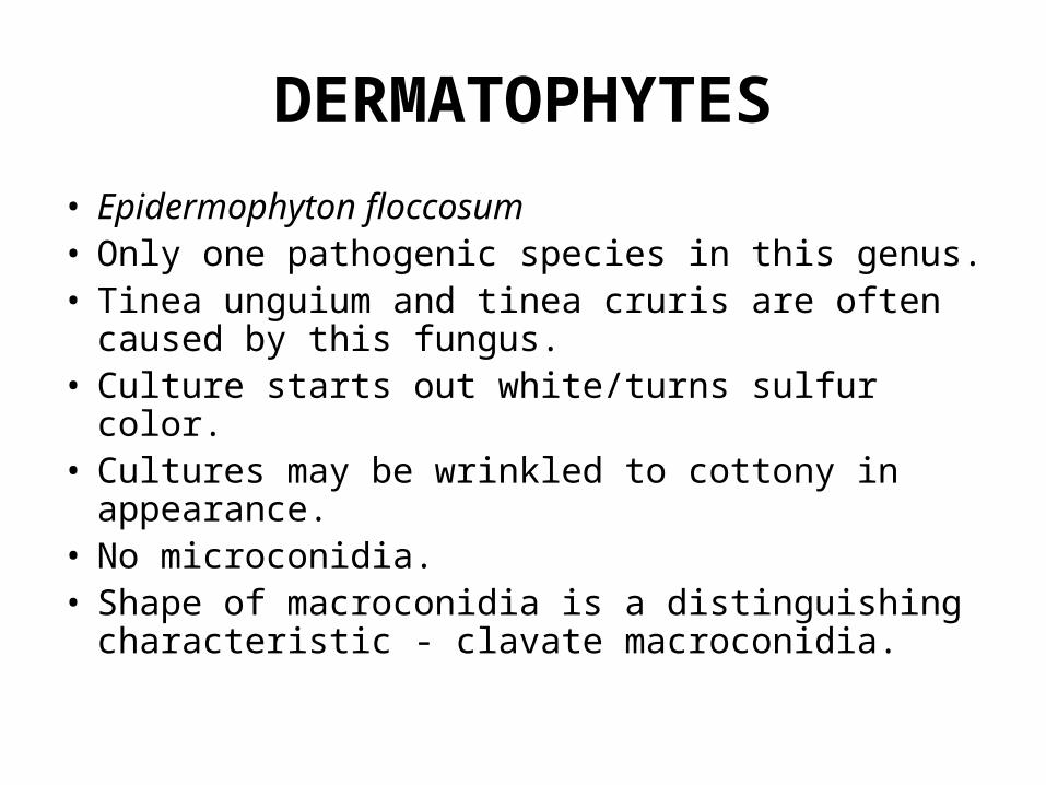
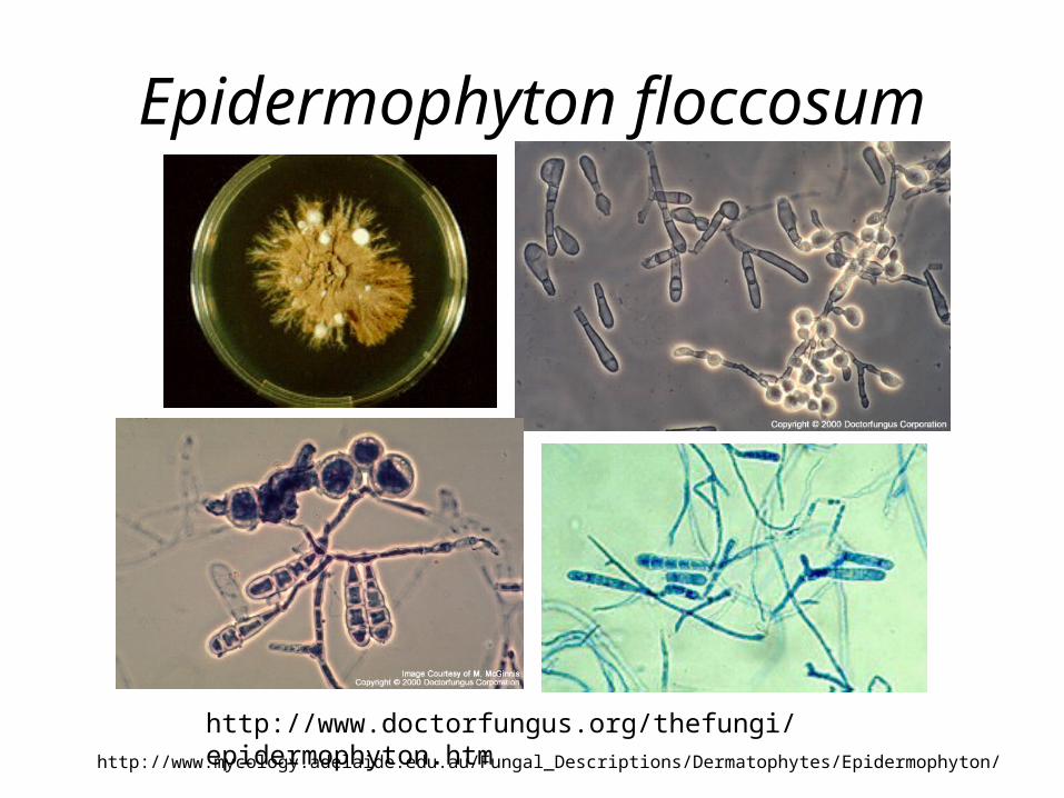
• Epidermophyton floccosum • Only one pathogenic species in this genus. • Tinea unguium and tinea cruris are often caused
by this fungus. • Culture starts out white/turns sulfur color. • Cultures may be wrinkled to cottony in
appearance. • No microconidia. • Shape of macroconidia is a distinguishing
characteristic - clavate macroconidia.
Epidermophyton floccosum
http://www.mycology.adelaide.edu.au/Fungal_Descriptions/Dermatophytes/Epidermophyton/
http://www.doctorfungus.org/thefungi/epidermophyton.htm
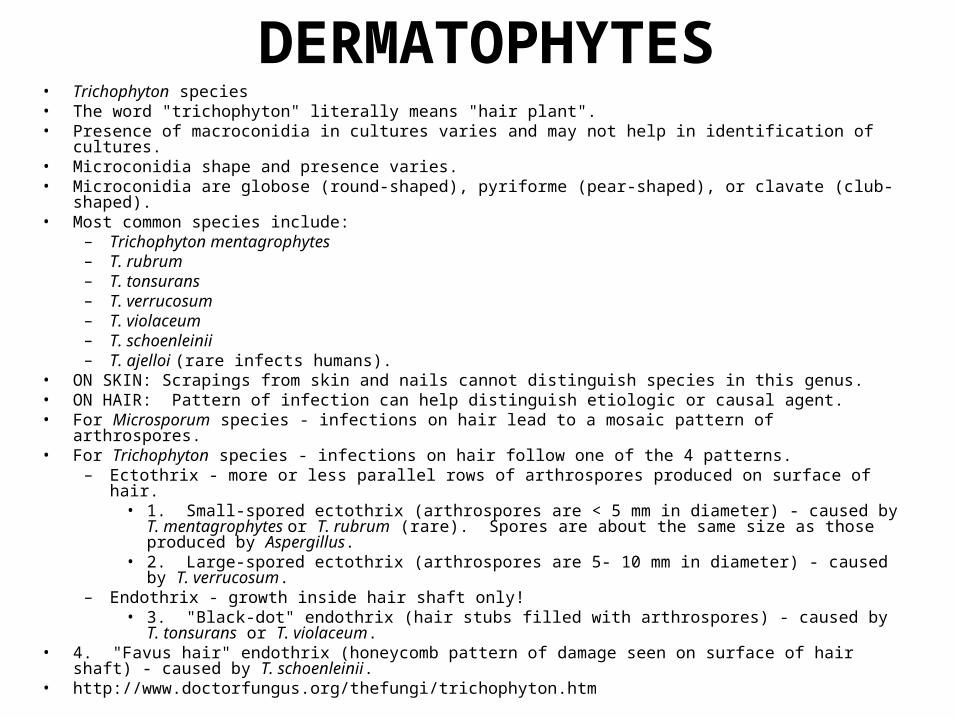
DERMATOPHYTES• Trichophyton species • The word "trichophyton" literally means "hair plant". • Presence of macroconidia in cultures varies and may not help in identification of cultures. • Microconidia shape and presence varies. • Microconidia are globose (round-shaped), pyriforme (pear-shaped), or clavate (club-shaped). • Most common species include:
– Trichophyton mentagrophytes – T. rubrum – T. tonsurans – T. verrucosum – T. violaceum – T. schoenleinii – T. ajelloi (rare infects humans).
• ON SKIN: Scrapings from skin and nails cannot distinguish species in this genus. • ON HAIR: Pattern of infection can help distinguish etiologic or causal agent. • For Microsporum species - infections on hair lead to a mosaic pattern of arthrospores. • For Trichophyton species - infections on hair follow one of the 4 patterns.
– Ectothrix - more or less parallel rows of arthrospores produced on surface of hair. • 1. Small-spored ectothrix (arthrospores are < 5 mm in diameter) - caused by T.
mentagrophytes or T. rubrum (rare). Spores are about the same size as those produced by Aspergillus.
• 2. Large-spored ectothrix (arthrospores are 5- 10 mm in diameter) - caused by T. verrucosum.
– Endothrix - growth inside hair shaft only! • 3. "Black-dot" endothrix (hair stubs filled with arthrospores) - caused by T. tonsurans or T.
violaceum. • 4. "Favus hair" endothrix (honeycomb pattern of damage seen on surface of hair shaft) - caused by
T. schoenleinii. • http://www.doctorfungus.org/thefungi/trichophyton.htm
DERMATOPHYTES
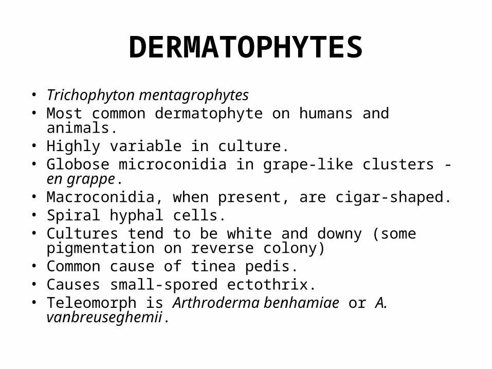
• Trichophyton mentagrophytes • Most common dermatophyte on humans and animals. • Highly variable in culture. • Globose microconidia in grape-like clusters - en grappe. • Macroconidia, when present, are cigar-shaped. • Spiral hyphal cells. • Cultures tend to be white and downy (some pigmentation
on reverse colony) • Common cause of tinea pedis. • Causes small-spored ectothrix. • Teleomorph is Arthroderma benhamiae or A.
vanbreuseghemii.
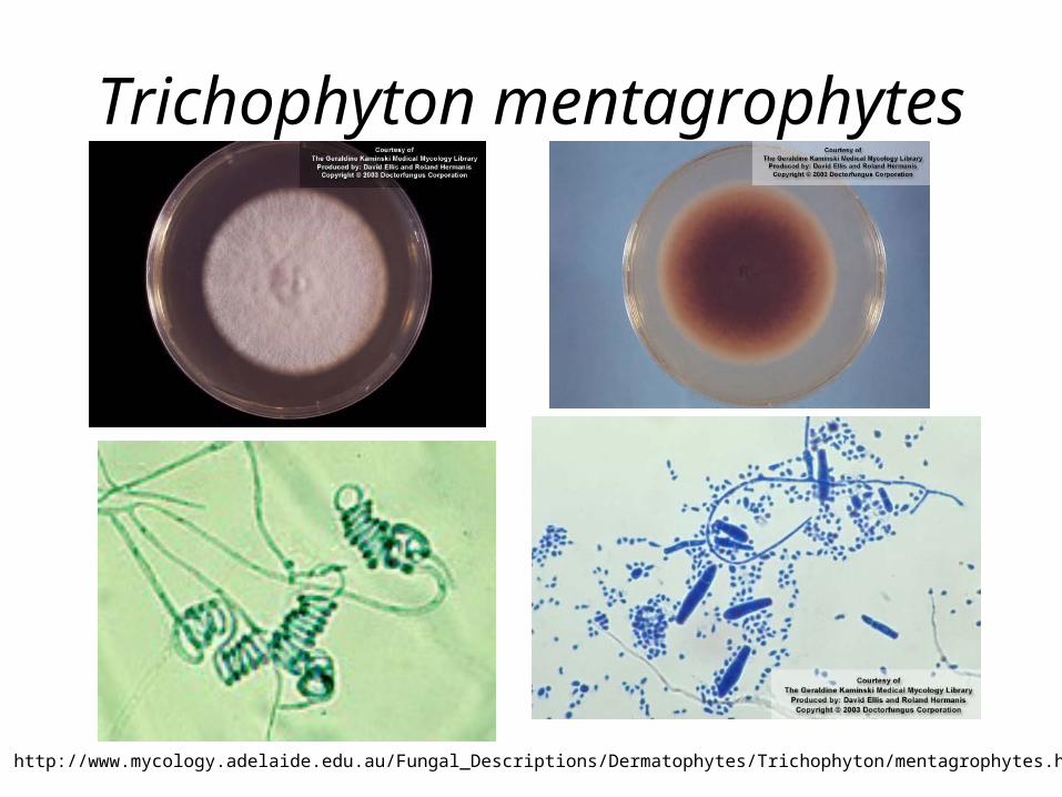
Trichophyton mentagrophytes
http://www.mycology.adelaide.edu.au/Fungal_Descriptions/Dermatophytes/Trichophyton/mentagrophytes.html
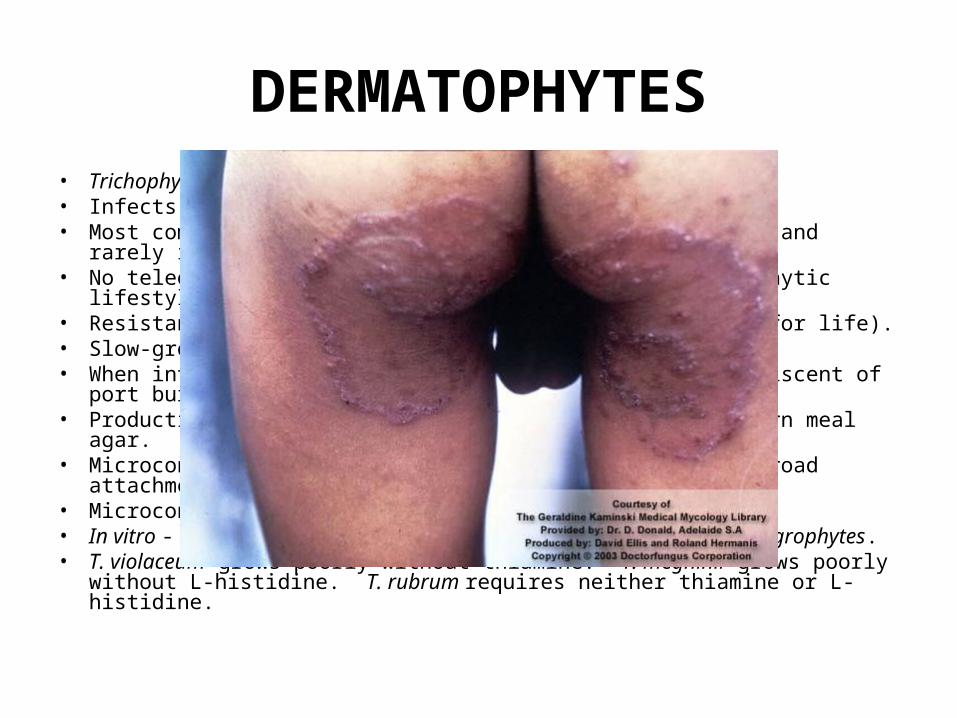
DERMATOPHYTES• Trichophyton rubrum • Infects nails and smooth skin (rarely found on hair). • Most common and widely distributed dermatophyte on man and rarely
isolated from animals, never from soils. • No teleomorph (possibly lost in transition from saprophytic lifestyle to man). • Resistant and persistent (some people become carriers for life). • Slow-growing in culture. • When intensely pigmented in culture the color is reminiscent of port
burgundy wine or venous blood. • Production of pigment increased, if fungus grown on corn meal agar. • Microconidium are clavate or "teardrop" shape with a broad attachment
point of the hyphae. • Microconidia may develop on sides of macroconidium. • In vitro - lack of hair penetrating organs, unlike T. mentagrophytes. • T. violaceum grows poorly without thiamine. T. megninii grows poorly
without L-histidine. T. rubrum requires neither thiamine or L- histidine.
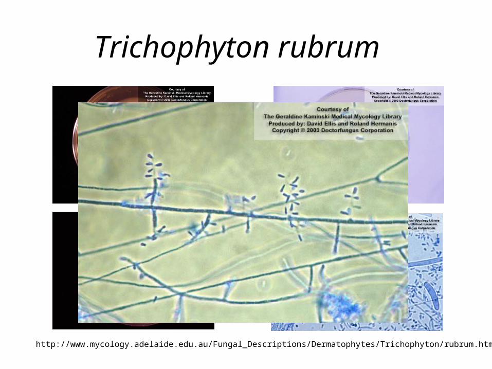
Trichophyton rubrum
http://www.mycology.adelaide.edu.au/Fungal_Descriptions/Dermatophytes/Trichophyton/rubrum.htm
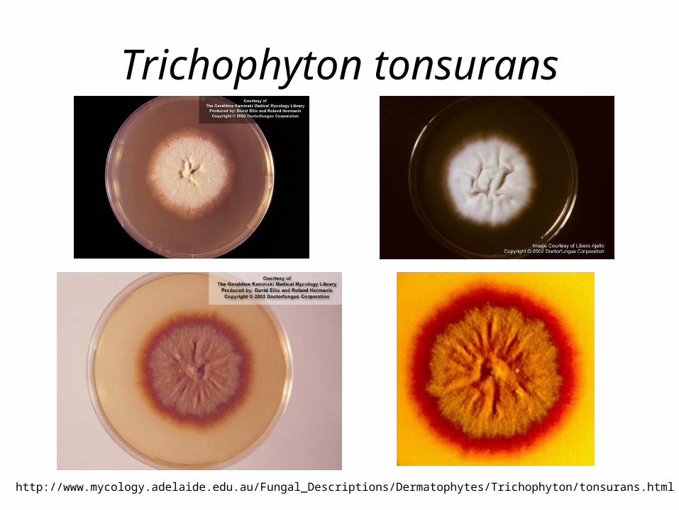
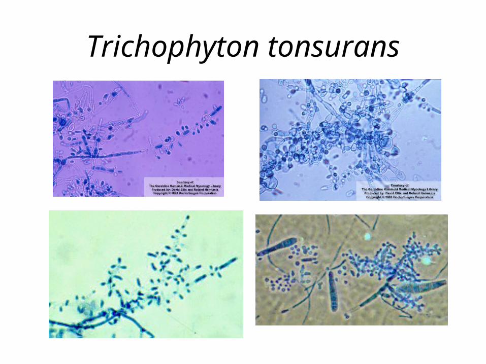
DERMATOPHYTES• Trichophyton tonsurans • Anthropophilic and on hair causes endothrix. • Third most common cause of tinea capitis • Other leading causes of tinea capitis are M.
audouinii (transmission is generally from child to child) and M. canis (transmission is from animal to human).
• Colonies whitish and folded. • Colonies are yellowish-brown color on reverse of
colony. • Microconidia are longer and larger than in T.
rubrum. • Intercalary and terminal chlamydoconidia
common in older cultures. • Macroconidia not common, irregular in form.
Trichophyton tonsurans
http://www.mycology.adelaide.edu.au/Fungal_Descriptions/Dermatophytes/Trichophyton/tonsurans.html
Trichophyton tonsurans
DERMATOPHYTES• Trichophyton violaceum • Attacks hair, scalp, skin and nails. • Nail infections are persistent. • Endothrix (black dot infection of scalp). • Found in humans, rarely in animals. • Disease has been reported in horses, cats, dogs, mice
and pigeons. • Very slow growing in culture with a waxy appearance. • Colony deep violent in color, purplish pigment diffuses into
media. • Rarely produces microconidia and macroconidia. • In culture this species requires thiamine for proper growth. • Hyphae coarser in appearance than seen in other
dermatophytes. • Chlamydoconidia are seen in culture.
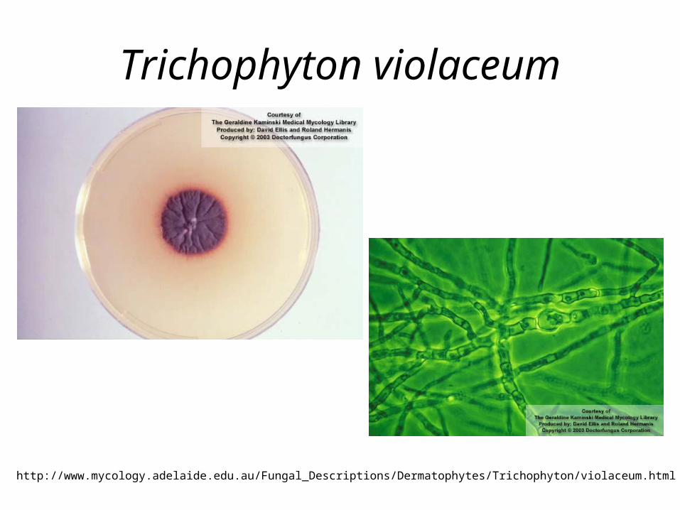
Trichophyton violaceum
http://www.mycology.adelaide.edu.au/Fungal_Descriptions/Dermatophytes/Trichophyton/violaceum.html

DERMATOPHYTES
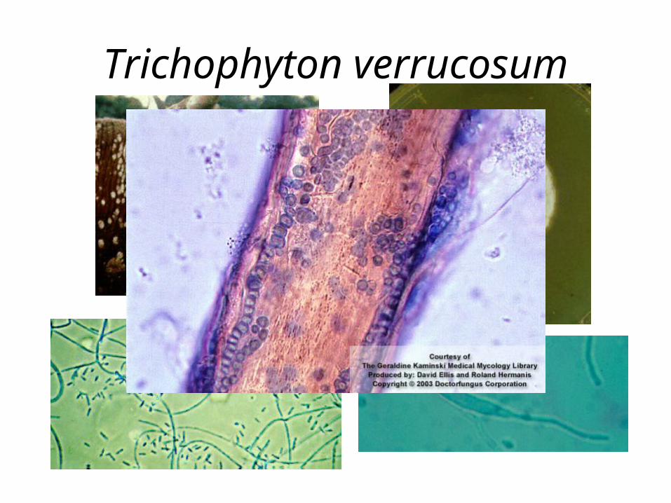
• Trichophyton verrucosum• Associated with cattle ("barn itch"). • Large-spored ectothrix. • Causes severer infections in humans on the scalp and
beard. • Very slow growing, no pigment on reverse to yellow. • Grows best at 37 C. • On unenriched media - chains of chlamydoconidia and
antler-like hyphae. • On thiamine-enriched media, produces many small
microconidia and occasionally macroconidia are produced.
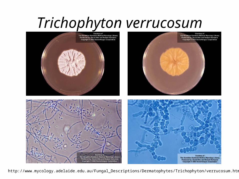
Trichophyton verrucosum
http://www.mycology.adelaide.edu.au/Fungal_Descriptions/Dermatophytes/Trichophyton/verrucosum.html
Trichophyton verrucosum
DERMATOPHYTES
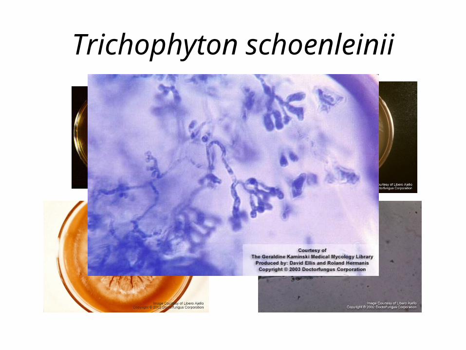
• Trichophyton schoenleinii • Endothrix infection of hair. • Causes tinea favosa (cup-shaped crusts on scalp called
favus). • tinea favosa may lead to alopecia or permanent
baldness. • Colonies waxy to suede-like; off white in color. • Colony may become convoluted from folds that develop • No conidia (micro- or macro-) even on enriched media . • Grows will at 37 C.
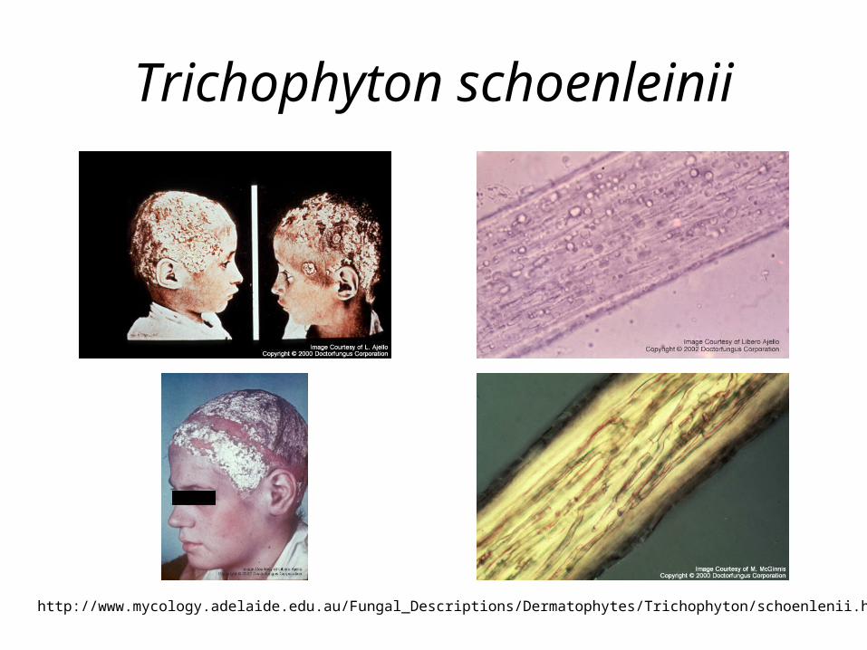
Trichophyton schoenleinii
http://www.mycology.adelaide.edu.au/Fungal_Descriptions/Dermatophytes/Trichophyton/schoenlenii.html
Trichophyton schoenleinii
DERMATOPHYTES
• Trichophyton ajelloi
• Teleomorph - Arthroderma uncinatum.
• Common soil dermatophilic fungus.
• Rarely causes infection in man or animals (cattle, dogs, horses, squirrels).
• Readily isolated from soil by hair baiting.
• Cigar-shaped macroconidia with smooth ends.
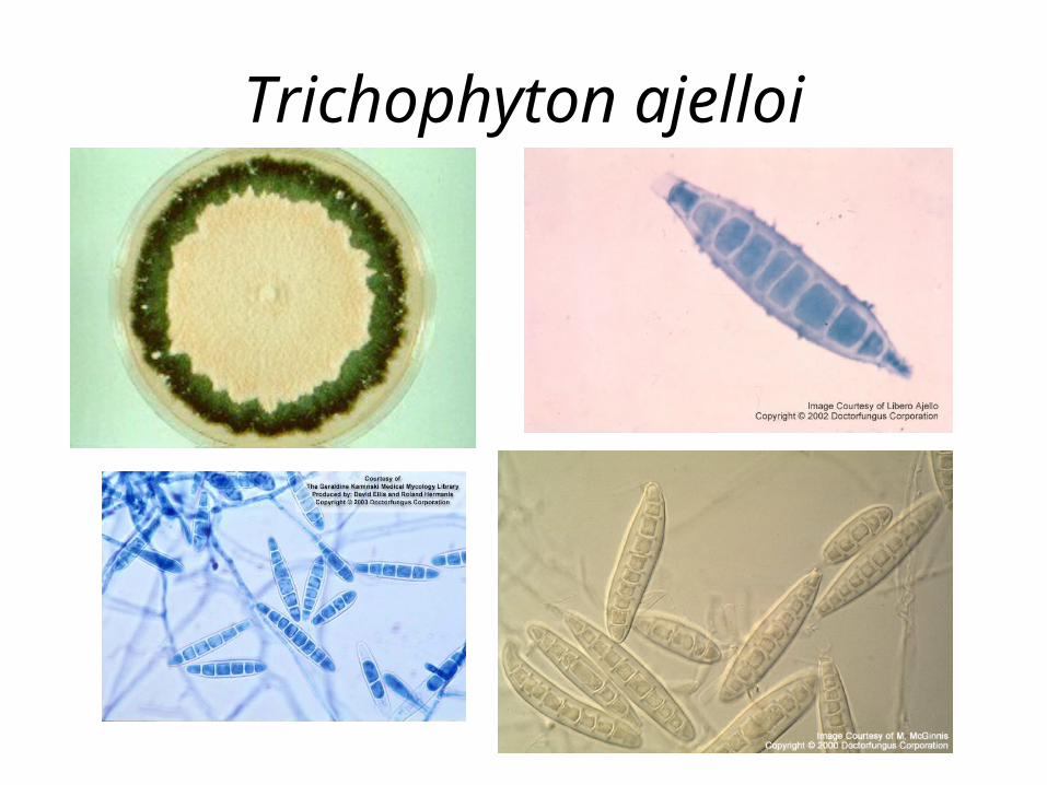
Trichophyton ajelloi