current and future radiopharmaceuticals for brain imaging with single photon emission computed...
TRANSCRIPT
Current and Future Radiopharmaceuticals for Brain Imaging With Single Photon Emission Computed Tomography
Hank F. Kung, Yoshiro Ohmomo, and Mei-Ping Kung
Development of radiopharmaceuticals for functional brain imaging has progressed rapidly in recent years. Measurement of regional cerebral blood flow in humans can be achieved by using [1231]-iodoamphet- amine or [ssmTC]-HMPAO. Several other lipid-soluble [sSmTc]-technetium complexes are currently undergo- ing clinical trials. New lz31-1abeled agents designed to measure central nervous system receptors, including
D1 and D2 dopamine, serotonin, muscarinic, and benzodiazepine receptors, have been developed. In conjunction with single photon emission computed tomography, they may provide useful tools to evalu- ate brain function related to changes in receptor concentration. �9 1990 by W.B. Saunders Company.
T RADITIONAL brain imaging agents, which have been used in nuclear medicine for the
past 40 years, have been essential in the detection of the breakdown of the blood-brain barrier. The radiopharmaceuticals used in this type of proce- dure customarily are agents that can detect the breakdown of the barrier and therefore show a positive uptake in the region. These types of studies are generally effective in localizing focal diseases such as neoplasma, abscesses, and infarc- tions. However, over the past 10 or 15 years, other modalities in radiology have developed, such as radiography, computed tomography, and, more recently, nuclear magnetic resonance imag- ing, which are more effective in detecting these types of anatomical defects. Therefore, nuclear medicine procedures have to compete with these newer types of imaging modalities instead of looking at the morphology of the brain; the detection of brain function based on tracer princi- ples will be the major application in the future. The traditional brain imaging agents using tech- netium-labeled pertechnetate have not been used in nuclear medicine for the past few years. However, radiopharmaceuticals that have been recently developed for functional imaging, such
From the Department of Radiology, University of Pennsyl- vania, Philadelphia, PA; Division of Radiopharmaceutical Chemistry, Osaka University of Pharmaceutical Sciences, Osaka, Japan; and the Department of Psychiatry, University of Pennsylvania, PA.
Supported by National Institutes of Health Grants No. NS-15809 and NS-24538, and a grant from Nihon Medi- Physics, Tokyo, Japan.
Address reprint requests to Hank F. Kung, PhD, Depart- ment of Radiology, Division of Nuclear Medicine, Hospital of the University of Pennsylvania, 3400 Spruce St, Philadel- phia, PA 19104.
�9 1990 by W.B. Saunders Company. 0001-2998/90/2004-0002505.00/0
as iodine 123 and technetium 99m regional perfusion agents, have been introduced into regu- lar clinical use. This type of brain imaging could find extended use in the brain imaging studies typically performed in nuclear medicine. ]'2
BRAIN PERFUSION IMAGING AGENTS
Measurement of regional cerebral blood flow in humans has been achieved by using xenon 133 gas. The technique is based on the Kety-Schmidt technique. 3 The noninvasive technique involves the inhalation of xenon gas while the radioactiv- ity in each specific region of the brain is moni- tored with gamma detector probes. The xenon technique is limited by the poor resolution due to the less than optimal gamma ray energy (80 keV) emitted by 133Xe. The method could also be used to accumulate data for single photon emis- sion computed tomography (SPECT) imaging; however, due to the physical characteristic of the gamma ray and the fast washout of xenon from the normal brain tissue, the image quality of xenon blood flow with SPECT is generally very poor. 4'5
In the past 10 years, a large number of iodine-labeled monoamines and diamines have been developed. 6-16 The first iodinated amine reported in the literature for measuring regional brain perfusion was iodoantipyrine. 6 This com- pound is very lipid soluble and therefore is easily passed through the intact blood-brain barrier. Radioactive labeled 123I-iodoantipyrine can eas- ily cross the cell membrane by a simple diffusion mechanism. However, this compound does not have a trapping mechanism for brain retention. Therefore, it is washed out from the brain very rapidly. The trapping mechanism is especially crucial in SPECT imaging, which requires ] 0 to 60 minutes of data acquisition time with the
290 Seminars in Nuclear Medicine, Vol XX, No 4 (October), 1990: pp 290-302
BRAIN RADIOPHARMACEUTICALS FOR SPECT 291
current single- or dual-headed gamma camera- based equipment. The current status of SPECT instrumentation places stringent requirements on the biologic properties of tracers in order for it to be useful in the measurement of regional cerebral perfusion. In addition to showing high uptake and retention in the brain, the tracer has to remain fixed (no change in regional distribution) for a period of time, preferably 30 to 60 minutes, to allow for adequate data collection. Tracer development based on 123I-labeled compounds has been concentrated on the development of a trapping mechanism for long brain retention, fixed regional distribution reflecting regional blood perfusion, and minimal in vivo metabolism.
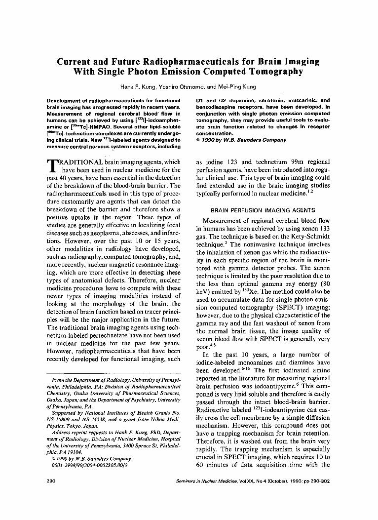
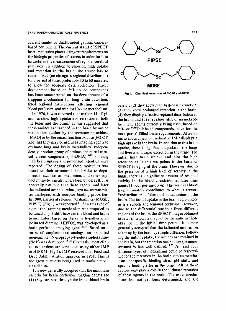
In 1976, it was reported that carbon 11 alkyl- amines show high uptake and retention in both the lungs and the brain. 7 It was suggested that these amines are trapped in the brain by amine metabolism (either by the monoamine oxidase [MAO] or by the mixed function oxidase [MFO]) and that they may be useful as imaging agents to evaluate lung and brain metabolism. Indepen- dently, another group of amines, iodinated cata- col amine congeners (4-I-DPIA), s'l~ showing high brain uptake and prolonged retention were reported. The design of these molecules was based on their structural similarities to dopa- mine, mescaline, amphetamine, and other psy- chotomimetic agents. Therefore, by default, it is generally assumed that these agents, and later the iodinated amphetamines, are neurotransmit- ter analogues with receptor binding properties. In 1980, a series of selenium 75 diamines (MOSE, PIPSE) (Fig 1) was reported. 11'12 In this type of agent, the trapping mechanism was proposed to be based on pH shift between the blood and brain tissue. Later, based on the same hypothesis, an iodinated diamine, HIPDM, was developed as a brain perfusion imaging agent. 13'14 Based on a series of amphetamine analogs, an iodinated monoamine N-isopropyl-4-iodo-amphetamine (IMP) was developed. 15-1s Currently, most clini- cal evaluations are conducted using either IMP or H1PDM (Fig 2). IMP received final Food and Drug Administration approval in 1988. This is the agent currently being used in nuclear medi- cine clinics.
It is now generally accepted that the minimum criteria for brain perfusion imaging agents are (1) they can pass through the intact blood-brain
PIPSE /---X /--X
MOSE
Fig 1. Chemical structures of MOSE and PIPSE.
barrier; (2) they show high first-pass extraction; (3) they show prolonged retention in the brain; (4) they display effective regional distribution in the brain; and (5) they show little or no metabo- lism. The agents currently being used, based on 1231- or 99mTc-labeled compounds, have for the most part fulfilled these requirements. After an intravenous injection, iodinated IMP displays a high uptake in the brain. In addition to this brain uptake, there is significant uptake in the lungs and liver and a rapid excretion in the urine. The initial high brain uptake and also the high retention at later time points is the basis of SPECT imaging of the brain. However, due to the presence of a high level of activity in the lungs, there is a significant amount of residual activity in the blood circulation at later time points (1 hour postinjection). This residual blood level ultimately contributes to what is termed "redistribution" of these iodinated amines in the brain. The initial uptake in the brain region more or less reflects the regional perfusion. However, due to the differential washout from different regions of the brain, the SPECT images obtained at later time points may not be the same as those obtained in the initial time period. It is now generally accepted that the iodinated amines are taken up by the brain by simple diffusion. Follow- ing the initial uptake, the amines are retained in the brain, but the retention mechanism (or mech- anisms) is less well defined. 19'2~ At least four different types of mechanisms could be responsi- ble for the retention in the brain: amine metabo- lism, nonspecific binding sites, pH shift, and specific binding sites in the brain. All of these factors may play a role in the ultimate retention of these agents in the brain. The exact mecha- nism has not yet been determined, and the
292 KUNG, OHMOMO, AND KUNG
OH
C H 3 ~ N ~ N t C H 3
I
CH 3 I
CH2CHNHCH(CH3) 2
HIPDM IMP Fig 2. Chemical structures of
HIPDM and IMP.
retention mechanism in the disease state may also be changing. The pattern of distribution may also be changed because of the differences in the retention mechanism.
Iodinated compounds have provided initial success in routine regional brain perfusion stud- ies. However, the iodinated compounds are not as convenient as the 99mTc-labeled compounds with comparable properties. In the 1970s, several neutral and lipid soluble technetium compounds that pass through the blood-brain barrier were demonstrated. 21"23 However, despite the high ini- tial brain uptake in this type of neutral techne- tium complex, the retention time is not long enough for evaluation of regional brain perfusion with SPECT.
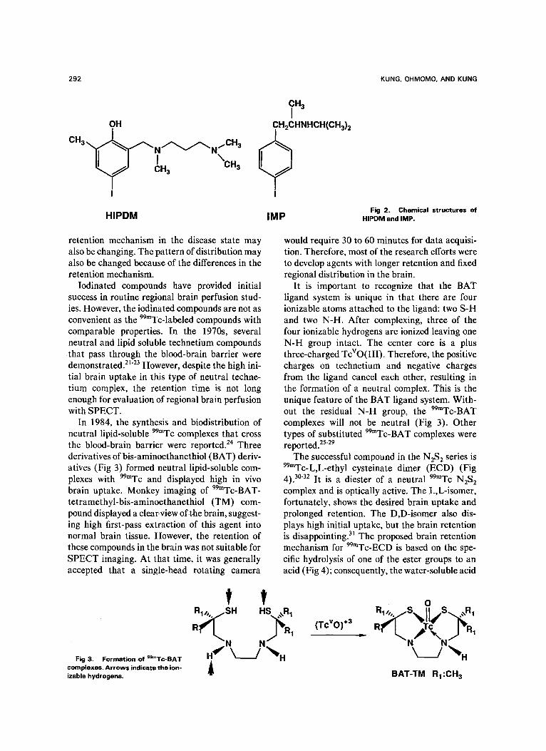
In 1984, the synthesis and biodistribution of neutral lipid-soluble 99roTe complexes that cross the blood-brain barrier were reported, 24 Three derivatives of bis-aminoethanethiol (BAT) deriv- atives (Fig 3) formed neutral lipid-soluble com- plexes with 99mTc and displayed high in vivo brain uptake. Monkey imaging of 99mTc-BAT- tetramethyl-bis-aminoethanethiol (TM) com- pound displayed a clear view of the brain, suggest- ing high first-pass extraction of this agent into normal brain tissue. However, the retention of these compounds in the brain was not suitable for SPECT imaging. At that time, it was generally accepted that a single-head rotating camera
would require 30 to 60 minutes for data acquisi- tion. Therefore, most of the research efforts were to develop agents with longer retention and fixed regional distribution in the brain.
It is important to recognize that the BAT ligand system is unique in that there are four ionizable atoms attached to the ligand: two S-H and two N-H. After complexing, three of the four ionizable hydrogens are ionized leaving one N-H group intact. The center core is a plus three-charged TcVO(III). Therefore, the positive charges on technetium and negative charges from the ligand cancel each other, resulting in the formation of a neutral complex. This is the unique feature of the BAT ligand system. With- out the residual N-H group, the 99mTc-BAT complexes will not be neutral (Fig 3). Other types of substituted 99mTc-BAT complexes were reported. 25-29
The successful compound in the N2S 2 series is 99mTc-L,L-ethyl cysteinate dimer (ECD) (Fig 4). 30.32 It is a diester of a neutral 99mTc N2S 2 complex and is optically active. The L,L-isomer, fortunately, shows the desired brain uptake and prolonged retention. The D,D-isomer also dis- plays high initial uptake, but the brain retention is disappointing. 31 The proposed brain retention mechanism for 99mTc-ECD is based on the spe- cific hydrolysis of one of the ester groups to an acid (Fig 4); consequently, the water-soluble acid
Fig 3. Formation of SS'Tc-BAT i complexes. Arrows indicate the ion- �9 izable hydrogens.
JlP
t R l h " SH
H 4 \
t HS ,~R 1
(TcVO) § RlyS,.l~ "r k
\ I
BAT-TM R I :CH 3
BRAIN RADIOPHARMACEUTICALS FOR SPECT 293
C2HsOO c N COOC2H s .4\ / [99mTc]ECD
Fig 4. Chemical structure of S~"Tc-ECD.
is trapped in the brain. Apparently, the hydroly- sis process is a very specific enzymatic reaction. The most interesting aspect of the enzymatic process is that it is stereo-specific and only happens in the brains of primates (monkeys and humans), but not in rats or rabbits. The specific mechanism is worth noting because the perfusion images obtained by this agent are probably a combination of perfusion and the specific enzyme reaction.
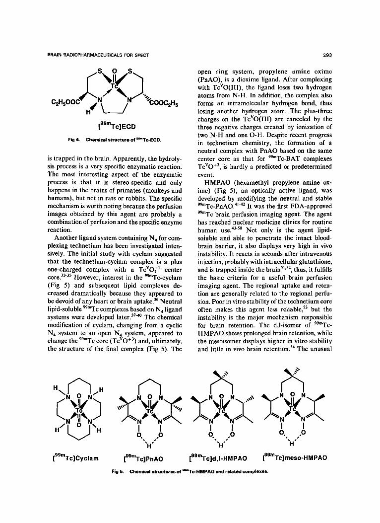
Another ligand system containing N 4 for com- plexing technetium has been investigated inten- sively. The initial study with cyclam suggested that the technetium-cyclam complex is a plus one-charged complex with a TcVO~ ] center core. "35 However, interest in the 99~Tc-cyclam (Fig 5) and subsequent lipid complexes de- creased dramatically because they appeared to be devoid of any heart or brain uptake. 36 Neutral lipid-soluble 99mTc complexes based on N 4 ligand systems were developed later. 37"40 The chemical modification of cyclam, changing from a cyclic N 4 system to an open N 4 system, appeared to change the 99mTC core (TcVO +3) and, ultimately, the structure of the final complex (Fig 5). The
open ring system, propylene amine oxime (PnAO), is a dioxime ligand. After complexing with TcVO(III), the ligand loses two hydrogen atoms from N-H. In addition, the complex also forms an intramolecular hydrogen bond, thus losing another hydrogen atom. The plus-three charges on the TcVO(III) are canceled by the three negative charges created by ionization of two N-H and one O-H. Despite recent progress in technetium chemistry, the formation of a neutral complex with PnAO based on the same center core as that for 99mTc-BAT complexes TcVO § is hardly a predicted or predetermined event.
HMPAO (hexamethyl propylene amine ox- ime) (Fig 5), an optically active ligand, was developed by modifying the neutral and stable 99mTc-PnAO.41-42 It was the first FDA-approved 99mTc brain perfusion imaging agent. The agent has reached nuclear medicine clinics for routine human use. 435~ Not only is the agent lipid- soluble and able to penetrate the intact blood- brain barrier, it also displays very high in vivo instability. It reacts in seconds after intravenous injection, probably with intracellular glutathione, and is trapped inside the brainS~'s2; thus, it fulfills the basic criteria for a useful brain perfusion imaging agent. The regional uptake and reten- tion are generally related to the regional perfu- sion. Poor in vitro stability of the technetium core often makes this agent less reliable, 53 but the instability is the major mechanism responsible for brain retention. The d,l-isomer of 99mTc- HMPAO shows prolonged brain retention, while the mesoisomer displays higher in vitro stability and little in vivo brain retention. 54 The unusual
"H" "H" [99mTc]Cyclam [99mTc]PnA O [99mTc]d,I-HM PAO [99mTc]meso-HMPAO
Fig 5. Chemical structures of sS"~rc-HMPAO and related complexes.
294 KUNG, OHMOMO, AND KUNG
.. 'O:" "H . / / " / t
.," \ s
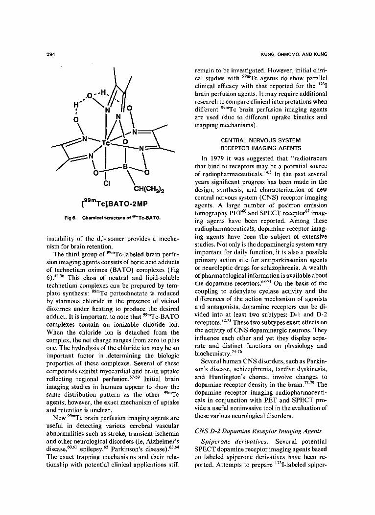
CH(CH3)2 [99mTc]BATO-2M P
Fig 6. Chemical structure of 99mTc-BATO.
instability of the d,l-isomer provides a mecha- nism for brain retention.
The third group of 99mTc-labeled brain perfu- sion imaging agents consists of boric acid adducts of technetium oximes (BATO) complexes (Fig 6). 5s'56 This class of neutral and lipid-soluble technetium complexes can be prepared by tem- plate synthesis: 99roTe pertechnetate is reduced by stannous chloride in the presence of vicinal dioximes under heating to produce the desired adduct. It is important to note that 99mTc-BATO complexes contain an ionizable chloride ion. When the chloride ion is detached from the complex, the net charge ranges from zero to plus one. The hydrolysis of the chloride ion may be an important factor in determining the biologic properties of these complexes. Several of these compounds exhibit myocardial and brain uptake reflecting regional perfusion. 5749 Initial brain imaging studies in humans appear to show the same distribution pattern as the other 99mTc agents; however, the exact mechanism of uptake and retention is unclear.
New 99mTc brain perfusion imaging agents are useful in detecting various cerebral vascular abnormalities such as stroke, transient ischemia and other neurological disorders (ie, Alzheimer's disease, 6~ epilepsy, 62 Parkinson's disease). 63'64 The exact trapping mechanisms and their rela- tionship with potential clinical applications still
remain to be investigated. However, initial clini- cal studies with 99mTc agents do show parallel clinical efficacy with that reported for the mI brain perfusion agents. It may require additional research to compare clinical interpretations when different 99mTc brain perfusion imaging agents are used (due to different uptake kinetics and trapping mechanisms).
CENTRAL NERVOUS SYSTEM RECEPTOR IMAGING AGENTS
In 1979 it was suggested that "radiotracers that bind to receptors may be a potential source of radiopharmaceuticals. ''65 In the past several years significant progress has been made in the design, synthesis, and characterization of new central nervous system (CNS) receptor imaging agents. A large number of positron emission tomography PET 66 and SPECT receptor 67 imag- ing agents have been reported. Among these radiopharmaceuticals, dopamine receptor imag- ing agents have been the subject of extensive studies. Not only is the dopaminergic system very important for daily function, it is also a possible primary action site for antiparkinsonian agents or neuroleptic drugs for schizophrenia. A wealth of pharmacological information is available about the dopamine receptors. 6871 On the basis of the coupling to adenylate cyclase activity and the differences of the action mechanism of agonists and antagonists, dopamine receptors can be di- vided into at least two subtypes: D-1 and D-2 receptors. 72'73 These two subtypes exert effects on the activity of CNS dopaminergic neurons. They influence each other and yet they display sepa- rate and distinct functions on physiology and biochemistry. 74-76
Several human CNS disorders, such as Parkin- son's disease, schizophrenia, tardive dyskinesia, and Huntington's chorea, involve changes to dopamine receptor density in the brain. 7779 The dopamine receptor imaging radiopharmaceuti- cals in conjunction with PET and SPECT pro- vide a useful noninvasive tool in the evaluation of those various neurological disorders.
CNS D-2 Dopamine Receptor Imaging Agents
Spiperone derivatives. Several potential SPECT dopamine receptor imaging agents based on labeled spiperone derivatives have been re- ported. Attempts to prepare 123I-labeled spiper-
BRAIN RADIOPHARMACEUTICALS FOR SPECT
one were inspired by the successful D-2 dopa- mine receptor images of the human brain with N-nC-methylspiperone with PET. a~ Prelimi- nary studies of an iodinated 2'-iodo-spiperone (2'-ISP) (Fig 7) 82'83 indicated that the spiperone analogue displayed excellent D-2 specificity (Kd = 0.25 nmol/L in rat striatum) and in vivo stability compared with the 4-iodospiperone (Fig 7) 84 reported earlier. Recently, Mertens et al s5 reported the imaging studies of 2'-ISP in hu- mans. Initial SPECT images showed less favor- able quality compared with those obtained by IBZM, a close analogue of raclopride. In addi- tion, a new analogue of spiperone, N-iodoallyl spiperone (Fig 7), has been reported, s6.s7 The agent showed good striatum to cerebellum ratio in mice.
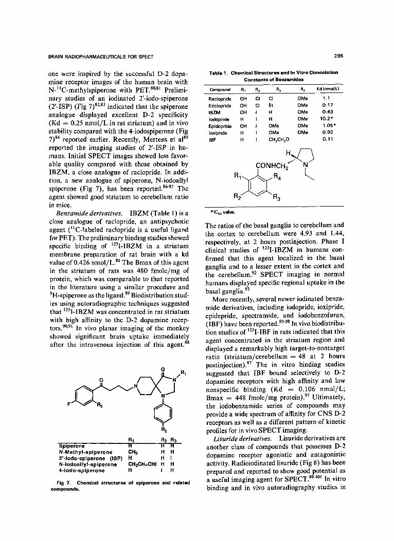
Benzamide derivatives. IBZM (Table 1) is a close analogue of raclopride, an antipsychotic agent (ltC-labeled raclopride is a useful ligand for PET). The preliminary binding studies showed specific binding of ~25I-IBZM in a striatum membrane preparation of rat brain with a kd value of 0.426 nmol/L. 8s The Bmax of this agent in the striatum of rats was 480 fmole/mg of protein, which was comparable to that reported in the literature using a similar procedure and 3H-spiperone as the ligand.S9 Biodistribution stud- ies using autoradiographic techniques suggested that ~25I-IBZM was concentrated in rat striatum with high affinity to the D-2 dopamine recep- tors. 9~ In vivo planar imaging of the monkey showed significant brain uptake immediately after the intravenous injection of this agent. 8s
O 0 ~ / ~ ' N / R 1
R2
R1 R2 R3 Spiperone H H H N-Methyl-spiperone CH3 H H 2'-Iodo-spiperone (ISP) H H I N - I o d o a l l y l - s p i p e r o n e CH2CH=CHI H H 4-10do-spiperone H I H
Fig 7. Chemical structures of spiperone and related compounds.
295
Table 1. Chemical Structures and In Vitro Dissociation Constants of Benzamides
Compound R~ R 2 R 3 R4 Kd (nmol/L)
Raclopride OH CI CI OMe 1.1 Eticlopride OH CI Et OMe O. 17 IBZM OH I H OMe 0.43 Iodopride H I H OMe 10.2" Epidopride OH I OMe OMe 1.05* Ioxipride H I OMe OMe 0.92 IBF H I CH2CH20 O. 11
CON H C H ~ ' " " ~
* ICso value.
The ratios of the basal ganglia to cerebellum and the cortex to cerebellum were 4.93 and 1.44, respectively, at 2 hours postinjection. Phase I clinical studies of 123I-IBZM in humans con- firmed that this agent localized in the basal ganglia and to a lesser extent in the cortex and the cerebellum. 92 SPECT imaging in normal humans displayed specific regional uptake in the basal ganglia. 92
More recently, several newer iodinated benza- mide derivatives, including iodopride, ioxipride, epidepride, spectramide, and iodobenzofuran, (IBF) have been reported. 93"9s In vivo biodistribu- tion studies of ~25I-IBF in rats indicated that this agent concentrated in the striatum region and displayed a remarkably high target-to-nontarget ratio (striatum/cerebellum = 48 at 2 hours postinjection). 97 The in vitro binding studies suggested that IBF bound selectively to D-2 dopamine receptors with high affinity and low nonspecific binding (Kd = 0.106 nmol/L; Bmax = 448 fmole/mg protein). 97 Ultimately, the iodobenzamide series of compounds may provide a wide spectrum of affinity for CNS D-2 receptors as well as a different pattern of kinetic profiles for in vivo SPECT imaging.
Lisuride derivatives. Lisuride derivatives are another class of compounds that possesses D-2 dopamine receptor agonistic and antagonistic activity. Radioiodinated lisuride (Fig 8) has been prepared and reported to show good potential as a useful imaging agent for SPECT. 99q~ In vitro binding and in vivo autoradiography studies in
296 KUNG, OHMOMOoAND KUNG
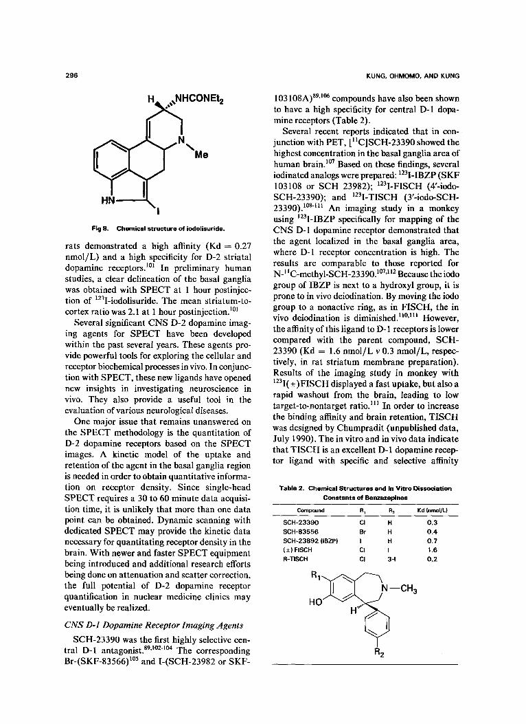
H..,r
N\ Me
Fig 8. Chemical structure of iodolisuride.
rats demonstrated a high affinity (Kd ----- 0.27 nmol/L) and a high specificity for D-2 striatal dopamine receptors. 1~ In preliminary human studies, a clear delineation of the basal ganglia was obtained with SPECT at 1 hour postinjec- tion of 123I-iodolisuride. The mean striatum-to- cortex ratio was 2.1 at 1 hour postinjection. 1~
Several significant CNS D-2 dopamine imag- ing agents for SPECT have been developed within the past several years. These agents pro- vide powerful tools for exploring the cellular and receptor biochemical processes in vivo. In conjunc- tion with SPECT, these new ligands have opened new insights in investigating neuroscience in vivo. They also provide a useful tool in the evaluation of various neurological diseases.
One major issue that remains unanswered on the SPECT methodology is the quantitation of D-2 dopamine receptors based on the SPECT images. A kinetic model of the uptake and retention of the agent in the basal ganglia region is needed in order to obtain quantitative informa- tion on receptor density. Since single-head SPECT requires a 30 to 60 minute data acquisi- tion time, it is unlikely that more than one data point can be obtained. Dynamic scanning with dedicated SPECT may provide the kinetic data necessary for quantitating receptor density in the brain. With newer and faster SPECT equipment being introduced and additional research efforts being done on attenuation and scatter correction, the full potential of D-2 dopamine receptor quantification in nuclear medicine clinics may eventually be realized.
CNS l)-1 Dopamine Receptor Imaging Agents
SCH-23390 was the first highly selective cen- tral D-1 antagonist, s9"1~176 The corresponding Br-(SKF-83566) 1~ and I-(SCH-23982 or SKF-
103108A) 89'1~ compounds have also been shown to have a high specificity for central D-1 dopa- mine receptors (Table 2).
Several recent reports indicated that in con- junction with PET, [11C] SCH-23390 showed the highest concentration in the basal ganglia area of human brain. ]~ Based on these findings, several iodinated analogs were prepared: 123I-IBZP (SKF 103108 or SCH 23982); 123I-FISCH (4'-iodo- SCH-23390); and ]23I-TISCH (3'-iodo-SCH- 23390). 1~ An imaging study in a monkey using 123I-IBZP specifically for mapping of the CNS D-1 dopamine receptor demonstrated that the agent localized in the basal ganglia area, where D-1 receptor concentration is high. The results are comparable to those reported for N j 1C_methyl_SCH_23390.107,112 Because the iodo group of IBZP is next to a hydroxyl group, it is prone to in vivo deiodination. By moving the iodo group to a nonactive ring, as in FISCH, the in vivo deiodination is diminished. 11~ However, the affinity of this ligand to D- 1 receptors is lower compared with the parent compound, SCH- 23390 (Kd = 1.6 nmol/L v 0.3 nmol/L, respec- tively, in rat striatum membrane preparation). Results of the imaging study in monkey with 1231( _+)FISCH displayed a fast uptake, but also a rapid washout from the brain, leading to low target-to-nontarget ratio. H~ In order to increase the binding affinity and brain retention, TISCH was designed by Chumpradit (unpublished data, July 1990). The in vitro and in vivo data indicate that TISCH is an excellent D-1 dopamine recep- tor ligand with specific and selective affinity
Table 2. Chemical Structures and In Vi t ro Dissociation Constants of Benzazepines
Compound R 1 R2 Kd (nmol/L)
SCH-23390 CI H 0.3
SCH-83556 Br H 0.4
SCH-23892 (IBZP) I H 0.7 (+-) FISCH CI I 1.6 R-TISCH CI 3-1 0.2
R I " ~ / / N --CH 3 H ,,.. O" V H , ~
R2
BRAIN RADIOPHARMACEUTICALS FOR SPECT 297
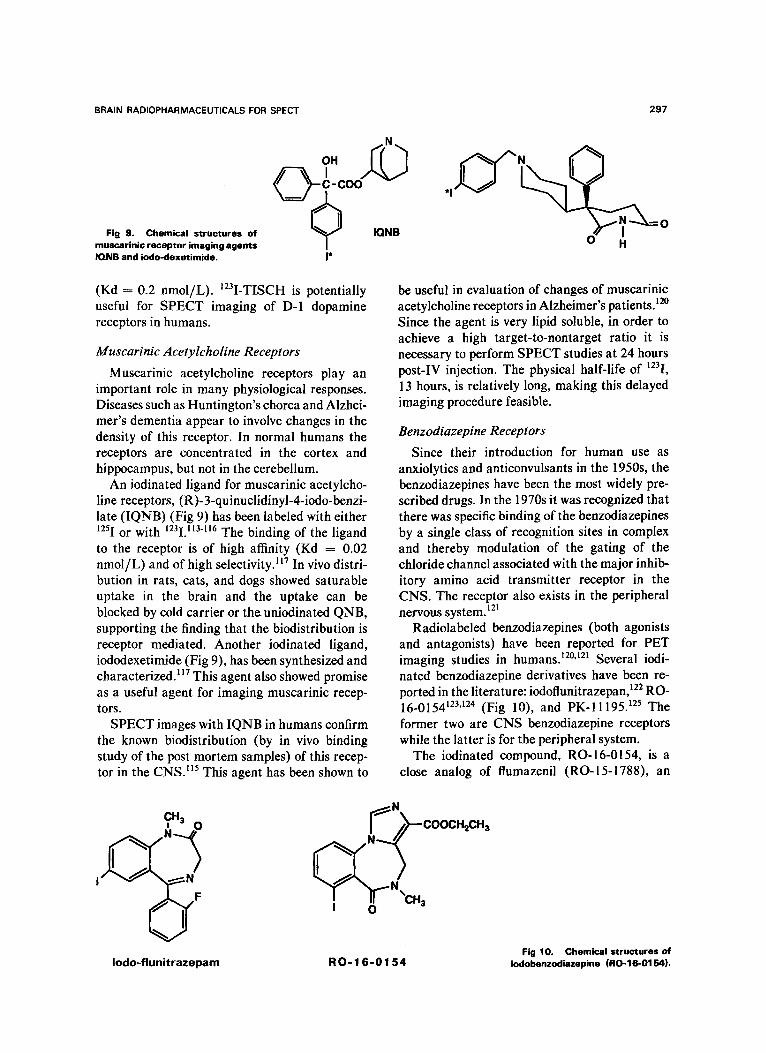
Fig 9. Chemical structures of muscarinir receptor imaging agents IQNB and iodo-dexctimide.
N
I*
O / - O
(Kd = 0.2 nmol/L). 123I-TISCH is potentially useful for SPECT imaging of D-1 dopamine receptors in humans.
Muscarinic Acetylcholine Receptors
Muscarinic acetylcholine receptors play an important role in many physiological responses. Diseases such as Huntington's chorea and Alzhei- mer's dementia appear to involve changes in the density of this receptor. In normal humans the receptors are concentrated in the cortex and hippocampus, but not in the cerebellum.
An iodinated ligand for muscarinic acetylcho- line receptors, (R)-3-quinuclidinyl-4-iodo-benzi- late (IQNB) (Fig 9) has been labeled with either 125I or with 123[.113-116 The binding of the ligand to the receptor is of high affinity (Kd = 0.02 nmol/L) and of high selectivity. 117 In vivo distri- bution in rats, cats, and dogs showed saturable uptake in the brain and the uptake can be blocked by cold carrier or the uniodinated QNB, supporting the finding that the biodistribution is receptor mediated. Another iodinated ligand, iododexetimide (Fig 9), has been synthesized and characterized. 117 This agent also showed promise as a useful agent for imaging muscarinic recep- tors.
SPECT images with IQNB in humans confirm the known biodistribution (by in vivo binding study of the post mortem samples) of this recep- tor in the CNS. H5 This agent has been shown to
be useful in evaluation of changes of muscarinic acetylcholine receptors in Alzheimer's patients.12~ Since the agent is very lipid soluble, in order to achieve a high target-to-nontarget ratio it is necessary to perform SPECT studies at 24 hours post-IV injection. The physical half-life of 123I, 13 hours, is relatively long, making this delayed imaging procedure feasible.
Benzodiazepine Receptors
Since their introduction for human use as anxiolytics and anticonvulsants in the 1950s, the benzodiazepines have been the most widely pre- scribed drugs. In the 1970s it was recognized that there was specific binding of the benzodiazepines by a single class of recognition sites in complex and thereby modulation of the gating of the chloride channel associated with the major inhib- itory amino acid transmitter receptor in the CNS. The receptor also exists in the peripheral nervous system. 121
Radiolabeled benzodiazepines (both agonists and antagonists) have been reported for PET imaging studies in humans. 12~ Several iodi- nated benzodiazepine derivatives have been re- ported in the literature: iodoflunitrazepan, 122 RO- 16-0154123'124 (Fig 10), and PK-11195) 25 The former two are CNS benzodiazepine receptors while the latter is for the peripheral system.
The iodinated compound, RO-16-0154, is a close analog of flumazenil (RO-15-1788), an
I o d o - f l u n i t r a z e p a m
[ ~ ~/~--- COOCH2CH3
l . "c.,
R O - 1 6 - 0 1 5 4 Fig 10. Chemical structures of
iodobenzodiazepine (R0-16-0154).
298 KUNG, OHMOMO, AND KUNG
antagonis t tha t is used as an an t ido te in the t r e a t m e n t of benzodiazepine intoxicat ion. Brain images of RO-16-0154 indicate tha t the agent binds to the receptor in a specific and reversible fashion. 126'127 I t is possible tha t this agent m a y be useful to evalua te changes of receptor densi ty a n d / o r measur ing receptor occupancy re la ted to therapy .
BRAIN TUMOR IMAGING AGENTS
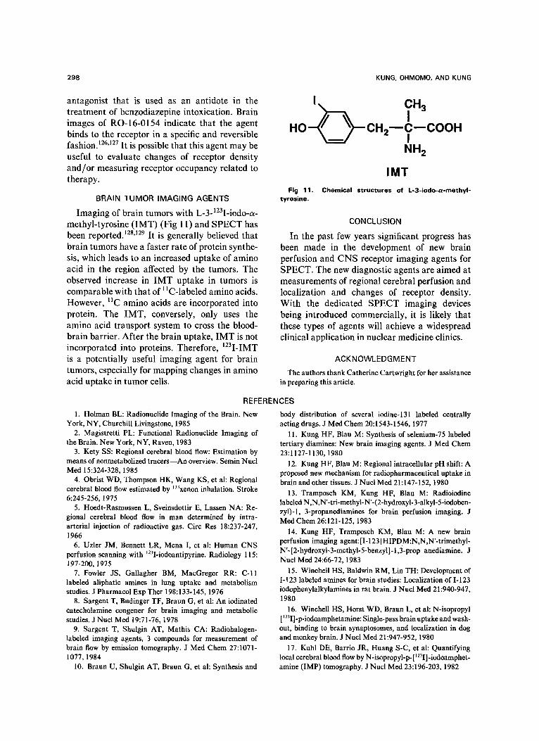
Imag ing of b ra in tumors with L-3-123I-iodo-a-
methyl - ty ros ine ( I M T ) (Fig 11) and S P E C T has been repor ted. 128'129 I t is genera l ly bel ieved tha t
b ra in tumors have a fas ter ra te of protein synthe- sis, which leads to an increased up take of amino acid in the region affected by the tumors. The observed increase in I M T up take in tumors is comparab le with tha t of l~C-labeled amino acids. However , HC amino acids a re incorpora ted into protein. The I M T , conversely, only uses the amino acid t r anspor t sys tem to cross the blood- bra in barr ier . A f t e r the bra in uptake , I M T is not incorpora ted into proteins. Therefore , ]z3I-IMT is a potent ia l ly useful imaging agent for bra in tumors , especial ly for mapping changes in amino acid up take in tumor cells.
. o
NH 2
I M T
Fig 11. Chemical structures of L-3-iodo-a-methyl- tyrosine.
CONCLUSION
In the past few years significant progress has been made in the development of new bra in perfusion and C N S receptor imaging agents for S P E C T . The new diagnost ic agents a re a imed at measurements of regional ce rebra l perfusion and local izat ion and changes of receptor densi ty. W i t h the ded ica ted S P E C T imaging devices being in t roduced commerc ia l ly , it is l ikely tha t these types of agents will achieve a widespread cl inical appl ica t ion in nuclear medic ine clinics.
ACKNOVVLEDGMENT
The authors thank Catherine Cartwright for her assistance in preparing this article.
REFERENCES
1. Holman BL: Radionuclide Imaging of the Brain. New York, MY, Churchill Livingstone, 1985
2. Magistretti PL: Functional Radionuclide Imaging of the Brain. New York, MY, Raven, 1983
3. Kety SS: Regional cerebral blood flow: Estimation by means of nonmetabolized tracers--An overview. Semin Nucl Med 15:324-328, 1985
4. Obrist WD, Thompson HK, Wang KS, et al: Regional cerebral blood flow estimated by ~33xenon inhalation. Stroke 6:245-256, 1975
5. Hoedt-Rasmussen L, Sveinsdottir E, Lassen NA: Re- gional cerebral blood flow in man determined by intra- arterial injection of radioactive gas. Circ Res 18:237-247, 1966
6. Uzler JM, Bennett LR, Mena I, et al: Human CNS perfusion scanning with ~3I-iodoantipyrine. Radiology 115: 197-200, 1975
7. Fowler JS, Gallagher BM, MacGregor RR: C-11 labeled aliphatic amines in lung uptake and metabolism studies. J Pharmacol Exp Ther 198:133-145, 1976
8. Sargent T, Budinger TF, Braun G, et al: An iodinated catechotamine congener for brain imaging and metabolic studies. J Nucl Med 19:71-76, 1978
9. Sargent T, Shulgin AT, Mathis CA: Radiohalogen- labeled imaging agents, 3 compounds for measurement of brain flow by emission tomography. J Med Chem 27:1071- 1077, 1984
10. Braun U, Shulgin AT, Braun G, et al: Synthesis and
body distribution of several iodine-131 labeled centrally acting drugs. J Med Chem 20:1543-1546, 1977
11. Kung HF, Blau M: Synthesis of selenium-75 labeled tertiary diamines: New brain imaging agents. J Med Chem 23:1127-1130, 1980
12. Kung HF, Blau M: Regional intracellular pH shift: A proposed new mechanism for radiopharmaceutical uptake in brain and other tissues. J Nucl Med 21:147-152, 1980
13. Tramposch KM, Kung HF, Blau M: Radioiodine labeled N,N,N'-tri-methyl-N'-(2-hydroxyl- 3-alkyl-5-iodoben- zyl)-l, 3-propanediamines for brain perfusion imaging. J Med Chem 26:121-125, 1983
14. Kung HF, Tramposch KM, Blau M: A new brain perfusion imaging agent:[I-123]HIPDM:N,N,N'-trimethyl- N'-[2-hydroxyl-3-methyl-5-benzyl]-l,3-prop anediamine. J Nucl Med 24:66-72, 1983
15. Winchell HS, Baldwin RM, Lin TH: Development of I- 123 labeled amines for brain studies: Localization of I- 123 iodophenylalkylamines in rat brain. J Nucl Med 21:940-947, 1980
16. Winchell HS, Horst WD, Braun L, et al: N-isopropyl [I2~I]-p-iodoamphetamine: Single-pass brain uptake and wash- out, binding to brain synaptosomes, and localization in dog and monkey brain. J Nucl Med 21:947-952, 1980
17. Kuhl DE, Barrio JR, Huang S-C, et al: Quantifying local cerebral blood flow by N-isopropyl-p- [I:3I]-iodoamphet- amine (IMP) tomography. J Nucl Med 23:196-203, 1982
BRAIN RADIOPHARMACEUTICALS FOR SPECT 299
18. Hill TC, Holman BL, Lovett R, et al: Initial experi- ence with SPECT (single-photon computerized tomography) of the brain using N-isopropyl-I-123-p-iodoamphetamine: Concise communication. J Nucl Med 23:191-195, 1982
19. Som P, Oster ZH, Yamamoto K, et al: Some factors affecting the cerebral and extracerebral accumulation of N-isopropyl-p-iodo-amphetamine (IAMP). lnt J Nucl Med Biol 12:185-196, 1985
20. Moretti JL, Holman BL, Delmon L, et al: Effect of antidepressant and narcoleptic drugs on N-isopropyl p-iodo- amphetamine biodistribution in animals. J Nucl Med 28:354- 359, 1987
21. Burns HD, Manspeaker H, Miller R, et al: Prepara- tion and biodistribution of neutral, lipid soluble Tc-99m complexes of bis (2-mercaptoethyl) amine ligands. J Nucl Med 20:654, 1979 (abstr)
22. Burns HD, Dannals RF, Dannals TE, et al: Synthesis of tetradentate aminothiol ligands and their technetium complexes. J Lab Comp Radiopharm 18:54-55, 1981
23. Loberg MD, Corder EH, Fields AT, et al: Membrane transport of Tc-99m-labeled radiopharmaceuticals. I. Brain uptake by passive transport. J Nucl Med 20:1181-1188, 1979
24. Kung HF, Molnar M, Billings J, et al: Synthesis and biodistribution of neutral lipid-soluble Tc-99m complexes which cross the blood brain barrier. J Nucl Med 25:326-332, 1984
25. Lever SZ, Burns HD, Kervitsky TM, et al: Design, preparation and biodistribution of a technetium-99m tri- aminedithiol complex to access regional cerebral blood flow. J Nucl Med 26:1287-1294, 1985
26. Kung HF, Guo Y-Z, Yu C-C, et al: New brain perfusion imaging agents based on Tc-99m bis-aminoet- hanethiol (BAT) complexes: Stereoisomers and biodistribu- tion. J Med Chem 32:433-437, 1989
27. Efange SMN, Kung HF, Billings J, et al: Tc-99m bis(amino-ethanethiol) (BAT) complexes with amine side- chains--Potential brain perfusion imaging agents for SPECT. J Nucl Med 28:1012-1019, 1987
28. Efange SMN, Kung HF, Billings J, et al: The synthe- sis and biodistribution of [99mTe]piperidinyl bis(aminoet- hanethiol) complexes: Potential brain perfusion imaging agents for SPECT. J Med Chem 31:1043-1047, 1988
29. Scheffel U, Goldfarb HW, Lever SZ, et al: Compari- son of technetium-99m aminoalkyl diaminodithiol analogs as potential brain blood flow imaging agents. J Nucl Med 29:73-82, 1988
30. Holman BL, Hellman RS, Goldsmith S J, et al: Biodis- tribution, dosimetry, and clinical evaluation of technetium- 99m ethyl cysteinate dimer in normal subjects and in patients with chronic cerebral infarction. J Nuci Med 30:1018-1024, 1989
31. Walovitch RC, Hill TC, Garrity ST, et al: Character- ization of technetium-99m-L,L-ECD for brain perfusion imaging, part 1: Pharmacology of technetium-99m ECD in nonhuman primates. J Nucl Med 30:1892-1901, 1989
32. L6veill6 J, Demonceau G, De Roo M, et al: Character- ization of technetium-99m-L,L-ECD for brain perfusion imaging, part 2: Biodistribution and brain imaging in hu- mans. J Nucl Med 30:1902-10, 1989
33. Troutner DE, Simon J, Ketring AR, et al: Complexing
of Tc-99m with cyclam: Concise communication. J Nucl Med 21:443-448, 1980
34. Simon J, Zuckman S, Troutner DE, et al: Tc-99- Cyclam. Third International Symposium on Radiopharmaceu- tical Chemistry. St. Louis, MO, Washington University, 1981, pp 151-152
35. Zuckman SA, Freeman GM, Troutner DE, et al: Preparation and x-ray structure of trans-dioxo (1, 4, 8, 11-tetraazacylotetradecane) technetium (V) perchlorate hy- drate. Inorg Chem 20:2386-2389, 1981
36. Ketring AR, Troutner DE, Volkert WA, et al: Tc-99m complexes of lipophilic cyclam derivatives: Assessment as potential myocardial imaging agents. J Nucl Med 23:P17, 1982 (abstr)
37. Volkert WA, Hoffman T J, Seger RM, et al: 99mTc- propylene amine oxime (99mTc-PBAO): A potential brain radiopharmaceutical. Enr J Nucl Med 9:51 I-516, 1984
38. Troutner DE, Volkert WA, Hoffman TJ, et al: A neutral lipophilic complex of 99roTe with a multidentate amine oxime. Int J Appl Radiat Isotope 35:46%470, 1984
39. Chaplin SB, Oberle PA, McKenzie EH, et al: Re- gional cerebral uptake and retention of 99mTc-tetramethyl- and pentamethyl- propyleneamine oxime chelates. Nucl Med Biol 13:261-267, 1986
40. Jurisson S, Aston K, Faiz CK, et al: Effect of ring size on properties of technetium amine oxime complexes. X-ray structures of TcO2Pen(AO)2. Inorg Chem 26:3576-3583, 1987
41. Nechvatal G, Canning LR, Cumming S, et al: New derivatives of Tc-99m PnAO as potential regional cerebral blood flow agents, in Nicolini M, Bandoli G, Mazzi U (eds): Technetium in Chemistry and Nuclear Medicine, vol 2. New York, NY, Raven, 1984, pp 187-192
42. Nowotnik DP, Canning LR, Cumming SA, et al: Development of a 99roTe-labelled radiopharmaceutical for cerebral blood flow imaging. Nucl Med Commun 6:499-506, 1985
43. Ncirinckx RD, Canning LR, Piper IM, et al: Techne- tium-99m-d,l-HM-PAO: A new radiopharmaceutical for SPECT imaging of regional cerebral blood perfusion. J Nucl Med 28:191-202, 1987
44. Podreka I, Suess E, Goldenberg G, et al: Initial experience with technetium-99m HM-PAO brain SPECT. J Nucl Med 28:1657-1666, 1987
45. Sharp PF, Smith FW, Gemmell HG, et al: Technetium- 99m HMPAO stereoisomers as potential agents for imaging regional cerebral blood flow: Human volunteer studies. J Nucl Med 27:171-177, 1986
46. Leonard J, Nowotnik DP, Neirinckx RD: Tc-99m d,l HMPAO: A new radiopharmaceutical for imaging regional brain perfusion using SPECT A comparison with iodine- 123 HIPDM. J Nucl Med 27:1819-1823, 1986
47. Lear JL: Initial cerebral HM-PAO distribution com- pared to LCBF: Use of a model which considers cerebral HM-PAO trapping kinetics. J Cereb Blood Flow Metab 8:$31-$37, 1988 (suppl)
48. Andersen AR, Friberg HH, Schmidt JF, et al: Quanti- tative measurements of cerebral blood flow using SPECT and [99mTc]-d,I-HM-PAO compared to xenon- 133. J Cereb Blood Flow Metab 8:$69-$81, 1988 (suppl)
49. Andersen AR, Friberg HH, Knudson KBM, et al:
300 KUNG, OHMOMO, AND KUNG
Extraction of [99mTc]-d,I-HM-PAO across the blood-brain barrier. J Cereb Blood Flow Metab 8:$44-$51, 1988 (suppl)
50. Andersen AR, Friberg HH, Lassen NA, et al: Assess- ment of the arterial input curve for [99mTc]-d,l-HM-PAO by rapid octanol extraction. J Cereb Blood Flow Metab 8:$23- $30, 1988 (suppl)
51. Ballinger JR, Reid RH, Gnlenchyn KY: Technetium- 99m HM-PAO stereoisomers: Differences in interaction with glutathione. J Nucl Med 29:1998-2000, 1988
52. Neirinckx RD, Burke JF, Harrison RC, et al: The retention mechanism of technetium-99m-HM-PAO: Intracel- lular reaction with glutathione. J Cereb Blood Flow Metab 8:$4-S12, 1988 (suppl)
53. Hung JC, Corlija M, Volkert WA, et al: Kinetic analysis of technetium-99m d,I-HM-PAO decomposition in aqueous media. J Nucl Med 29"1568-1576, 1988
54. Hoffman T J, Corlija M, Chaplin SB, et al: Retention of [99mTc]-d,I-HM-PAO in rat brain: An autoradiographie study. J Cereb Blood Flow Metab 8:$38-$43, 1988
55. Treher EN, Francesconi LC, Gougoutas J, et al: The first monocapped tris dioxime complexes of a transition metal: Synthesis and structural characterization of [Tc- X(DIOXIMEH)2(DIOXIME)(B-R)]. Inorg Chem 28:3411- 3416, 1989
56. Thompson M, Nunn AD, Treher EN: X-ray photoelec- tron spectroscopy of potential technetium-based organ imag- ing agents. Anal Chem 58:3100-3103, 1986
57. Coleman RE, Maturi M, Nunn AD, et al: Imaging of myocardial porfusion with Tc-99m SQ 30217: Dog and human studies. J Nucl Med 27:893, 1986 (abstr)
58. Kuczynski BL, Silva D, Feld T, et al: Autoradiography of monkey brain with SQ 32097: A new Tc-99m-labeled brain imaging agent. J Nucl Med 28:1081, 1987 (abstr)
59. Narra RK, Nunn AD, Kuczynski BL, et al: A neutral technetium-99m complex for myocardial imaging. J Nncl Med 30:1830-1837, 1989
60. Smith FW, Besson JAO, Gemmell HG, et al: The use of technetium-99m-HM-PAO in the assessment of patients with dementia and other neuropsychiatric conditions. J Cereb Blood Flow Metab 8:S116-S122, 1988 (suppl)
61. Testa H J, Snowden JS, Neary D, et al. The use of [99mTc]-HM-PAO in the diagnosis of primary degenerative dementia. J Cereb Blood Flow Metab 8:S123-S126, 1988 (snppl)
62. Ryding E, Rosen I, Emqvist D, et al: SPECT measure- ments with 99mTc-HM-PAO in focal epilepsy. J Cereb Blood Flow Metab 8:$95-S100, 1988 (suppl)
63. Costa DC, Ell P J, Burns A, et al: CBF tomograms with [99mTc]-HM-PAO in patient with dementia (Alzheimer type and HIV) and Parkinson's disease--initial results. J Cereb Blood Flow Metab 8:S 109-S 115, 1988 (suppl)
64. Pizzolato G, Dam M, Borsato N, et al: [99mTc]-HM- PAO SPECT in Parkinson's disease. J Cereb Blood Flow Metab 8:S101-S108, 1988 (suppl)
65. Eckelman WC, Reba RC, Gibson RE, et al: Receptor binding radiotracers: A class of potential radiopharmaceuti- eals. J Nucl Med 20:350-357, 1979
66. Fowler JS, Wolf AP: New directions in positron emission tomography. Ann Rep Med Chem 24:277-286, 1989
67. Kung HF, Billings J, Guo YZ, et al: Preparation and biodistribution of [~25I]-IBZP: A potential CNS D-I dopa-
mine receptor imaging agent. Nucl Med Biol 15:187-193, 1988
68. Creese I, Fraser CM: Dopamine Receptors. New York, NY, Liss, 1987
69. SeemanP, NiznikHB:DopamineD1 receptor pharma- cology. ISI Atlas of Science: Pharmacology, 1988, pp 161- 170
70. Beaulieu M: Clinical importance of D-1 and D-2 receptors. Can J Neurol Sci 14:402-406, 1987
71. Waddington JL, O'Boyle KM: The D-1 dopamine receptor and the search for its functional role: From neuro- chemistry to behaviour. Rev Neurosci 1:157, 1987
72. Kebabian JW, Calne DB: Multiple receptors for dopamine. Nature 277:93-96, 1979
73. Sailer CF, Salama AI: Dopamine receptor subtypes: In vivo biochemical evidence for functional interaction. Eur J Pharmacol 109:297-300, 1985
74. Waiters JR, Bergstrom DA, Carlson JH, et al: Dj dopamine receptor activation required for post synaptic expression of D 2 agonist effects. Science 236:719-722, 1987
75. Robertson GS, Robertson HA: D1 and D2 dopamine agonist synergism: Separate sites of action. Trends Pharma- col Sci 8:295-299, 1987
76. Breese GR, Creese I (eds): Neurobiology of Central D-1 Dopamine Receptors. New York, NY, Plenum, 1986
77. Rinne JO, Rinne JK, Laakso K, et al: Dopamine D-1 receptors in the parkinsonian brain. Brain Res 359:306-310, 1985
78. Raisman R, Cash R, Ruberg M, et al: Binding of [3H]SCH 23390 to D-1 receptors in the putamen of control and parkinsonian subjects. Eur J Pharmacol 113:467-468, 1985
79. Bokobza B, Ruberg M, Seatton B, et al: [3H] spiper- one binding, dopamine and HVA concentrations in Parkin- son's disease and supranuclear palsy. Eur J Pharmacol 99:167-175, 1984
80. Wagner HN, Burns HD, Dannals R J, et al: Imaging dopamine receptors in the human brain by positron tomogra- phy. Science 221:1264-1266, 1983
81. Wong DF, Wagner HN, Dannals R J, et al: Effects of age on dopamine and serotonin receptors measured by positron tomography in the human brain. Science 226:1393- 1396, 1984
82. Saji H, Nakatzuka I, Shiba K, et al: Radioiodinated 2-iodospiperone: A new radioligand for in vivo dopamine receptor study. Life Sci 41:1999-2006, 1987
83. Nakatsuka I, Saji H, Shiba K, et al: In vitro evaluation of radioiodinated butyrophenone as a radiotraeer for dopa- mine receptor study. Life Sci 41:1989-1997, 1987
84. Kilpatrick G J, Jenner P, Marsden CD: [125I]spiperone is not a useful ligand for studying the CHAPS solubilized dopamine D-2 receptor from rat striatum. J Pharm Pharma- col 38:406-408, 1986
85. Mertens J, Terriere D, Bossuyt A, et al: 4-1231- spiperone of high purity and high specific activity, a suitable tracer for imaging dopamine receptor sites in baboon brains with SPECT. J Labelled Comp Radiopharm 26:135-136, 1989
86. Lever JR, Musachio JL, Scheffel UA, et al: Synthesis of [l-125/123]-N-iodoallylspiperone for in vivo studies of dopamine D2 receptors. J Nucl Med 30:P803, 1989 (abstr)
BRAIN RADIOPHARMACEUTICALS FOR SPECT 301
87. Lisic EC, McPherson DW, Srivastava PC, et al: Radioiodinatod N-(w-iodoalkenyl) spiroperidol analogs for potential dopamine receptor imaging by SPECT. J Nucl Med 30:P925, 1989 (abstr)
88. Kung HF, Pan S, Kung M-P, et al: In vitro and in vivo evaluation of [tz3I]IBZM: A potential CNS D-2 dopamine receptor imaging agent. J Nuel Med 30:88-92, 1989
89. Manik CP, MolinoffPB, MeGonigle P: Comparison of JesI-SCH 23982 and [3H]SCH 23390 as ligands for the D-1 dopamine receptor. J Neuroehem 51:391-397, 1988
90. Kung HF, Billings J J, Guo Y-Z, et al: Preparation and biodistribution of [lesI]IBZM: A potential CNS D-2 dopa- mine receptor imaging agent. Intl Nucl Med Biol 15:195- 201, 1988
91. Kung HF, Billings J J, Guo Y-Z, et al: Comparison of in vivo 1)-2 dopamine receptor binding of IBZM and NMSP in rat brain. Int Nucl Med Biol 15:203-206, 1988
92. Kung HF, Aiavi A, Chang W, et al: In vivo SPECT imaging of D-2 dopamine receptors: Initial studies with [le~I]IBZM in humans. J Nuel Med 31:573-579, 1990
93. Janowsky A, de Paulis T, Clanton JA, et al: [*zsI]iodopride: A spoeific high affinity radioligand for label- ling striatal dopamine D-2 receptors. Eur J Pharmacol 150:203-205, 1988
94. de Paulis T, Janowsky A, Kessler RM, et al: (S)-N-[(1- ethyl-2-pyrrolidinyl) methyl] -5- [ '25I]iodo-2-methoxybenza - mide hydrechloride, a new selective radioligand for dopamine D-2 receptors. J Med Chem 31:2027-2033, t988
95. Kessler RM, de Paulis T, Ansari S, et al: High affinity substituted benzamides for SPECT. J Nucl Med 30:P803, 1989 (abstr)
96. Murphy RA, Kung HF, Kung M-P, et al: Synthesis and characterization of iodobenzamide analogues: Potential D-2 dopamine receptor imaging agents. J Med Chem 33:171- 178, 1990
97. Kung MP, Kung HF, Billings JB, et ai: The character- ization of IBF as a new selective dopamine D-2 receptor imaging agent. J Nucl Med 31:648-654, 1990
98. Sanehez-Roa PM, Grigoriadis DE, Wilson AA, et ai: ['2SI]speetramide: A novel benzamide displaying potent and selective effects at the De dopamine receptor. Life Sci 45:1821-t829, 1989
99. Kung HF, Kung MP, Pan S, et al: Radiolabeling and CNS D-2 dopamine receptor specificity of iodobenzamide (IBZM) and iodolisuride (ILLS). J Lab Comp Radiopbarm 26:98-99, 1988
100. Lec'h C, Maziere B: NCA synthesis of radiohaloge- hated derivatives of lisuride. J Labelled Comlxl Radiopharm 26:100-101, 1989
101. Maziere B, Loc'h C, Raynaud C, et al: I- 123 iodolisu- ride, a new SPECT imaging ligand for brain dopamine D-2 receptors. Proc Soc Nucl Med 30:731-732, 1989 (abstr)
102. Iorio LC, Barnett A, Leitz FH, et ai: SCH-23390, a potential benzazepine antipsychotic with unique interactions on dopaminergic systems. J Pharmacol Exp Ther 226:462- 468, 1983
103. Madras BK, Fahey MA, Canfield DR, et al: D] and De dopamine receptors in caudate putamen of nonhuman primates (Maeaca fascicularis). J Neurochem 51:934-943, 1988
104. O'Boyle KM, Waddington JL: Structural determi-
nants of selective affinity for brain D-I dopamine receptor within a series of l-pbenyl- IH-3-benzazepine analogues of SK&F38393 and SCH23390. Eur J Pharmacol 115:291-295, 1985
105. Friedman AM, Dejesus OT, Woolverton WL, et al: Positron tomography of a radic~brominated analog of the Dt/DA ~ antagonist, SCH23390. Eur J Pharmacol 108:327- 328, 1985
106. Sidbu A, Kebabian JW: An iodinated ligand identify- ing the D-1 dopamine receptor. Eur J Pharmacol 113:437- 440, 1985
107. Farde L, Haildin C, Stone-Elander S, et al: PET analysis of human dopamine receptor subtypes using "C- SCH-23390 and "C-raclopride. Psychopharmacol 92:278- 284, 1987
108. Thonoor CM, Couch MW, Greer DM, et al: Biodis- tribution and radiation dosimetry of radioiodinated-SCH 23982, a potential dopamine D-I receptor imaging agent. J Nucl Med 29:1668-1674, 1988
109. Chumpradit S, Billings JJ, Kung MP, et al: R-(+) and S - ( - ) TISCH: New CNS D-1 dopamine receptor ligands. J Nucl Med 31 :P899, 1990 (abstr)
110. Chumpradit S, Kung HF, Billings J, et al: (+)-7- cbloro-8-hydroxy- 1-(4'-['2sI]iodophenyl)-3-methyi-2,3,4,5- tetrahydro-lH-3- benzazepine: A potential CNS D-I dopa- mine receptor imaging agent. J Med Chem 32:1431-1435, 1989
111. Billings J, Kung MP, Chumpradit S, et al: [125I]FISCH: A new CNS D-1 dopamine receptor imaging agent. Life Sci 45:711-718, 1989
112. Sedvall G, Farde L, Stone-Elander S, et ai: Dopa- mine D-1 receptor binding in the living human brain, in Breese, GR, Creese I (eds): Neurobiology of Central D-I Dopamine Receptors. New York, NY, Plenum, 1986, pp 119-124
113. Gibson RE, Weckstein DJ, Jagoda EM, et al: The characteristics of 1-125 4-QNB and H-3 QNB in vivo and in vitro. J Nucl Med 25:214-222, 1984
114, Eckelman WC, Grissom M, Conldin J, et al: In vivo competition studies with analogues of quinuclidinyl benzi- late. J Pharm Sci 73:529-534, 1984
1t5. Eckelman WC, Reba RC, Rzeszotarsld WJ, et al: External imaging of cerebral musearinic acetylcholine recep- tors. Science 223:291-293, 1984
116. Gibson RE, Schneidau TA, Cohen VI, et al: In vitro and in vivo characteristics of [1251] 3-(R)-quinuclidinyl (S)-4- iodobenzilate. J Nucl MeAl 30:1079-1087, 1989
117. Wilson AA, Dannais R J, Ravert HT, et al: Synthesis and biological evaluation of [12sI]- and [tz3I]-4-iododexetim- ide, a potent musearinic cholinergic receptor antagonist. J Med Chem 32:1057-1062, 1989
118. Holman BL, Gibson RE, Hill TC, et al: Muscarinic acetylcholine receptors in Alzheimer's disease: In vivo imag- ing with iodine 123-labeled 3-quinuelidinyl-4-iodobenzilate and emission tomography. JAMA 254:3063-3066, 1985
119. Martin IL: The benzodiazepines and their receptors: 25 years of progress. Neuropharmacol 26:957-970, 1987
120. Comar D, Mazirre M, Cepeda C, et al: The kinetics displacement of ["C]flunitrazepam in the brain of living baboon. Eur J Pharmaco175:21-26, 1981
121. Frost JJ, Wagner HN Jr, Dannals RF, et al: Imaging
302 KUNG, OHMOMO, AND KUNG
benzodiazepine receptors in man with [11C]-suriclone by positron emission tomography. Eur J Pharmacol 122:381- 383, 1986
122. Zecca L, Ferrario P: Synthesis and biodistribution of an ~23I labelled flunitrazepam derivative: A potential in vivo tracer for benzodiazepine receptors. Appl Radiat Isotope 39:353-356, 1988
123. Maeda M, Komori H, Dohmoto H, et al: Synthesis of radioiodinated analogs of 2-phenylpyrazolo[4,3-c]-quinolin- 3 (5H)-one by a modified triazene method. J Lab Comp Radiopharm 22:487, 1985
124. Hoell K, Deisenhammer E, Dauth J, et al: SPECT mapping of human brain benzodiazepine receptors. J Nucl Med 29:P759, 1988 (abstr)
125. Van Dort ME, Ciliax BJ, Gidersleeve DL, et al:
Radioiodinated benzodiazepines: Agents for mapping glial tumors. J Med Chem 31:2081-2086, 1988
126. Bangerl I, Riccabona G, Bauer G, et al: 123I- iomazenil brain SPECT in various forms of epilepsy. Eur J Nucl Med 15:408, 1989
127. Beer HF, Blauenstein P, Hasler P, et al: First studies in humans with an 1-123 labeled benzodiazepine, a receptor binding tracer. Eur J Nucl Med 14:251, 1988
128. Biersack H J, Coenen HH, St~cklin G, et al: Imaging of brain tumors with L-3-[J23I]Iodo-a-methyl tyrosine and SPECT. J Nucl Med 30:110-112, 1989
129. Langen K J, Herzog H, Kuwert T, et al: Tomographic studies of rCBF with [99mTc]-HM-PAO SPECT in patients with brain tumors: Comparison with C~502 continuous inha- lation technique and PET. J Cereb Blood Flow Metab 8:$90-$94, 1988 (suppl)