crystal structures of the burkholderia multivorans ... · crystal structures of the burkholderia...
TRANSCRIPT
Crystal structures of the Burkholderia multivoranshopanoid transporter HpnNNitin Kumara,1, Chih-Chia Sub,1, Tsung-Han Choub, Abhijith Radhakrishnana, Jared A. Delmarb,Kanagalaghatta R. Rajashankarc,d, and Edward W. Yua,b,2
aDepartment of Chemistry, Iowa State University, Ames, IA 50011; bDepartment of Physics and Astronomy, Iowa State University, IA 50011; cNortheasternCollaborative Access Team, Argonne National Laboratory, Argonne, IL 60439; and dDepartment of Chemistry and Chemical Biology, Cornell University,Ithaca, NY 14850
Edited by Eric Gouaux, Oregon Health and Science University, Portland, OR, and approved May 15, 2017 (received for review November 30, 2016)
Strains of the Burkholderia cepacia complex (Bcc) are Gram-negativeopportunisitic bacteria that are capable of causing serious diseases,mainly in immunocompromised individuals. Bcc pathogens are in-trinsically resistant to multiple antibiotics, including β-lactams, ami-noglycosides, fluoroquinolones, and polymyxins. They are majorpathogens in patients with cystic fibrosis (CF) and can cause severenecrotizing pneumonia, which is often fatal. Hopanoid biosynthesisis one of the major mechanisms involved in multiple antimicrobialresistance of Bcc pathogens. The hpnN gene of B. multivorans en-codes an integral membrane protein of the HpnN family of trans-porters, which is responsible for shuttling hopanoids to the outermembrane. Here, we report crystal structures of B. multivoransHpnN, revealing a dimeric molecule with an overall butterfly shape.Each subunit of the transporter contains 12 transmembrane helicesand two periplasmic loops that suggest a plausible pathway forsubstrate transport. Further analyses indicate that HpnN is capableof shuttling hopanoid virulence factors from the outer leaflet of theinner membrane to the periplasm. Taken together, our data suggestthat the HpnN transporter is critical for multidrug resistance and cellwall remodeling in Burkholderia.
hopanoid transport | HpnN transporter | Burkholderia multivorans |multidrug resistance | cell wall remodeling
The successful human pathogen Burkholderia multivorans is amember of the Burkholderia cepacia complex (Bcc) that
causes pneumonia in immunocompromised individuals with un-derlying lung diseases, such as cystic fibrosis (CF) and chronicgranulomatous disease (CGD) (1, 2). Bcc consists of a group ofat least 17 closely related Gram-negative bacteria with extremegenetic capacity and metabolic diversity. All Bcc members cantrigger chronic airway infections in patients with CF and haveemerged as opportunistic pulmonary pathogens (3). Burkholderiacenocepacia and B. multivorans are the two most commonlyisolated species (4, 5), which are threats for outbreaks. Bcc in-fections in patients with CF are associated with enhanced mor-bidity and mortality. They also have the capacity to cause rapidclinical deterioration with septicemia that leads to death. Severaloutbreaks of B. multivorans causing severe morbidity and mor-tality in both patients with CF and patients without CF haveoccurred (6–8). In 2013, the rapid emergence of a ceftazidime-resistant strain of B. multivorans in a patient with CF was iden-tified in the United States (7). It was found that the resistantstrain maintained dominance, resulting in an overall decline inpatient health and treatment efficiency. Subsequently, a wide-spread outbreak of infection caused by B. multivorans wasreported from the Czech Republic (8). Surprisingly, this out-break of B. multivorans affected patients without CF with 24%mortality rate, indicating that B. multivorans is a significant andemerging threat beyond patients with CF.Bcc pathogens are intrinsically resistant to a broad range of
antimicrobials, including β-lactams, fluoroquinolones, amino-glycosides, polymyxins, and cationic peptides, creating a majorchallenge to the treatment of Bcc pulmonary infections (9–12). A
critical line of defense against antimicrobial agents in Burkholderiaspecies is the permeability barrier of the outer membrane. Most ofthese species contain a modified lipopolysaccharide, which results inpolymyxin resistance (13). In addition, the permeability of the majorouter membrane porin channel Omp38 appears to be low for an-tibiotics (14). The presence of a large number of multidrug effluxpumps, belonging to the resistance-nodulation-cell division (RND)superfamily, also plays a major role in the intrinsic resistance to avariety of antimicrobials. It has been found that some Bcc bacteriaharbor between 11 and 16 hydrophobic/amphiphile efflux 1(HAE1)-RND pumps that are responsible for drug efflux (13).In B. multivorans, it has been reported that hopanoids play a
predominant role in supporting membrane stability and barrierfunction, thus participating in multidrug resistance (9, 11). Amutant strain of B. multivorans lacking the ability to synthesizehopanoids exhibits hypersensitivity to polymyxin B and colistin (9).Hopanoids are pentacyclic triterpenoid lipids that are sterol ana-logs in prokaryotic membranes (15–17). Like cholesterols ineukaryotic membranes, hopanoids are capable of inserting inbacterial membranes and contributing to their stability and stiff-ness (18). Hopanoids help membranes withstand damaging stressconditions, including high temperature, low pH, and the presenceof antibiotics (9, 11, 12, 18). Not all bacteria produce hopanoids,but they play a vital role in those that do make them. It has beenshown that hopanoid production plays an important role in thephysiology and pathogenesis of B. cenocepacia (12, 19).
Significance
Bcc bacteria are intrinsically resistant to multiple antibiotics. Theyare major pathogens in patients with cystic fibrosis (CF) and cancause severe necrotizing pneumonia, which is often fatal. Hopa-noid biosynthesis is one of the major mechanisms involved inmultiple antimicrobial resistance of Bcc pathogens. The hpnN geneof B. multivorans encodes an integral membrane protein of theHpnN family of transporters, which is responsible for shuttlinghopanoids to the outer membrane. Here, we report crystal struc-tures of B. multivorans HpnN that indicate a plausible pathway forhopanoid transport. Overall our data suggest a novel mechanismfor hopanoid transport involved in cell wall remodeling, which iscritical for mediating multidrug resistance in Burkholderia.
Author contributions: C.-C.S. and E.W.Y. designed research; N.K., C.-C.S., and E.W.Y. per-formed research; N.K., C.-C.S., T.-H.C., A.R., J.A.D., K.R.R., and E.W.Y. analyzed data; andC.-C.S. and E.W.Y. wrote the paper.
The authors declare no conflict of interest.
This article is a PNAS Direct Submission.
Freely available online through the PNAS open access option.
Data deposition: The atomic coordinates and structure factors have been deposited in theProtein Data Bank, www.pdb.org [PDB ID code 5KHN (form I) and 5KHS (form II)].1N.K. and C.-C.S. contributed equally to this work.2To whom correspondence should be addressed. Email: [email protected].
This article contains supporting information online at www.pnas.org/lookup/suppl/doi:10.1073/pnas.1619660114/-/DCSupplemental.
www.pnas.org/cgi/doi/10.1073/pnas.1619660114 PNAS | June 20, 2017 | vol. 114 | no. 25 | 6557–6562
BIOCH
EMISTR
Y
Despite the importance of hopanoids in bacteria, the mecha-nism of intracellular hopanoid trafficking for cell wall remodel-ing has not been explored. A subfamily of the RND superfamilyof transporters (20), termed hopanoid biosynthesis-associatedRND (HpnN) transporters (21), are responsible for shuttlinghopanoids from the cytoplasmic membrane to the outer mem-brane of Gram-negative bacteria (22). Typically, an RND effluxpump works in conjunction with a periplasmic membrane fusionprotein (MFP) (23–25). However, the HpnN-subfamily trans-porters do not seem to associate with any of these MFPs. As aninitial step to elucidate the mechanism of hopanoid transport, wehere present the crystal structure of the B. multivorans HpnNtransporter that is essential for cell wall biogenesis in this path-ogen. A combination of the 3D structure and genetic analysisallows us to identify important residues for the function of thismembrane protein.
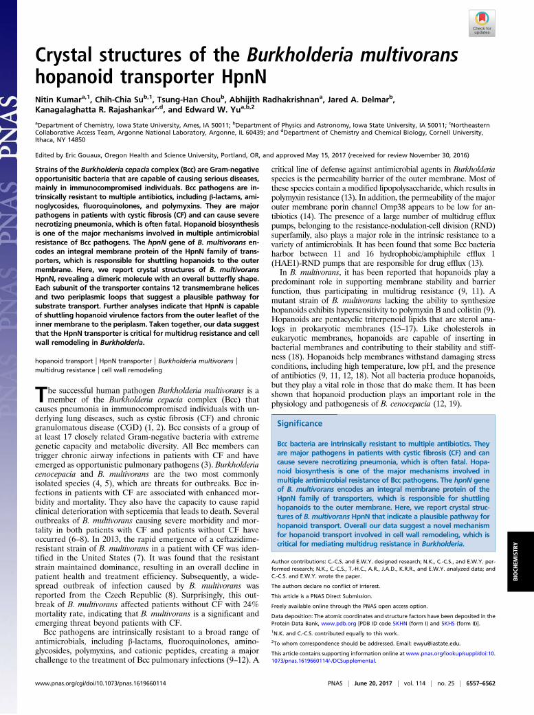
ResultsOverall Structure of B. multivorans HpnN. B. multivorans HpnNconsists of 877 amino acids (SI Appendix, Fig. S1). Two distinctconformations of HpnN (forms I and II) were captured in twodifferent forms of crystals (SI Appendix, Table S1). In each struc-ture, two monomers were found in the asymmetric unit arranged asa dimer (SI Appendix, Figs. S2 and S3). The dimeric form of HpnNin the crystal lattice is in good agreement with the oligomerizationstate of this membrane protein in detergent solution, in which itassembles as a dimer (SI Appendix, Fig. S4). Overall, the topologyof HpnN is unique. The HpnN dimer is butterfly shaped with atwofold symmetry axis perpendicular to the membrane plane (Fig.1A). The overall structure of HpnN indicates that this membraneprotein mainly constitutes the transmembrane and periplasmicdomains. Viewed in parallel to the membrane, the dimer is about110 Å tall, 100 Å wide, and 52 Å thick.Each protomer of HpnN in the dimer contains 12 trans-
membrane helices (TMs 1–12 and TMs 1′–12′, respectively). Inaddition, the monomer possesses a large periplasmic domainformed by two periplasmic loops between TMs 1 and 2 (loop 1),and between TMs 7 and 8 (loop 2). Loop 1 is composed of 11α-helices and four β-strands, whereas loop 2 constitutes 10α-helices and three β-strands. The N-terminal and C-terminalhalves of the transmembrane region are assembled in a pseu-dosymmetrical fashion, although the periplasmic loops 1 and2 are quite asymmetrical. Interestingly, the transmembrane do-main of HpnN can be superimposed to those of AcrB (26), CusA(27), and SecDE (28) with root mean square deviation (rmsd)values of 2.8 Å, 2.7 Å, and 2.1 Å (SI Appendix, Fig. S5). However,the architectures of their periplasmic domains are completelydifferent. The TMs are membrane embedded, but TM8 is sig-nificantly longer and protrudes into the periplasm. TM2 andTM8 directly tether the periplasmic subdomains PD1 and PD2,respectively (Fig. 1 B and C). A hairpin is formed in the middlesection of each periplasmic loop (loops 1 and 2). These twoα-helical hairpins contact one another through a coiled-coil in-teraction and form a four α-helix bundle, contributing to thehairpin subdomain PD4. PD4 is connected to PD1 and PD2 throughan elongated α-helical subdomain PD3, which is composed of threeα-helices (Fig. 1). Several long flexible loops are found to link eachperiplasmic subdomain, suggesting that the periplasmic domain ofHpnN is quite flexible in nature.The crystal structure reveals that TMs 7–9 are involved in the
formation of the dimer. In addition, α6, α14, α18, α21, andβ6 contribute to form the dimer interface. Dimerization occursmainly through van der Waals interactions, as the interactionsurface is mostly hydrophobic in nature. The buried surface areasat the interface between the two monomers of form I and form IIwere estimated to be ∼2,610 Å2 and 2,070 Å2 (SI Appendix, Fig.S6) with the shape complementarity index scores of 0.66 and0.65, suggesting that the oligomerization interface is extensive for
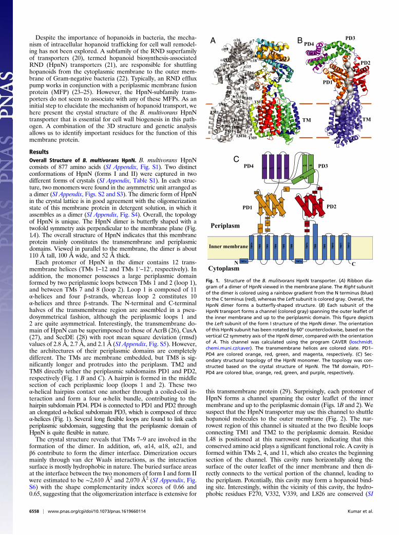
this transmembrane protein (29). Surprisingly, each protomer ofHpnN forms a channel spanning the outer leaflet of the innermembrane and up to the periplasmic domain (Figs. 1B and 2). Wesuspect that the HpnN transporter may use this channel to shuttlehopanoid molecules to the outer membrane (Fig. 2). The nar-rowest region of this channel is situated at the two flexible loopsconnecting TM1 and TM2 to the periplasmic domain. ResidueL48 is positioned at this narrowest region, indicating that thisconserved amino acid plays a significant functional role. A cavity isformed within TMs 2, 4, and 11, which also creates the beginningsection of the channel. This cavity runs horizontally along thesurface of the outer leaflet of the inner membrane and then di-rectly connects to the vertical portion of the channel, leading tothe periplasm. Potentially, this cavity may form a hopanoid bind-ing site. Interestingly, within the vicinity of this cavity, the hydro-phobic residues F270, V332, V339, and L826 are conserved (SI
TM1a
TM1b
α1
α2
α3
β1
α4β2
α5
α6α7α8
β3α9β4
α10
α11
TM2
TM3
TM4
TM5
TM6
TM7a
TM7b
α12
α13
β5α14 β6
α15
α16α17α18
α19
α20
β7α21
TM8
TM9
TM10
TM11
TM12
Cytoplasm
Periplasm
Inner membrane
N C
PD1 PD2
PD3PD4
TM9TM10
TM11
TM5TM4
TM6
TM1
TM3
TM2TM12 TM7
α21
α12α1
α2α11
TM8
α13α9
α14α15
α4 α20
α3 β6β4
β1β2β3
β5β7
α10α19
α17
α16 α7
α6
α18α8
α5
TM TM
PD1
PD2
PD3PD4A
C
B
Fig. 1. Structure of the B. mulitvorans HpnN transporter. (A) Ribbon dia-gram of a dimer of HpnN viewed in the membrane plane. The Right subunitof the dimer is colored using a rainbow gradient from the N terminus (blue)to the C terminus (red), whereas the Left subunit is colored gray. Overall, theHpnN dimer forms a butterfly-shaped structure. (B) Each subunit of theHpnN transport forms a channel (colored gray) spanning the outer leaflet ofthe inner membrane and up to the periplasmic domain. This figure depictsthe Left subunit of the form I structure of the HpnN dimer. The orientationof this HpnN subunit has been rotated by 60° counterclockwise, based on thevertical C2 symmetry axis of the HpnN dimer, compared with the orientationof A. This channel was calculated using the program CAVER (loschmidt.chemi.muni.cz/caver). The transmembrane helices are colored slate. PD1–PD4 are colored orange, red, green, and magenta, respectively. (C) Sec-ondary structural topology of the HpnN monomer. The topology was con-structed based on the crystal structure of HpnN. The TM domain, PD1–PD4 are colored blue, orange, red, green, and purple, respectively.
6558 | www.pnas.org/cgi/doi/10.1073/pnas.1619660114 Kumar et al.
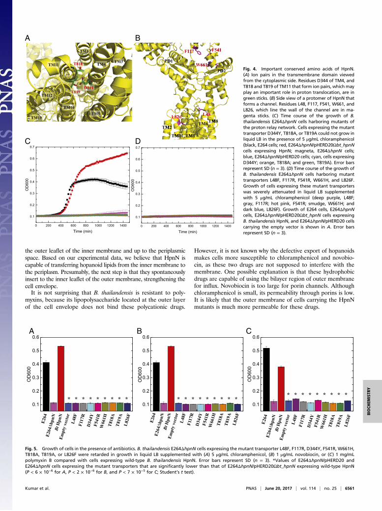
Appendix, Fig. S1). These residues may play a crucial role in rec-ognizing hopanoids. The end of this channel is located at the topportion of PD1 and PD2, where the short helix α5 is also involved informing this exit. Alignment of protein sequences indicates thatseveral conserved aromatic residues, including F117, F541, andW661(SI Appendix, Fig. S1), are found to line the wall of this exiting site.These conserved residues may play an important functional role fortransporting hopanoids in this membrane protein.HpnN is a proton-motive-force (PMF)–dependent transporter
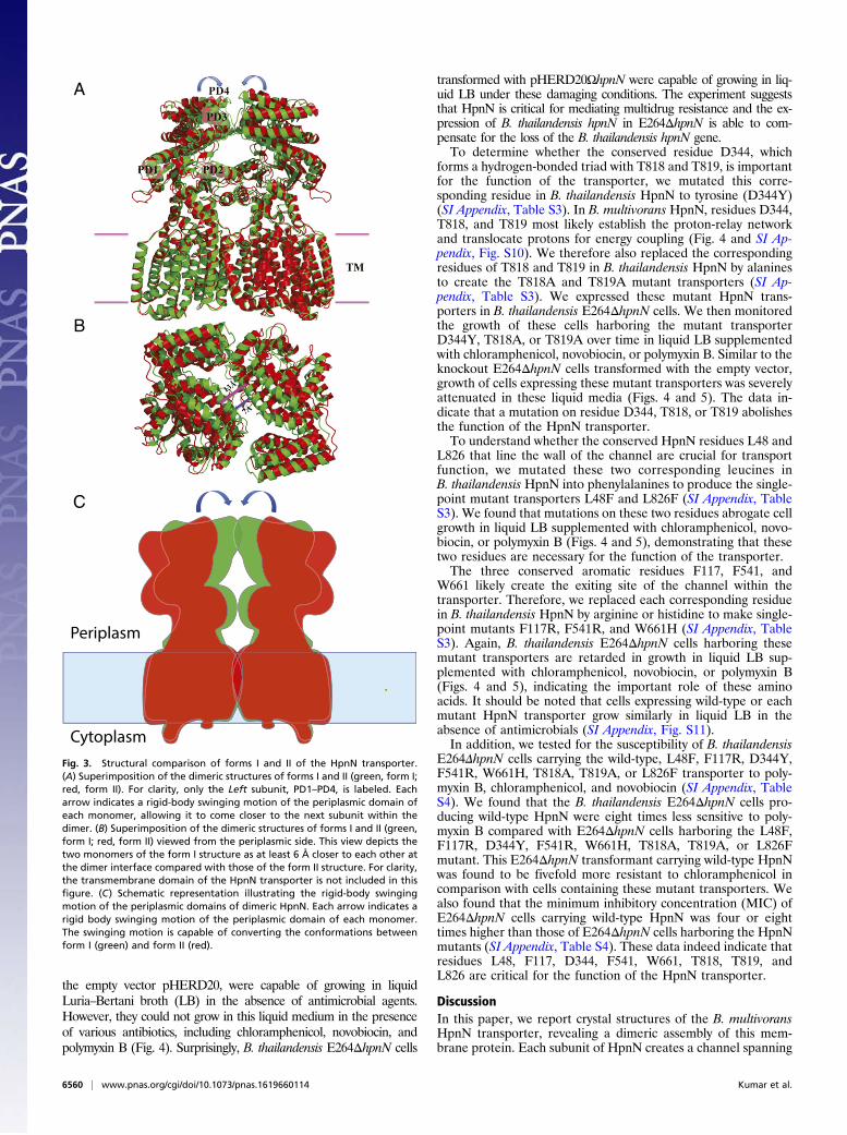
(17, 30). Within the transmembrane region, we found that theconserved residues D344, T818, and T819 (SI Appendix, Fig. S1)of HpnN are in close proximity and seem to interact with eachother to form a triad. It has been reported that threonine canparticipate in forming part of a proton translocation pathway (31).These three residues are less than 4 Å away from each other.Thus, they may be involved in creating a proton-relay network totranslocate protons for energy coupling. As we crystallized HpnNat pH 3.5, residue D344 should be protonated under this acidicenvironment. In addition, it has been found by NMR spectroscopythat threonine can act as a proton donor or acceptor to form ahydrogen bond (32). Therefore, it is possible that one of thethreonines, probably T819, may donate its hydroxyl proton to in-teract with the side chain oxygen of D344 to form a hydrogenbond. If this is the case, then T818 may participate in the proton-relay network as a proton acceptor to receive the acidic protonfrom D344 in this triad. During proton translocation, other nearbyamino acids or solvent molecules may contribute to help transferprotons from the periplasm to the cytosol.
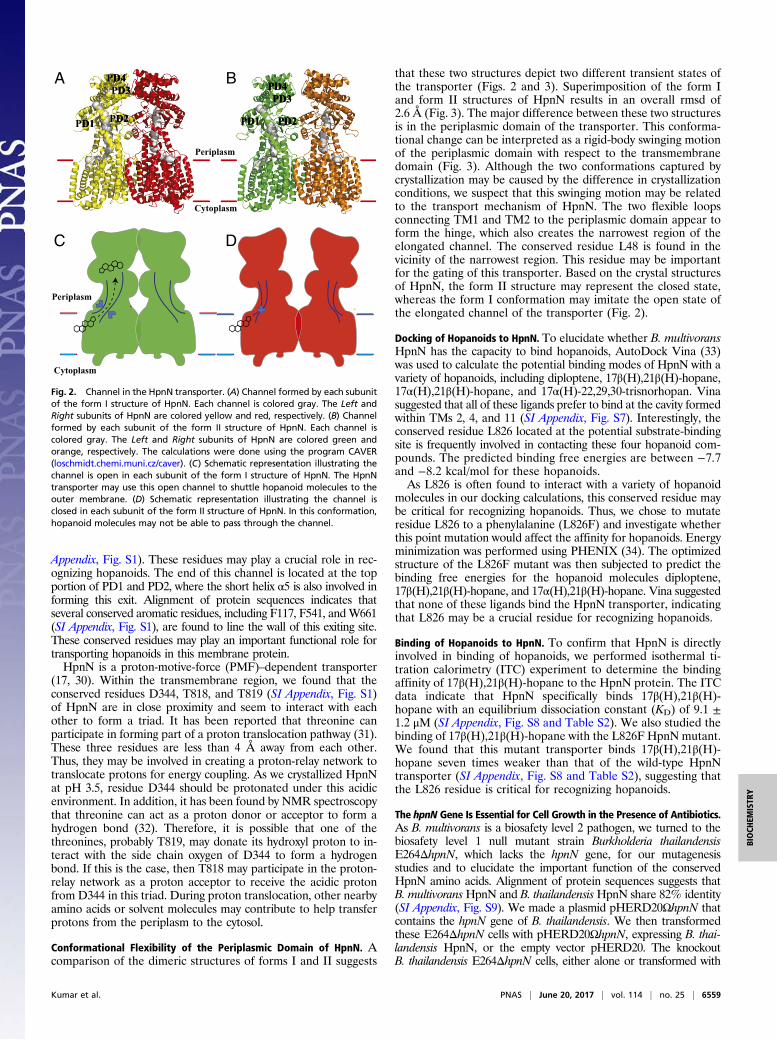
Conformational Flexibility of the Periplasmic Domain of HpnN. Acomparison of the dimeric structures of forms I and II suggests
that these two structures depict two different transient states ofthe transporter (Figs. 2 and 3). Superimposition of the form Iand form II structures of HpnN results in an overall rmsd of2.6 Å (Fig. 3). The major difference between these two structuresis in the periplasmic domain of the transporter. This conforma-tional change can be interpreted as a rigid-body swinging motionof the periplasmic domain with respect to the transmembranedomain (Fig. 3). Although the two conformations captured bycrystallization may be caused by the difference in crystallizationconditions, we suspect that this swinging motion may be relatedto the transport mechanism of HpnN. The two flexible loopsconnecting TM1 and TM2 to the periplasmic domain appear toform the hinge, which also creates the narrowest region of theelongated channel. The conserved residue L48 is found in thevicinity of the narrowest region. This residue may be importantfor the gating of this transporter. Based on the crystal structuresof HpnN, the form II structure may represent the closed state,whereas the form I conformation may imitate the open state ofthe elongated channel of the transporter (Fig. 2).
Docking of Hopanoids to HpnN. To elucidate whether B. multivoransHpnN has the capacity to bind hopanoids, AutoDock Vina (33)was used to calculate the potential binding modes of HpnN with avariety of hopanoids, including diploptene, 17β(H),21β(H)-hopane,17α(H),21β(H)-hopane, and 17α(H)-22,29,30-trisnorhopan. Vinasuggested that all of these ligands prefer to bind at the cavity formedwithin TMs 2, 4, and 11 (SI Appendix, Fig. S7). Interestingly, theconserved residue L826 located at the potential substrate-bindingsite is frequently involved in contacting these four hopanoid com-pounds. The predicted binding free energies are between −7.7and −8.2 kcal/mol for these hopanoids.As L826 is often found to interact with a variety of hopanoid
molecules in our docking calculations, this conserved residue maybe critical for recognizing hopanoids. Thus, we chose to mutateresidue L826 to a phenylalanine (L826F) and investigate whetherthis point mutation would affect the affinity for hopanoids. Energyminimization was performed using PHENIX (34). The optimizedstructure of the L826F mutant was then subjected to predict thebinding free energies for the hopanoid molecules diploptene,17β(H),21β(H)-hopane, and 17α(H),21β(H)-hopane. Vina suggestedthat none of these ligands bind the HpnN transporter, indicatingthat L826 may be a crucial residue for recognizing hopanoids.
Binding of Hopanoids to HpnN. To confirm that HpnN is directlyinvolved in binding of hopanoids, we performed isothermal ti-tration calorimetry (ITC) experiment to determine the bindingaffinity of 17β(H),21β(H)-hopane to the HpnN protein. The ITCdata indicate that HpnN specifically binds 17β(H),21β(H)-hopane with an equilibrium dissociation constant (KD) of 9.1 ±1.2 μM (SI Appendix, Fig. S8 and Table S2). We also studied thebinding of 17β(H),21β(H)-hopane with the L826F HpnN mutant.We found that this mutant transporter binds 17β(H),21β(H)-hopane seven times weaker than that of the wild-type HpnNtransporter (SI Appendix, Fig. S8 and Table S2), suggesting thatthe L826 residue is critical for recognizing hopanoids.
The hpnN Gene Is Essential for Cell Growth in the Presence of Antibiotics.As B. multivorans is a biosafety level 2 pathogen, we turned to thebiosafety level 1 null mutant strain Burkholderia thailandensisE264ΔhpnN, which lacks the hpnN gene, for our mutagenesisstudies and to elucidate the important function of the conservedHpnN amino acids. Alignment of protein sequences suggests thatB. multivoransHpnN and B. thailandensisHpnN share 82% identity(SI Appendix, Fig. S9). We made a plasmid pHERD20ΩhpnN thatcontains the hpnN gene of B. thailandensis. We then transformedthese E264ΔhpnN cells with pHERD20ΩhpnN, expressing B. thai-landensis HpnN, or the empty vector pHERD20. The knockoutB. thailandensis E264ΔhpnN cells, either alone or transformed with
PD4D4PD3D3
PD1D1 PD2D2 PD1PD1 PD2PD2
PD4PD4PD3PD3
A B
C D
Periplasm
Cytoplasm
Periplasm
Cytoplasm
Fig. 2. Channel in the HpnN transporter. (A) Channel formed by each subunitof the form I structure of HpnN. Each channel is colored gray. The Left andRight subunits of HpnN are colored yellow and red, respectively. (B) Channelformed by each subunit of the form II structure of HpnN. Each channel iscolored gray. The Left and Right subunits of HpnN are colored green andorange, respectively. The calculations were done using the program CAVER(loschmidt.chemi.muni.cz/caver). (C) Schematic representation illustrating thechannel is open in each subunit of the form I structure of HpnN. The HpnNtransporter may use this open channel to shuttle hopanoid molecules to theouter membrane. (D) Schematic representation illustrating the channel isclosed in each subunit of the form II structure of HpnN. In this conformation,hopanoid molecules may not be able to pass through the channel.
Kumar et al. PNAS | June 20, 2017 | vol. 114 | no. 25 | 6559
BIOCH
EMISTR
Y
the empty vector pHERD20, were capable of growing in liquidLuria–Bertani broth (LB) in the absence of antimicrobial agents.However, they could not grow in this liquid medium in the presenceof various antibiotics, including chloramphenicol, novobiocin, andpolymyxin B (Fig. 4). Surprisingly, B. thailandensis E264ΔhpnN cells
transformed with pHERD20ΩhpnN were capable of growing in liq-uid LB under these damaging conditions. The experiment suggeststhat HpnN is critical for mediating multidrug resistance and the ex-pression of B. thailandensis hpnN in E264ΔhpnN is able to com-pensate for the loss of the B. thailandensis hpnN gene.To determine whether the conserved residue D344, which
forms a hydrogen-bonded triad with T818 and T819, is importantfor the function of the transporter, we mutated this corre-sponding residue in B. thailandensis HpnN to tyrosine (D344Y)(SI Appendix, Table S3). In B. multivorans HpnN, residues D344,T818, and T819 most likely establish the proton-relay networkand translocate protons for energy coupling (Fig. 4 and SI Ap-pendix, Fig. S10). We therefore also replaced the correspondingresidues of T818 and T819 in B. thailandensis HpnN by alaninesto create the T818A and T819A mutant transporters (SI Ap-pendix, Table S3). We expressed these mutant HpnN trans-porters in B. thailandensis E264ΔhpnN cells. We then monitoredthe growth of these cells harboring the mutant transporterD344Y, T818A, or T819A over time in liquid LB supplementedwith chloramphenicol, novobiocin, or polymyxin B. Similar to theknockout E264ΔhpnN cells transformed with the empty vector,growth of cells expressing these mutant transporters was severelyattenuated in these liquid media (Figs. 4 and 5). The data in-dicate that a mutation on residue D344, T818, or T819 abolishesthe function of the HpnN transporter.To understand whether the conserved HpnN residues L48 and
L826 that line the wall of the channel are crucial for transportfunction, we mutated these two corresponding leucines inB. thailandensis HpnN into phenylalanines to produce the single-point mutant transporters L48F and L826F (SI Appendix, TableS3). We found that mutations on these two residues abrogate cellgrowth in liquid LB supplemented with chloramphenicol, novo-biocin, or polymyxin B (Figs. 4 and 5), demonstrating that thesetwo residues are necessary for the function of the transporter.The three conserved aromatic residues F117, F541, and
W661 likely create the exiting site of the channel within thetransporter. Therefore, we replaced each corresponding residuein B. thailandensis HpnN by arginine or histidine to make single-point mutants F117R, F541R, and W661H (SI Appendix, TableS3). Again, B. thailandensis E264ΔhpnN cells harboring thesemutant transporters are retarded in growth in liquid LB sup-plemented with chloramphenicol, novobiocin, or polymyxin B(Figs. 4 and 5), indicating the important role of these aminoacids. It should be noted that cells expressing wild-type or eachmutant HpnN transporter grow similarly in liquid LB in theabsence of antimicrobials (SI Appendix, Fig. S11).In addition, we tested for the susceptibility of B. thailandensis
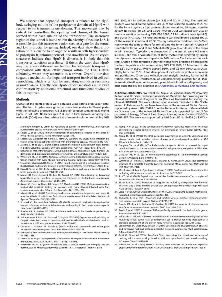
E264ΔhpnN cells carrying the wild-type, L48F, F117R, D344Y,F541R, W661H, T818A, T819A, or L826F transporter to poly-myxin B, chloramphenicol, and novobiocin (SI Appendix, TableS4). We found that the B. thailandensis E264ΔhpnN cells pro-ducing wild-type HpnN were eight times less sensitive to poly-myxin B compared with E264ΔhpnN cells harboring the L48F,F117R, D344Y, F541R, W661H, T818A, T819A, or L826Fmutant. This E264ΔhpnN transformant carrying wild-type HpnNwas found to be fivefold more resistant to chloramphenicol incomparison with cells containing these mutant transporters. Wealso found that the minimum inhibitory concentration (MIC) ofE264ΔhpnN cells carrying wild-type HpnN was four or eighttimes higher than those of E264ΔhpnN cells harboring the HpnNmutants (SI Appendix, Table S4). These data indeed indicate thatresidues L48, F117, D344, F541, W661, T818, T819, andL826 are critical for the function of the HpnN transporter.
DiscussionIn this paper, we report crystal structures of the B. multivoransHpnN transporter, revealing a dimeric assembly of this mem-brane protein. Each subunit of HpnN creates a channel spanning
Periplasm
Cytoplasm
A
B
C
Fig. 3. Structural comparison of forms I and II of the HpnN transporter.(A) Superimposition of the dimeric structures of forms I and II (green, form I;red, form II). For clarity, only the Left subunit, PD1–PD4, is labeled. Eacharrow indicates a rigid-body swinging motion of the periplasmic domain ofeach monomer, allowing it to come closer to the next subunit within thedimer. (B) Superimposition of the dimeric structures of forms I and II (green,form I; red, form II) viewed from the periplasmic side. This view depicts thetwo monomers of the form I structure as at least 6 Å closer to each other atthe dimer interface compared with those of the form II structure. For clarity,the transmembrane domain of the HpnN transporter is not included in thisfigure. (C) Schematic representation illustrating the rigid-body swingingmotion of the periplasmic domains of dimeric HpnN. Each arrow indicates arigid body swinging motion of the periplasmic domain of each monomer.The swinging motion is capable of converting the conformations betweenform I (green) and form II (red).
6560 | www.pnas.org/cgi/doi/10.1073/pnas.1619660114 Kumar et al.
the outer leaflet of the inner membrane and up to the periplasmicspace. Based on our experimental data, we believe that HpnN iscapable of transferring hopanoid lipids from the inner membrane tothe periplasm. Presumably, the next step is that they spontaneouslyinsert to the inner leaflet of the outer membrane, strengthening thecell envelope.It is not surprising that B. thailandensis is resistant to poly-
myxins, because its lipopolysaccharide located at the outer layerof the cell envelope does not bind these polycationic drugs.
However, it is not known why the defective export of hopanoidsmakes cells more susceptible to chloramphenicol and novobio-cin, as these two drugs are not supposed to interfere with themembrane. One possible explanation is that these hydrophobicdrugs are capable of using the bilayer region of outer membranefor influx. Novobiocin is too large for porin channels. Althoughchloramphenicol is small, its permeability through porins is low.It is likely that the outer membrane of cells carrying the HpnNmutants is much more permeable for these drugs.
F11711 F54141
W661661
L8262 L484
PD1D
PD2D
TM2M
TM11M1 TM1M1
TM6TM8M
0 200 400 600 800 1000 1200 1400
0.1
0.2
0.3
0.4
0.5
0.6
0.7
OD
600
Time (min)0 200 400 600 800 1000 1200 1400
0.1
0.2
0.3
0.4
0.5
0.6
0.7
OD
600
Time (min)
A B
C D
Fig. 4. Important conserved amino acids of HpnN.(A) Ion pairs in the transmembrane domain viewedfrom the cytoplasmic side. Residues D344 of TM4, andT818 and T819 of TM11 that form ion pairs, which mayplay an important role in proton translocation, are ingreen sticks. (B) Side view of a protomer of HpnN thatforms a channel. Residues L48, F117, F541, W661, andL826, which line the wall of the channel are in ma-genta sticks. (C) Time course of the growth of B.thailandensis E264ΔhpnN cells harboring mutants ofthe proton relay network. Cells expressing the mutanttransporter D344Y, T818A, or T819A could not grow inliquid LB in the presence of 5 μg/mL chloramphenicol(black, E264 cells; red, E264ΔhpnN/pHERD20Ωbt_hpnNcells expressing HpnN; magneta, E264ΔhpnN cells;blue, E264ΔhpnN/pHERD20 cells; cyan, cells expressingD344Y; orange, T818A; and green, T819A). Error barsrepresent SD (n = 3). (D) Time course of the growth ofB. thailandensis E264ΔhpnN cells harboring mutanttransporters L48F, F117R, F541R, W661H, and L826F.Growth of cells expressing these mutant transporterswas severely attenuated in liquid LB supplementedwith 5 μg/mL chloramphenicol (deep purple, L48F;gray, F117R; hot pink, F541R; smudge, W661H; anddark blue, L826F). Growth of E264 cells, E264ΔhpnNcells, E264ΔhpnN/pHERD20Ωbt_hpnN cells expressingB. thailandensis HpnN, and E264ΔhpnN/pHERD20 cellscarrying the empty vector is shown in A. Error barsrepresent SD (n = 3).
A B C
E264
E264Δh
pnN
Bt H
pnN
Empt
y ve
ctor
L48F
F117
RD
344Y
F541
RW
661H
T818
AT8
19A
L826
F
0.1
0.2
0.3
0.4
0.5
0.6
OD
600
* *** * * * * *0.1
0.2
0.3
0.4
0.5
0.6
OD
600
E264
E264Δh
pnN
Bt H
pnN
Empt
y ve
ctor
L48F
F117
RD
344Y
F541
RW
661H
T818
AT8
19A
L826
F
* *** * * * * *
E264
E264Δh
pnN
Bt H
pnN
Empt
y ve
ctor
L48F
F117
RD
344Y
F541
RW
661H
T818
AT8
19A
L826
F
0.1
0.2
0.3
0.4
0.5
0.6
OD
600
* *** * * * * *
Fig. 5. Growth of cells in the presence of antibiotics. B. thailandensis E264ΔhpnN cells expressing the mutant transporter L48F, F117R, D344Y, F541R, W661H,T818A, T819A, or L826F were retarded in growth in liquid LB supplemented with (A) 5 μg/mL chloramphenicol, (B) 1 μg/mL novobiocin, or (C) 1 mg/mLpolymyxin B compared with cells expressing wild-type B. thailandensis HpnN. Error bars represent SD (n = 3). *Values of E264ΔhpnN/pHERD20 andE264ΔhpnN cells expressing the mutant transporters that are significantly lower than that of E264ΔhpnN/pHERD20Ωbt_hpnN expressing wild-type HpnN(P < 6 × 10⁻⁶ for A, P < 2 × 10⁻⁶ for B, and P < 7 × 10⁻⁵ for C; Student’s t test).
Kumar et al. PNAS | June 20, 2017 | vol. 114 | no. 25 | 6561
BIOCH
EMISTR
Y
We suspect that hopanoid transport is related to the rigid-body swinging motion of the periplasmic domain of HpnN withrespect to its transmembrane domain. This movement may becritical for controlling the opening and closing of the tunnelformed within each subunit of the transporter. The narrowestregion of the tunnel is found within the vicinity of residue L48. Itis possible that this narrowest region creates a gate for the tunneland L48 is crucial for gating. Indeed, our data show that a mu-tation of this leucine to an arginine results in cells hypersensitiveto polymyxin B, chloramphenicol, and novobiocin. As the crystalstructures indicate that HpnN is dimeric, it is likely that thistransporter functions as a dimer. If this is the case, then HpnNmay use a very different mechanism for substrate transport incomparison with those efflux pumps within the HAE1-RNDsubfamily, where they assemble as a trimer. Overall, our datasuggest a mechanism for hopanoid transport involved in cell wallremodeling, which is critical for mediating multidrug resistancein Burkholderia. Exactly how HpnN export substrates must awaitconfirmation by additional structural and functional studies ofthis transporter.
MethodsCrystals of the HpnN protein were obtained using sitting-drop vapor diffu-sion. The form I crystals were grown at room temperature in 24-well plateswith the following procedures: A 2-μL protein solution containing 20 mg/mLHpnN in 20 mM Na-Hepes (pH 7.5) and 0.05% (wt/vol) n-dodecyl-β-D-maltoside (DDM) was mixed with a 2 μL of reservoir solution containing 16%
PEG 2000, 0.1 M sodium citrate (pH 3.5) and 0.2 M Li2SO4. The resultantmixture was equilibrated against 500 μL of the reservoir solution at 25 °C.For the form II crystals, a 2-μL protein solution containing 20 mg/mL HpnN in20 mM Na-Hepes (pH 7.5) and 0.05% (wt/vol) DDM was mixed with 2 μL ofreservoir solution containing 15% PEG 2000, 0.1 M sodium citrate (pH 4.0),and 0.2 M (NH4)2SO4. The resultant mixture was equilibrated against 500 μLof the reservoir solution at 25 °C. The crystallization conditions for SeMet-HpnN were the same as those for crystallizing the form II crystals. Crystals ofHpnN (both forms I and II) and SeMet-HpnN grew to a full size in the dropswithin a month. Typically, the dimensions of the crystals were 0.2 mm ×0.2 mm × 0.2 mm. Cryoprotection of these crystals was achieved by raisingthe glycerol concentration stepwise to 30% with a 5% increment in eachstep. Crystals of the tungsten cluster derivative were prepared by incubatingthe form I crystals in solution containing 18% PEG 2000, 0.1 M sodium citrate(pH 3.5), 0.2 M Li2SO4, 0.05% (wt/vol) DDM, and 0.5 mM (NH4)2W6(μ-O)6(μ-Cl)6Cl6 for 5 h at 25 °C. A full description of methods for protein expressionand purification, X-ray data collection and analysis, docking, isothermal ti-tration calorimetry, construction of complementing plasmid for B. thai-landensis, site-directed mutagenesis, plasmid mobilization, cell growth, anddrug susceptibility are described in SI Appendix, SI Materials and Methods.
ACKNOWLEDGMENTS. We thank Dr. Miguel A. Valvano (Queen’s UniversityBelfast) and Dr. Silvia Cardona (University of Manitoba) for providing us theplasmid pScrhaB2 and Hongwei D. Yu (Marshall University) for sharing theplasmid pHERD20T. This work is based upon research conducted at the North-eastern Collaborative Access Team beamlines of the Advanced Photon Source,supported by Award GM103403 from the National Institutes of General Med-ical Sciences. Use of the Advanced Photon Source is supported by the US De-partment of Energy, Office of Basic Energy Sciences, under Contract DE-AC02-06CH11357. This work was supported by NIH Grant R01AI114629 (to E.W.Y.).
1. Mahenthiralingam E, Urban TA, Goldberg JB (2005) The multifarious, multirepliconBurkholderia cepacia complex. Nat Rev Microbiol 3:144–156.
2. Sajjan U, et al. (2001) Immunolocalisation of Burkholderia cepacia in the lungs ofcystic fibrosis patients. J Med Microbiol 50:535–546.
3. Ledson MJ, Gallagher MJ, Corkill JE, Hart CA, Walshaw MJ (1998) Cross infection be-tween cystic fibrosis patients colonised with Burkholderia cepacia. Thorax 53:432–436.
4. Zlosnik JE, et al. (2015) Burkholderia species infections in patients with cystic fibrosisin British Columbia, Canada. 30 years’ experience. Ann Am Thorac Soc 12:70–78.
5. Drevinek P, Mahenthiralingam E (2010) Burkholderia cenocepacia in cystic fibrosis: Epi-demiology and molecular mechanisms of virulence. Clin Microbiol Infect 16:821–830.
6. Whiteford ML, et al. (1995) Outcome of Burkholderia (Pseudomonas) cepacia colonisa-tion in children with cystic fibrosis following a hospital outbreak. Thorax 50:1194–1198.
7. Stokell JR, Gharaibeh RZ, Steck TR (2013) Rapid emergence of a ceftazidime-resistantBurkholderia multivorans strain in a cystic fibrosis patient. J Cyst Fibros 12:812–816.
8. Hanulik V, et al. (2013) An outbreak of Burkholderia multivorans beyond cystic fi-brosis patients. J Hosp Infect 84:248–251.
9. Malott RJ, Steen-Kinnaird BR, Lee TD, Speert DP (2012) Identification of hopanoidbiosynthesis genes involved in polymyxin resistance in Burkholderia multivorans.Antimicrob Agents Chemother 56:464–471.
10. Aaron SD, Ferris W, Henry DA, Speert DP, Macdonald NE (2000) Multiple combinationbactericidal antibiotic testing for patients with cystic fibrosis infected with Bur-kholderia cepacia. Am J Respir Crit Care Med 161:1206–1212.
11. Malott RJ, et al. (2014) Fosmidomycin decreases membrane hopanoids and potenti-ates the effects of colistin on Burkholderia multivorans clinical isolates. AntimicrobAgents Chemother 58:5211–5219.
12. Schmerk CL, Bernards MA, Valvano MA (2011) Hopanoid production is required forlow-pH tolerance, antimicrobial resistance, and motility in Burkholderia cenocepacia.J Bacteriol 193:6712–6723.
13. Rhodes KA, Schweizer HP (2016) Antibiotic resistance in Burkholderia species. DrugResist Updat 28:82–90.
14. Siritapetawee J, Prinz H, Krittanai C, Suginta W (2004) Expression and refolding ofOmp38 from Burkholderia pseudomallei and Burkholderia thailandensis, and itsfunction as a diffusion porin. Biochem J 384:609–617.
15. Ourisson G, Rohmer M, Poralla K (1987) Prokaryotic hopanoids and other poly-terpenoid sterol surrogates. Annu Rev Microbiol 41:301–333.
16. Mahato SB, Sen S (1997) Advances in triterpenoid research, 1990-1994. Phytochemistry44:1185–1236.
17. Saenz JP, et al. (2015) Hopanoids as functional analogues of cholesterol in bacterialmembranes. Proc Natl Acad Sci USA 112:11971–11976.
18. Welander PV, et al. (2009) Hopanoids play a role in membrane integrity and pHhomeostasis in Rhodopseudomonas palustris TIE-1. J Bacteriol 191:6145–6156.
19. Tseng SP, et al. (2014) The contribution of antibiotic resistance mechanisms in clinicalBurkholderia cepacia complex isolates: An emphasis on efflux pump activity. PLoSOne 9:e104986.
20. Tseng TT, et al. (1999) The RND permease superfamily: an ancient, ubiquitous anddiverse family that includes human disease and development proteins. J MolMicrobiol Biotechnol 1:107–125.
21. Doughty DM, et al. (2011) The RND-family transporter, HpnN, is required for hopa-noid localization to the outer membrane of Rhodopseudomonas palustris TIE-1. ProcNatl Acad Sci USA 108:E1045–E1051.
22. Daligault HE, et al. (2014) Whole-genome assemblies of 56 burkholderia species.Genome Announc 2: pii: e01106-14.
23. Symmons MF, Bokma E, Koronakis E, Hughes C, Koronakis V (2009) The assembledstructure of a complete tripartite bacterial multidrug efflux pump. Proc Natl Acad SciUSA 106:7173–7178.
24. Mikolosko J, Bobyk K, Zgurskaya HI, Ghosh P (2006) Conformational flexibility in themultidrug efflux system protein AcrA. Structure 14:577–587.
25. Su CC, et al. (2011) Crystal structure of the CusBA heavy-metal efflux complex ofEscherichia coli. Nature 470:558–562.
26. Eicher T, et al. (2012) Transport of drugs by the multidrug transporter AcrB involvesan access and a deep binding pocket that are separated by a switch-loop. Proc NatlAcad Sci USA 109:5687–5692.
27. Long F, et al. (2010) Crystal structures of the CusA efflux pump suggest methionine-mediated metal transport. Nature 467:484–488.
28. Tsukazaki T, et al. (2011) Structure and function of a membrane component SecDFthat enhances protein export. Nature 474:235–238.
29. Duarte JM, Biyani N, Baskaran K, Capitani G (2013) An analysis of oligomerizationinterfaces in transmembrane proteins. BMC Struct Biol 13:21.
30. Perrin E, et al. (2013) A census of RND superfamily proteins in the Burkholderia genus.Future Microbiol 8:923–937.
31. Takatsuka Y, Nikaido H (2006) Threonine-978 in the transmembrane segment of themultidrug efflux pump AcrB of Escherichia coli is crucial for drug transport as aprobable component of the proton relay network. J Bacteriol 188:7284–7289.
32. Brockerman JA, Okon M, McIntosh LP (2014) Detection and characterization of serineand threonine hydroxyl protons in Bacillus circulans xylanase by NMR spectroscopy.J Biomol NMR 58:17–25.
33. Trott O, Olson AJ (2010) AutoDock Vina: Improving the speed and accuracy ofdocking with a new scoring function, efficient optimization, and multithreading.J Comput Chem 31:455–461.
34. Adams PD, et al. (2002) PHENIX: Building new software for automated crystallo-graphic structure determination. Acta Crystallogr D Biol Crystallogr 58:1948–1954.
6562 | www.pnas.org/cgi/doi/10.1073/pnas.1619660114 Kumar et al.