cronicon open access dental science … · 4oral and dental research institute, school of...
TRANSCRIPT

CroniconO P E N A C C E S S DENTAL SCIENCE
Research Article
V Tamara Perchyonoka1, John Souzab2, Shengmiao Zhangc3, Desigar Moodleyd4 and Sias Groblerd4
1VTPCHEM PTY LTD, Glenhuntly, Melbourne, Australia2Department of Prosthetics, TAFE Queensland, South Brisbane, Australia3School of Material Science and Engineering, East China University of Science and Technology, China4Oral and Dental Research Institute, School of Dentistry, The University of Western Cape, South Africa
Received: November 03, 2015; Published: November 17, 2015
*Corresponding Author: V Tamara Perchyonoka, VTPCHEM PTY LTD, Glenhuntly, Melbourne, 4215, Australia.
Bio-Active Nano Diamond Designer Materials and Dentures: From Design to Application
Abstract
Objective: The present study aims to design and evaluate performance of Nano diamond: chitosan based bio-active containing acryl-ic materials and investigations towards application for the development of novel bio-active materials with build in capabilities for treatment and prevention of denture stomatitis and associated conditions in denture wearers in vitro.
Methods: The bio-active Nano diamond modified PMMA were prepared by dispersion of the corresponding component in glycerol and acetic acid with the addition of chitosan gelling agent. The release behaviors at physiological pH and also under acidic condi-tions and stability of the antioxidant-chitosan-Nano diamond were also evaluated. Mechanical performance such as tensile strength and compressive strength were measured as well bio-adhesive studies were investigated in order to assess the suitability of these designer materials.
Results: The bioactive: Nano diamond modified PMMA materials showed a high adhesive force and were only slightly swelled in the aqueous medium. Bioactive release suggested prolonged release of the therapeutic agent from the hydrogels. The hydrogels also had significant free radical defense capability.
Conclusion: In this study we demonstrated that the newly prepared bio-active modified PMMA resins are suitable novel bio-active materials capable of comparable performance with the conventional PMMA materials with additional benefit of therapeutic bioactive release as well as potential antimicrobial properties to be demonstrated in vitro. Our findings might be thus a step forward towards the development of alternative non antibiotic based strategies targeting bacterial infections.
Keywords: Chitosan; Nano diamond; Bio-active; PMMA; Drug delivery systems; Free radicals
Citation: V Tamara Perchyonoka., et al. “Bio-Active Nano Diamond Designer Materials and Dentures: From Design to Application”. EC Dental Science 3.2 (2015): 483-495.
IntroductionThe initial interaction between pathogenic bacteria and host cells is one of the critical steps leading to host colonization [1]. These
first adhesion events allow host-pathogen recognition and prevent bacteria from being washed out by the host and are generally medi-ated by species proteins called adhesions. C. albicans binds various bacterial species, participating in polymicrobial interactions in the normally healthy host [2]. One of these is the oral commensal bacterium Streptococcus gordonii [3]. Als3 is one of eight C. albicans Als proteins (Als1-Als7, Als9), large cell-surface glycoprotein that primarily function in adhesive interactions [4].
Nano diamond particles (NDs) are emerging as particularly well-suited for biological applications due to their biocompatibility [6-10]. It is indeed because of this feature that diamond Nano particles should also be explored for anti-bacterial applications. While Nano
Bio-Active Nano Diamond Designer Materials and Dentures: From Design to Application484
Citation: V Tamara Perchyonoka., et al. “Bio-Active Nano Diamond Designer Materials and Dentures: From Design to Application”. EC Dental Science 3.2 (2015): 483-495.
The copaiba tree is native to Latin America and Occidental Africa [19]. There are more than 20 species of copaiba in Brazil, and the most commonly described effects are anti-inflammatory, analgesic, antibacterial and antitumoral activities [20-22].
The present study aims to design and evaluate performance of chitosan: Nano diamond based bio-active containing acrylic materials and investigations towards application for the development of novel bio-active materials with build in capabilities for treatment and pre-vention of denture stomatitis and associated conditions in denture wearers, while the performance of the material is not compromised in vitro.
Propolis Brazilian (Red, Natura Nectar), Copaiba Oil (Laboratorio Sao Lucas, Brazil) and Shiitake powder (Border herbal health, Aus-tralia) were purchased from a commercial supplier (Wholesale Chemist, QLD, Australia) and used without further purification. (_)-Epi-gallocatechingallate (EGCG) from green tea, 95%, (_)-epicatechingallate (ECG) from green tea, 98%, (-)-epigallocatechin (EGC) from green tea, 95%, (_)-epicatechin (EC), 90%, gallic acid (GA) purity not specified, 6-hydroxy-2,5,7,8-tetramethylchromane-2-carboxylic acid (trolox), 95%, Folin-Ciocalteu reagent, ferric chloride, Tween-20 and 2,20-azino-bis (3-ethylbenzothiazoline-6-sulfonic acid) diam-monium salt (ABTS) were purchased from Sigma-Aldrich Company Ltd. (Australia).
The bio active containing gel was prepared by dispersion of 0.2 gm of commercially available bioactives [Propolis (Red Brazilian), Copaiba oil or Shiitake powder] and Nano diamond powder (0.08g) in glycerol (5% w/w) (1 ml) using a mortar and a pestle follow-ing the earlier reported generic protocol [9-11]. Ten milliliters of glacial acetic acid (3% w/w) was then added with continuous mixing and finally chitosan (10% chitosan w/w) polymer was spread on the surface of the dispersion and mixed well to form the required gel and then mixed into a PMMA resin prior to setting. The strength of the prepared gel (10 gm) is 0.2 gm of propolis, copaibaoilor shiitake mushrooms extract in each gram of the base and 0.08g of Nano-diamond in each gram of the base. The total phenolic concentration was quantified using Rocha., et al. [23] and described by Waterman & Mole [24] with some modifications.
The swelling/weight loss tests were performed when triplicates of each samples composition (approximately 2 cm2, weight normal-ized) were immersed in 2 mL of different fluids at 37°C for each time interval studied (1, 2, 4, 24, and 96h). Two different media were
Chitosan (Aldrich, Australia), glycerol (Sigma, USA), glacial acetic acid (E. Merck, Germany) were used as received. The degree of de-acetylation of typical commercial chitosan used in this study is 87%. Chitosan with molecular weight 2.5 x 10³ KD was used in the study. Gelatin in powder form was purchased from Shanghai Chemical Reagent Co., (Shanghai, China) with the number-average molecular weight (Mn) of about 8.7 x 10⁴. The isoelectric point is 4.0-5.0. Nano-diamonds where purchased from Ebersoles, (25 carats, size 0-2 microns, Grit 14,000) and used as received.
Materials
Preparation of Nano-diamond bioactive containing methyl methacrylate materials: general protocol
Swelling/Weight Loss Tests and Bioactive Release
diamond conjugates have shown their interest for the detection and removal of bacteria in solution [11], their potential for the efficient inhibition of Als-mediated binding phenomena has however not been fully explored.
Chitosan, which is a biologically safe biopolymer as well as an antioxidant, has been proposed as a bio-adhesive polymer and is of continuous interest to us due to its unique properties and flexibility in a broad range of oral applications reported by others and our-selves recently [12-15]. Lentinusedodes, known as shiitake mushroom, has received great attention due to positive health effects, includ-ing anti-tumour and hypo-cholesterolemic activity [12], related to the presence of β-glucans [13].
Propolis is a resinous substance produced by bees with antibacterial, antifungal, antiviral, and anti-inflammatory activities [14,15]. Propolis antibacterial activity is bacteriostatic and, in high concentration, bactericidal [16]. Propolis has antimicrobial activity against gram-positive bacteria, egS. aureus, but limited action against gram-negative bacteria and also against some fungi, e.g., C. albicans [17,18].
Bio-Active Nano Diamond Designer Materials and Dentures: From Design to Application485
Citation: V Tamara Perchyonoka., et al. “Bio-Active Nano Diamond Designer Materials and Dentures: From Design to Application”. EC Dental Science 3.2 (2015): 483-495.
Tensile strength testing of the material
Compressive strength
Bio-adhesive investigation
SEM Images
To analyze the bio-active release (propolis (Brazilian), Copaiba oil and Shiitake mushrooms) based on the total phenolic concentra-tion, the swelling media was analyzed after 1, 2, 24, and 96h of immersion via UV-Vis spectrometer, from 300 to 800 nm, using polysty-rene cuvettes [21]. For quantification of the amount of propolis released, a standard curve was created by diluting the original propolis in isopropanol resulting in several aliquots of known concentration, which were then analyzed in the same wavelength range. The area of the peak of these aliquots (of known concentration of bio-additive) was calculated and used to compare with those of the bio-additive released by the samples.
Tensile testing was conducted using Instron 5565. Following American Standardized Testing Materials Standard D3039, rectangular samples were approximately 6-8 mm in length, 1 mm in width an 1 mm in thickness, and tested with a gauge length of 3.5 ± 0.4 mm [24]. Samples were elongated at a rate of 1% of gauge length per second. The cross-sectional area of samples was evaluated using Image J image analysis software [25].
Compressive strength test samples were placed in the measuring apparatus in an appropriate manner and cross-sectional area of each sample (mm2) was determined. A compressive load (N) was applied at a crosshead speed of 1.3 mm/min [26]. The compressive strength (MPa) was measured at the sample fracture point. Mean, average, and mode in each group were calculated and normal distri-bution curve was evaluated. One-way ANOVA, followed by multiple comparison test (Scheffé’s test), was used for statistical analysis. Statistical significance was set at P < 0.05.
Bio-adhesion studies were done using a Chatillon apparatus for force measurement [27]. This method determines the maximum force and work needed to separate two surfaces in intimate contact [27]. The modified Nano-diamond biomaterials (0.1g) were homo-geneously spread on a 1 cm2 disk and then the disks were fixed to the support of the tensile strength tester using double sided adhesive. The bio active modified PMMA material was brought into contact with a slice of pig ear skin was established in order to imitate adhe-sion of the gel to the “oral mucosa prototype system” structure. After a preset contact time of 1 min under contact strength of 0.5N, the 2 surfaces were separated at a constant rate of displacement of 1 mm/s. The strength was recorded as a function of the displacement, which allowed to determine the maximal detachment force, Fmax, and the work of adhesion, W, which was calculated from the area under the strength-displacement curve [27].
Results and Discussion
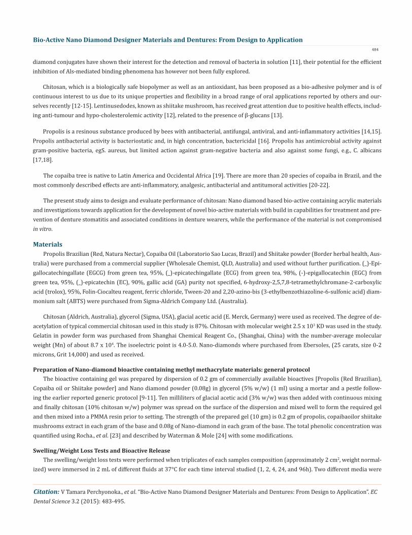
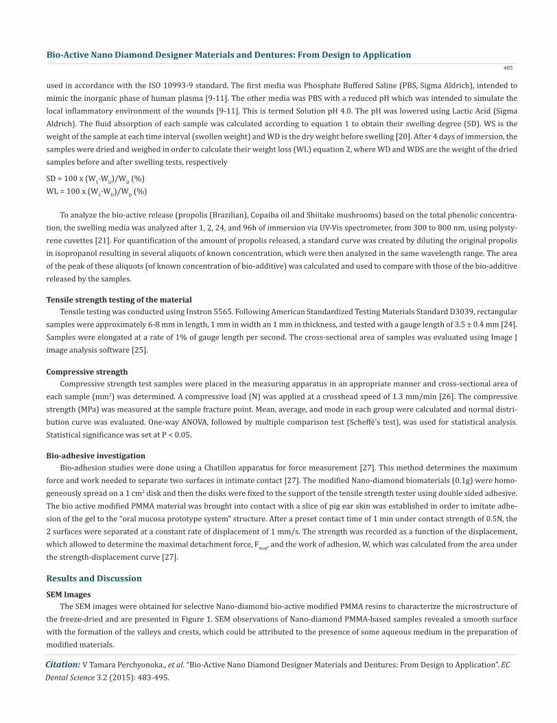
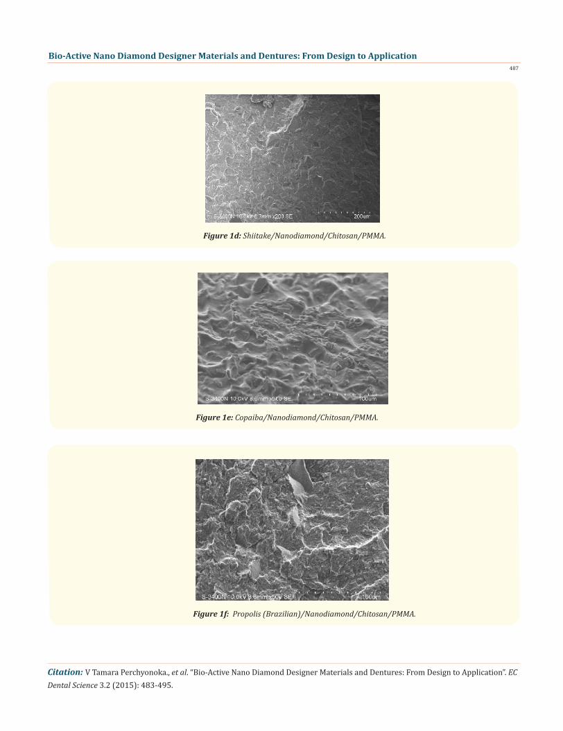
The SEM images were obtained for selective Nano-diamond bio-active modified PMMA resins to characterize the microstructure of the freeze-dried and are presented in Figure 1. SEM observations of Nano-diamond PMMA-based samples revealed a smooth surface with the formation of the valleys and crests, which could be attributed to the presence of some aqueous medium in the preparation of modified materials.
used in accordance with the ISO 10993-9 standard. The first media was Phosphate Buffered Saline (PBS, Sigma Aldrich), intended to mimic the inorganic phase of human plasma [9-11]. The other media was PBS with a reduced pH which was intended to simulate the local inflammatory environment of the wounds [9-11]. This is termed Solution pH 4.0. The pH was lowered using Lactic Acid (Sigma Aldrich). The fluid absorption of each sample was calculated according to equation 1 to obtain their swelling degree (SD). WS is the weight of the sample at each time interval (swollen weight) and WD is the dry weight before swelling [20]. After 4 days of immersion, the samples were dried and weighed in order to calculate their weight loss (WL) equation 2, where WD and WDS are the weight of the dried samples before and after swelling tests, respectively
SD = 100 x (WS-WD)/WD (%)WL = 100 x (WS-WD)/WD (%)

Bio-Active Nano Diamond Designer Materials and Dentures: From Design to Application486
Citation: V Tamara Perchyonoka., et al. “Bio-Active Nano Diamond Designer Materials and Dentures: From Design to Application”. EC Dental Science 3.2 (2015): 483-495.
Figure 1a: Shiitake/Nanodiamond/PMMA.
Figure 1b: Copaiba/Nanodiamond/PMMA.
Figure 1c: Propolis (Brazilian)/Nanodiamond/PMMA.
Bio-Active Nano Diamond Designer Materials and Dentures: From Design to Application487
Citation: V Tamara Perchyonoka., et al. “Bio-Active Nano Diamond Designer Materials and Dentures: From Design to Application”. EC Dental Science 3.2 (2015): 483-495.
Figure 1d: Shiitake/Nanodiamond/Chitosan/PMMA.
Figure 1e: Copaiba/Nanodiamond/Chitosan/PMMA.
Figure 1f: Propolis (Brazilian)/Nanodiamond/Chitosan/PMMA.

Bio-Active Nano Diamond Designer Materials and Dentures: From Design to Application488
Citation: V Tamara Perchyonoka., et al. “Bio-Active Nano Diamond Designer Materials and Dentures: From Design to Application”. EC Dental Science 3.2 (2015): 483-495.
Compression Test
Tensile strength of the bioactive functionalized materials
Swelling/Weight Loss Tests and Bioactive Release
Mechanical properties investigated
The poly-methyl-methacrylate has adequate tensile and compressive strength for complete and partial dentures [28]. The com-pression behavior for composite prosthetic dentures represents the important mechanical properties specialization when using the polymer matrix materials. The compression strength values results obtained from compression tests are carried out for all bioactive prepared materials and results are summarized in the Figure 2. The addition of the bioactive compounds such as Shiitake extract, Co-paiba oil or Brazilian propolis in combination with Nano-diamond had significant influenced into the compression strength of the bio-active PMMA material. Also upon incorporation of Nano diamond: chitosan: bioactive combination (10% w/w) into the PMMA material the compressive strength of the new bioactive material was significantly increased in comparison to the standard PMMA material.
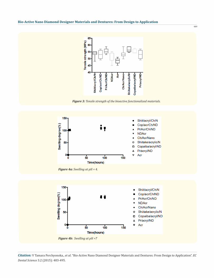
The tensile behavior for composite prosthetic dentures represents the important mechanical properties specialization when using the polymer matrix materials and is summarized in Figure 3. The addition of the bioactive compounds such as Shiitake extract, Co-paiba oil or Brazilian propolis in combination with Nano diamond had not influenced significantly the tensile strength of the bioactive PMMA material. Also upon incorporation of Nano diamond: chitosan: bioactive combination (10% w/w) into the PMMA material the tensile strength of the new bioactive material was significantly increased in comparison to the standard PMMA material. The slight increase of tensile strength is probably due to the potential action interference of the bio-actives and their antioxidant capacity with the polymerization rates of the PMMA presence of chitosan acts as the protective host of the excess of free radical formation and there for some increase in the tensile as well as compressive strength is observed. The results are basically consistent with the literature [29,30] and more detailed investigations into mechanistic interaction of chitosan/bioactive/PMMA resin are currently on the way in our laboratory.
The swelling characteristics of the bio-active modified materials at pH 7.0 and pH 4 are shown in Figure 4a and 4b respectively. Swelling has not been affected significantly in case of either incorporation of bioactive/Nano diamond or bioactive/chitosan/Nano diamond did not increased in the case at either pH. No definite trend in swelling with composition was observed. Though PMMA is a hydrophobic polymer, the acidic environment mayhydrolyze the methacrylate group to some extent conferring hydrophilic nature to the copolymer. The in depth investigation between the nature of the functional surface interaction of Nano-diamond: chitosan: bioac-tive is currently under investigation in our laboratory.
Figure 2: Compressive strength measurements for the bio-active materials for investigation.
Bio-Active Nano Diamond Designer Materials and Dentures: From Design to Application489
Citation: V Tamara Perchyonoka., et al. “Bio-Active Nano Diamond Designer Materials and Dentures: From Design to Application”. EC Dental Science 3.2 (2015): 483-495.
Figure 3: Tensile strength of the bioactive functionalized materials.
Figure 4a: Swelling at pH = 4.
Figure 4b: Swelling at pH =7
Bio-Active Nano Diamond Designer Materials and Dentures: From Design to Application490
Citation: V Tamara Perchyonoka., et al. “Bio-Active Nano Diamond Designer Materials and Dentures: From Design to Application”. EC Dental Science 3.2 (2015): 483-495.
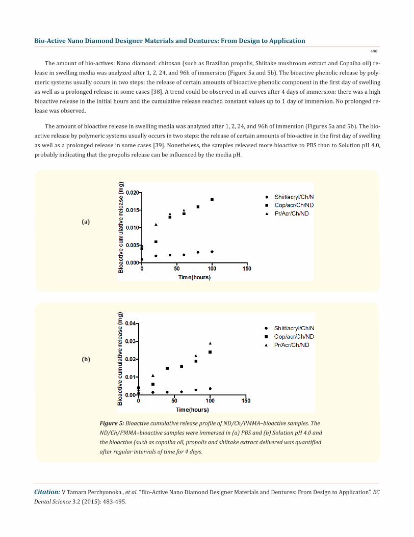
The amount of bio-actives: Nano diamond: chitosan (such as Brazilian propolis, Shiitake mushroom extract and Copaiba oil) re-lease in swelling media was analyzed after 1, 2, 24, and 96h of immersion (Figure 5a and 5b). The bioactive phenolic release by poly-meric systems usually occurs in two steps: the release of certain amounts of bioactive phenolic component in the first day of swelling as well as a prolonged release in some cases [38]. A trend could be observed in all curves after 4 days of immersion: there was a high bioactive release in the initial hours and the cumulative release reached constant values up to 1 day of immersion. No prolonged re-lease was observed.
The amount of bioactive release in swelling media was analyzed after 1, 2, 24, and 96h of immersion (Figures 5a and 5b). The bio-active release by polymeric systems usually occurs in two steps: the release of certain amounts of bio-active in the first day of swelling as well as a prolonged release in some cases [39]. Nonetheless, the samples released more bioactive to PBS than to Solution pH 4.0, probably indicating that the propolis release can be influenced by the media pH.
(a)
(b)
Figure 5: Bioactive cumulative release profile of ND/Ch/PMMA–bioactive samples. The ND/Ch/PMMA–bioactive samples were immersed in (a) PBS and (b) Solution pH 4.0 and the bioactive (such as copaiba oil, propolis and shiitake extract delivered was quantified after regular intervals of time for 4 days.

Bio-Active Nano Diamond Designer Materials and Dentures: From Design to Application491
Citation: V Tamara Perchyonoka., et al. “Bio-Active Nano Diamond Designer Materials and Dentures: From Design to Application”. EC Dental Science 3.2 (2015): 483-495.
The first step for successful colonization of mucosal surfaces or any other surface by C. albicans is adhesion [41]. Although among the functions of Propolis, the fungicidal property has already been shown, in this study, we aimed to verify its effectiveness in inhibiting the adhesion of C. albicans biofilm on a denture surface.
Bio-adhesion of PMMA modified materials
Discussion
Higher adhesiveness of the modified PMMA resins is desired to maintain an intimate contact oral mucosa and the prosthetic device such as full or partial denture, there for bio-adhesion between the newly prepared modified bio-active containing PMMA was tested against pig ear skin structure and results are summarized in Table 1. Chitosan hydrogels and chitosan/Nano diamond combinations showed the highest adhesive force and the work of adhesion this can be expected because of the well known intrinsic bio adhesive properties of chitosan as well as favorable functional surface of Nano-diamond: chitosan and its favorable interaction with the bio-active components [40]. The adequate water absorption capacity together with the cationic nature which promotes binding to the negative surface of skin structure can also interpret this results. The additional benefits of the incorporation of the Nano-diamonds as a highly functionalized surface which is able to interact with the surface of the skin are currently under investigation in our laboratory.
Bio-materials Adhesive Force(N) ± SD (skin) Work of Adhesion (Ncm) ± SD (Skin)Shiitake/PMMA/ND 1.16 ± 0.320 4.35 ± 0.48Shiit/PMMA/Chi/ND 1.23 ± 0.32 4.83 ± 0.42Cop/PMMA/ND 1.26 ± 0.29 4.28 ± 0.31Cop/PMMA/Ch/ND 1.35 ± 0.40 4.35 ± 0.65Prop/PMMA/ND 1.52 ± 0.28 4.87 ± 0.42Pr/PMMA/Ch/ND 1.24 ± 0.22 4.81 ± 0.39PMMA 1.10 ± 0.34 4.41 ± 0.36Ch/PMMA 0.97 ± 0.30 4.97 ± 0.42
Table 1: Bio-adhesion table.
(6a)

Bio-Active Nano Diamond Designer Materials and Dentures: From Design to Application492
Citation: V Tamara Perchyonoka., et al. “Bio-Active Nano Diamond Designer Materials and Dentures: From Design to Application”. EC Dental Science 3.2 (2015): 483-495.
(6b)
(6c)
(6d)
Bio-Active Nano Diamond Designer Materials and Dentures: From Design to Application493
Citation: V Tamara Perchyonoka., et al. “Bio-Active Nano Diamond Designer Materials and Dentures: From Design to Application”. EC Dental Science 3.2 (2015): 483-495.
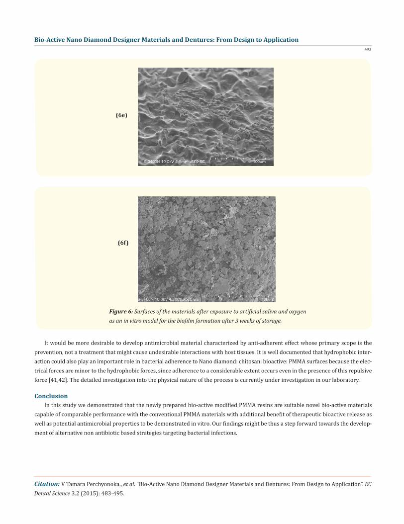
It would be more desirable to develop antimicrobial material characterized by anti-adherent effect whose primary scope is the prevention, not a treatment that might cause undesirable interactions with host tissues. It is well documented that hydrophobic inter-action could also play an important role in bacterial adherence to Nano diamond: chitosan: bioactive: PMMA surfaces because the elec-trical forces are minor to the hydrophobic forces, since adherence to a considerable extent occurs even in the presence of this repulsive force [41,42]. The detailed investigation into the physical nature of the process is currently under investigation in our laboratory.
In this study we demonstrated that the newly prepared bio-active modified PMMA resins are suitable novel bio-active materials capable of comparable performance with the conventional PMMA materials with additional benefit of therapeutic bioactive release as well as potential antimicrobial properties to be demonstrated in vitro. Our findings might be thus a step forward towards the develop-ment of alternative non antibiotic based strategies targeting bacterial infections.
(6e)
(6f)
Figure 6: Surfaces of the materials after exposure to artificial saliva and oxygen as an in vitro model for the biofilm formation after 3 weeks of storage.
Conclusion

Bio-Active Nano Diamond Designer Materials and Dentures: From Design to Application494
Citation: V Tamara Perchyonoka., et al. “Bio-Active Nano Diamond Designer Materials and Dentures: From Design to Application”. EC Dental Science 3.2 (2015): 483-495.
Bibliography
1. Blignaut E., et al. “Ca3 finger printing of Candida albicans isolates from human immuno-deficiency virus-positive and healthy individuals reveals a new clade in South Africa”. Journal of Clinical Microbiology 40 (2002): 826-836.2. Chen MH., et al. “Structure of Saccharomyces cerevisiae alpha-agglutinin. Evidence for a yeast cell wall protein with multiple immunoglobulin-like domains with atypical disulfides”. The Journal of Biological Chemistry 270 (1995): 26168-26177.3. De Bernardis F., et al. “Aspartyl proteinases of Candida albicans and their role in pathogenicity”. Med Mycol 39.4 (2001): 303-313.4. Fu Y., et al. “Candida albicans Als1p: an adhesin that is a downstream effector of the EFG1 filamentation pathway”. Molecular Microbiology 44.1 (2002): 61-72.5. Gaur N K., et al. “Overexpression of the Candida albicans ALA1 gene in Saccharomyces cerevisiae results in aggregation following attachment of yeast cells to extracellular matrix proteins, adherence properties similar to those of Candida albicans”. Infection and Immunity 67 (1999): 6040-6047.6. Barras., et al. “Glycan-functionalized diamond nanoparticles as potent E. coli anti-adhesives”. Nanoscale 5.6 (2013): 2307.7. PC Ray., et al. “Nanomaterials for targeted detection and photothermal killing of bacteriaChem”. Chemical Society Reviews 41.8 (2012): 3193.8. Herman and AP Herman. “Nanoparticles as antimicrobial agents: their toxicity and mechanisms of action “. Journal of Nanosci- ence and Nanotechnology 14.1 (2014): 946-957.9. RP Allaker and K Memarzadeh. “Nanoparticles and the control of oral infections”. International Journal of Antimicrobial Agents 43.2 (2014): 95-104.10. L Marcon., et al. “Cellular and in vivo toxicity of functionalized nanodiamond in Xenopus embryos”. Journal of Materials Chemistry 20.37 (2010): 8064-8069.11. Perchyonok., et al. “Bioinspired-Interpenetrating Network (IPNs) Hydrogel (BIOF-INPs) and TMD in Vitro: Bioadhesion, Drug Release and Build in Free Radical Detection and Defense”. Open Journal of Stomatology 5.3 (2015): 53-6.12. Lindequist U., et al. “The pharmacological potential of mushrooms”. eCAM 2.3 (2005): 285-299.13. Chu KT., et al. “Pleurostrin, an antifungal peptide from the oyster mushroom”. Peptides 26 (2005): 2098-2103.14. Zumla A and Lulat. “A Honey - a remedy rediscovered”. Journal of the Royal Society of Medicine 82 (1989): 384-385.15. Havsteen B. “Flavenoids, a class of natural products of high pharmacological potency”. Biochemical Pharmacology 32.7 (1983): 1141-1148. 16. Lepekhin VN and Leonova TA. “Some data concerning the study of antimicrobial properties of propolis”. Stomatologzja (Moscow) 49 (1970): 16-19.17. Gronstol H and Aspoy E. “A new selective medium for the isolation of Listeria monocytogenes”. Nordisk Veterinaer medicin 29.10 (1977): 446-451.18. Cizmarik J and Trupl J. “Propolis-Wirkung auf Hautpilze”. Pharmnazie 31 (1976): 55.19. Vasconcelos KRF., et al. “In vitro assessment of antibacterial activity of a dental cement constituted of a Copaifera multijuga Hayne oil-resin”. Brazil Journal of Pharmacognosy 18 (2008): 733-738.20. Santos AO., et al. “Antimicrobial activity of Brazilian copaiba oils obtained from different species of the Copaifera genus”. Memórias do Instituto Oswaldo Cruz 103.3 (2008): 277-281.21. Ferrari M., et al. “Terpenoids from Copaiferalangsdorffi”. Phytochemistry 10 (1971): 905-907.22. Basile AC., et al. “Antiinflamatory activity of oleoresin from Brazilian Copaifera”. Journal of Ethnopharmacology 22.1 (1988): 101-109.23. Rocha A and Morais A. “Polyphenoloxidase activity and total phenolic content as related to browning of minimally processed ‘Jonagored’ apple”. Journal of the Science of Food and Agriculture 82.1 (2001): 120-126.24. Waterman PG and S Mole. “Analysis of Phenolic Plant Metabolites”. Blackwell Scientific Publications, Oxford.25. Tugut F., et al. “Strength of the bond between a silicone lining material and denture resin after Er:YAG laser treatments with dif- ferent pulse durations and levels of energy”. Lasers in Medical Science 27.2 (2012): 281-285.
Bio-Active Nano Diamond Designer Materials and Dentures: From Design to Application495
Citation: V Tamara Perchyonoka., et al. “Bio-Active Nano Diamond Designer Materials and Dentures: From Design to Application”. EC Dental Science 3.2 (2015): 483-495.
26. Al-Athel M., et al. “Effect of ageing on the bond strength of a permanent denture soft lining material”. Journal of Oral Rehabilita- tion 29.10 (2002): 992-996.27. Takahashi JM., et al. “Effect of accelerated aging on permanent deformation and tensile bond strength of autopolymerizing soft denture liners”. Journal of Prosthodontics 20.3 (2011): 200-204.28. Senna PM., et al. “Microwave disinfection: cumulative effect of different power levels on physical properties of denture base resins”. Journal of Prosthodontics 20.8 (2011): 606-612.29. Minami H., et al. “Effect of surface treatment on the bonding of an autopolymerizing soft denture liner to a denture base resin”. The International Journal of Prosthodontics 17.3 (2004): 297-301.30. Burdock GA. “Review of the biological properties and toxicity of bee propolis (propolis)”. Food and Chemical Toxicology 36.4 (1998): 347–363.31. Bankova V., et al. “Chemical composition and antibacterial activity of Brazilian propolis”. Zeitschrift für Naturforschung 50.3-4 (1995): 167-172.32. Zhang Z., et al. “Effects of Different Drying Methods and Extraction Condition on Antioxidant Properties of Shiitake (Lentinuse- dodes)”. Food Science and Technology Research 15.5 (2009): 547-552. 33. Veiga Junior., et al. “Phytochemical and antioedematogenic studies of commercial copaiba oils available in Brazil”. Phytotherapy Research 15.6 (2001): 476-480.34. Maeda T., et al. “Durability of peel bond of resilient denture liners to acrylic denture base resin”. Journal of Prosthodontic Re- search 56.2 (2012): 136-141.35. Gonçalves LM., et al. “Effects of undecylenic acid released from denture liner on Candida biofilms”. Journal of Dental Research 91.10 (2012): 985-989.36. Uludamar A., et al. “Clinical and microbiological efficacy of three different treatment methods in the management of denture stomatitis”. Gerodontology 28.2 (2011):104-110.37. Pinto JR., et al. “Effect of thermocycling on bond strength and elasticity of 4 long-term soft denture liners”. Journal of Prosthetic Dentistry 88.5 (2002): 516-521. 38. Bulad K., et al. “Colonization and penetration of denture soft lining materials by Candida albicans”. Dental Materials 20.2 (2004): 167-175.39. Rodger G., et al. “In vitro colonization of an experimental silicone by Candida albicans”. Journal of Biomedical Materials Research 92.1 (2010): 226-235.40. Garcia RM., et al. “Effect of a denture cleanser on weight, surface roughness, and tensile bond strength of two resilient denture liners”. Journal of Prosthetic Dentistry 89.5 (2003): 489-494.41. Fletcher M and Loeb GI. “Influence of substratum characteristics on the attachment of a marine pseudomonad to solid surfaces”. Applied and Environmental Microbiology 37.1 (1979): 67-72. 42. Liu J and Hurt RH. “Ion release kinetics and particle persistence in aqueous nano-silver colloids”. Environmental Science & Tech- nology 44.6 (2010): 2169-2175.
Volume 3 Issue 2 November 2015© All rights are reserved by V Tamara Perchyonoka., et al.