cronicon · cronicon open access ec anaesthesia case report anaesthetic management of neonate with...
TRANSCRIPT
CroniconO P E N A C C E S S EC ANAESTHESIA
Case Report
Anaesthetic Management of Neonate with Omphalocele and Congenital Heart Disease: A Case Report
Anil Kumar Bhiwal1*, Atul Kumar Mishra2, Karishma Johari3, Kunal Chauhan3 and Sunanda Gupta4
1Assistant Professor, Department of Anaesthesiology, Geetanjali Medical College and Hospital, Udaipur, Rajasthan, India2Assistant Professor, Department of Paediatric Surgery, Geetanjali Medical College and Hospital, Udaipur, Rajasthan, India3Resident, Department of Anaesthesiology, Geetanjali Medical College and Hospital, Udaipur, Rajasthan, India4Professor, Department of Anaesthesiology, Geetanjali Medical College and Hospital, Udaipur, Rajasthan, India
Citation: Anil Kumar Bhiwal., et al. “Anaesthetic Management of Neonate with Omphalocele and Congenital Heart Disease: A Case Report”. EC Anaesthesia 4.8 (2018): 283-287.
*Corresponding Author: Anil Kumar Bhiwal, Assistant Professor, Department of Anaesthesiology, Geetanjali Medical College and Hospi-tal, Udaipur, Rajasthan, India.
Received: May 17, 2018; Published: July 11, 2018
AbstractThe common congenital malformations of the anterior abdominal wall are gastroschisis and omphalocele. The herniation of ab-
dominal contents through umbilical cord is omphalocele which are covered by sac and associated with congenital anomalies. We describe here perioperative management of a neonate presented with a giant omphalocele along with small Atrial Septal Defect with left to right shunt, multiple mid muscular Ventricular Septal Defect and small Pulmonary Ductus Arteriosus with Left to Right shunt, posted for its repair on 4th day of life.
Keywords: Omphalocele; Congenital Heart Disease; Anesthetic Management; Neonatal Intensive Care Unit
Introduction
The common congenital malformations of anterior abdominal wall are gastroschisis and omphalocele. The incidence of omphalocele ranges between 1.5 and 3 per 10,000 births and are usually associated in elderly mothers where the maternal age is often > 40 years [1]. The herniation of abdominal contents through the umbilical cord is omphalocele and is differentiated from gastroschisis by presence of the covering sac. Omphalocele is two type: omphalocele minor in which the wall defect is 5 - 8 cms and omphalocele major in which defect is large that included liver, bowel and abdominal and thoracic cavities are poorly developed. Neonates with omphalocele have high incidence of congenital anomalies including cardiac malformations (15 - 54%), chromosomal abnormalities (trisomy 13, 14, 15, 18, 21), genitourinary abnormalities and Beckwith Wiedemann syndrome (10%) [2].
We describe here perioperative management of a neonate presenting with a giant omphalocele associated with small Atrial Septal Defect with left to right shunt, multiple mid muscular Ventricular Septal Defect and small Pulmonary Ductus Arteriosus with Left to Right shunt posted for its repair on 4th day of life.
Case Report
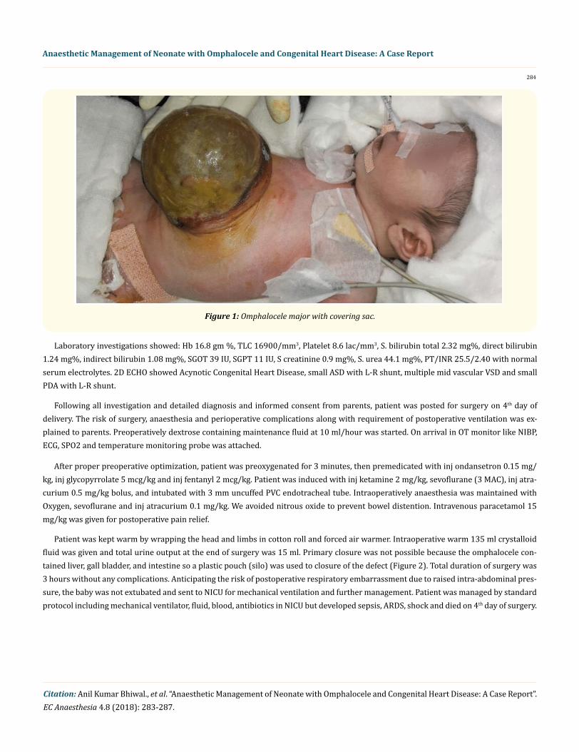
A 2 days old male baby, vaginally delivered to term, weighing 3 kg was presented to pediatric surgery OPD with a single huge mass over the umbilical region (Figure 1). A diagnosis of omphalocele major was made following clinical and radiological investigation and the patient was posted for repair of the defect on 4th day of delivery.
284
Anaesthetic Management of Neonate with Omphalocele and Congenital Heart Disease: A Case Report
Citation: Anil Kumar Bhiwal., et al. “Anaesthetic Management of Neonate with Omphalocele and Congenital Heart Disease: A Case Report”. EC Anaesthesia 4.8 (2018): 283-287.
After proper preoperative optimization, patient was preoxygenated for 3 minutes, then premedicated with inj ondansetron 0.15 mg/kg, inj glycopyrrolate 5 mcg/kg and inj fentanyl 2 mcg/kg. Patient was induced with inj ketamine 2 mg/kg, sevoflurane (3 MAC), inj atra-curium 0.5 mg/kg bolus, and intubated with 3 mm uncuffed PVC endotracheal tube. Intraoperatively anaesthesia was maintained with Oxygen, sevoflurane and inj atracurium 0.1 mg/kg. We avoided nitrous oxide to prevent bowel distention. Intravenous paracetamol 15 mg/kg was given for postoperative pain relief.
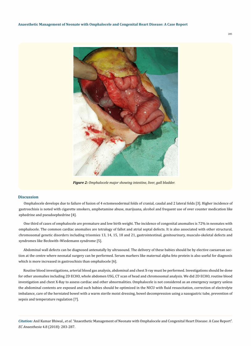
Patient was kept warm by wrapping the head and limbs in cotton roll and forced air warmer. Intraoperative warm 135 ml crystalloid fluid was given and total urine output at the end of surgery was 15 ml. Primary closure was not possible because the omphalocele con-tained liver, gall bladder, and intestine so a plastic pouch (silo) was used to closure of the defect (Figure 2). Total duration of surgery was 3 hours without any complications. Anticipating the risk of postoperative respiratory embarrassment due to raised intra-abdominal pres-sure, the baby was not extubated and sent to NICU for mechanical ventilation and further management. Patient was managed by standard protocol including mechanical ventilator, fluid, blood, antibiotics in NICU but developed sepsis, ARDS, shock and died on 4th day of surgery.
Figure 1: Omphalocele major with covering sac.
Laboratory investigations showed: Hb 16.8 gm %, TLC 16900/mm3, Platelet 8.6 lac/mm3, S. bilirubin total 2.32 mg%, direct bilirubin 1.24 mg%, indirect bilirubin 1.08 mg%, SGOT 39 IU, SGPT 11 IU, S creatinine 0.9 mg%, S. urea 44.1 mg%, PT/INR 25.5/2.40 with normal serum electrolytes. 2D ECHO showed Acynotic Congenital Heart Disease, small ASD with L-R shunt, multiple mid vascular VSD and small PDA with L-R shunt.
Following all investigation and detailed diagnosis and informed consent from parents, patient was posted for surgery on 4th day of delivery. The risk of surgery, anaesthesia and perioperative complications along with requirement of postoperative ventilation was ex-plained to parents. Preoperatively dextrose containing maintenance fluid at 10 ml/hour was started. On arrival in OT monitor like NIBP, ECG, SPO2 and temperature monitoring probe was attached.
Citation: Anil Kumar Bhiwal., et al. “Anaesthetic Management of Neonate with Omphalocele and Congenital Heart Disease: A Case Report”. EC Anaesthesia 4.8 (2018): 283-287.
Anaesthetic Management of Neonate with Omphalocele and Congenital Heart Disease: A Case Report
285
Abdominal wall defects can be diagnosed antenatally by ultrasound. The delivery of these babies should be by elective caesarean sec-tion at the centre where neonatal surgery can be performed. Serum markers like maternal alpha feto protein is also useful for diagnosis which is more increased in gastroschisis than omphalocele [6].
Discussion
Routine blood investigations, arterial blood gas analysis, abdominal and chest X-ray must be performed. Investigations should be done for other anomalies including 2D ECHO, whole abdomen USG, CT scan of head and chromosomal analysis. We did 2D ECHO, routine blood investigation and chest X-Ray to assess cardiac and other abnormalities. Omphalocele is not considered as an emergency surgery unless the abdominal contents are exposed and such babies should be optimized in the NICU with fluid resuscitation, correction of electrolyte imbalance, care of the herniated bowel with a warm sterile moist dressing, bowel decompression using a nasogastric tube, prevention of sepsis and temperature regulation [7].
Figure 2: Omphalocele major showing intestine, liver, gall bladder.
Omphalocele develops due to failure of fusion of 4 ectomesodermal folds of cranial, caudal and 2 lateral folds [3]. Higher incidence of gastroschisis is noted with cigarette smokers, amphetamine abuse, marijuana, alcohol and frequent use of over counter medication like ephedrine and pseudoephedrine [4].
One third of cases of omphalocele are premature and low birth weight. The incidence of congenital anomalies is 72% in neonates with omphalocele. The common cardiac anomalies are tetralogy of fallot and atrial septal defects. It is also associated with other structural, chromosomal genetic disorders including trisomies 13, 14, 15, 18 and 21, gastrointestinal, genitourinary, musculo-skeletal defects and syndromes like Beckwith–Wiedemann syndrome [5].
Citation: Anil Kumar Bhiwal., et al. “Anaesthetic Management of Neonate with Omphalocele and Congenital Heart Disease: A Case Report”. EC Anaesthesia 4.8 (2018): 283-287.
Anaesthetic Management of Neonate with Omphalocele and Congenital Heart Disease: A Case Report
286
Dextrose containing fluids should be given as maintenance fluid to prevent hypoglycemia, antibiotics should be given preoperatively to prevent intra-abdominal sepsis and intravenous access should be avoided in lower limb because venous return may be obstructed after closure of sac. We used dextrose containing fluid, antibiotics (linezolid, ceftriaxone, metronidazole) intravenously by upper limb IV access.
Hypothermia should be prevented during transfer and in the operating theater. Neonate should be transfer in a warm incubator, OT temperature should be 25 - 26°C, forced air warmer and warming mattress should be used in OT, head should be covered by cotton roll to prevent loss of heat, bowel should be with swabs and warmed fluids should be used intraoperatively [8].
The treatment of omphalocele minor is done in single stage with reduction of intestine and closure of sac but staged surgery is required in omphalocele major and gastroschisis. When intragastric or intravesical pressures increases > 20 mm Hg or the inspiratory pressures increases > 30 mm Hg then staged surgery should be done to prevent abdominal compartment syndrome by application of silo to cover the abdominal contents and definitive defect should be closed after 4 - 7 days [9].
We avoided nitrous oxide to prevent bowel distention which can cause increase intra-abdominal pressure during closure. Oxygenation and ventilation may be compromised due to elevation of diaphragm and increased intra-abdominal pressure so peak airway pressure should be monitored. So in this case surgeon planned to close the defect by staged surgery using silo to avoid such complications because the defect is major which contained liver, gall bladder and intestine and patient had congenital heart disease. We ventilate the patient manually by Jackson and Rees circuit because minimal limit of tidal volume of our anaesthesia ventilator is 50 ml and our patient has only 3 kg weight.
Invasive blood pressure monitoring should be done in patients with cardiac anomalies like PDA because they are prone to coronary ischemia due to pulmonary runoff and vasopressor should be used to prevent diastolic hypotension and to maintain systemic vascular resistance [10].
After surgery elective postoperative ventilation is required because of compromised respiratory function secondary to raised intra-ab-dominal pressure and peak airway pressure along with signs of increased abdominal pressure should be monitored. Prevention of infec-tion, hypothermia, hypoglycemia and fluid-electrolyte imbalance is major part of postoperative management. This patient was managed postoperatively by all these standard protocol but could not survived and died on 4th day of surgery due to sepsis, ARDS.
Neonates with omphalocele major and neonates with associated chromosomal, structural and cardiac defects have poor prognosis while neonate with gastroschisis and omphalocele minor have a good survival rate [11].
Management of neonates with major omphalocele are challenge to paediatric surgeon as well as to the anaesthesiologist. Fluid re-suscitation, temperature control, electrolyte maintenance, infection control, cardiovascular stability are the key point in managing this case. Postoperative ventilation is mandatory as increased intraabdominal pressure may cause respiratory complications with a negative outcome.
Conclusion
Bibliography
1. Ledbetter DJ. “Gastroschisis and omphalocele”. Surgical Clinics of North America 86.2 (2006): 249-260.
2. Stoll C., et al. “Omphalocele and gastroschisis and associated malformations”. American Journal of Medical Genetics Part A 146A.10 (2008): 1280-1285.
3. Vermeij-keers C., et al. “Embryonic development of ventral body wall and its congenital malformations”. Seminars in Pediatric Surgery 5.2 (1996): 82-89.
Citation: Anil Kumar Bhiwal., et al. “Anaesthetic Management of Neonate with Omphalocele and Congenital Heart Disease: A Case Report”. EC Anaesthesia 4.8 (2018): 283-287.
Anaesthetic Management of Neonate with Omphalocele and Congenital Heart Disease: A Case Report
287
4. Hoyme HE., et al. “Gastroschisis: Abdominal wall disruption secondary to early gestational interruption of the omphalomesenteric artery”. Seminars in Perinatology 7.4 (1983): 294-298.
5. Brett CM., et al. “Anaesthesia for neonates and premature infants”. In: Moyotyama EK, Davis PJ, editors. Smith’s Anesthesia for Infants and Children. 7th edition. Philadelphia: Mosby (2006): 521-570.
6. Martin RW. “Screening for fetal abdominal wall defects”. Obstetrics and Gynecology Clinics of North America 25.3 (1998): 517-526.
7. Pai VK., et al. “Neonate with Omphalocele and Dextrocardia: Anaesthetic Goals and Challenges”. Journal of Medical Sciences 36.2 (2016): 81-83.
8. McNair C., et al. “Caring for the newborn with an omphalocele”. Neonatal Network 25 (2006): 319-327.
9. Schwartz MZ, et al. “Staged reduction using a silastic sac is the treatment of choice for large congenital abdominal wall defects”. Jour-nal of Pediatric Surgery 18.6 (1983): 713-719.
10. Yaster M., et al. “Hemodynamic effects of primary closure of omphalocele/gastroschisis in human newborns”. Anesthesiology 69.1 (1988): 84-88.
11. Driver CP., et al. “The contemporary outcome of gastroschisis”. Journal of Pediatric Surgery 35.12 (2000): 1719-1723.
Volume 4 Issue 8 August 2018©All rights reserved by Anil Kumar Bhiwal., et al.