critical event checklists
TRANSCRIPT
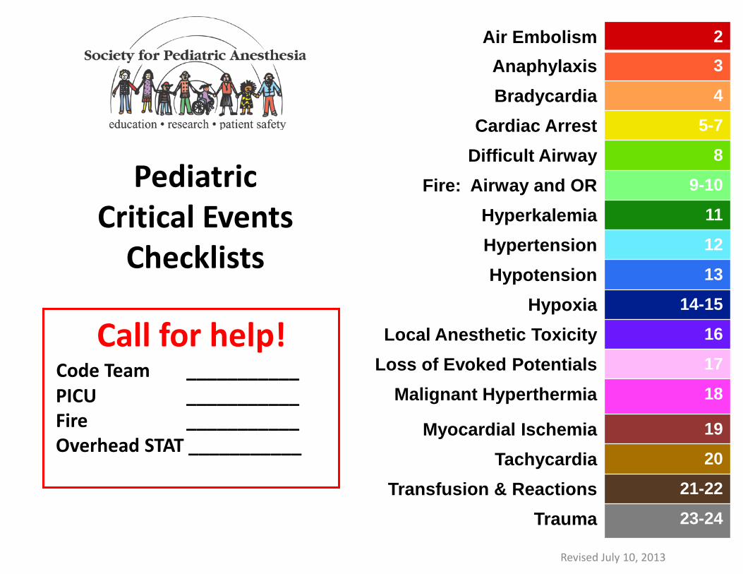
Pediatric Critical Events
Checklists
Air Embolism 2
Anaphylaxis 3
Bradycardia 4
Cardiac Arrest 5-7
Difficult Airway 8
Fire: Airway and OR 9-10
Hyperkalemia 11
Hypertension 12
Hypotension 13
Hypoxia 14-15
Local Anesthetic Toxicity 16
Loss of Evoked Potentials 17
Malignant Hyperthermia 18
Myocardial Ischemia 19
Tachycardia 20
Transfusion & Reactions 21-22
Trauma 23-24
Call for help! Code Team ___________ PICU ___________ Fire ___________ Overhead STAT ___________
Revised July 10, 2013
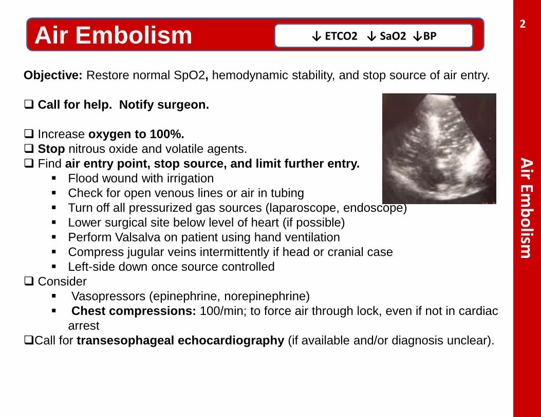
Objective: Restore normal SpO2, hemodynamic stability, and stop source of air entry. Call for help. Notify surgeon. Increase oxygen to 100%. Stop nitrous oxide and volatile agents. Find air entry point, stop source, and limit further entry.
Flood wound with irrigation Check for open venous lines or air in tubing Turn off all pressurized gas sources (laparoscope, endoscope) Lower surgical site below level of heart (if possible) Perform Valsalva on patient using hand ventilation Compress jugular veins intermittently if head or cranial case Left-side down once source controlled
Consider Vasopressors (epinephrine, norepinephrine) Chest compressions: 100/min; to force air through lock, even if not in cardiac
arrest Call for transesophageal echocardiography (if available and/or diagnosis unclear).
Air Embolism Air Em
bolism
↓ ETCO2 ↓ SaO2 ↓BP 2
Anaphylaxis
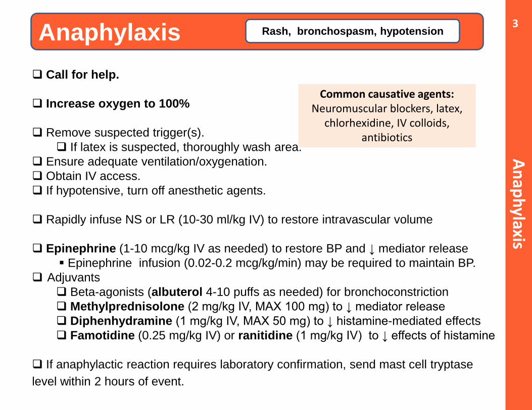
Call for help. Increase oxygen to 100%
Remove suspected trigger(s).
If latex is suspected, thoroughly wash area. Ensure adequate ventilation/oxygenation. Obtain IV access. If hypotensive, turn off anesthetic agents.
Rapidly infuse NS or LR (10-30 ml/kg IV) to restore intravascular volume Epinephrine (1-10 mcg/kg IV as needed) to restore BP and ↓ mediator release
Epinephrine infusion (0.02-0.2 mcg/kg/min) may be required to maintain BP. Adjuvants
Beta-agonists (albuterol 4-10 puffs as needed) for bronchoconstriction Methylprednisolone (2 mg/kg IV, MAX 100 mg) to ↓ mediator release Diphenhydramine (1 mg/kg IV, MAX 50 mg) to ↓ histamine-mediated effects Famotidine (0.25 mg/kg IV) or ranitidine (1 mg/kg IV) to ↓ effects of histamine
If anaphylactic reaction requires laboratory confirmation, send mast cell tryptase level within 2 hours of event.
Anaphylaxis Rash, bronchospasm, hypotension 3
Common causative agents: Neuromuscular blockers, latex,
chlorhexidine, IV colloids, antibiotics
Bradycardia - Unstable
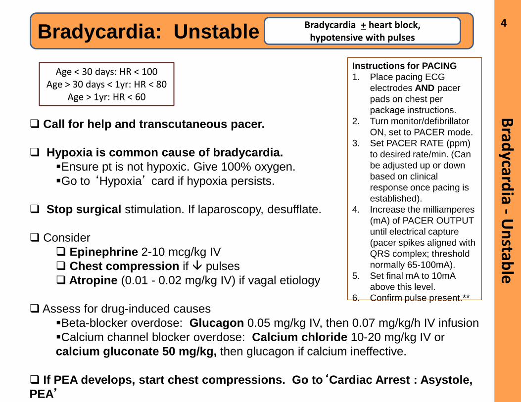
Call for help and transcutaneous pacer. Hypoxia is common cause of bradycardia.
Ensure pt is not hypoxic. Give 100% oxygen. Go to ‘Hypoxia’ card if hypoxia persists.
Stop surgical stimulation. If laparoscopy, desufflate. Consider
Epinephrine 2-10 mcg/kg IV Chest compression if pulses Atropine (0.01 - 0.02 mg/kg IV) if vagal etiology
Assess for drug-induced causes
Beta-blocker overdose: Glucagon 0.05 mg/kg IV, then 0.07 mg/kg/h IV infusion Calcium channel blocker overdose: Calcium chloride 10-20 mg/kg IV or calcium gluconate 50 mg/kg, then glucagon if calcium ineffective.
If PEA develops, start chest compressions. Go to‘Cardiac Arrest : Asystole, PEA’
Bradycardia: Unstable Bradycardia + heart block, hypotensive with pulses
4
Age < 30 days: HR < 100 Age > 30 days < 1yr: HR < 80
Age > 1yr: HR < 60
Instructions for PACING 1. Place pacing ECG
electrodes AND pacer pads on chest per package instructions.
2. Turn monitor/defibrillator ON, set to PACER mode.
3. Set PACER RATE (ppm) to desired rate/min. (Can be adjusted up or down based on clinical response once pacing is established).
4. Increase the milliamperes (mA) of PACER OUTPUT until electrical capture (pacer spikes aligned with QRS complex; threshold normally 65‐100mA).
5. Set final mA to 10mA above this level.
6. Confirm pulse present.**
Cardiac Arrest: Asystole, PEA
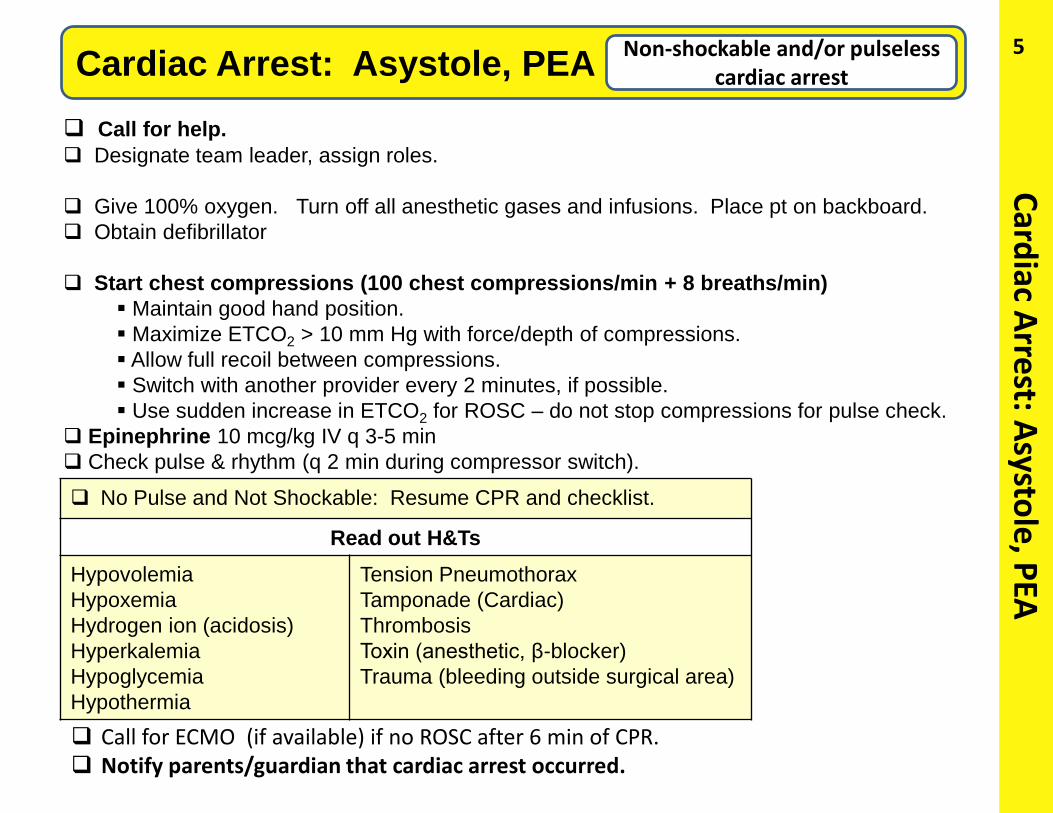
Call for help. Designate team leader, assign roles. Give 100% oxygen. Turn off all anesthetic gases and infusions. Place pt on backboard. Obtain defibrillator
Start chest compressions (100 chest compressions/min + 8 breaths/min)
Maintain good hand position. Maximize ETCO2 > 10 mm Hg with force/depth of compressions. Allow full recoil between compressions. Switch with another provider every 2 minutes, if possible. Use sudden increase in ETCO2 for ROSC – do not stop compressions for pulse check.
Epinephrine 10 mcg/kg IV q 3-5 min Check pulse & rhythm (q 2 min during compressor switch).
Cardiac Arrest: Asystole, PEA Non-shockable and/or pulseless cardiac arrest
No Pulse and Not Shockable: Resume CPR and checklist.
Read out H&Ts Hypovolemia Hypoxemia Hydrogen ion (acidosis) Hyperkalemia Hypoglycemia Hypothermia
Tension Pneumothorax Tamponade (Cardiac) Thrombosis Toxin (anesthetic, β-blocker) Trauma (bleeding outside surgical area)
5
Call for ECMO (if available) if no ROSC after 6 min of CPR. Notify parents/guardian that cardiac arrest occurred.
Cardiac Arrest: VF/VT
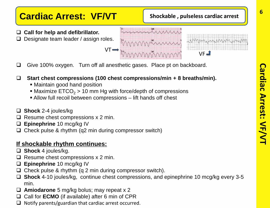
Call for help and defibrillator. Designate team leader / assign roles.
Give 100% oxygen. Turn off all anesthetic gases. Place pt on backboard.
Start chest compressions (100 chest compressions/min + 8 breaths/min).
Maintain good hand position Maximize ETCO2 > 10 mm Hg with force/depth of compressions Allow full recoil between compressions – lift hands off chest
Shock 2-4 joules/kg Resume chest compressions x 2 min. Epinephrine 10 mcg/kg IV Check pulse & rhythm (q2 min during compressor switch)
If shockable rhythm continues: Shock 4 joules/kg. Resume chest compressions x 2 min. Epinephrine 10 mcg/kg IV Check pulse & rhythm (q 2 min during compressor switch). Shock 4-10 joules/kg, continue chest compressions, and epinephrine 10 mcg/kg every 3-5
min. Amiodarone 5 mg/kg bolus; may repeat x 2 Call for ECMO (if available) after 6 min of CPR Notify parents/guardian that cardiac arrest occurred.
Cardiac Arrest: VF/VT Shockable , pulseless cardiac arrest 6
VT VF
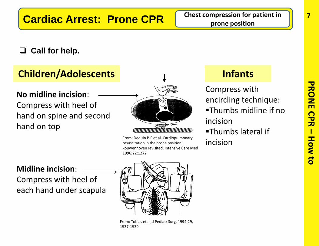
Call for help.
Cardiac Arrest: Prone CPR Chest compression for patient in prone position
No midline incision: Compress with heel of hand on spine and second hand on top
Midline incision: Compress with heel of each hand under scapula
Compress with encircling technique: Thumbs midline if no incision Thumbs lateral if incision
Children/Adolescents Infants PRON
E CPR – How to
7
From: Tobias et al, J Pediatr Surg. 1994:29, 1537-1539
From: Dequin P-F et al. Cardiopulmonary resuscitation in the prone position: kouwenhoven revisited. Intensive Care Med 1996;22:1272
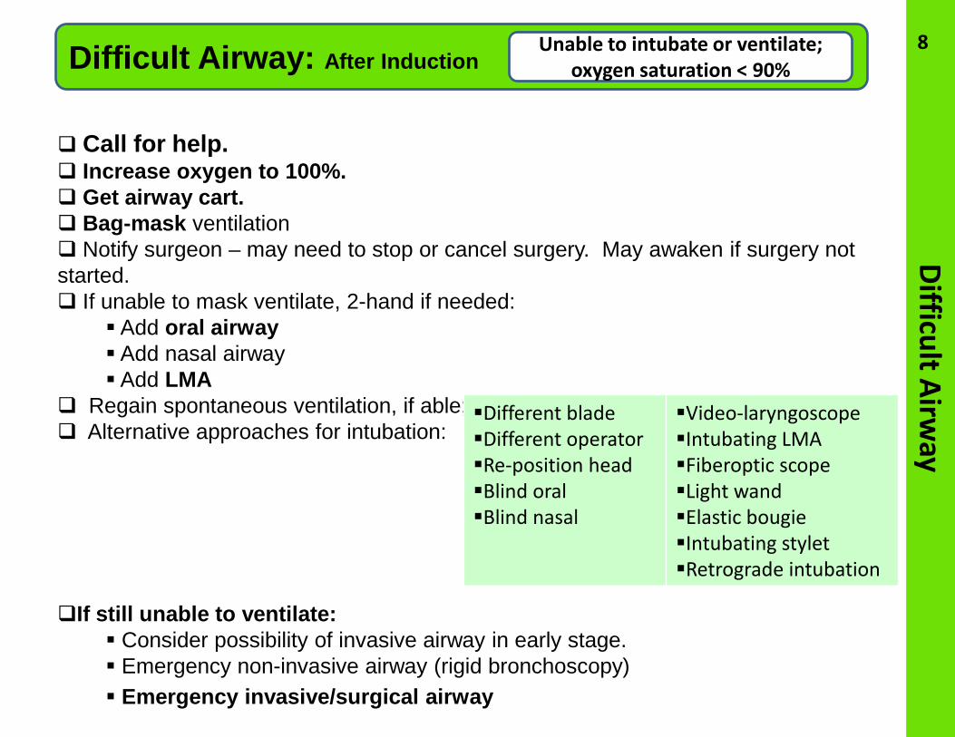
Difficult Airway
Call for help. Increase oxygen to 100%. Get airway cart. Bag-mask ventilation Notify surgeon – may need to stop or cancel surgery. May awaken if surgery not started. If unable to mask ventilate, 2-hand if needed:
Add oral airway Add nasal airway Add LMA
Regain spontaneous ventilation, if able; reverse neuromuscular blocker Alternative approaches for intubation:
If still unable to ventilate:
Consider possibility of invasive airway in early stage. Emergency non-invasive airway (rigid bronchoscopy) Emergency invasive/surgical airway
Difficult Airway: After Induction Unable to intubate or ventilate; oxygen saturation < 90%
Different blade Different operator Re-position head Blind oral Blind nasal
Video-laryngoscope Intubating LMA Fiberoptic scope Light wand Elastic bougie Intubating stylet Retrograde intubation
8
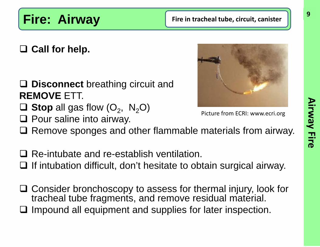
Airway Fire
Fire: Airway Fire in tracheal tube, circuit, canister
Call for help.
Disconnect breathing circuit and REMOVE ETT. Stop all gas flow (O2, N2O) Pour saline into airway. Remove sponges and other flammable materials from airway.
Re-intubate and re-establish ventilation. If intubation difficult, don’t hesitate to obtain surgical airway.
Consider bronchoscopy to assess for thermal injury, look for
tracheal tube fragments, and remove residual material. Impound all equipment and supplies for later inspection.
9
Picture from ECRI: www.ecri.org
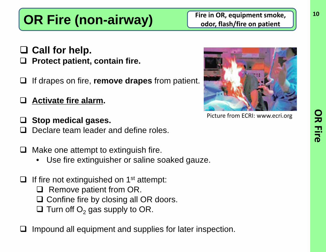
OR Fire
OR Fire (non-airway) Fire in OR, equipment smoke, odor, flash/fire on patient
Call for help. Protect patient, contain fire. If drapes on fire, remove drapes from patient.
Activate fire alarm.
Stop medical gases. Declare team leader and define roles.
Make one attempt to extinguish fire.
• Use fire extinguisher or saline soaked gauze.
If fire not extinguished on 1st attempt: Remove patient from OR. Confine fire by closing all OR doors. Turn off O2 gas supply to OR.
Impound all equipment and supplies for later inspection.
10
Picture from ECRI: www.ecri.org
Hyperkalemia
Hyperkalemia Serum K+ > 6 meq/L
Causes: Excessive intake: massive or “old” blood transfusion, cardioplegia, “K+ runs” Shift of K+ from tissues to plasma: crush injury, burns, succinylcholine, malignant hyperthermia, acidosis Inadequate excretion: renal failure
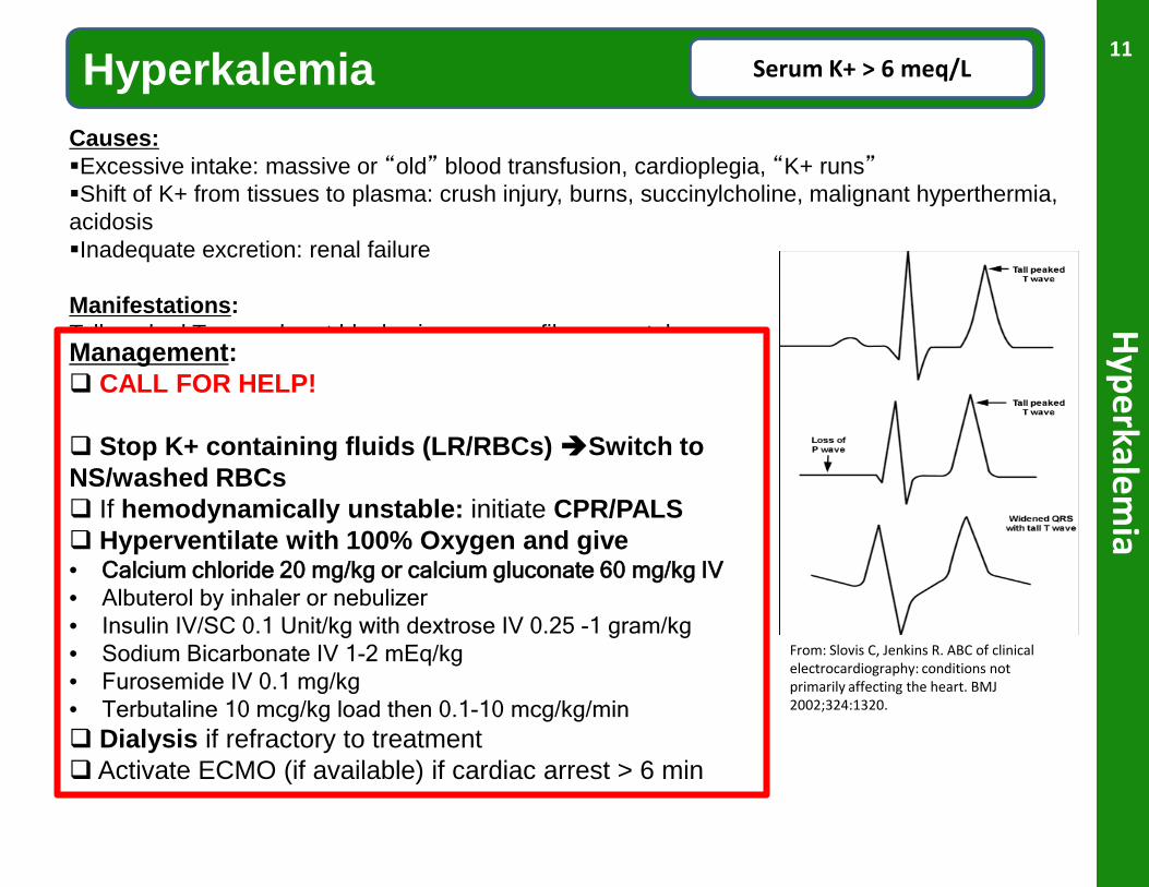
Manifestations: Tall peaked T wave, heart block, sine wave, v fib or asystole
Management: CALL FOR HELP! Stop K+ containing fluids (LR/RBCs) Switch to NS/washed RBCs If hemodynamically unstable: initiate CPR/PALS Hyperventilate with 100% Oxygen and give • Calcium chloride 20 mg/kg or calcium gluconate 60 mg/kg IV • Albuterol by inhaler or nebulizer • Insulin IV/SC 0.1 Unit/kg with dextrose IV 0.25 -1 gram/kg • Sodium Bicarbonate IV 1-2 mEq/kg • Furosemide IV 0.1 mg/kg • Terbutaline 10 mcg/kg load then 0.1-10 mcg/kg/min Dialysis if refractory to treatment Activate ECMO (if available) if cardiac arrest > 6 min
11
From: Slovis C, Jenkins R. ABC of clinical electrocardiography: conditions not primarily affecting the heart. BMJ 2002;324:1320.
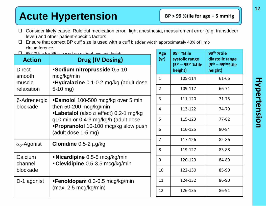
Hypertension Acute Hypertension BP > 99 %tile for age + 5 mmHg
Consider likely cause. Rule out medication error, light anesthesia, measurement error (e.g. transducer level) and other patient-specific factors.
Ensure that correct BP cuff size is used with a cuff bladder width approximately 40% of limb circumference.
99th %tile for BP is based on patient age and height.
12
Action Drug (IV Dosing) Direct smooth muscle relaxation
Sodium nitroprusside 0.5-10 mcg/kg/min Hydralazine 0.1-0.2 mg/kg (adult dose 5-10 mg)
β-Adrenergic blockade
Esmolol 100-500 mcg/kg over 5 min then 50-200 mcg/kg/min Labetalol (also α effect) 0.2-1 mg/kg q10 min or 0.4-3 mg/kg/h (adult dose Propranolol 10-100 mcg/kg slow push (adult dose 1-5 mg)
α2-Agonist Clonidine 0.5-2 µg/kg
Calcium channel blockade
Nicardipine 0.5-5 mcg/kg/min Clevidipine 0.5-3.5 mcg/kg/min
D-1 agonist Fenoldopam 0.3-0.5 mcg/kg/min (max. 2.5 mcg/kg/min)
Age (yr)
99th %tile systolic range (5th – 95th %tile height)
99th %tile diastolic range (5th – 95th%tile height)
1 105-114 61-66
2 109-117 66-71
3 111-120 71-75
4 113-122 74-79
5 115-123 77-82
6 116-125 80-84
7 117-126 82-86
8 119-127 83-88
9 120-129 84-89
10 122-130 85-90
11 124-132 86-90
12 126-135 86-91
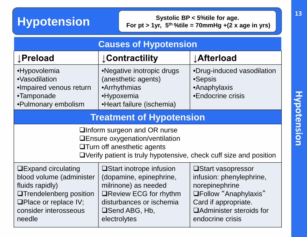
Hypotension Systolic BP < 5%tile for age. For pt > 1yr, 5th %tile = 70mmHg +(2 x age in yrs)
Hypotension
Causes of Hypotension ↓Preload ↓Contractility ↓Afterload •Hypovolemia •Vasodilation •Impaired venous return •Tamponade •Pulmonary embolism
•Negative inotropic drugs (anesthetic agents) •Arrhythmias •Hypoxemia •Heart failure (ischemia)
•Drug-induced vasodilation •Sepsis •Anaphylaxis •Endocrine crisis
Treatment of Hypotension Inform surgeon and OR nurse Ensure oxygenation/ventilation Turn off anesthetic agents Verify patient is truly hypotensive, check cuff size and position
Expand circulating blood volume (administer fluids rapidly) Trendelenberg position Place or replace IV; consider interosseous needle
Start inotrope infusion (dopamine, epinephrine, milrinone) as needed Review ECG for rhythm disturbances or ischemia Send ABG, Hb, electrolytes
Start vasopressor infusion: phenylephrine, norepinephrine Follow “Anaphylaxis” Card if appropriate. Administer steroids for endocrine crisis
13
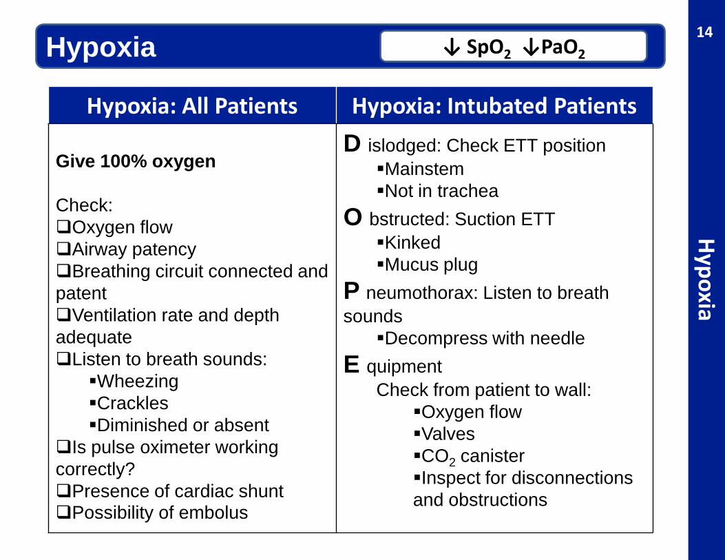
Hypoxia Hypoxia
↓ SpO2 ↓PaO2
Hypoxia: All Patients Hypoxia: Intubated Patients Give 100% oxygen Check: Oxygen flow Airway patency Breathing circuit connected and patent Ventilation rate and depth adequate Listen to breath sounds:
Wheezing Crackles Diminished or absent
Is pulse oximeter working correctly? Presence of cardiac shunt Possibility of embolus
D islodged: Check ETT position Mainstem Not in trachea
O bstructed: Suction ETT Kinked Mucus plug
P neumothorax: Listen to breath sounds
Decompress with needle E quipment
Check from patient to wall: Oxygen flow Valves CO2 canister Inspect for disconnections and obstructions
14
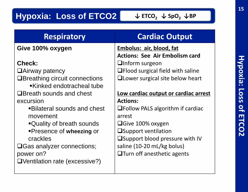
Hypoxia: Loss of ETCO2 Hypoxia: Loss of ETCO
2 ↓ ETCO2 ↓ SpO2 ↓BP
Respiratory Cardiac Output Give 100% oxygen Check: Airway patency Breathing circuit connections
Kinked endotracheal tube Breath sounds and chest excursion
Bilateral sounds and chest movement Quality of breath sounds Presence of wheezing or crackles
Gas analyzer connections; power on? Ventilation rate (excessive?)
Embolus: air, blood, fat Actions: See Air Embolism card Inform surgeon Flood surgical field with saline Lower surgical site below heart
Low cardiac output or cardiac arrest Actions: Follow PALS algorithm if cardiac arrest Give 100% oxygen Support ventilation Support blood pressure with IV saline (10-20 mL/kg bolus) Turn off anesthetic agents
15
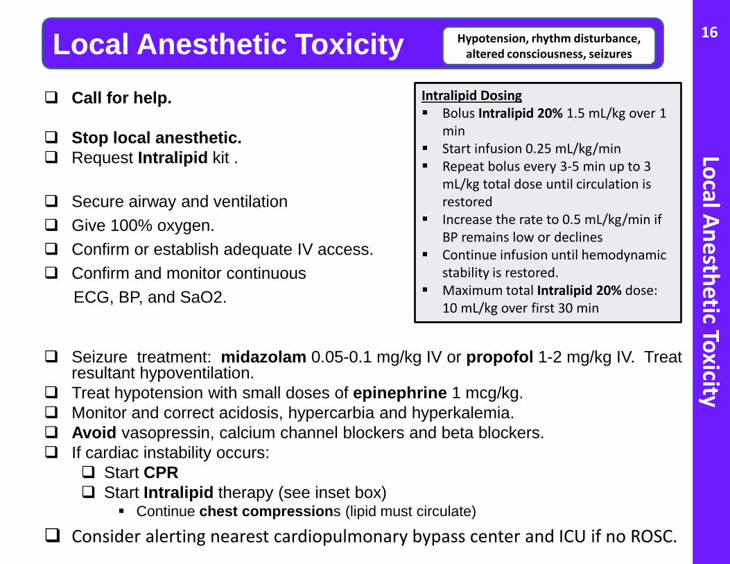
Local Anesthetic Toxicity Local Anesthetic Toxicity Hypotension, rhythm disturbance,
altered consciousness, seizures
Call for help.
Stop local anesthetic. Request Intralipid kit .
Secure airway and ventilation Give 100% oxygen. Confirm or establish adequate IV access. Confirm and monitor continuous
ECG, BP, and SaO2.
Seizure treatment: midazolam 0.05-0.1 mg/kg IV or propofol 1-2 mg/kg IV. Treat
resultant hypoventilation. Treat hypotension with small doses of epinephrine 1 mcg/kg. Monitor and correct acidosis, hypercarbia and hyperkalemia. Avoid vasopressin, calcium channel blockers and beta blockers. If cardiac instability occurs:
Start CPR Start Intralipid therapy (see inset box)
Continue chest compressions (lipid must circulate)
Consider alerting nearest cardiopulmonary bypass center and ICU if no ROSC.
16
Intralipid Dosing Bolus Intralipid 20% 1.5 mL/kg over 1
min Start infusion 0.25 mL/kg/min Repeat bolus every 3-5 min up to 3
mL/kg total dose until circulation is restored
Increase the rate to 0.5 mL/kg/min if BP remains low or declines
Continue infusion until hemodynamic stability is restored.
Maximum total Intralipid 20% dose: 10 mL/kg over first 30 min
Loss of Evoked Potentials
Notify surgeon. Turn off inhalation agent/N2O and switch to propofol/ketamine infusion. Turn off or reverse neuromuscular blockers Increase perfusion pressure (MAP > 70 mmHg) using ephedrine (0.2 – 0.3 mg/kg IV) and/or phenylephrine (1-10 mcg/kg IV). Check Hb; transfuse RBC (10-15 mL/kg IV) if anemic. Ensure normocarbia: ↑ I/E ratio, ↓ PEEP Ensure normothermia. Consider wake-up test. Consider high-dose steroid for spinal cord injury: Methylprednisolone 30 mg/kg IV over 15 min, then 5.4 mg/kg/h IV infusion.
Loss of Evoked Potentials Management of signal changes during spine surgery
17
Malignant Hypertherm
ia Malignant Hyperthermia Temp, HR, CO2, Acidosis
Call for help. Get Malignant Hyperthermia (MH) Kit. Stop procedure if possible Stop volatile anesthetic. Transition to non-triggering anesthetic Request chilled IV saline. Hyperventilate pt to reduce CO2: 2-4 times patient’s minute ventilation Dantrolene 2.5 mg/kg IV every 5 min until symptoms resolve. Assign dedicated person to mix dantrolene (20 mg/vial) with 60 mL sterile water. Bicarbonate 1-2 meq/kg IV for suspected metabolic acidosis; maintain pH > 7.2. Cool patient if temperature > 38.5°C.
NG lavage with cold water. Apply ice externally. Infuse cold saline intravenously. ** Stop cooling if temperature < 38°C.
Hyperkalemia treatment: (See ‘Hyperkalemia’ card) Ca gluconate 30 mg/kg IV or Ca chloride 10 mg/kg IV; Sodium bicarbonate 1-2 mEq/kg IV; Regular insulin 10 Units IV with 1-2 amps D50 (0.1 units insulin/kg and 1 mL/kg D50)
Dysrhythmia treatment: Standard anti-arrhythmics; do NOT use calcium channel blocker Send labs: ABG or VBG, electrolytes, serum CK, serum/urine myoglobin, coagulation Place Foley catheter to monitor urine output. Call ICU to arrange disposition.
MH hotline 1-800-644-9737
18
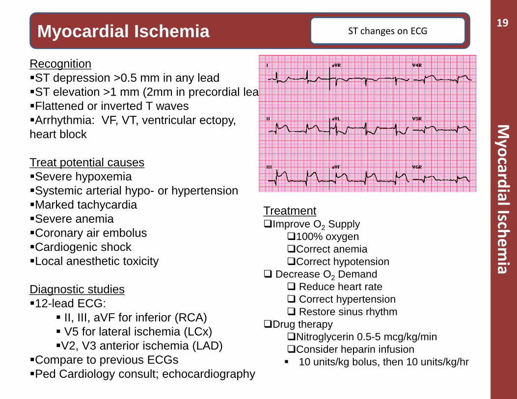
Myocardial Ischem
ia Myocardial Ischemia ST changes on ECG
Recognition ST depression >0.5 mm in any lead ST elevation >1 mm (2mm in precordial leads) Flattened or inverted T waves Arrhythmia: VF, VT, ventricular ectopy, heart block
Treat potential causes Severe hypoxemia Systemic arterial hypo- or hypertension Marked tachycardia Severe anemia Coronary air embolus Cardiogenic shock Local anesthetic toxicity
Diagnostic studies 12-lead ECG:
II, III, aVF for inferior (RCA) V5 for lateral ischemia (LCx) V2, V3 anterior ischemia (LAD)
Compare to previous ECGs Ped Cardiology consult; echocardiography
19
Treatment Improve O2 Supply
100% oxygen Correct anemia Correct hypotension
Decrease O2 Demand Reduce heart rate Correct hypertension Restore sinus rhythm
Drug therapy Nitroglycerin 0.5-5 mcg/kg/min Consider heparin infusion 10 units/kg bolus, then 10 units/kg/hr
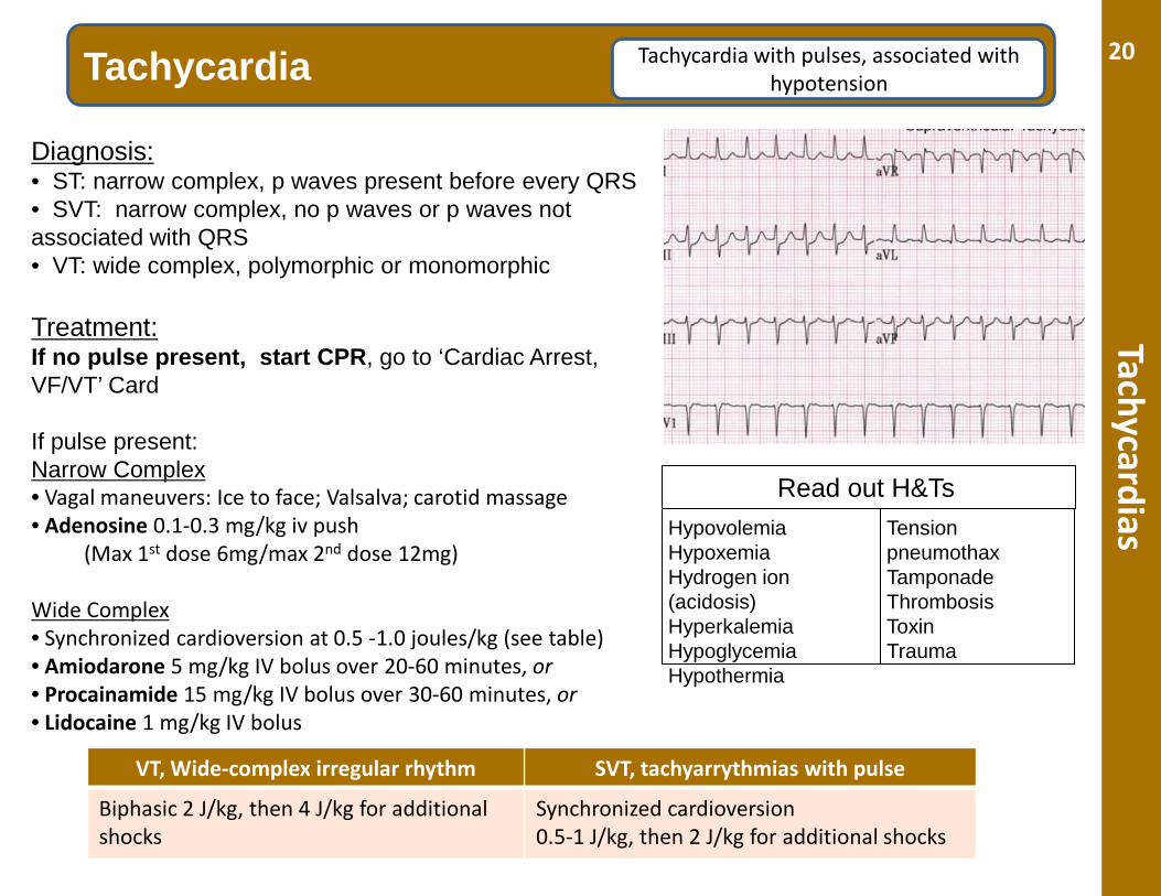
Tachycardias Tachycardia Tachycardia with pulses, associated with
hypotension
Diagnosis: • ST: narrow complex, p waves present before every QRS • SVT: narrow complex, no p waves or p waves not associated with QRS • VT: wide complex, polymorphic or monomorphic
Treatment: If no pulse present, start CPR, go to ‘Cardiac Arrest, VF/VT’ Card If pulse present: Narrow Complex • Vagal maneuvers: Ice to face; Valsalva; carotid massage • Adenosine 0.1-0.3 mg/kg iv push
(Max 1st dose 6mg/max 2nd dose 12mg)
Wide Complex • Synchronized cardioversion at 0.5 -1.0 joules/kg (see table) • Amiodarone 5 mg/kg IV bolus over 20-60 minutes, or • Procainamide 15 mg/kg IV bolus over 30-60 minutes, or • Lidocaine 1 mg/kg IV bolus
VT, Wide-complex irregular rhythm SVT, tachyarrythmias with pulse
Biphasic 2 J/kg, then 4 J/kg for additional shocks
Synchronized cardioversion 0.5-1 J/kg, then 2 J/kg for additional shocks
Read out H&Ts Hypovolemia Hypoxemia Hydrogen ion (acidosis) Hyperkalemia Hypoglycemia Hypothermia
Tension pneumothax Tamponade Thrombosis Toxin Trauma
20
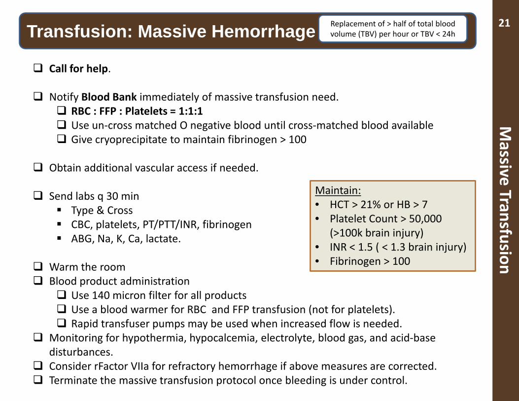
Massive Transfusion
Transfusion: Massive Hemorrhage Replacement of > half of total blood volume (TBV) per hour or TBV < 24h
Call for help.
Notify Blood Bank immediately of massive transfusion need. RBC : FFP : Platelets = 1:1:1 Use un-cross matched O negative blood until cross-matched blood available Give cryoprecipitate to maintain fibrinogen > 100
Obtain additional vascular access if needed. Send labs q 30 min
Type & Cross CBC, platelets, PT/PTT/INR, fibrinogen ABG, Na, K, Ca, lactate.
Warm the room Blood product administration
Use 140 micron filter for all products Use a blood warmer for RBC and FFP transfusion (not for platelets). Rapid transfuser pumps may be used when increased flow is needed.
Monitoring for hypothermia, hypocalcemia, electrolyte, blood gas, and acid-base disturbances.
Consider rFactor VIIa for refractory hemorrhage if above measures are corrected. Terminate the massive transfusion protocol once bleeding is under control.
21
Maintain: • HCT > 21% or HB > 7 • Platelet Count > 50,000
(>100k brain injury) • INR < 1.5 ( < 1.3 brain injury) • Fibrinogen > 100
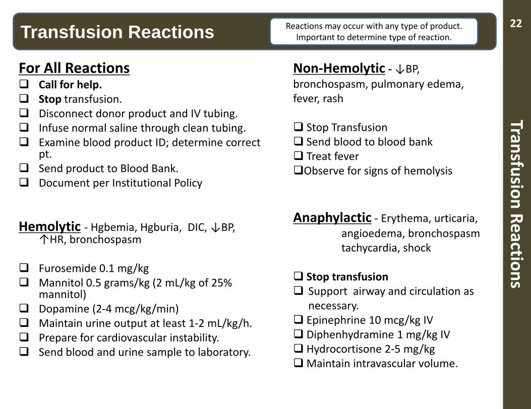
Transfusion Reactions Transfusion Reactions Reactions may occur with any type of product.
Important to determine type of reaction.
For All Reactions Call for help. Stop transfusion. Disconnect donor product and IV tubing. Infuse normal saline through clean tubing. Examine blood product ID; determine correct
pt. Send product to Blood Bank. Document per Institutional Policy Hemolytic - Hgbemia, Hgburia, DIC, ↓BP,
↑HR, bronchospasm Furosemide 0.1 mg/kg Mannitol 0.5 grams/kg (2 mL/kg of 25%
mannitol) Dopamine (2-4 mcg/kg/min) Maintain urine output at least 1-2 mL/kg/h. Prepare for cardiovascular instability. Send blood and urine sample to laboratory.
Non-Hemolytic - ↓BP, bronchospasm, pulmonary edema, fever, rash Stop Transfusion Send blood to blood bank Treat fever Observe for signs of hemolysis
Anaphylactic - Erythema, urticaria, angioedema, bronchospasm tachycardia, shock Stop transfusion Support airway and circulation as
necessary. Epinephrine 10 mcg/kg IV Diphenhydramine 1 mg/kg IV Hydrocortisone 2-5 mg/kg Maintain intravascular volume.
22
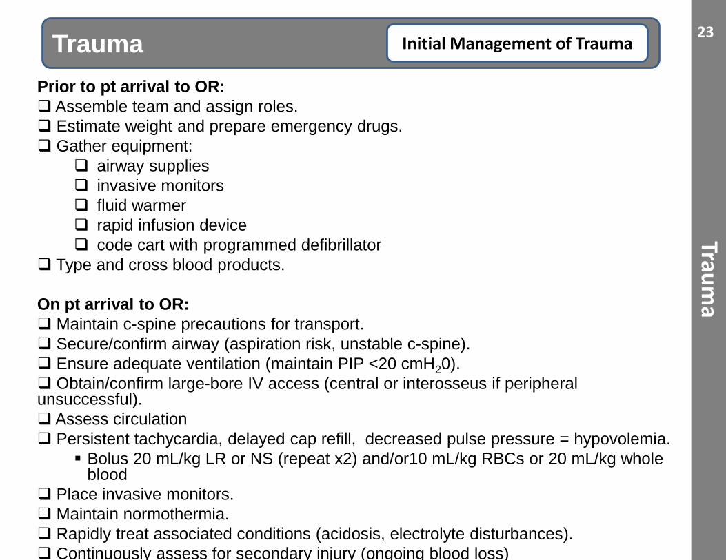
ith Prior to pt arrival to OR: Assemble team and assign roles. Estimate weight and prepare emergency drugs. Gather equipment:
airway supplies invasive monitors fluid warmer rapid infusion device code cart with programmed defibrillator
Type and cross blood products. On pt arrival to OR: Maintain c-spine precautions for transport. Secure/confirm airway (aspiration risk, unstable c-spine). Ensure adequate ventilation (maintain PIP <20 cmH20). Obtain/confirm large-bore IV access (central or interosseus if peripheral unsuccessful). Assess circulation Persistent tachycardia, delayed cap refill, decreased pulse pressure = hypovolemia.
Bolus 20 mL/kg LR or NS (repeat x2) and/or10 mL/kg RBCs or 20 mL/kg whole blood
Place invasive monitors. Maintain normothermia. Rapidly treat associated conditions (acidosis, electrolyte disturbances). Continuously assess for secondary injury (ongoing blood loss)
Trauma
Trauma Initial Management of Trauma 23
Trauma
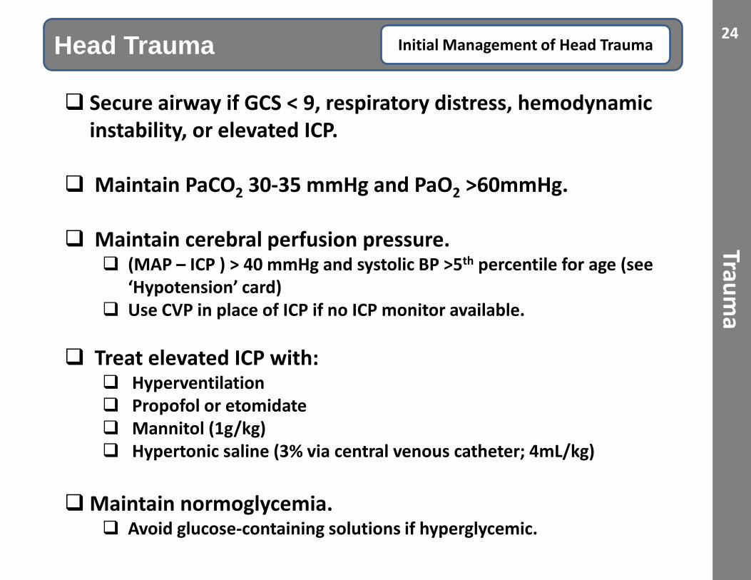
Head Trauma Initial Management of Head Trauma
Secure airway if GCS < 9, respiratory distress, hemodynamic instability, or elevated ICP.
Maintain PaCO2 30-35 mmHg and PaO2 >60mmHg. Maintain cerebral perfusion pressure.
(MAP – ICP ) > 40 mmHg and systolic BP >5th percentile for age (see ‘Hypotension’ card)
Use CVP in place of ICP if no ICP monitor available. Treat elevated ICP with:
Hyperventilation Propofol or etomidate Mannitol (1g/kg) Hypertonic saline (3% via central venous catheter; 4mL/kg)
Maintain normoglycemia. Avoid glucose-containing solutions if hyperglycemic.
24