crispr/cas9-mediated genome editing in a reef-building coral · emergence of the crispr/cas9...
TRANSCRIPT
CRISPR/Cas9-mediated genome editing in areef-building coralPhillip A. Clevesa,1, Marie E. Straderb,1, Line K. Bayc, John R. Pringlea,2, and Mikhail V. Matzb,2
aDepartment of Genetics, Stanford University School of Medicine, Stanford, CA 94305; bDepartment of Integrative Biology, The University of Texas atAustin, Austin, TX 78712; and cAustralian Institute of Marine Science, Townsville, QLD 4810, Australia
Contributed by John R. Pringle, March 26, 2018 (sent for review December 20, 2017; reviewed by Denis Allemand and Ann M. Tarrant)
Reef-building corals are critically important species that are threat-ened by anthropogenic stresses including climate change. In attemptsto understand corals’ responses to stress and other aspects of theirbiology, numerous genomic and transcriptomic studies have beenperformed, generating a variety of hypotheses about the roles ofparticular genes and molecular pathways. However, it has not gener-ally been possible to test these hypotheses rigorously because of thelack of genetic tools for corals. Here, we demonstrate efficient ge-nome editing using the CRISPR/Cas9 system in the coral Acroporamillepora. We targeted the genes encoding fibroblast growth factor1a (FGF1a), green fluorescent protein (GFP), and red fluorescent pro-tein (RFP). After microinjecting CRISPR/Cas9 ribonucleoprotein com-plexes into fertilized eggs, we detected induced mutations in thetargeted genes using changes in restriction-fragment length, Sangersequencing, and high-throughput Illumina sequencing. We observedmutations in ∼50% of individuals screened, and the proportions ofwild-type and various mutant gene copies in these individuals indi-cated that mutation induction continued for at least several cell cyclesafter injection. Although multiple paralogous genes encoding greenfluorescent proteins are present in A. millepora, appropriate design ofthe guide RNA allowed us to induce mutations simultaneously inmore than one paralog. Because A. millepora larvae can be inducedto settle and begin colony formation in the laboratory, CRISPR/Cas9-based gene editing should allow rigorous tests of gene function inboth larval and adult corals.
Acropora millepora | coral | CRISPR/Cas9 | genome editing
Reef-building corals are ecologically, economically, and aes-thetically important species that provide critical habitat,
primary production, and biodiversity in the oceans (1, 2). Theability of corals to grow in nutrient-poor waters and to depositcalcium carbonate-based reef materials depends on their symbiosiswith photosynthetic dinoflagellate algae in the genus Symbiodinium,which supply most of the necessary energy (3–5). Corals are cur-rently under threat worldwide due to anthropogenic environmentalstresses including those related to climate change (2, 6), many ofwhich lead to a breakdown in the critical symbiosis (called “coralbleaching” because of the loss of the algal pigments). These globaldeclines have heightened the need for a deeper understanding ofcoral biology, leading to a recent surge in research that has includednumerous genomic and transcriptomic studies (e.g., refs. 7–16). Inparticular, studies of gene expression in corals and other cnidarianshave suggested many plausible hypotheses about the genes andmolecular pathways controlling fundamental processes such aspartner selection during symbiosis establishment, metabolic ex-change between the symbiotic partners, biomineralization, localadaptation and physiological plasticity, and the response to stress.However, the evidence in support of such hypotheses has remainedlargely correlational because of a lack of genetic tools that wouldallow more rigorous testing.Reverse-genetic methods producing knockout or knockdown
of genes of interest have successfully elucidated gene function inmany model and nonmodel organisms. Recently, the range andpower of such methods has been dramatically expanded by theemergence of the CRISPR/Cas9 genome-editing technology,
which can be applied to diverse organisms and has facilitated notonly the generation of loss-of-function mutations but also the in-troduction of more subtly modified genes, the tagging of proteins,and large-scale genomic restructuring (17–19). In cnidarians, thistool has been used in the sea anemoneNematostella vectensis and thehydrozoan Hydractinia echinata by microinjecting single-guide RNA(sgRNA)/Cas9 ribonucleoprotein complexes into one-cell zygotes(20–22), a powerful method for producing genetic changes at anearly developmental stage (19, 23, 24). An obstacle to applying thistechnique to corals is the limited availability of gametes to generatezygotes. Most reef-building corals reproduce seasonally throughbroadcast spawning, releasing gametes once or a few times a year inresponse to temperature and lunar cues (25–27). Nonetheless, formany coral species, there are well-established methods for obtaininggametes, achieving fertilization, culturing larvae in the laboratory,and inducing larval settlement and metamorphosis (26, 28). Thesemethods make it feasible both to microinject one-cell zygotes withgenome-modification reagents and to analyze the resulting pheno-types despite the undoubted logistical challenges.In an initial attempt to generate loss-of-function mutations using
CRISPR/Cas9 in corals, we targeted the Acropora millepora genesencoding fibroblast growth factor 1a (FGF1a), green fluorescentprotein (GFP), and red fluorescent protein (RFP). FGF1a appearsto be single copy in the genome and encodes an extracellular
Significance
Coral reefs are biodiversity hotspots of great ecological, eco-nomic, and aesthetic importance. Their global decline due toclimate change and other anthropogenic stressors has in-creased the urgency to understand the molecular bases ofvarious aspects of coral biology, including the interactions withalgal symbionts and responses to stress. Recent genomic andtranscriptomic studies have yielded many hypotheses aboutgenes that may be important in such processes, but rigoroustesting of these hypotheses will require the generation ofmutations affecting these genes. Here, we demonstrate theefficient production of mutations in three target genes usingthe recently developed CRISPR/Cas9 gene-editing technique. Byclarifying aspects of basic coral biology, such genetic ap-proaches should also provide a more solid foundation for coral-conservation efforts.
Author contributions: P.A.C., M.E.S., J.R.P., and M.V.M. designed research; P.A.C., M.E.S.,and L.K.B. performed research; P.A.C., M.E.S., L.K.B., J.R.P., and M.V.M. analyzed data;and P.A.C., M.E.S., L.K.B., J.R.P., and M.V.M. wrote the paper.
Reviewers: D.A., Centre Scientifique de Monaco; and A.M.T., Woods Hole OceanographicInstitution.
The authors declare no conflict of interest.
Published under the PNAS license.1P.A.C. and M.E.S. contributed equally to this work.2To whom correspondence may be addressed. Email: [email protected] or [email protected].
This article contains supporting information online at www.pnas.org/lookup/suppl/doi:10.1073/pnas.1722151115/-/DCSupplemental.
Published online April 25, 2018.
www.pnas.org/cgi/doi/10.1073/pnas.1722151115 PNAS | May 15, 2018 | vol. 115 | no. 20 | 5235–5240
GEN
ETICS
Dow
nloa
ded
by g
uest
on
Apr
il 19
, 202
0
FGF-signaling ligand; it was chosen because experiments in bothNematostella and A. millepora have suggested that FGF signalingplays a role in sensing the environment and/or in modulating geneexpression during larval settlement and metamorphosis (29–31).Both GFP and RFP genes are multicopy in this species; they arehighly expressed in larvae and responsive to environmental pertur-bations (32–37). The visual conspicuousness of GFP and RFP pro-teins in larvae and their putative ecological importance made thesegenes promising candidates for such tests despite their multiple
copies, particularly as the high nucleotide-sequence similarityof these copies appeared likely to allow the targeting of multipleparalogs with one sgRNA.To our knowledge, the only report to date of successful gene-
knockout or gene-knockdown experiments in corals is one in whicha morpholino was used to perform transient loss-of-function ex-periments in larvae of Acropora digitifera (38). We report here theefficient induction of mutations in each of the three targetedA. millepora genes after microinjection of appropriate sgRNA/Cas9 complexes, showing the potential of CRISPR/Cas9 toallow reverse-genetic approaches in corals.
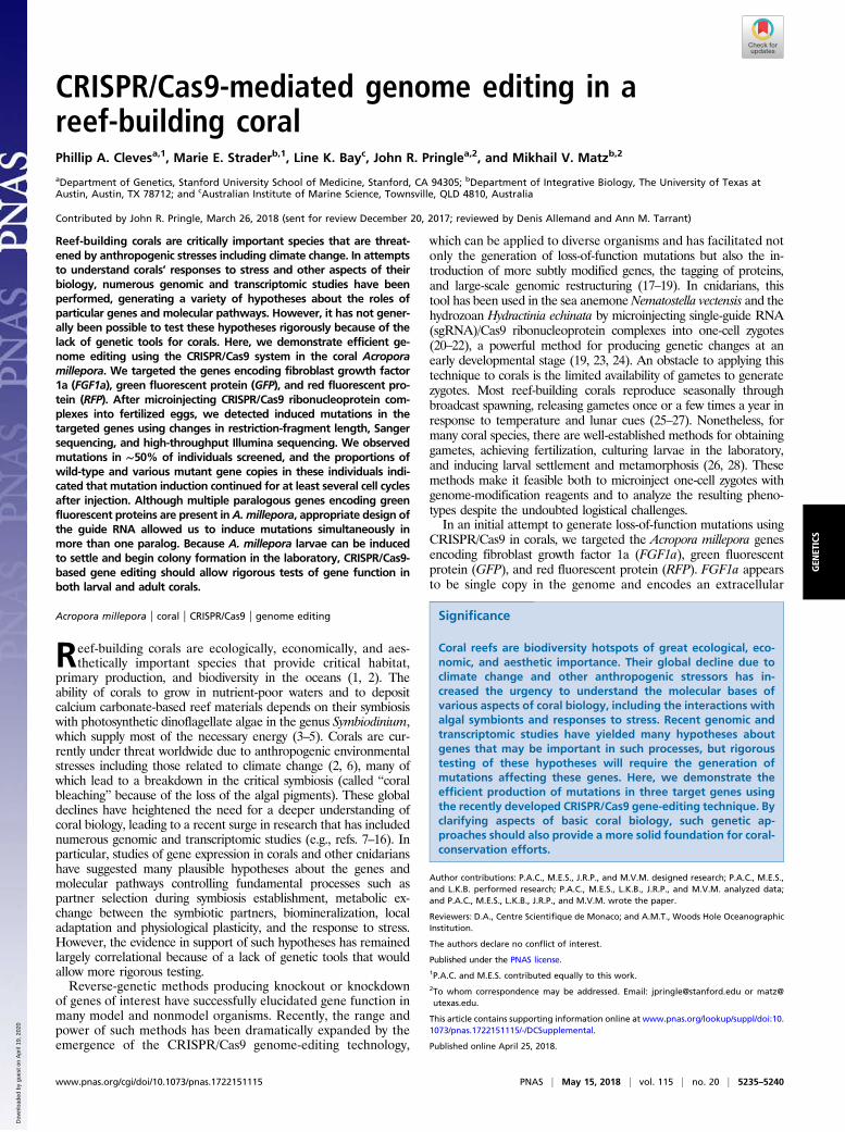
ResultsDesign and in Vitro Testing of sgRNAs. To explore the use of CRISPR/Cas9 to generate mutations in corals, we designed sgRNAs target-ing the A. millepora FGF1a, GFP, and RFP genes (Introduction).The sgRNAs were designed to minimize the chances of off-targeteffects and to encompass endogenous restriction sites that wouldfacilitate the detection of mutations (Fig. 1A and SI Appendix, SIMaterials and Methods). We also targeted positions that should befar enough upstream within the genes’ coding sequences that mu-tations altering the reading frame would knock out gene functionbut also far enough downstream that it seemed unlikely that func-tional gene products could be generated by the use of an alternativetranscription start site downstream of the induced mutation (Fig.1A). In vitro tests showed that some of the sgRNAs could effectivelyguide cutting by Cas9 of PCR-amplified fragments of genomicDNA, whereas no cleavage was observed when the DNA fragmentswere incubated with either Cas9 or the sgRNA alone (Fig. 1B).
Induction of Mutations After Microinjection of Larvae. To avoid thepossible toxicity and/or temporal delay in mutation inductionthat can be seen when vector-driven expression of sgRNA andCas9 is used (39), we injected in vitro transcribed sgRNAs thathad been precomplexed with Cas9 protein (20, 21, 40–42). Oneach of two nights of spawning, >400 freshly fertilized zygotes(Materials and Methods) were injected either with a set of two orthree sgRNA/Cas9 complexes (SI Appendix, SI Materials andMethods) or with Cas9 protein alone as a negative control (Table1). Survival and continued development of the embryos intolarvae was seen in ∼50–75% of the injected individuals (Table 1).We used several methods to assess mutation induction in the
surviving larvae. First, we used the restriction-fragment-length-polymorphism (RFLP) assays allowed by the restriction sites inthe sgRNA-binding sites to estimate the fractions of larvae car-rying mutations. For each gene, some injected larvae displayedincomplete digestion with the relevant restriction enzyme (Fig.2A, arrows), indicating that at least some copies of the gene inthose larvae had lost the restriction site because of inducedmutations. These assays suggested that mutations had been in-duced in roughly half the larvae that appeared to have beensuccessfully injected (Fig. 2B, column 2). Second, to determinethe types of mutations induced, we cloned and sequenced PCR-amplified fragments from one larva for each target gene that had
Fig. 1. Design and activity in vitro of sgRNAs targeting A. millepora genes.(A) sgRNAs targeting exon 2 (of at least four) of FGF1a, exon 3 (of five) ofGFP genes, and exon 3 (of five) of RFP genes were designed to inducedouble-strand breaks near endogenous restriction-enzyme sites that couldbe used to detect induced mutations. Colored bars, approximate locations ofthe sgRNA-binding sites; asterisks, predicted Cas9 cleavage sites and thenearby restriction sites. (B) Digestion in vitro of FGF1a, GFP, or RFP targetDNA (SI Appendix, SI Materials and Methods) incubated with Cas9 protein,the appropriate sgRNA (as transcribed in vitro from the pDR274-basedconstruct), or both. Fragments were analyzed by gel electrophoresis; outsidelanes of each gel show molecular-size markers.
Table 1. Numbers of injected and surviving A. millepora zygotes
Night 1 Night 2
Target gene(s)* FGF1a GFP RFP FGF1a None (Cas9-only)
No. of individuals injected successfully† 146 123 147 246 227No. of individuals surviving until 12 h postfertilization‡ 74 88 79 116 176Percent of individuals surviving until 12 h postfertilization, %‡ 51 72 54 47 78
*For each gene, a mixture of sgRNA/Cas9 complexes was injected (SI Appendix, SI Materials and Methods); there were two suchcomplexes for FGF1a, three for GFP, and two for RFP.†As judged by the phenol-red injection marker (SI Appendix, SI Materials and Methods).‡As judged by the numbers of larvae that were seemingly intact at the time of observation.
5236 | www.pnas.org/cgi/doi/10.1073/pnas.1722151115 Cleves et al.
Dow
nloa
ded
by g
uest
on
Apr
il 19
, 202
0
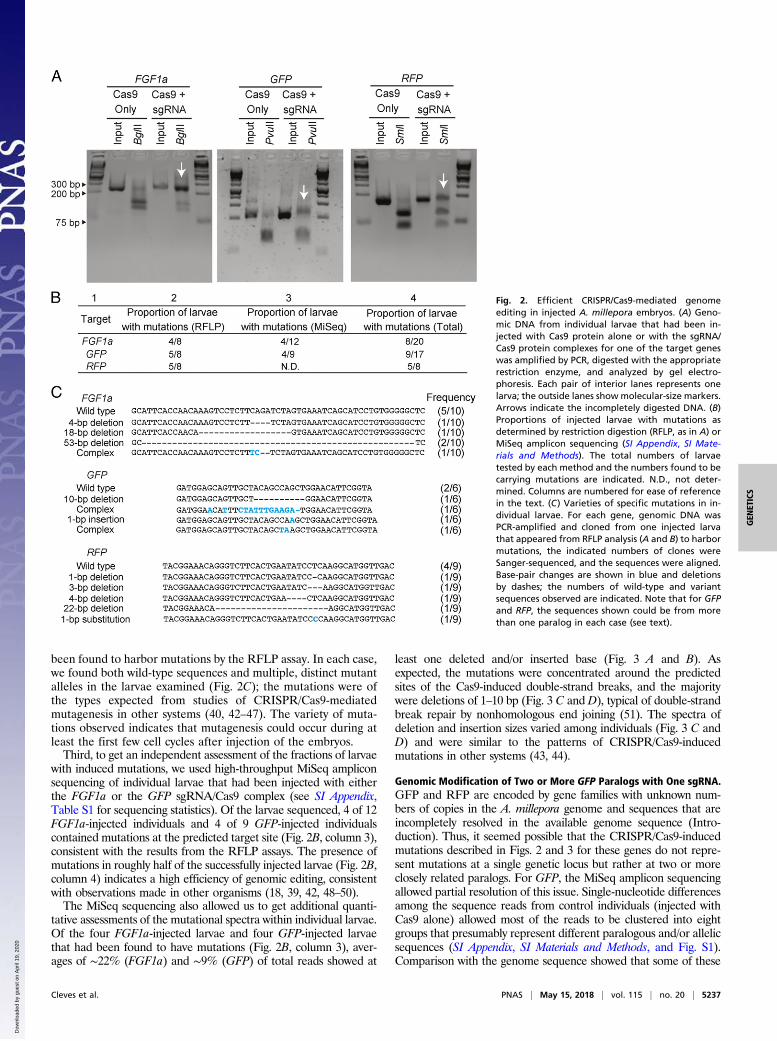
been found to harbor mutations by the RFLP assay. In each case,we found both wild-type sequences and multiple, distinct mutantalleles in the larvae examined (Fig. 2C); the mutations were ofthe types expected from studies of CRISPR/Cas9-mediatedmutagenesis in other systems (40, 42–47). The variety of muta-tions observed indicates that mutagenesis could occur during atleast the first few cell cycles after injection of the embryos.Third, to get an independent assessment of the fractions of larvae
with induced mutations, we used high-throughput MiSeq ampliconsequencing of individual larvae that had been injected with eitherthe FGF1a or the GFP sgRNA/Cas9 complex (see SI Appendix,Table S1 for sequencing statistics). Of the larvae sequenced, 4 of 12FGF1a-injected individuals and 4 of 9 GFP-injected individualscontained mutations at the predicted target site (Fig. 2B, column 3),consistent with the results from the RFLP assays. The presence ofmutations in roughly half of the successfully injected larvae (Fig. 2B,column 4) indicates a high efficiency of genomic editing, consistentwith observations made in other organisms (18, 39, 42, 48–50).The MiSeq sequencing also allowed us to get additional quanti-
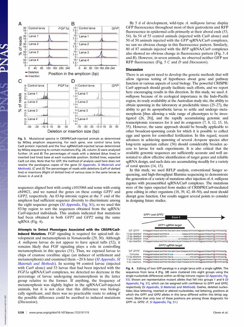
tative assessments of the mutational spectra within individual larvae.Of the four FGF1a-injected larvae and four GFP-injected larvaethat had been found to have mutations (Fig. 2B, column 3), aver-ages of ∼22% (FGF1a) and ∼9% (GFP) of total reads showed at
least one deleted and/or inserted base (Fig. 3 A and B). Asexpected, the mutations were concentrated around the predictedsites of the Cas9-induced double-strand breaks, and the majoritywere deletions of 1–10 bp (Fig. 3 C and D), typical of double-strandbreak repair by nonhomologous end joining (51). The spectra ofdeletion and insertion sizes varied among individuals (Fig. 3 C andD) and were similar to the patterns of CRISPR/Cas9-inducedmutations in other systems (43, 44).
Genomic Modification of Two or More GFP Paralogs with One sgRNA.GFP and RFP are encoded by gene families with unknown num-bers of copies in the A. millepora genome and sequences that areincompletely resolved in the available genome sequence (Intro-duction). Thus, it seemed possible that the CRISPR/Cas9-inducedmutations described in Figs. 2 and 3 for these genes do not repre-sent mutations at a single genetic locus but rather at two or moreclosely related paralogs. For GFP, the MiSeq amplicon sequencingallowed partial resolution of this issue. Single-nucleotide differencesamong the sequence reads from control individuals (injected withCas9 alone) allowed most of the reads to be clustered into eightgroups that presumably represent different paralogous and/or allelicsequences (SI Appendix, SI Materials and Methods, and Fig. S1).Comparison with the genome sequence showed that some of these
Fig. 2. Efficient CRISPR/Cas9-mediated genomeediting in injected A. millepora embryos. (A) Geno-mic DNA from individual larvae that had been in-jected with Cas9 protein alone or with the sgRNA/Cas9 protein complexes for one of the target geneswas amplified by PCR, digested with the appropriaterestriction enzyme, and analyzed by gel electro-phoresis. Each pair of interior lanes represents onelarva; the outside lanes showmolecular-size markers.Arrows indicate the incompletely digested DNA. (B)Proportions of injected larvae with mutations asdetermined by restriction digestion (RFLP, as in A) orMiSeq amplicon sequencing (SI Appendix, SI Mate-rials and Methods). The total numbers of larvaetested by each method and the numbers found to becarrying mutations are indicated. N.D., not deter-mined. Columns are numbered for ease of referencein the text. (C) Varieties of specific mutations in in-dividual larvae. For each gene, genomic DNA wasPCR-amplified and cloned from one injected larvathat appeared from RFLP analysis (A and B) to harbormutations, the indicated numbers of clones wereSanger-sequenced, and the sequences were aligned.Base-pair changes are shown in blue and deletionsby dashes; the numbers of wild-type and variantsequences observed are indicated. Note that for GFPand RFP, the sequences shown could be from morethan one paralog in each case (see text).
Cleves et al. PNAS | May 15, 2018 | vol. 115 | no. 20 | 5237
GEN
ETICS
Dow
nloa
ded
by g
uest
on
Apr
il 19
, 202
0
sequences aligned best with contig c1015068 and some with contigc849821, and we named the genes on these contigs GFP1 andGFP2, respectively. An 83-bp intronic region at the 3′ end of theamplicon had sufficient sequence diversity to discriminate amongthe eight sequence groups (SI Appendix, Fig. S1), so we used this83-bp region to sort the sequences obtained from the sgRNA/Cas9-injected individuals. This analysis indicated that mutationshad been obtained in both GFP1 and GFP2 using the samesgRNA (Fig. 4).
Attempts to Detect Phenotypes Associated with the CRISPR/Cas9-Induced Mutations. FGF signaling is required for apical-tuft de-velopment and metamorphosis in Nematostella (29, 30). AlthoughA. millepora larvae do not appear to have apical tufts (52), itremains likely that FGF signaling plays a role in controllingmetamorphosis in this species (31). Thus, we exposed larvae tochips of crustose coralline algae (an inducer of settlement andmetamorphosis) and examined them ∼20 h later (SI Appendix, SIMaterials and Methods). In scoring 94 control larvae (injectedwith Cas9 alone) and 53 larvae that had been injected with theFGF1a sgRNA/Cas9 complexes, we detected no decrease in thepercentage of larvae undergoing metamorphosis in the lattergroup relative to the former. If anything, the frequency ofmetamorphosis was slightly higher in the sgRNA/Cas9-injectedanimals, but it is not clear that this difference was biologi-cally significant, and there was no practicable route to asking ifthe possible difference could be ascribed to induced mutations(Discussion).
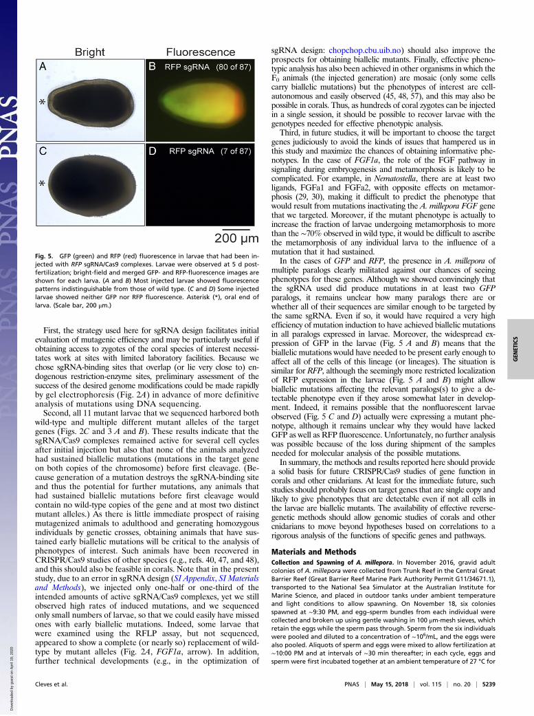
By 5 d of development, wild-type A. millepora larvae displayGFP fluorescence throughout most of their gastroderm and RFPfluorescence in epidermal cells primarily at their aboral ends (53,54). In 54 of 55 control animals (injected with Cas9 alone) and50 of 50 animals injected with the GFP sgRNA/Cas9 complexes,we saw no obvious change in this fluorescence pattern. Similarly,80 of 87 animals injected with the RFP sgRNA/Cas9 complexesalso showed no obvious change in fluorescence pattern (Fig. 5 Aand B). However, in seven animals, we observed neither GFP norRFP fluorescence (Fig. 5 C and D and Discussion).
DiscussionThere is an urgent need to develop the genetic methods that willallow rigorous testing of hypotheses about gene and pathwayfunction in various aspects of coral biology. The powerful CRISPR/Cas9 approach should greatly facilitate such efforts, and we reporthere encouraging results in this direction. In this study, we used A.millepora because of its ecological importance in the Indo-Pacificregion, its ready availability at the Australian study site, the ability toobtain spawning in the laboratory at predictable times (25–27), theability to get its aposymbiotic larvae to settle and undergo meta-morphosis [thus allowing a wide range of phenotypes to be inves-tigated (26, 28)], and the rapidly accumulating genomic andtranscriptomic resources for it and its congeners (7, 8, 12, 15, 16,55). However, the same approach should be broadly applicable toother broadcast-spawning corals for which it is possible to collecteggs and sperm for controlled fertilization. In this regard, recentadvances in achieving spawning of several Acropora species afterlong-term aquarium culture (56) should considerably broaden ac-cess to larvae for such experiments. It is also critical that theavailable genomic sequences are sufficiently accurate and well an-notated to allow effective identification of target genes and reliablesgRNA design, and such data are accumulating steadily for a varietyof coral species (14, 15).In this study, we used RFLP analysis, conventional Sanger se-
quencing, and high-throughput Illumina sequencing to demonstratethe generation of a variety of mutations after injection of A. milleporazygotes with preassembled sgRNA/Cas9 complexes. The mutationswere of the types expected from studies of CRISPR/Cas9-mediatedgene editing in other organisms (18, 39, 42, 48–50), and most shoulddisrupt gene function. Our results suggest several points to considerin designing future studies.
Fig. 3. Mutational spectra in CRISPR/Cas9-injected animals as determinedby MiSeq amplicon sequencing. For each gene, one control larva (onlyCas9 protein injected) and the four sgRNA/Cas9-injected larvae determinedby MiSeq sequencing to contain mutations (Fig. 2B, column 3) were analyzedfurther. (A and B) The percentages of reads with a deleted (blue lines) orinserted (red lines) base at each nucleotide position. Dotted lines, expectedCas9 cut sites. Note that for GFP, the method of analysis used here does notresolve the paralogous copies of the gene (SI Appendix, SI Materials andMethods). (C and D) The percentages of reads with deletions (Left of dottedline) or insertions (Right of dotted line) of various sizes in the same larvae asshown in A and B.
Fig. 4. Editing of two GFP paralogs in a single larva with a single sgRNA. Thesequences from larva 4 (Fig. 3B) were clustered into eight groups using thesingle-nucleotide differences within an 83-bp intronic region (SI Appendix, Fig.S1). Shown are representative mutant alleles that fell into groups 1 and 6 (SIAppendix, Fig. S1), which can be assigned with confidence to GFP1 and GFP2,respectively (SI Appendix, SI Materials and Methods). Dashes, deleted nucleo-tides; blue lettering, inserted or altered nucleotides; red lettering, positions atwhich the GFP1 and GFP2 alleles in this larva differed within this 64-bp seg-ment. (Note that only two of these positions are among those diagnostic forGFP1 vs. GFP2: cf. SI Appendix, Fig. S1.)
5238 | www.pnas.org/cgi/doi/10.1073/pnas.1722151115 Cleves et al.
Dow
nloa
ded
by g
uest
on
Apr
il 19
, 202
0
First, the strategy used here for sgRNA design facilitates initialevaluation of mutagenic efficiency and may be particularly useful ifobtaining access to zygotes of the coral species of interest necessi-tates work at sites with limited laboratory facilities. Because wechose sgRNA-binding sites that overlap (or lie very close to) en-dogenous restriction-enzyme sites, preliminary assessment of thesuccess of the desired genome modifications could be made rapidlyby gel electrophoresis (Fig. 2A) in advance of more definitiveanalysis of mutations using DNA sequencing.Second, all 11 mutant larvae that we sequenced harbored both
wild-type and multiple different mutant alleles of the targetgenes (Figs. 2C and 3 A and B). These results indicate that thesgRNA/Cas9 complexes remained active for several cell cyclesafter initial injection but also that none of the animals analyzedhad sustained biallelic mutations (mutations in the target geneon both copies of the chromosome) before first cleavage. (Be-cause generation of a mutation destroys the sgRNA-binding siteand thus the potential for further mutations, any animals thathad sustained biallelic mutations before first cleavage wouldcontain no wild-type copies of the gene and at most two distinctmutant alleles.) As there is little immediate prospect of raisingmutagenized animals to adulthood and generating homozygousindividuals by genetic crosses, obtaining animals that have sus-tained early biallelic mutations will be critical to the analysis ofphenotypes of interest. Such animals have been recovered inCRISPR/Cas9 studies of other species (e.g., refs. 40, 47, and 48),and this should also be feasible in corals. Note that in the presentstudy, due to an error in sgRNA design (SI Appendix, SI Materialsand Methods), we injected only one-half or one-third of theintended amounts of active sgRNA/Cas9 complexes, yet we stillobserved high rates of induced mutations, and we sequencedonly small numbers of larvae, so that we could easily have missedones with early biallelic mutations. Indeed, some larvae thatwere examined using the RFLP assay, but not sequenced,appeared to show a complete (or nearly so) replacement of wild-type by mutant alleles (Fig. 2A, FGF1a, arrow). In addition,further technical developments (e.g., in the optimization of
sgRNA design: chopchop.cbu.uib.no) should also improve theprospects for obtaining biallelic mutants. Finally, effective pheno-typic analysis has also been achieved in other organisms in which theF0 animals (the injected generation) are mosaic (only some cellscarry biallelic mutations) but the phenotypes of interest are cell-autonomous and easily observed (45, 48, 57), and this may also bepossible in corals. Thus, as hundreds of coral zygotes can be injectedin a single session, it should be possible to recover larvae with thegenotypes needed for effective phenotypic analysis.Third, in future studies, it will be important to choose the target
genes judiciously to avoid the kinds of issues that hampered us inthis study and maximize the chances of obtaining informative phe-notypes. In the case of FGF1a, the role of the FGF pathway insignaling during embryogenesis and metamorphosis is likely to becomplicated. For example, in Nematostella, there are at least twoligands, FGFa1 and FGFa2, with opposite effects on metamor-phosis (29, 30), making it difficult to predict the phenotype thatwould result from mutations inactivating the A. millepora FGF genethat we targeted. Moreover, if the mutant phenotype is actually toincrease the fraction of larvae undergoing metamorphosis to morethan the ∼70% observed in wild type, it would be difficult to ascribethe metamorphosis of any individual larva to the influence of amutation that it had sustained.In the cases of GFP and RFP, the presence in A. millepora of
multiple paralogs clearly militated against our chances of seeingphenotypes for these genes. Although we showed convincingly thatthe sgRNA used did produce mutations in at least two GFPparalogs, it remains unclear how many paralogs there are orwhether all of their sequences are similar enough to be targeted bythe same sgRNA. Even if so, it would have required a very highefficiency of mutation induction to have achieved biallelic mutationsin all paralogs expressed in larvae. Moreover, the widespread ex-pression of GFP in the larvae (Fig. 5 A and B) means that thebiallelic mutations would have needed to be present early enough toaffect all of the cells of this lineage (or lineages). The situation issimilar for RFP, although the seemingly more restricted localizationof RFP expression in the larvae (Fig. 5 A and B) might allowbiallelic mutations affecting the relevant paralogs(s) to give a de-tectable phenotype even if they arose somewhat later in develop-ment. Indeed, it remains possible that the nonfluorescent larvaeobserved (Fig. 5 C and D) actually were expressing a mutant phe-notype, although it remains unclear why they would have lackedGFP as well as RFP fluorescence. Unfortunately, no further analysiswas possible because of the loss during shipment of the samplesneeded for molecular analysis of the possible mutations.In summary, the methods and results reported here should provide
a solid basis for future CRISPR/Cas9 studies of gene function incorals and other cnidarians. At least for the immediate future, suchstudies should probably focus on target genes that are single copy andlikely to give phenotypes that are detectable even if not all cells inthe larvae are biallelic mutants. The availability of effective reverse-genetic methods should allow genomic studies of corals and othercnidarians to move beyond hypotheses based on correlations to arigorous analysis of the functions of specific genes and pathways.
Materials and MethodsCollection and Spawning of A. millepora. In November 2016, gravid adultcolonies of A. millepora were collected from Trunk Reef in the Central GreatBarrier Reef (Great Barrier Reef Marine Park Authority Permit G11/34671.1),transported to the National Sea Simulator at the Australian Institute forMarine Science, and placed in outdoor tanks under ambient temperatureand light conditions to allow spawning. On November 18, six coloniesspawned at ∼9:30 PM, and egg–sperm bundles from each individual werecollected and broken up using gentle washing in 100 μm-mesh sieves, whichretain the eggs while the sperm pass through. Sperm from the six individualswere pooled and diluted to a concentration of ∼106/mL, and the eggs werealso pooled. Aliquots of sperm and eggs were mixed to allow fertilization at∼10:00 PM and at intervals of ∼30 min thereafter; in each cycle, eggs andsperm were first incubated together at an ambient temperature of 27 °C for
Fig. 5. GFP (green) and RFP (red) fluorescence in larvae that had been in-jected with RFP sgRNA/Cas9 complexes. Larvae were observed at 5 d post-fertilization; bright-field and merged GFP- and RFP-fluorescence images areshown for each larva. (A and B) Most injected larvae showed fluorescencepatterns indistinguishable from those of wild type. (C and D) Some injectedlarvae showed neither GFP nor RFP fluorescence. Asterisk (*), oral end oflarva. (Scale bar, 200 μm.)
Cleves et al. PNAS | May 15, 2018 | vol. 115 | no. 20 | 5239
GEN
ETICS
Dow
nloa
ded
by g
uest
on
Apr
il 19
, 202
0

15 min and then moved to a 24 °C room for microinjection. This staggeredfertilization allowed time for microinjection of each batch of zygotes beforefirst cleavage, which occurred 30–60 min after fertilization. On November19, four other individuals spawned, and their eggs and sperm were collectedand processed as on the preceding day.
Other Methods. SI Appendix, SI Materials and Methods, describes the gen-eration and testing of sgRNAs, the method for microinjection, and themethods used for molecular analysis of mutations and assessment ofpossible phenotypes.
ACKNOWLEDGMENTS. We thank S. Sanders, B. Mason, and B. Fu forhelpful suggestions on the design of the sgRNAs; H. Elder and I. Bjørnsbofor valuable assistance during coral spawning; and Eppendorf South Pacificfor loan of the microinjection system. MiSeq sequencing was performed bythe Genomic Sequencing and Analysis Facility at the University of Texas atAustin. Some bioinformatic analyses were carried out using the computa-tional resources of the Texas Advanced Computing Center. Funding forthis study was provided by NSF Doctoral Dissertation Improvement GrantDEB-1501463 (to M.E.S. and M.V.M.); the Simons Foundation Grant LIFE336932 (to J.R.P.); and internal Australian Institute for Marine Sciencefunding (to L.K.B.).
1. Moberg F, Folke C (1999) Ecological goods and services of coral reef ecosystems. EcolEcon 29:215–233.
2. Hughes TP, et al. (2017) Coral reefs in the Anthropocene. Nature 546:82–90.3. Muscatine L, Cernichiari E (1969) Assimilation of photosynthetic products of zoo-
xanthellae by a reef coral. Biol Bull 137:506–523.4. Muscatine L, Falkowski PG, Porter JW, Dubinsky Z (1984) Fate of photosynthetic fixed
carbon in light- and shade-adapted colonies of the symbiotic coral Stylophora pis-tillata. Proc R Soc B 222:181–202.
5. Davy SK, Allemand D, Weis VM (2012) Cell biology of cnidarian-dinoflagellate sym-biosis. Microbiol Mol Biol Rev 76:229–261.
6. Hughes TP, et al. (2017) Global warming and recurrent mass bleaching of corals.Nature 543:373–377.
7. Shinzato C, et al. (2011) Using the Acropora digitifera genome to understand coralresponses to environmental change. Nature 476:320–323.
8. Moya A, et al. (2012) Whole transcriptome analysis of the coral Acropora milleporareveals complex responses to CO2-driven acidification during the initiation of calci-fication. Mol Ecol 21:2440–2454.
9. Lehnert EM, et al. (2014) Extensive differences in gene expression between symbioticand aposymbiotic cnidarians. G3 (Bethesda) 4:277–295.
10. Baumgarten S, et al. (2015) The genome of Aiptasia, a sea anemone model for coralsymbiosis. Proc Natl Acad Sci USA 112:11893–11898.
11. Dixon GB, et al. (2015) Genomic determinants of coral heat tolerance across latitudes.Science 348:1460–1462.
12. Barshis DJ (2015) Genomic potential for coral survival of climate change. Coral Reefsin the Anthropocene, ed Birkeland C (Springer, Heidelberg, Germany), pp 133–145.
13. Kenkel C, Matz MV (2016) Enhanced gene expression plasticity as a mechanism ofadaptation to a variable environment in a reef-building coral. Nat Ecol Evol 1:59667.
14. Bhattacharya D, et al. (2016) Comparative genomics explains the evolutionary successof reef-forming corals. eLife 5:e13288.
15. Liew YJ, Aranda M, Voolstra CR (2016) Reefgenomics.Org: A repository for marinegenomics data. Database (Oxford) 2016:baw152.
16. Traylor-Knowles N, Rose NH, Sheets EA, Palumbi SR (2017) Early transcriptional re-sponses during heat stress in the coral Acropora hyacinthus. Biol Bull 232:91–100.
17. Ran FA, et al. (2013) Genome engineering using the CRISPR-Cas9 system. Nat Protoc 8:2281–2308.
18. Doudna JA, Charpentier E (2014) Genome editing. The new frontier of genome en-gineering with CRISPR-Cas9. Science 346:1258096.
19. Momose T, Concordet JP (2016) Diving into marine genomics with CRISPR/Cas9 systems.Mar Genomics 30:55–65.
20. Ikmi A, McKinney SA, Delventhal KM, Gibson MC (2014) TALEN and CRISPR/Cas9-mediated genome editing in the early-branching metazoan Nematostella vectensis.Nat Commun 5:5486.
21. Servetnick MD, et al. (2017) Cas9-mediated excision of Nematostella brachyury dis-rupts endoderm development, pharynx formation and oral-aboral patterning.Development 144:2951–2960.
22. Gahan JM, et al. (2017) Functional studies on the role of Notch signaling in Hydrac-tinia development. Dev Biol 428:224–231.
23. Zhang Y, Yu LC (2008) Single-cell microinjection technology in cell biology. BioEssays30:606–610.
24. Gilles AF, Averof M (2014) Functional genetics for all: Engineered nucleases, CRISPRand the gene editing revolution. Evodevo 5:43.
25. Harrison PL, et al. (1984) Mass spawning in tropical reef corals. Science 223:1186–1189.
26. Baird AH, Guest JR, Willis BL (2009) Systematic and biogeographical patterns in thereproductive biology of scleractinian corals. Annu Rev Ecol Evol Syst 40:551–571.
27. Keith SA, et al. (2016) Coral mass spawning predicted by rapid seasonal rise in oceantemperature. Proc Biol Sci 283:20160011.
28. Pollock FJ, et al. (2017) Coral larvae for restoration and research: A large-scale methodfor rearing Acropora millepora larvae, inducing settlement, and establishing symbi-osis. PeerJ 5:e3732.
29. Rentzsch F, Fritzenwanker JH, Scholz CB, Technau U (2008) FGF signalling controlsformation of the apical sensory organ in the cnidarian Nematostella vectensis.Development 135:1761–1769.
30. Sinigaglia C, Busengdal H, Lerner A, Oliveri P, Rentzsch F (2015) Molecular charac-terization of the apical organ of the anthozoan Nematostella vectensis. Dev Biol 398:120–133.
31. Strader ME, Aglyamova GV, Matz MV (2018) Molecular characterization of larvaldevelopment from fertilization to metamorphosis in a reef-building coral. BMCGenomics 19:17.
32. Kelmanson IV, Matz MV (2003) Molecular basis and evolutionary origins of color di-versity in great star coral Montastraea cavernosa (Scleractinia: Faviida). Mol Biol Evol20:1125–1133.
33. Alieva NO, et al. (2008) Diversity and evolution of coral fluorescent proteins. PLoS One3:e2680.
34. D’Angelo C, et al. (2008) Blue light regulation of host pigment in reef-building corals.Mar Ecol Prog Ser 364:97–106.
35. D’Angelo C, et al. (2012) Locally accelerated growth is part of the innate immuneresponse and repair mechanisms in reef-building corals as detected by green fluo-rescent protein (GFP)-like pigments. Coral Reefs 31:1045–1056.
36. Shinzato C, Shoguchi E, Tanaka M, Satoh N (2012) Fluorescent protein candidategenes in the coral Acropora digitifera genome. Zool Sci 29:260–264.
37. Gittins JR, D’Angelo C, Oswald F, Edwards RJ, Wiedenmann J (2015) Fluorescentprotein-mediated colour polymorphism in reef corals: Multicopy genes extend theadaptation/acclimatization potential to variable light environments. Mol Ecol 24:453–465.
38. Yasuoka Y, Shinzato C, Satoh N (2016) The mesoderm-forming gene brachyury reg-ulates ectoderm-endoderm demarcation in the coral Acropora digitifera. Curr Biol 26:2885–2892.
39. Paix A, Folkmann A, Rasoloson D, Seydoux G (2015) High efficiency, homology-directedgenome editing in Caenorhabditis elegans using CRISPR-Cas9 ribonucleoprotein com-plexes. Genetics 201:47–54.
40. Cho SW, Lee J, Carroll D, Kim J-S, Lee J (2013) Heritable gene knockout in Caeno-rhabditis elegans by direct injection of Cas9-sgRNA ribonucleoproteins. Genetics 195:1177–1180.
41. Kim S, Kim D, Cho SW, Kim J, Kim J-S (2014) Highly efficient RNA-guided genomeediting in human cells via delivery of purified Cas9 ribonucleoproteins. Genome Res24:1012–1019.
42. Sung YH, et al. (2014) Highly efficient gene knockout in mice and zebrafish with RNA-guided endonucleases. Genome Res 24:125–131.
43. Hwang WY, et al. (2013) Efficient genome editing in zebrafish using a CRISPR-Cassystem. Nat Biotechnol 31:227–229.
44. Ansai S, Kinoshita M (2014) Targeted mutagenesis using CRISPR/Cas system in me-daka. Biol Open 3:362–371.
45. Guo X, et al. (2014) Efficient RNA/Cas9-mediated genome editing in Xenopus tropi-calis. Development 141:707–714.
46. Liang X, et al. (2015) Rapid and highly efficient mammalian cell engineering viaCas9 protein transfection. J Biotechnol 208:44–53.
47. Zu Y, et al. (2016) Biallelic editing of a lamprey genome using the CRISPR/Cas9 system.Sci Rep 6:23496.
48. Jao L-E, Wente SR, Chen W (2013) Efficient multiplex biallelic zebrafish genome ed-iting using a CRISPR nuclease system. Proc Natl Acad Sci USA 110:13904–13909.
49. Wang H, et al. (2013) One-step generation of mice carrying mutations in multiplegenes by CRISPR/Cas-mediated genome engineering. Cell 153:910–918.
50. Kistler KE, Vosshall LB, Matthews BJ (2015) Genome engineering with CRISPR-Cas9 inthe mosquito Aedes aegypti. Cell Rep 11:51–60.
51. Rodgers K, McVey M (2016) Error-prone repair of DNA double-strand breaks. J CellPhysiol 231:15–24.
52. Hayward DC, Grasso LC, Saint R, Miller DJ, Ball EE (2015) The organizer in evolution-gastrulation and organizer gene expression highlight the importance of Brachyuryduring development of the coral, Acropora millepora. Dev Biol 399:337–347.
53. Kenkel CD, et al. (2011) Development of gene expression markers of acute heat-lightstress in reef-building corals of the genus Porites. PLoS One 6:e26914.
54. Strader ME, Davies SW, Matz MV (2015) Differential responses of coral larvae to thecolour of ambient light guide them to suitable settlement microhabitat. R Soc OpenSci 2:150358.
55. Meyer E, et al. (2009) Sequencing and de novo analysis of a coral larval transcriptomeusing 454 GSFlx. BMC Genomics 10:219.
56. Craggs J, et al. (2017) Inducing broadcast coral spawning ex situ: Closed systemmesocosm design and husbandry protocol. Ecol Evol 7:11066–11078.
57. Mazo-Vargas A, et al. (2017) Macroevolutionary shifts of WntA function potentiatebutterfly wing-pattern diversity. Proc Natl Acad Sci USA 114:10701–10706.
5240 | www.pnas.org/cgi/doi/10.1073/pnas.1722151115 Cleves et al.
Dow
nloa
ded
by g
uest
on
Apr
il 19
, 202
0