correction serial fluorescein fundus photography of retinal circulation
TRANSCRIPT
360 CORRESPONDENCE
vout Catholic and a leading member of a number of Catholic organizations, he was yet without bigotry or sentimentality. He was the embodiment of Christian charity. One could never meet him without his pulling a slip of paper from his pocket and asking what could or should be done for the person whose name was on the slip and who was in need of some help. Race, creed, ethnic origin did not matter—there was a human being in some sort of distress. The number of those indebted to Regan, not a few younger ophthalmologists among them, is very large indeed.
A great profession such as ophthalmology is not the creation of one man or one generation. Many varied personalities are required to build and develop it. What is achieved today is, in the broadest sense, coming down to us from those who have gone before. The beneficial effect of the life and work of James J. Regan will be felt in American ophthalmology for a long time to come.
Hermann M. Burian
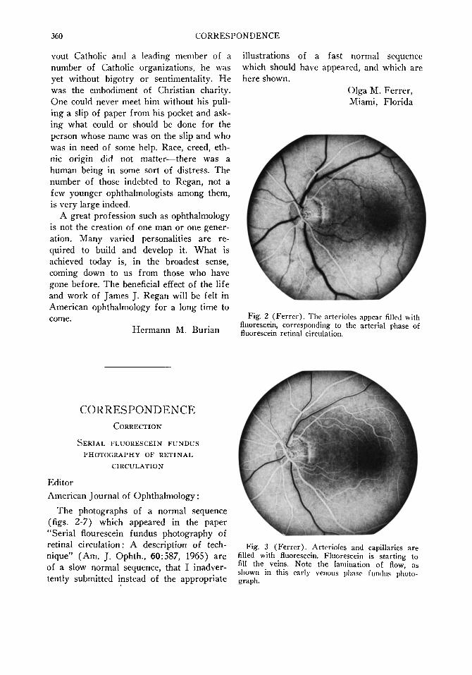
illustrations of a fast normal sequence which should have appeared, and which are here shown.
Olga M. Ferrer, Miami, Florida
Fig. 2 (Fer re r ) . The arterioles appear filled with fluorescein, corresponding to the arterial phase of fluorescein retinal circulation.
CORRESPONDENCE CORRECTION
SERIAL FLUORESCEIN FUNDUS PHOTOGRAPHY OF RETINAL
CIRCULATION
Editor American Journal of Ophthalmology :
The photographs of a normal sequence (figs. 2-7) which appeared in the paper "Serial flourescein fundus photography of retinal circulation: A description of technique" (Am. J. Ophth., 60:587, 1965) are of a slow normal sequence, that I inadvertently submitted instead of the appropriate
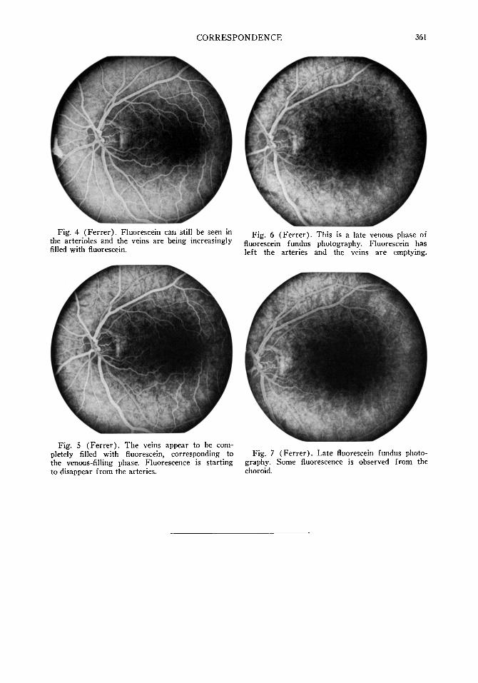
Fig. 3 (Fe r re r ) . Arterioles and capillaries are filled with fluorescein. Fluorescein is starting to fill the veins. Note the lamination of flow, as shown in this early venous phase fundus photograph.
CORRESPONDENCE 361
Fig. 4 (Ferrer). Fluorescein can still be seen in the arterioles and the veins are being increasingly filled with fluorescein.
Fig. 6 (Ferrer). This is a late venous phase of fluorescein fundus photography. Fluorescein has left the arteries and the veins are emptying.
Fig. 5 (Ferrer). The veins appear to be completely filled with fluorescein, corresponding to the venous-filling phase. Fluorescence is starting to disappear from the arteries.
Fig. 7 (Ferrer). Late fluorescein fundus photography. Some fluorescence is observed from the choroid.