corneal vascularization in the florida...
TRANSCRIPT

CORNEAL VASCULARIZATION IN THE FLORIDA MANATEE
(Trichechus manatus latirostris)
By
JENNIFER YOUNG HARPER
A DISSERTATION PRESENTED TO THE GRADUATE SCHOOL OF THE UNIVERSITY OF FLORIDA IN PARTIAL FULFILLMENT
OF THE REQUIREMENTS FOR THE DEGREE OF DOCTOR OF PHILOSOPHY
UNIVERSITY OF FLORIDA
2004

Copyright 2004
by
Jennifer Young Harper

ACKNOWLEDGMENTS
I would like to thank my mentor, Dr. Don Samuelson. He has been a wonderful
source of knowledge, inspiration, and motivation. Without his help and guidance, I could
not have accomplished any of this work. I would also like to thank Dr. Roger Reep for
all of his help and support along the way. He too has acted as a rock and support system.
My additional committee members (Dr. Peter McGuire, Dr. Dennis Brooks, and
Dr. Gordon Bauer) have been tremendous support and I thank them for all they have
offered. Their guidance has been appreciated beyond belief.
Laboratory technologists Pat Lewis and Maggie Stoll were extremely helpful and
supportive during much of my work. I would have not been able to accomplish the first
procedure without their help. I thank these fine ladies from the bottom of my heart.
My parents, Jim and Marion Young, have meant more to me than I can ever
describe or explain. I appreciate all their love and support.
Finally, I thank my husband Ridge Harper. He has been the strongest support
system I could ever ask for and makes me happier than I ever knew I could be.
iii

TABLE OF CONTENTS page ACKNOWLEDGMENTS ................................................................................................. iii
LIST OF TABLES............................................................................................................. vi
LIST OF FIGURES .......................................................................................................... vii
ABSTRACT.........................................................................................................................x
CHAPTER
1 LITERATURE REVIEW .............................................................................................1
The Florida Manatee.....................................................................................................1 The Tactile Sensory System of Marine Mammals .......................................................2 The Auditory System of Marine Mammals ..................................................................5 The Olfaction and Gustation of Marine Mammals.......................................................7 The Vision of Marine Mammals...................................................................................8 The Cornea..................................................................................................................13 Angiogenesis...............................................................................................................17 Objectives ...................................................................................................................22
2 CORNEAL VASCULARIZATION IN THE FLORIDA MANATEE AND
THREE-DIMENSIONAL RECONSTRUCTION .....................................................23
Introduction.................................................................................................................23 Materials and Methods ...............................................................................................32
Specimen Collection............................................................................................32 Results.........................................................................................................................36
Anatomical Description.......................................................................................36 Morphometry.......................................................................................................39 Statistics...............................................................................................................40
Discussion...................................................................................................................42 3 VASCULAR ENDOTHELIAL GROWTH FACTOR RECEPTOR-1 AND -2 IN
THE FLORIDA MANATEE CORNEA ....................................................................96
Introduction.................................................................................................................96 Materials and Methods .............................................................................................106
iv

Specimen Collection..........................................................................................106 Preparation of Eyes............................................................................................106 Histology ...........................................................................................................107 Immunohistochemistry ......................................................................................107
Results.......................................................................................................................108 Vascular endothelial growth factor receptor-1 (VEGFR-1) ..............................108 Vascular endothelial growth factor receptor-2 (VEGFR-2) ..............................109
4 ADDITIONAL STUDIES........................................................................................127
Corneal Vascularization in the Antillean Manatee...................................................127 Ophthalmic Examinations in Two Captive Florida Manatees..................................128
5 GENERAL CONCLUSIONS...................................................................................139
LIST OF REFERENCES.................................................................................................142
BIOGRAPHICAL SKETCH ...........................................................................................154
v

LIST OF TABLES
Table page 2-1 Information on manatee tissue used and volume of vasculature at 25 X.................41
2-2 Volume of vasculature at 200 X...............................................................................42
vi

LIST OF FIGURES
Figure page 2-1 Florida manatee eye with ocular tissue and fat removed revealing the eye and
optic nerve..............................................................................................................55
2-2 Histological pictures of MNW-9111 .....................................................................56
2-3 These pictures were taken of MSW-9114 of the same blood vessel at varying magnifications to show the penetration of this vessel into the anterior epithelium ..............................................................................................................57
2-4 UCF-9114 shows the smaller vessels that were outlined within the stroma for three-dimensional reconstruction (200X). .............................................................58
2-5 Pictures from MNW-9016 .....................................................................................59
2-6 These pictures were taken from the unknown specimen. (A) Shows the edge of the limbal vascular arrangement at the peripheral cornea (200X), and (B) larger vessels that go right up to the anterior epithelium at 200X. ..................................60
2-7 Anatomical orientation of manatee eyes as they are described in three-dimensional reconstruction ....................................................................................61
2-8 MNW-9016 ............................................................................................................62
2-9 MNW-9022(R).......................................................................................................63
2-10 MNW-9111. ...........................................................................................................64
2-11 MNW-9116 ............................................................................................................65
2-12 MSE-9101. .............................................................................................................66
2-13 MSE-9105..............................................................................................................67
2-14 MSE-9107. .............................................................................................................68
2-15 MSE-9115. .............................................................................................................69
2-16 MSE-9131. .............................................................................................................70
vii

2-17 MSW-9114.............................................................................................................71
2-18 MSW-9131(R). ......................................................................................................72
2-19 MSW-9137(L)........................................................................................................73
2-20 MSW-9137(R). ......................................................................................................74
2-21 MSW-9141.............................................................................................................75
2-22 MSW-9143.............................................................................................................76
2-23 TM-8619(L). ..........................................................................................................77
2-24 TM-8619(R)...........................................................................................................78
2-25 TM-8630(L) ...........................................................................................................79
2-26 TM-8630(R)...........................................................................................................80
2-27 UCF-9114. .............................................................................................................81
2-28 UCF-9137 ..............................................................................................................82
2-29 UCF-9138 ..............................................................................................................83
2-30 UCF-9141. .............................................................................................................84
2-31 Unknown................................................................................................................85
2-32 Fetus(L)..................................................................................................................86
2-33 Fetus(R)..................................................................................................................87
2-34 Dorsal quadrant of MNW-9016 at 200X ...............................................................88
2-35 Medial quadrant of MNW-9016 at 200X...............................................................89
2-36 Dorsal quadrant of MSW-9114 at 200X................................................................90
2-37 Ventral quadrant of MSW-9114 at 200X...............................................................91
2-38 Dorsal quadrant of TM-8619(L) at 200X ..............................................................92
2-39 Medial quadrant of TM-8619(L) at 200X..............................................................93
2-40 Ventral quadrant of TM-8619(L) at 200X.............................................................94
2-41 Lateral quadrant of TM-8619(L) at 200X..............................................................95
viii

3-1 Florida manatee eye .............................................................................................118
3-2 Pictures taken from TM-0310 showing a positive reaction of VEGFR-1 ...........119
3-3 VEGFR-1 positive reactions in TM-0310 showing the prevalence of the positive reaction at 200X with 1:50. ....................................................................120
3-4 VEGFR-1 positive reactions in TM-0310 (A) at 400X with 1:100 .....................121
3-5 VEGFR-1 reactions in TM-0310 of control samples...........................................122
3-6 Pictures taken from TM-0311 showing a positive reaction of VEFGR-1 ...........123
3-7 VEGFR-1 positive reactions in TM-0311............................................................124
3-8 VEGFR-1 reactions in TM-0311 of control samples...........................................125
3-9 Pictures taken of TM-0310 showing no positive reactions of VEGFR-2 that appears to be different from the control samples.................................................126
4-1 The cornea from an Antillean manatee eye.. .......................................................130
4-2 This corneal section was cut at 10 microns and stained using Masson’s Trichrome stain. ...................................................................................................131
4-3 These pictures show the unusually nonorganized, wavy stroma observed in the Antillean manatee cornea...............................................................................132
4-4 Extensive vasculature was observed in the (A) stroma of the Antillean manatee cornea at 100X and (B) as the stroma approaches the anterior epithelium at 100X.....................................................................................................................133
4-5 These pictures illustrate the vessels in the anterior epithelium............................134
4-6 Vasculature can be observed (A) beneath the anterior epithelium at 200X and (B) penetrating the anterior epithelium at 400X. .................................................135
4-7 The unusual material thought to be mucus surrounding the anterior epithelium ............................................................................................................136
4-8 Unusual projections were observed in the posterior stroma and adjacent endothelium..........................................................................................................137
4-9 A PAS stain of the Antillean manatee cornea showing no reaction to a fungus in the anterior epithelium at (A) 100X and (B) 200X..........................................138
ix
Abstract of Dissertation Presented to the Graduate School of the University of Florida in Partial Fulfillment of the Requirements for the Degree of Doctor of Philosophy
CORNEAL VASCULARIZATION IN THE FLORIDA MANATEE (Trichechus manatus latirostris)
By
Jennifer Young Harper
May 2004
Chair: Don A. Samuelson Cochair: Roger L. Reep Major Department: Veterinary Medical Sciences
The cornea of the Florida manatee is anatomically unique because blood vessels are
found throughout the cornea. In all other species of animals, this is considered a
pathological condition that impedes vision and is usually caused by injury or trauma of
some sort. The purpose of this study was to describe more clearly corneal vascularization
by (1) examining the architecture through three-dimensional reconstruction in order to
find possible patterns in size, distribution, and location of blood vessels that may be
related to age, gender, location found, and environment, and (2) examining vascular
endothelial growth factor receptors-1 and -2 in the cornea. Twenty-six eyes from 22
individuals were prepared for histological examination and subsequent three-dimensional
reconstruction. Every specimen examined possessed blood vessels in the cornea, mostly
comprising 0.3% of total surface density (volume) of the cornea. However, two
individuals had a greater density of vascularization (1.2% and 0.7%). The size and
x

distribution of vessels appeared to be greatest toward the mid-periphery of the cornea.
No differences were found among individuals based on age, gender, and site of recovery.
Environmental influences were not a significant factor either, which was not originally
anticipated. The presence of vessels at the level of the anterior epithelium was surprising,
and it appeared that the vascularization was directed more anteriorly than originally
thought. Blood vessels were also found in fetal eyes. In all the eyes examined, no signs
of injury or trauma could be observed. Vascular endothelial growth factor receptor-1
(VEGFR-1) was found in the corneas of 2 individuals examined through
immunohistochemistry. However, vascular endothelial growth factor receptor-2
(VEGFR-2) was not, indicating that a noninflammatory and nonpathological process of
angiogenesis might be taking place in the Florida manatee cornea. The presence of blood
vessels appears to impair vision minimally based on the blood vessels’ low density, size,
and location. The penetration of vessels into the anterior epithelium and development of
vessels within the fetus points to an evolutionary adaptation, possibly due to the
manatee’s unique ability to move freely in salt, brackish, and fresh water.
xi

CHAPTER 1 LITERATURE REVIEW
The Florida Manatee
The Florida manatee, Trichechus manatus latirostris, is perhaps one of the most
interesting marine mammals. It is a subspecies of the West Indian manatee and is in the
order Sirenia, which includes two additional species, the Amazonian manatee and the
West African manatee. The order Sirenia is made up of two families, Trichechidae
(manatees) and Dugongidae (dugongs). Manatees are the only marine mammal
herbivores (Beck and Barros 1991; Reynolds and Odell 1991) and the Florida manatee
feeds on over 60 different species of aquatic vegetation (Provancha and Hall 1991).
Trichechids are also unique among marine mammals in that they are able to live in
different types of aquatic habitats including fresh water, salt water and brackish water
(Hartman 1979). During warmer months of the year, manatees can be found in
subtropical and tropical coastal waters of the Southeastern portion of the United States.
However, during winter months, manatees are limited to the warmer waters of Florida
because they have relatively low metabolic rates that prevent them from producing heat
efficiently in cooler water (Gallivan et al. 1983; Irvine 1983). Therefore, Florida
manatees are particularly vulnerable to water temperatures that fall below 20ºC (Bossart
et al. 2002; Bossart 2001).
Florida manatees are listed as endangered species in the United States (United
States Marine Mammal Commission 2001) and their survival is jeopardized by many
environmental and human factors (United States Fish and Wildlife Service 2001).
1

2
Environmental influences include infection caused by epizootic outbreaks (Bossart et al.
1998) and exchange of morbillivirus between individuals (Duignan et al. 1995).
However, human influences are more often the cause of death or injury to manatees,
including the ingestion of debris such as monofilament fishing line (Beck and Barros
1991), oil spills and habitat alteration (Lefebvre and O’Shea 1996). Perhaps the most
direct human impact on manatees’ survival is collision with watercraft. Mortality rates
from 1974 to 1996 indicate that 23.4% of manatee deaths were caused by watercraft
(Reynolds 1999).
Due to the endangered species status of the manatee, the United States Fish and
Wildlife Service (2001) designed a recovery plan for the Florida manatee. This recovery
plan outlines actions that need to be taken in order to reduce threats to Florida manatees.
Included are four primary objectives: (1) minimize causes of manatee disturbance,
harassment, injury and mortality; (2) determine and monitor the status of manatee
populations; (3) protect, identify, evaluate, and monitor manatee habitats; and (4)
facilitate manatee recovery through public awareness and education. In efforts to reduce
the threats to manatees using the recovery plan, the scientific community has taken quite
an interest in learning as much as possible about these unique gentle giants. Establishing
a better understanding of the five sensory systems, touch (tactile), hearing (auditory),
smell (olfaction), taste (gustation), and vision, is a step in the right direction in order to
discover methods to conserve the species, learn about their biology, and how they interact
in their environment.
The Tactile Sensory System of Marine Mammals
Approximately 120 million years ago mammals began to develop the one of their
sensory systems, that of touch through the use of whiskers or vibrissae. These tactile
3
structures have evolved to increase the search area for an animal (Brecht et al. 1997).
Vibrissae can be found on many areas of animals, but most commonly the feet or pads
and face. The vibrissal follicle-sinus complexes of the mystacial vibrissae, located on the
face, are very well innervated (Ebara et al. 2002) and are used during active touch to
discriminate objects (Bachteler and Dehnhardt 1999). It has been shown that rats use
their mystacial vibrissae exclusively to discriminate textured surfaces. Additionally,
walruses and California sea lions use their mystacial vibrissae to distinguish different
shapes (Bachteler and Dehnhardt 1999).
Mammals living in aquatic environments often need to interpret information about
their surrounding conditions when visibility is very poor if not impossible. Therefore,
several species of mammals living in these conditions have developed the sensory
abilities to compensate for visual losses (Dehnhardt et al. 2001). Most marine mammals
have some mechanism for tactile sensation in which they interpret information about their
environment, and for these aquatic animals, vibrissae are one of the ways to do so. In
marine mammals, vibrissae are most commonly associated with pinnipeds, being what
appear to be the whiskers of the animal. The vibrissae of pinnipeds are innervated
peripherally and are found in large sections of the animals’ bodies to maximize the
amount of information gained about the surrounding environment. Vibrissae are not
limited to pinnipeds, however, as all marine mammals have some form of vibrissae
varying in tactile sensitivity (Wartzok and Ketten 1999). The Florida manatee has thick,
dense lips with attached rigid vibrissae that create a unique anatomy of the face (Marshall
et al. 1998a). These vibrissae flare out and are used to grab vegetation and to manipulate
objects in their environment (Reep et al. 1998). Additional vibrissae are found in the oral

4
cavity, being smaller and less noticeable, and still concerned with feeding behaviors
(Marshall et al. 1998b). Florida manatees also have facial hairs, as well as stiff hairs
found on the oral disk (Reep et al. 1998). The perioral bristles found on the manatees’
muscular snouts, which are used in a prehensile manner to forage on aquatic vegetation,
are distinctive to Sirenians (Marshall et al. 2000).
Reep et al. (2001) examined the follicles of the face and found that they showed
characteristics of vibrissae that included a well-defined blood sinus, a connective tissue
capsule, and dense innervation. This finding suggests that the anatomy of these follicles
is comparable to that of many other mammals that possess vibrissae used for tactile
exploration, such as the closest living land relative of the manatee, the elephant. The tip
of the trunk of elephants is referred to as the finger and is extremely mobile in that it can
be used for a wide range of behaviors such as grasping food, tactile sensitivity, and
chemosensory recognition. The skin of the trunk tip has a high density of nerve endings,
several convoluted branched corpuscles, and vibrissae that are densely innervated. The
unique innervation found in the trunk tip of Asian elephants is similar to that of the
mystacial vibrissae found in rodents and the lip tissue found in some monkey species
(Rasmussen and Munger 1996). A study conducted by Bachteler and Dehnhardt (1999)
investigating the tactile hairs on the face of the Antillean manatee found that it had a
tactile sensitivity that compared closely to the trunk tip of Asian elephants, but was less
sensitive than that of the vibrissae of pinnipeds.
More recent studies conducted by Reep et al. (2002) suggest that in addition to
sensory vibrissae located on the facial region, Florida manatees also possess sensory hairs
located on the body that may be analogous to the lateral line found on fish. The follicles
5
of these sensory hairs were examined histologically and also showed characteristics of
follicle-sinus complexes. It was found that the hairs varied in follicle size and there
appeared to be no major differences in follicle geometry. The follicles found on the body
differed from those on the face in that body follicles lacked a ring sinus, were less
innervated, and the capsules were thinner in body follicles than those on the face. Reep
and his colleagues suggested that these hairs are actually vibrissae used to detect water
displacements connected to environment stimuli, such as other manatees, currents and
water flows. The hairs found on the body of the manatee might be similar to the whiskers
of harbor seals (Phoca vitulina), which also act as the fish lateral line by detecting water
movements and other information about the surrounding environment (Dehnhardt et al.
2001). If these body hairs of the manatee are indeed tactile hairs, then this information
combined with the studies of the facial vibrissae, indicates that manatees’ most developed
sensory system would be the tactile sensory system (Reep et al. 2002).
The Auditory System of Marine Mammals
Hearing in mammals is a remarkable process that involves several very small bones
in the ear. The malleus, incus, and stapes in the middle ear transmit sound to the inner
ear from the tympanic membrane. In land mammals, vibrations occur in the tympanic
membrane where sound is received and then the sound is passed on to the malleus. The
malleus and incus then rotate together producing a back and forth movement at the
vestibular window where sound pressure is amplified (Kinkel et al. 2001; Mallo et al.
2000). This process that takes place in the middle ear of land mammals allows them to
efficiently receive airborne sounds. However, for marine mammals, this process is not
efficient because once sound has passed through water into the air-filled middle ear, it
has been obstructed. Most marine mammals have developed unique middle ears to
6
compensate for this loss and are well adapted to hearing sounds in their underwater world
(Ketten 1991). The ossicles in the middle ears of cetaceans have evolved to transmit
vibrations to the oval window where the velocity is amplified and not the pressure, as in
land mammals. The ossicles of cetaceans are also inflated and much denser than that of
any other mammals (Ridgway et al. 2001; Nummela et al. 1999).
The Florida manatee also adapted to hearing underwater. The ears of the manatee
are located just behind the eyes and have very small openings. Like pinnipeds, the ear
bones of manatees are in direct contact with the cranium (Wartzok and Ketten 1999).
Because of this anatomical positioning, it has been suggested that sound passes through
the lower jaw and is carried to the ear bones (Reynolds and Odell 1991). Experiments
conducted by Gerstein et al. (1999) tested the auditory abilities of two captive manatees
to measure the underwater hearing sensitivity with regard to frequency and hearing
threshold. When measuring hearing sensitivity, an audiogram is often performed. An
audiogram is also referred to as a hearing curve and graphs the range of frequencies that
can be heard. Additionally, an audiogram measures an individual’s sensitivity within its
hearing range. Most mammals have a U-shaped plot on a hearing curve because the
intensity of a signal at its lowest detection threshold is measured. The highest threshold
areas indicate the lowest sensitivity and are found on either side of the frequency range
(Gerstein 2002). The experimenters discovered that manatees displayed an inverted U-
shaped hearing range from 500 Hz to 38 kHz. This information suggested that manatees
hear low frequency sounds below 16 kHz (i.e., the frequency at which they can discern
the lowest amplitude or quietest sound) as well as other marine mammals (Gerstein et al.
1999). Audiograms conducted on other marine mammals have produced U-shaped

7
hearing ranges also. The arctic white whale, Delphinapterus leucas, has a hearing range
of 40 Hz to 150 kHz (Ridgway et al. 2001); the pacific walrus, Odobenus rosmarus
divergens, has a range of 1 to 12 kHz (Kastelein et al. 2002a); the harbor porpoise,
Phocoena phocoena, has a range between 250 Hz and 180 kHz (Kastelein et al. 2002b);
and the striped dolphin has a range of 29 to 123 kHz (Kastelein et al. 2003).
Compared with other marine mammals, manatees appear to have a similar hearing
sensitivity with pinnipeds and some cetaceans of a range between 10 and 26 kHz. This
range of frequency seems to allow these marine mammals to hear best in low noise
conditions underwater (Gerstein et al. 1999; Ketten 1991). It also appears that manatees
and killer whales (Orcinus orca) share a peak hearing sensitivity around 16 kHz
(Gerstein et al. 1999). To put this in terms relative to humans, normal hearing range in
adults is between 0.04 and 16 kHz (Wartzok and Ketten 1999).
The Olfaction and Gustation of Marine Mammals
In order to detect materials in the water and in the air, smell and taste are usually
closely associated senses. Neither smell nor taste has been examined thoroughly in
marine mammals. Smell sensations decreased in marine mammals as they adapted to
their aquatic environment, and while taste sensations have been observed in dolphins,
there is a lack of experimentation to support this with other marine mammals (Wartzok
and Ketten 1999). Anatomical studies have shown that taste and smell of terrestrial
mammals are far more developed than that of certain marine mammals including
mysticetes, odontocetes, and sirenians. Manatees have a rudimentary olfactory system
and both cetaceans and manatees are deficient of a vomeronasal organ, which is an
essential factor of the olfactory system (Wartzok and Ketten 1999; Mackay-Sim et al.

8
1985). Studies done by Hartman (1979) indicated that manatees might use their sense of
smell more through their mouth than their nose.
Manatees have taste buds at the back of their tongues (Levin and Pfeiffer 2002;
Reynolds and Odell 1991), located on the dorsal and posterior lateral swellings (Levin
and Pfeiffer 2002; Wartzok and Ketten 1999). The taste responses in manatees appear to
be more developed than those in cetaceans. Manatees may be using taste and smell to
avoid eating certain plants, as well as for recognition of other manatees and determining
if a female is in estrous (Reynolds and Odell 1991). Overall, the taste and smell
capabilities of manatees are not well known and require further investigation.
The Vision of Marine Mammals
The amount of light present in any environment affects the way a terrestrial or
marine mammal will view its world. However, for marine mammals, the amount of light
in their aquatic environment can be very different from that of the environment of land
mammals. Light passing through the water is absorbed, refracted, and scattered, and can
then be affected by its wavelength, chlorophyll concentration, and organic matter
concentration in the water. All of these factors can potentially influence visual detection
and visual acuity (Wartzok and Ketten 1999). Visual detection for marine mammals
refers to adaptations that facilitate its becoming aware of predators or prey in the aquatic
environment. When an animal identifies a predator or prey, it must take some type of
action. If a marine mammal is to find prey at deep depths in the water, then it needs to
develop visual adaptations such as corresponding receptor pigment sensitivity to low
light environments, having an increased number of photoreceptors, and being able to
increase the light intensity captured through the tapetum (Wartzok and Ketten 1999).

9
Most marine mammals’ photoreceptors will have the highest sensitivity in the light
range of water where they usually feed. For example, the pigments of rod receptors of
pinnipeds and cetaceans have their highest sensitivity near the blue end of the spectrum
because this is the same as the wavelengths of light piercing the open ocean (Wartzok
and Ketten 1999). The number of photons captured dictates what visual signals will be
detected (Saari 1992). Marine mammals are able to detect objects in their environment in
a special way, especially in the open ocean. Their well-developed tapetum reflects
photons and this reflection process gives the visual pigment further opportunity to secure
a photon, increasing the chance for detection of objects in the environment (Wartzok and
Ketten 1999). Not surprisingly, marine mammals have more highly developed tapeta
than any other mammal (Supin et al. 2001; Walls 1942).
Visual acuity measures an animal’s ability to resolve features in its environment
and is measured in degrees or minutes of arc (Wartzok and Ketten 1999; Westheimer
1992). The refraction of light rays depends on differences in refractive indices and the
angle of light. The lens of terrestrial mammals depends on this refractive index and the
radius of curvature, which can generate a lens with a power of 25 to 40 diopters (a diopter
is the measure within the lens of refractive power). As marine mammals adapted to their
aquatic environment, the strength of their principal refractive structure, the lens, was
markedly reduced. This loss of refractive strength occurred because as light passed
through water into the corneas, there was a very small change in refractive index due to
the refractive index of water and the interior of the eye being so similar. To compensate
for this, marine mammals developed spherical lenses, which are more similar to fish than
to terrestrial mammals (Wartzok and Ketten 1999).

10
Within the last century, manatee eye anatomy and vision have not been studied
extensively. Early studies conducted by Walls (1942) suggested that manatees have
limited visual capabilities and acuity. Other research conducted (West et al. 1991;
Piggins et al. 1983) supported Walls’ theory that manatee eyes were adapted for low light
conditions and contained very few ganglion cells, as well as no mechanism for
accommodation, thus limiting vision. Behavioral research conducted by Hartman (1979)
suggested that vision is the sensory mechanism which manatees rely on the most for
receiving information about their environment and that manatees were most likely
hypermetropic (vision for distant objects is better than vision for near objects). However,
the research of Piggins et al. (1983) suggested that manatees were emmetropic (an eye
with normal vision that sharply focuses vision at all distances). Research conducted by
Cohen et al. (1982) further supports the idea that manatees’ vision is not as poor as
previously thought. Cohen and his colleagues found two types of photoreceptors, rod-
like and cone-like, in the eyes of the West Indian manatee. The photoreceptors were
found in the retina at low rod:cone ratios suggesting that the occurrence of a large
number of cone cells in the rod-dominated retina would indicate relatively good visual
acuity. They also suggested that by having both rods and cones in the retina, manatees
were likely to have vision functioning more on a diurnal level than nocturnal.
Mass et al. (1997) studied the topographic order of ganglion cells in the retina of
the Florida manatee. They found that the ganglion-cell distribution did not appear to be
even and was diverse across the retina with higher ganglion cell densities near the center
of the retina. Based on these findings using ganglion cell densities, the investigators were
able to calculate retinal resolution. Retinal resolution can be used as an accurate measure

11
for visual acuity because the retinal resolution in the normal eye has evolved to have a
similar resolution as the eye optics (Supin et al. 2001). Visual acuity can be measured in
minutes or degrees of visual arc, which is also called cycles per degree. Mass and his
team (1997) found that the Florida manatee had limited visual resolution; 20’ of visual
arc, suggesting manatees are myopic (nearsightedness or when blurred vision results as
objects are moved further away). These anatomical findings are consistent with
behavioral studies conducted by Bauer et al. (1999) and Colbert et al. (1999). Two
manatees were trained with two-choice simultaneous discrimination tasks. The manatees
were trained to discriminate vertical black and white bands equated for brightness.
During each training trial, a standard and a target with wider vertical stripes were
presented. The manatees were then reinforced with food for choosing the target with
wider stripes. Visual acuity of the manatees was measured in degrees of visual arc
depending on the size of the vertical bands and the distance the eyes were from the target.
Based on these visual discrimination tasks that the two manatees were trained to do, the
research team found that one manatee had a visual arc of 23’ and the other manatee had a
much lower level of visual acuity at 1 degree of visual arc. Compared with other
mammals, both terrestrial and marine, there is a wide range of visual acuity results. It has
been found that mixed breed kittens have a visual acuity measure of 3.86 cycles per
degrees (Whittle et al. 1987); horses have a measure of 23.3 cycles per degree (Timney
and Keil 1992); the bottlenose dolphin, Tursiops truncates, measures at 9’ in water (Mass
and Supin 1995); the harbor porpoise measures between 2.2 and 2.6 cycles per degree;
the gray whale, Eschrichtius gibbosus, has a visual acuity of 2.4 to 2.9 cycles per degree;
12
and the Amazon river dolphin, Inia geoffrensis, has a very poor visual acuity of 0.6 to
0.75 cycles per degree (Supin et al. 2001).
Recent studies conducted by Griebel and Schmid (1996) found that manatees do
have limited color vision. Manatees were trained in captivity to discriminate a gray
target during a two-fold simultaneous choice test. Three different shades of blue, green,
and red were tested against varying gray colors. A color would first be presented with a
gray that was almost black. As trials continued, the brighter grays were presented until a
gray almost white in color was reached. If the manatee discriminated the color from
gray, then it was assumed that color was perceived. Discrimination between some shades
of gray and a color was not possible if they had a similar brightness. The manatees were
able to discriminate blue and green from several gray hues, but could not distinguish red
and blue-green from gray hues. Griebel and Schmid suggested that the color vision of
manatees was similar to that of fur seals and California sea lions in which similar
methodologies were used to test color vision. The method used to test color vision of
manatees suggests they possess dichromatic color vision. In addition, manatees were also
able to distinguish a color independently of the shape of the stimulus and other colors.
Additionally, the spotted seal seems to possess some capacity for color discrimination
based on behavioral studies (Peichl et al. 2001). However, another marine mammal, the
bottlenose dolphin, does not seem to have the ability to discriminate colors based on
behavioral tests (Madsen and Herman 1980).
An additional study conducted by Griebel and Schmid (1997) investigated
brightness discrimination in the manatee. Two manatees were trained to discriminate
gray targets that varied in brightness in a two-fold simultaneous choice test. Thirty

13
different shades of gray were used and the manatee made a decision between different
targets by touching its choice with its snout. The Weber fraction, which is a formula used
to calculate the difference in relative reflection, was used to determine how well the
manatees discriminated differing shades of brightness. It was found that manatees have
brightness discrimination ability three times less than that of humans. This additional
study further suggested that manatees do possess dichromatic color vision and that the
brightness discrimination ability is similar to that of the fur seal.
A histological examination conducted by Samuelson et al. (1994) examining the
entire eye of four Florida manatees found some very interesting and unique results. They
found that the corneas were small and round with anterior epithelium of 14-18 cell layers.
A diffused capillary bed was found under the anterior epithelium in each eye. A large
majority of the stroma in the cornea appeared to be vascularized. This was an amazing
find because in no other species of animal have blood vessels been consistently found in
the cornea with no signs of injury or infection (Klintworth 1991). An additional
discovery of interest made by Samuelson et al. (1994) was that manatees lack a
nontapetal choroid. Based on their histological and anatomical findings, they suggested
that manatees’ visual systems function diurnally and are unique from other marine
mammals because they have no tapetum.
The Cornea
Several interesting discoveries were made by Samuelson et al. (1994) upon
histologically examining the eye of the Florida manatee, especially the cornea. In order
to understand the significance of finding blood vessels in several corneas without injury,
it is important to understand the morphology and anatomy of the normal cornea in most
mammalian species. The cornea is the anterior-most, transparent avascular layer of the

14
eye. Its primary functions are to provide support for the contents within the globe, refract
light, and transmit light. Corneal shape is elliptical, and in most species, the horizontal
diameter is larger than the vertical diameter. Cats and dogs have a diameter that is very
similar, but ungulates have a much wider horizontal diameter, which aids in their
horizontal field of view. The thickness of the cornea varies between species. Usually the
thickest area of the cornea is along the peripheral edges and the thinnest area of the
cornea is in the center (Samuelson 1999). The cornea is a transparent tissue because it
lacks blood vessels, is supplied with sensory nerves, has a nonkeratinized surface
epithelium, and lacks pigmentation (Muller et al. 2003).
The cornea is composed of four (and sometimes five) layers including the anterior
epithelium, Bowman’s layer, stroma, Descemet’s membrane, and the endothelium
(Samuelson 1999). The anterior epithelium covers the corneal anterior surface. It is
nonkeratinized and made up of stratified squamous cells. In carnivores, the epithelium is
25 to 40 µm thick, while in ungulates the epithelium can be two to four times thicker.
The epithelium in cats, dogs, and birds consists of a single layer of columnar basal cells,
which lie on a thin basement membrane and give rise to several layers of wing-shaped
cells and two to three layers of nonkeratinized squamous cells. Larger animals have
more layers of wing-shaped and squamous cells (Agrawal and Tsai 2003). A basement
membrane lies under the epithelium. The basement membrane is usually 30 to 55 nm
thick and is anchored to the stroma by hemidesmosomes. The hemidesmosomes vary
among species; they are linear in mammals and amphibians, rosettes in birds and reptiles,
and without arrangement or absent in fish. Regenerative capabilities of epithelial cells
are good (Buck 1983).

15
The stroma, which comprises approximately 90% of the corneal thickness, is made
up of sheets of lamellae tissue that are transparent. Cells called keratocytes can be found
among the lamellae sheets. These keratocytes aid in the maintenance and formation of
the lamellae, and can form into fibroblasts if injury within the cornea takes place. The
lamellae run parallel with the diameter of the cornea. Maintenance of corneal clarity is
the primary function of the stroma. The stroma’s organization allows 99% of light to
pass through the cornea without scattering and the primary support structure of the
stroma is made up of collagen fibrils, proteoglycans, and glycoproteins, which constitute
15 to 25% of the stroma (Joyce 2003; Hogan et al. 1971).
In humans, nonhuman primates, and avian species, an additional corneal layer
exists. This layer is called Bowman’s layer and is found in the anterior-most region of
the stroma. Collagen fibrils in Bowman’s layer are found randomly and are small in
diameter compared to the stroma. The Bowman’s layer is 10 to 15 µm thick and is
acellular. The collagen found in Bowman’s layer is produced by the anterior epithelium
(Hayashi et al. 2002). Posteriorly, there is another membrane, Descemet’s membrane,
which is an acellular membrane that forms a protective border within the cornea.
Descemet’s membrane is produced by the posterior endothelium and has been found to
be elastic with fine collagen fibrils. The membrane has a thin anterior zone next to the
stroma and two broad zones located in a posterior region next to the endothelium
(Hayashi et al. 2002). The innermost cornea is lined with one layer of flattened cells
called the corneal endothelium. Mitosis takes place in immature animals within the
endothelium usually, but in older individuals the ability to divide is lost. In adult eyes,
the surface of the corneal endothelium has small microvillae and pores spotted on it with
16
hexagonally shaped cells. With age, there is a loss in hexagonal shape due to a decrease
in the density of the epithelium (Laing et al. 1976).
Even though the normal corneas of terrestrial and aquatic mammals are similar,
there are some differences. Studies conducted by Dawson et al. (1987) with the corneas
of cetacean species found few differences from the corneas of terrestrial mammals.
However, a more current study conducted by Miles (2002, unpublished) found several
differences between the corneas of marine and terrestrial mammals. Miles found through
histological examination and three-dimensional reconstruction that cetacean corneas were
thinner centrally than that of terrestrial mammals. Miles also found that cetacean corneas
lacked a Descemet’s membrane. It was postulated that this membrane was absent to
allow the cornea to change its curvature to enhance refractive capabilities. Additionally,
Miles found that some species of cetaceans lacked a Bowman’s membrane (Kogia
breviceps and Kogia simus) while other species had a thick, prominent Bowman’s
membrane (Tursiops truncates and Globicephela macrorhyncus). Collins and Collins
(2000) found that the mean density of the anterior epithelium was thicker in aquatic
mammals than that of terrestrial ones. These variations are most likely due to differences
in environments. An additional study found that sea otters (Enhydra lutris) have an
extensively developed anterior epithelium. It is believed that the epithelium has
developed this way as an adaptation to the salinity of the aquatic environment in which it
lives (Murphy et al. 1990).
An investigation into the corneal anatomy of the Florida manatee conducted by
Samuelson et al. (1997) found blood vessels consistently present in eight eyes that were
examined histologically. The number, size, and depth of the vessels varied between the

17
eyes. In all the specimens examined, no signs of infection or injury were present. This
was a very unusual finding because normal mammalian corneas are avascular
(Samuelson 1999; Klintworth 1991). The presence of blood vessels in the cornea is
usually considered a pathological condition associated with infection or injury
(Klintworth 1991).
Angiogenesis
Angiogenesis is the biological process in which blood vessels form from existing
vasculature (Nicosia and Villaschi 1999; Battegay 1995, Folkman 1995; Schultz and
Grant 1991). Angiogenesis occurs in almost every tissue and is a necessary process that
is critical in normal conditions such as development, reproduction, healing of wounds,
bone repair, ischemic heart disease, ischemic peripheral vascular disease, tumor growth
and metastasis, and diabetic retinopathy (Battegay 1995; Adamis et al. 1994).
Angiogenesis can also occur under pathological conditions such as cancer, rheumatoid
arthritis, trauma, or infection (Schultz and Grant 1991).
Early vessel formation occurs through a process called vasculogenesis, in which
endothelial cells separate and grow in an avascular tissue, and then unite to form a
primitive tubular network. This early network of vessels includes the aorta and major
veins. Interconnected branching patterns of vessels form through angiogenic remodeling
that are characteristic of mature vasculature. At this stage, endothelial cells join together
securely with supporting cells to form mature vessel walls (Yancopoulos et al. 2000).
New blood vessels form after the first capillary tubes have developed through
angiogenesis (Nicosia and Villaschi 1999). These vessels sprout into previously
avascular tissue (Yancopoulos et al. 2000) as a result of endothelial cell migration and
proliferation, and then further develop (Nicosia and Villaschi 1999). This process is

18
referred to as angiogenic sprouting and is responsible for vascularizing some tissues
throughout normal development (i.e., neural tube and retina) (Yancopoulos et al. 2000).
Mitosis of endothelial cells increases the sprout and a small lumen develops due to the
curve within each cell. Eventually a loop is formed and blood flow occurs when two
hollow sprouts join at the ends. The process of angiogenesis is complete when pericytes
travel within the newly formed loop structure (Schultz and Grant 1991).
Many factors, including cytokines, control the establishment and alteration of blood
vessels. Perhaps the most important determinant in vessel formation is vascular
endothelial growth factor, VEGF. It is distinguished for its capacity to cause vascular
leakage and permeability, in addition to its capacity to promote vascular endothelial cell
production (Yancopoulos et al. 2000). VEGF is a cytokine that is an endothelial cell
mitogen and heparin-binding, dimeric protein (Miller et al. 1994; Klagsbrun and
D’Amore 1991). This cytokine is necessary in initiating the formation of immature
vessels during vasculogenesis or angiogenic sprouting that occurs in development and in
adults (Yancopoulos et al. 2000). VEGF is able to stimulate endothelial migration,
proliferation, and proteolytic activity and has a signal peptide that is secreted through
numerous pathways. VEGF is made by many different cells, including endothelial cells,
smooth muscle cells, and fibroblasts (Nicosia and Villaschi 1999). Mesenchymal and
stromal cells also secrete VEGF (Beck and D’Amore 1997).
The VEGF gene is composed of eight exons that are separated by seven introns.
Alternative splicing of the VEGF gene has produced four isoforms, including VEGF121,
VEGF189, VEGF206, and VEGF165 (Nicosia and Villaschi 1999). Four VEGF-related
genes have been discovered (VEGF-B, VEGF-C, VEGF-D, and placenta growth factor),

19
and they produce glycosylated dimers that are secreted after cleavage of their signal
peptide. Two classes of high-affinity binding sites on endothelial cells have been found
for VEGF with the molecular mass of these binding sites of 180-200 kDa (Ortega et al.
1999). The tyrosine kinases primarily expressed by the endothelium, Flt-1 (VEGFR-1)
and Flt-1/KDR (VEGFR-2), are receptors for VEGF. The secretion of VEGF is
noticeably stimulated by hypoxic conditions (Nicosia and Villaschi 1999).
Both VEGFR-1 and VEGFR-2 bind VEGF with a high affinity and have seven
immunoglobulin-like domains, one transmembrane region, and a tyrosine kinase
sequence that is interrupted by a kinase insert domain. There are two promoters of
VEGFR-1 and VEGFR-2 that contain a 5’ flanking sequence that is needed for
endothelial specific expression. An additional receptor for VEGF is neuropilin-1. It
modulates the interaction between VEGF and VEGFR-2 (Ortega et al. 1999).
The expression of VEGFR-1 in naive cells mediates cell migration. Both VEGF
receptors are tyrosine phosphorylated. However, the phosphorylation of VEGFR-1 is not
required for function in vivo. Adaptators of the Src family, including Fyn and Yes, are
phosphorylated in response to VEGF/VEGFR-1 but not to VEGF/VEGFR-2 interactions.
In contrast, phosphorylated VEGFR-2 links with Shc, Grb2 and Nck, but also with
phosphatases of SHP-1 and SHP-2, and initiates the phosphorylation of the MAPK
cascade through the stimulation of Raf. VEGF has also been found to induce the tyrosine
phosphorylation and recruitment of focal adhesion kinase (Ortega et al. 1999).
Angiogenesis can occur in endothelial cells during events such as tumor
progression, diabetic retinopathy, and rheumatoid arthritis. Local angiogenesis may be
due to the release of soluble mediators by the involved tissues. At this point there is a

20
switch in the phenotype of the quiescent endothelial cell to an activated phenotype. The
endothelial cells are then able to respond to mitogenic signals (Ortega et al. 1999). There
is a larger increase in the upregulation of genes modulated by VEGFR-2 than with
VEGFR-1. These induced genes include growth factors, protease inhibitors, receptors,
transcription factors, and actin-binding proteins (Yang et al. 2002). At this point,
mitogenic growth factors are released and this changes the activated phenotype to an
angiogenic phenotype. Interactions between angiogenic growth factors and their
receptors provide the signals for cell migration, proliferation, and differentiation to form
new vessels (Ortega et al. 1999).
The normal cornea is usually an avascular structure. If blood vessels are present
within the cornea, this is referred to as corneal angiogenesis or vascularization and is
considered a pathological condition that can affect visual acuity. The causes of corneal
vascularization are varied and in humans can include infections (such as trachoma and
herpes), immunologic processes, alkali burns, toxic and nutritional deficiencies, and the
wearing of contact lenses. In experimental settings, corneal vascularization can be
induced through chemical, microbiological, and physical injuries (such as burns or
implantation of pellets containing a wide range of chemicals), nutritional deficiencies,
and immunological reactions (Klintworth 1991).
Rabbits have often been used to study experimentally induced corneal
vascularization. After an injury of some type has occurred in the cornea, cell migration
and mitosis are involved in healing of the epithelial corneal surface. If abrasion occurs,
cell migration takes place almost immediately. In order to return the epithelium to its
usual thickness, cell division follows. Cell migration can heal small injuries within 24

21
hours. If a larger portion of the anterior epithelium is injured, the limbus is the main
source for production of epithelial stem cells. Endothelial cells are not able to respond as
quickly to cell loss. In the absence of new cell formation, the presence of too few
endothelial cells may result in edema, which can result in corneal disease (Samuelson
1999).
The stromal edema that occurs with corneal vascularization aids in the growth of
blood vessels in the cornea and this is an inflammatory process. There are many
inflammatory mediators that are involved in this process including histamine, serotonin,
ADP, prostaglandins, many cytokines, free radicals, and immune aggregates. However,
several factors can help reverse the process including epinephrine, cortisone, and some
drugs such as antihistamines and glucocorticoids (Klintworth 1991).
When an injury of the cornea occurs, stroma edema takes place and neutrophils
move to the source of the injury to release enzymes and chemotaxic agents. Epithelial
wound healing begins within one hour of the injury and wing layer cells flatten and slide
next to the injury site in order to cover the area within 24 to 96 hours. The epithelium
returns to its full thickness by the action of basal stem cells of the limbus. Proliferation
of the stromal fibroblast takes place after 24 hours (Ahmadi and Jakobiec 2002).
Leukocytes move in the extravascular space within 24 hours and move through the
corneal stroma to the site of injury. The first new vessels appear after 27 hours.
Advancing new vessels remain permeable to fluid, plasma proteins, and smaller proteins.
Forming corneal blood vessels do not have firm intercellular complexes; whereas, well-
formed corneal vessels in advanced stages of wound healing do not leak. Once new
vessels have developed, the basal lamina breaks down allowing the endothelial cells to

22
move into the extracellular matrix for further vessel formation. Progression of these
endothelial cells at the end of the developing new blood vessels extends towards the
damaged area (Klintworth 1991). The movement of endothelial cells, which elongate and
form solid sprouts developing lumen, follows this. Two sprouts form a loop and blood
flows, and the process is repeated from the top of the loops (Pepose and Ubels 1992).
New blood vessels can emerge within 33 hours after injury. A network of vessels is
formed that becomes linked and these vessels then begin to circulate (Klintworth 1991).
Objectives
This study will examine several aspects of corneal vascularization in the Florida
manatee, Trichechus manatus latirostris. The purpose of the first part of this study is to
describe more clearly corneal vascularization by examining the architecture through
three-dimensional reconstruction in order to find possible patterns in size, distribution,
and location of blood vessels that may be related to age, gender, and environment.
Secondly, immunohistochemistry will be performed to determine if there is a presence or
absence of vascular endothelial growth factor (VEGF) receptors-1 and -2 in the corneas
of two manatees varying in age. This will be done in order to try and further explain
what is occurring in the corneas at a biochemical level.

CHAPTER 2 CORNEAL VASCULARIZATION IN THE FLORIDA MANATEE AND THREE-
DIMENSIONAL RECONSTRUCTION
Introduction
The eye is an amazing organ that allows organisms the ability to interpret light-
based information about their environment. Normal ocular functions are, in part,
supported by temporary and permanent vascular beds found throughout the developing
and mature eye. During embryonic development, temporary vascular beds are associated
with the lens. Posteriorly, the lens is supplied with blood from the hyaloid artery system
(Drohan et al. 2002), which consists of an extensive arterial, arteriolar and capillary
network throughout the vitreous body (Los et al. 2000). The hyaloid artery moves toward
the lens from the optic nerve head and provides a network of vessels that covers the
posterior side of the lens (tunica vasculosa lentis). The lens is also supplied anteriorly
with blood from vascular branches that form the pupillary membrane and are fed by long
ciliary arteries (Drohan et al. 2002) and are drained by vessels in the area of the future
ciliary body (Los et al. 2000). The vessels in the tunica vasculosa lentis connect with the
pupillary membrane (Strek et al. 1993). Later in gestation the hyaloid artery involutes,
resulting in a transparent remnant called the canal of Cloquet that leads to the lens and
becomes avascular (Drohan et al. 2002). The regression of the hyaloid artery begins
when cellular components of the blood vessel walls break down (Los et al. 2000).
In addition to temporary vascular beds, permanent vascular beds can also be found
in the eye. The ciliary body is the site for one of the key vascular structures occurring in
23

24
the eye by the major arterial circle (MAC). In most terrestrial mammals, the ciliary body
is supplied by the MAC, which originates from combinations of the anterior ciliary artery
and the long posterior ciliary arteries (Morrison et al. 1987). The most extensive vascular
bed within the ciliary body occurs within the ciliary processes and is fed by arterioles
derived from the MAC. In rats and guinea pigs the blood vessels within the ciliary
processes are concentrically parallel and travel posteriorly emptying into the choroidal
veins, forming a vascular pattern that is similar to that of primates (Morrison and Van
Buskirk 1984). The ciliary process in cats and dogs is supplied by one arteriole that also
travels posteriorly and sends out capillary branches that drain outwardly into venous
sinuses. Sheep, goats, pigs, and cows have ciliary processes that receive blood from
many arterioles and veins that empty into the choroidal circulation (Morrison et al. 1987).
In a study conducted by Miles (2002) examining the angioarchitecture of the ciliary
body of different whale species, similar vascular patterns were observed in the ciliary
body of all species. It was found that a single arteriole located in the apical area of the
ciliary body supplied the capillary bed in the bottlenose dolphin. In three other species
examined, Globicephela, K. simus, and K. breviceps, several arterioles supplied the
capillaries of each ciliary process. Other vessels in the ciliary body of the whale species
examined were hypothesized to aid in venous outflow, including many small vessels
located at the base of the processes, which collateralized with vessels responsible for
collecting aqueous humor. In addition to the smaller veins, larger veins were also found
that aided in the movement of blood of the iridal and ciliary processes to the choroidal
venous outflow system.

25
The ciliary body vasculature has also been investigated in the Florida manatee by
Natiello (1999). It was found that the ciliary process in the manatee contains many
capillaries, partly similar to that observed in cetaceans and pinnipeds. Abundant blood
vessels were observed on the outside folds that lead into the choroidal vasculature. The
major arterial circle (MAC), found at the base of the iris, fed the ciliary body’s
vasculature. The anterior ciliary artery and two long posterior ciliary arteries formed the
MAC. Venous outflow of the ciliary body consisted of a unique bifurcated system,
suggesting an interesting vascular dynamic associated with aqueous humor production.
The choriocapillaris, located in the posterior uvea, provides another example of a
permanent vascular bed in the eye, especially for animals with a tapetum lucidum. Blood
vessels penetrate the tapetum at right angles and are parallel to the incident light in order
to supply the choriocapillaris. The blood vessels go straight through the tapetum so as
not to interfere with light reflection. In carnivores such as the dog, cat, ferret, and grey
seal, the tapetum under the choriocapillaris is cellular (tapetum cellulosum) and indents
into the RPE so that the tapetum has a flat, reflective surface (Braekevelt 1986). With
some species of animals that have a tapetum fibrosum, such as the sheep, the
choriocapillaris is not indented into the RPE (Braekevelt 1986b).
The vasculature of the tapetum in most domestic species consists of medium-sized
vessels in the dorsal section of the choroid. Variations in the reflective color of the
tapetum can result from choroidal blood vessels. Many small vessels pass through the
tapetal layer and form the choriocapillaris, which is a single layered capillary bed. The
lumen of the choriocapillaris is wide, which allows red blood cells to go through easily
(Samuelson 1999). In the posterior pole of the choriocapillaris, alternating feeding

26
arterioles and draining venules are found (Alm 1992). The area including the RPE,
choriocapillaris and Bruch’s membrane is responsible for a number of functions
including moving metabolites to photoreceptors, stabilization of the structural design of
outer segments of the photoreceptors, storing vitamin A for visual pigments, and
phagocytosis and lysosomal break down of photoreceptor outer segments (Braekevelt
1983).
Bhutto and Amemiya (2001) conducted a study to investigate the architecture of
the rat choroid by using scanning electron microscopy of vascular corrosion casts. In this
study, it was found that the choriocapillaris viewed from the retinal side formed a
network of capillaries that ranged in diameter and appeared to resemble a dense
honeycomb pattern of vessels. Additionally, an irregular pattern of vessels was also
observed. The two different vascular patterns in the choriocapillaris were evenly
distributed throughout the choroid except in the peripheral area. When viewed from the
posterior side, the choriocapillaris’ vascular pattern resembled a palm-like pattern that
ended at the ora serrata. Of particular interest is that vision was not impeded even with a
dense network of blood vessels forming the choriocapillaris.
Permanent vascular beds can also be found in the mammalian retina. In order for
normal function to occur in a retina over 160 µm in thickness, ocular blood flow must be
strictly maintained. The central retinal vein controls blood flow from the retina and is
formed from retinal veins meeting on the optic nerve head. The central retinal vein has
two stems at the disc that connect while they are still in the optic nerve (Ruskell 1998).
Retinal perfusion for most mammals is carried out by the choroidal vessels, which are
found between the outer retina and sclera (Steinle et al. 2000). The vascular needs of the

27
outer retina are also met by choroidal circulation. Species that have a highly vascularized
inner retina usually have a wide range of vessel patterns and these vascular patterns are
determined by density of neural cells in the ganglion cell layer forming an area centralis
(Dunlop et al. 1997). Capillaries within the retina have been found to be very thin in
diameter and contain fewer corpuscular elements than the choriocapillaris (Ninomiya and
Kuno 2001). Major blood vessels in the retina avoid areas of high neural cell density.
By doing this, visual acuity is not negatively affected and many species of animals
display a similar retinal vascular pattern (Dunlop et al. 1997).
In some snake and lizard species, there is a unique ocular structure called the
spectacle or brille, which is a fused eyelid that forms a clear covering over the cornea. In
some species of snakes the spectacle is about the same size as the cornea, and in other
species the spectacle is much larger than the cornea and can even cover a portion of the
head (Sivak 1977). Walls (1942) originally believed that a sebaceous material entered
between the spectacle and cornea to act as a lubricant. Walls also thought that if a
spectacle had a flattened curvature, then the refractive involvement of the cornea would
be decreased. Sivak (1977) found that the spectacle essentially replaced the cornea as a
refractive component and that the spectacle plays the major refractive role.
Mead (1976) injected microsilicone into the vessels of the spectacle and found that
the spectacle was indeed vascularized, having vessels that transversed the stromal layer
of the spectacle. Visualization, using a slit lamp at a power of 32 X, was possible and
showed small vessels with transparency in the walls of the vessels. The vessels were
only visible because of the reflection of red blood cells that were circulating through
them. It was also found that there was a vertical arrangement of vessels in the spectacle

28
of members of some snake families. The most interesting finding of this study was that
the spectacle maintained a vascular pattern and still retained a high level of transparency.
The normal healthy cornea, unlike other regions such as the retina and
choriocapillaris, is an example of an ocular structure that is normally avascular.
However, if blood vessels are present within the cornea, this is referred to as corneal
angiogenesis or vascularization and is considered a pathological condition that affects
visual acuity. The causes of corneal vascularization are varied and in humans can include
infections (such as trachoma and herpes), immunologic processes, alkali burns, toxic and
nutritional deficiencies, and the wearing of contact lenses. Except for contact lenses,
identical causes have been observed in domestic species. In experimental settings,
corneal vascularization can be induced similarly through chemical, microbiological, and
physical injuries, nutritional deficiencies, and immunological reactions (Klintworth
1991).
Rabbits have often been used to study experimentally induced corneal
vascularization. After an injury of some type has occurred in the cornea, cell migration
and mitosis are involved in healing of the epithelial corneal surface. If abrasion occurs,
cell migration takes place almost immediately. In order to return the epithelium to its
usual thickness, cell division follows. Cell migration can heal small injuries within 24
hours. If a larger portion of the anterior epithelium is injured, the limbus is the main
source for production of epithelial stem cells. Endothelial cells are not able to respond as
quickly to cell loss. In the absence of new cell formation, existing endothelial cells
compensate for the loss. Too few endothelial cells are unable to provide adequate
deturgescence, resulting in corneal edema (Terry and Ousley 2003; Vemuganti et al.

29
2002). The cornea is a relatively dehydrated tissue being made up of 75 to 85% water.
Deturgescence is the state of dehydration in the cornea and is carried out by the
endothelium and epithelium (Samuelson 1999).
The stromal edema that occurs with corneal vascularization aids in the growth of
blood vessels in the cornea and this is an inflammatory process. There are many
inflammatory mediators that are involved in this process including histamine, serotonin,
ADP, prostaglandins, many cytokines, free radicals, and immune aggregates (Klintworth
1991). Angiogenesis is the biological process in which new blood vessels form from
existing vasculature (Nicosia and Villaschi 1999; Battegay 1995; Folkman 1995; Schultz
and Grant 1991). Angiogenesis occurs in almost every tissue and is a necessary process
that is critical in normal conditions such as development, reproduction, healing of
wounds, bone repair, ischemic heart disease, ischemic peripheral vascular disease, and
diabetic retinopathy (Battegay 1995; Adamis et al. 1994). Pathological conditions in
which angiogenesis can occur include cancer, rheumatoid arthritis, trauma, tumor growth
and metastazation, or infection (Schultz and Grant 1991).
Early vessel formation occurs through a process called vasculogenesis, in which
endothelial cells separate and grow in an avascular tissue, and then unite to form a
primitive tubular network. The aorta and major veins are examples of this early network
of vessels. Interconnected branching patterns of vessels that are characteristic of mature
vasculature form through angiogenic remodeling. At this stage, endothelial cells join
together securely with supporting cells to form mature vessel walls (Yancopoulos et al.
2000). New blood vessels form after the first capillary tubes have developed through
angiogenesis (Nicosia and Villaschi 1999). These vessels sprout into previously

30
avascular tissue as a result of endothelial cell migration and proliferation, and then further
develop (Yancopoulos et al. 2000; Nicosia and Villaschi 1999). This process is referred
to as angiogenic sprouting and is responsible for vascularizing some tissues throughout
normal development (i.e., neural tube and retina) (Yancopoulos et al. 2000). Mitosis of
endothelial cells increases the sprout and a small lumen develops due to the curve within
each cell. Eventually a loop is formed and blood flow occurs when two hollow sprouts
join at the ends (Schultz and Grant 1991).
When an injury of the cornea occurs it is accompanied with stromal edema and
eventually corneal angiogenesis as neutrophils move to the source of the injury to release
enzymes and chemotaxis agents. Epithelial wound healing begins within one hour of the
injury and wing layer cells flatten and slide next to the injury site in order to cover the
area within 24 to 96 hours. The epithelium returns to its full thickness by the action of
basal stem cells of the limbus. Proliferation of the stromal fibroblast takes place after 24
hours (Ahmadi and Jakobiec 2002). Leukocytes move in the extravascular space within
24 hours as well as traveling through the corneal stroma to the site of injury. The first
new vessels appear after 27 hours. Advancing new vessels remain permeable to fluid,
plasma proteins, and smaller proteins. Forming corneal blood vessels do not have firm
intercellular complexes, whereas well-formed corneal vessels in advanced stages of
wound healing do not leak. Once new vessels have developed, the basal lamina breaks
down at specific sites, allowing the endothelial cells to move into the extracellular matrix
for further vessel formation. Progression of these endothelial cells at the end of the
developing new blood vessels extends toward the damaged area (Klintworth 1991). The
movement of endothelial cells, which elongate and form solid sprouts developing lumen,

31
follows this. Two sprouts form a loop and blood flows, and the process is repeated from
the top of the loop (Pepose and Ubels 1992). New blood vessels can emerge within 33
hours after injury. A network of vessels is formed that become linked and these vessels
then begin to circulate (Klintworth 1991).
An investigation into the corneal anatomy of the Florida manatee (Trichechus
manatus latirostris) conducted by Samuelson et al. (1997) consistently found the
presence of blood vessels within the corneas of eight eyes that were examined
histologically. The number, size, and depth of the vessels varied among eyes. In all the
specimens examined, no signs of infection or injury were present. The interference of
corneal vascularization on the manatee’s ability to see, i.e., visual hindrance, had not
been assessed.
Within the last century, manatee eye anatomy and vision has not been studied
extensively. Early studies conducted by Walls (1942) suggested that manatees have
limited visual capabilities and acuity. Other research conducted supported Walls’ theory
that manatee eyes were adapted for low light conditions and contained very few ganglion
cells, as well as no mechanism for accommodation, thus limiting vision (West et al. 1991;
Piggins et al. 1983). However, behavioral research conducted by Hartman (1979)
suggested that vision is the sensory mechanism in which manatees rely on the most for
receiving information about their environment. More recent research carried out by Mass
et al. (1997) found that the ganglion-cell distribution in the retina did not appear to be
even and was diverse across the retina with higher ganglion cell densities near the center
of the retina suggesting a limited visual resolution. These anatomical findings were

32
consistent with behavioral studies conducted by Bauer et al. (1999) and Colbert et al.
(1999) where they also found that manatees had a limited visual ability.
We propose as our central hypothesis that corneal vascularization in the Florida
manatee affects the vision of these animals. The amount of vascularity from collected
specimens will be determined. The pattern(s) and amount of corneal vascularization
from previously collected specimens will be determined by light microscopic three-
dimensional reconstructions of the vascular elements found in the corneas from 22
deceased manatees. Correlations of the amount of vascularity with factors including age,
gender, time of year, and location where the animals were found will be made. This
study will lay the foundation for determining the extent to which corneal vascularization
exists in manatees and for understanding the potential impact of this unique condition on
their ability to survive.
Materials and Methods
Specimen Collection
Florida manatee eyes were collected from Dr. Gordon Bauer at New College in
Sarasota, Florida. Dr. Bauer received the eyes from Florida Marine Research Institute
(FMRI) in St. Petersburg, Florida. Twenty-six eyes from 22 individuals were used and
the eyes were taken from necropsied animals over a period of 5 years. Each eye was
labeled with an identification number given to it by FMRI. Information including
gender, size, county where the animal was found, and time when the animal was found
was recorded. Manatee eyes were placed in fresh 10% neutral buffered formalin solution
(Luna 1968).

33
Preparation of Eyes
All extraneous tissue surrounding the globe was removed using a scalpel (see
figure 2-1). Individual corneas were measured in millimeters from dorsal to ventral and
medial to lateral sections. An additional measurement was taken from the cornea to the
optic nerve. After measurements were taken and recorded, the corneas were removed
from the globe by cutting 1 mm from the limbus into the sclera with a sharp razor. A
notch was cut into the limbus at 12:00 in the dorsal section of each cornea in order to
know orientation during histological examination. The remainder of the eye was stored
and the cornea was placed in a tissue cassette, labeled and then returned to the 10%
neutral buffered formalin solution until processing.
Histology
In preparation for embedding, the corneas were dehydrated by placing them in the
following ethyl alcohols for an hour each: 70%, 80%, two changes of 95%, followed by
three changes of 100%. All dehydration steps were prepared by using ethyl alcohol. The
tissue was then cleared with two changes of xylene. The tissues were infiltrated with two
changes of Fisher Tissue Prep at 57º Celsius. The tissues were then embedded using
Fisher Tissue Prep T565 in metal molds and allowed to harden.
Using a Reichert-Jung 2030 microtome, the embedded corneas were cut in serial
dorsal/ventral sections at a thickness of 10 microns. The tissue ribbons were floated out
on a 60º Celsius water bath and then placed on 3 x 1” x 1 Superfrost microscope slides
(Fisher Scientific, Pittsburgh, Pennsylvania) with three sections per slide. Each slide was
labeled with the identification number of the animal, order of section, and orientation of
tissue. Approximately 35 slides and 105 sections per eye where cut. After cutting was

34
completed, the tissues were placed in a slide rack and into an oven at 60º Celsius
overnight to dry. After the slides had dried completely, they were deparaffinized and
then stained using a Masson’s Trichrome Stain (Humason 1972). When staining was
completed, the slides were coversliped using synthetic xylene based mounting media, Eu-
kitt.
Three-Dimensional Reconstruction
Three-dimensional reconstructions of corneal vasculature where carried out using
the Light Microscopy facility in the McKnight Brain Institute at the University of Florida.
Slides that had been histologically processed were placed on a Ludl-motorized scanning
stage of an AxioPlan 2 Zeiss Microscope. A Sony model DXC 970MD camera was used
to take individual pictures of each serial section in sequential order. Imaging Research
Incorporated (MCID) created the computer software used to produce the images.
Initially each section was digitized at a magnification of 25 X (2.5-Zeiss-Plan/NEO
Floar). The sections then were aligned based on landmarks (e.g., blood vessels or the
outline of the cornea) that were common with the previous section. The number of
images taken for each cornea varied from 16 to 48 sections (each section being 10
microns in thickness). Once all images per cornea were taken, the blood vessels were
outlined in red and the corneal tissue was outlined in yellow for each section using a
mouse driven color-coded screen cursor. The MCID program has an alignment feature
that allows the user to align the sections optically. Once the images of each cornea were
stacked and aligned, the MCID program then created a three-dimensional cube. The cube
could be rotated 360 degrees, as well as display the outlined structures (blood vessels and
corneal outline) within the stacked images that comprised each cornea. Other factors

35
including transparency, lighting, and viewing orientation could be manipulated allowing
for several images to be taken of each cornea. Due to these functions on the MCID
program, detailed maps of the spatial arrangement of blood vessels in the cornea could be
made. Three maps were made for each cornea that included a face-on view, a dorsal or
ventral view, and a lateral or medial view. For each map that was constructed, an image
was made that included both outlined features and an additional map was also made that
included only the blood vessels present in the cornea. The MCID program was also able
to calculate the volume and area of outlined features in each cornea, which allowed us to
analyze and measure the vascularization as a percent of the total volume of tissue
examined.
After examination of the maps constructed at 25 X, three additional corneal maps
were constructed at 200 X (20X-Zeiss-Plan/APO Chromat). Two of the corneas were
highly vascularized and one had a low percentage of vascularization. Each cornea was
divided into dorsal, ventral, medial and lateral quadrants. The same process was carried
out within each quadrant as previously described, but at 200 X instead of 25 X.
However, instead of outlining all the corneal tissue, only the stroma was outlined in order
to visualize vasculature that was passing from the stroma to the anterior epithelium. This
process of increasing the magnification and separating the stroma and anterior epithelium
was done in order to examine the vascular patterns of smaller, feeder vessels passing
between the two cornea layers, as well as determine the vascularization by these blood
vessels as a percent of the total volume. Additionally, the diameter of blood vessels at 25
X and 200 X was calculated in microns using a Nikon Labophot-2 microscope.

36
Statistics
One-way ANOVAs were carried out using the SAS computer program based on
blood vessel as a percent of the total volume in each cornea calculated for 20 individuals.
Four different parameters were analyzed including gender (male versus female), age class
(calf, juvenile, or adult), coast found (east versus west), and season found (Spring,
Summer, Fall, or Winter).
Results
Anatomical Description
The stroma of the cornea stained a light blue to medium blue color. Within the
stroma, blood vessels can be seen that are either red in color due to red blood cells or lack
color and can be seen because of their hollowed shape (Figures 2-2 through 2-6). The
border surrounding the blue stroma is stained a deep red to purple and is the anterior
epithelium. In Figures 2-2 and 2-3 vessel penetration between the two layers can be
easily observed. As Figures 2-2 through 2-6 illustrate, the vessels found in the cornea
vary in shape, size, and location. Diameters of these vessels were measured at 25 X in
microns using several specimens including MNW-9016, MNW-9114, MNW-9022, and
MSE-9105. The average vessel diameter for MNW-9016 was 60 µm, 75 µm for MSW-
9114, 82.1 µm for MNW-9022, and 121.4 µm for MSE-9105. The overall average for
vessel diameter at 25 X was 84.6 µm.
Orientation (left eye, right eye, dorsal, ventral, lateral, or medial) of the manatee
cornea as used for three-dimensional reconstruction can be viewed in Figure 2-7 and the
actual reconstructions of manatee corneal tissue at 25 X are represented in Figures 2-8
through 2-33. In Figures 2-8 through 2-33 A, a face-on view of both the corneal tissue
and vasculature can be seen. The tissue present represents approximately one-fourth of

37
the entire cornea. If the corneal tissue is removed, then only the vessels of the cornea can
be viewed (Figures 2-8 through 2-33 B). This allows for better visualization of the
vasculature present without it being obscured by corneal tissue. Figures 2-8 through 2-33
C represent a dorsal or ventral view of each tissue with the vessels present. The view is
dorsal or ventral depending on where most vasculature was observed through three-
dimensional reconstructions. In Figures 2-8 through 2-33 D, only the vasculature within
the cornea can be seen, which allows observations to be made of vascular patterns as they
pass through the tissue. In Figures 2-8 through 2-33 E and F, the same applies as with
Figures 2-8 through 2-33 C and D. However, the views represent a medial or lateral
(horizontal) perspective of the tissue and vasculature (Figures 2-8 through 2-33 E) or just
the vasculature within the corneal tissue (Figures 2-8 through 2-33 F). If left or right
orientation of an individual eye was not determined, then the medial or lateral perspective
was labeled horizontal instead.
The three-dimensional reconstructions of the corneal vascularization have the
appearance of spaghetti-like entangled networks. Tissues with less vascularization make
up the majority of the samples (Figures 2-10, 2-11, 2-14, 2-16, 2-18 through 2-30, and 2-
33) and appear to have randomly scattered vessels across the area of the tissue. No
apparent patterns can be seen with the vasculature and the vessels were found throughout
all parts of the cornea. Corneal tissues that are more highly vascularized (Figures 2-8, 2-
9, 2-12, 2-13, 2-15, 2-17, 2-31, and 2-32) have greater amounts of blood vessels that can
be seen in the reconstructions and cover more area of tissue. These tissue specimens
resemble the less vascularized tissue specimens in that they appear random in nature and
no apparent pattern can be observed.

38
It should be noted that the most highly vascularized cornea (Figure 2-17) had an
interesting vessel formation. This cornea appeared to contain a jumble of vessels that had
no discernable pattern. Vessels were seen criss-crossing throughout the entire area
represented by the three-dimensional reconstruction. Additional corneas of interest were
those of the fetus (Figures 2-32 and 2-33). A network of vessels was observed in both
corneas of the fetus, possibly indicating a developmental process. The vascularization in
the fetal corneas resembled that of the other corneas in that the vessels appeared random
in nature with no apparent pattern.
Additionally, three corneas were examined at a higher magnification of 200 X. The
tissue chosen for these examinations included MNW-9016, MSW-9114, and TM-8619
(left) because MNW-9016 and MSW-9114 were the two most highly vascularized
corneas and TM-8619 (left) was one of the least vascularized corneas. The area of
interest at 200 X was along the junction between the stroma and anterior epithelium of
the cornea. On the histologically processed slides, the stroma and anterior epithelium
were very similar in appearance, however; red blood cells were observed in the anterior
epithelium and not in the stroma. Additionally, the anterior epithelium stained a dark red
color and the stroma usually stained a light purple or blue color. The area between the
stroma and anterior epithelium was of interest because vessels not only came close to the
anterior epithelium via the stroma, but also penetrated into it without any apparent
connective tissue elements (see Figures 2-2 through 2-6).
Three-dimensional reconstructions were also carried out with these corneas at 200
X in order to examine the smaller, feeder vessels present in the stroma and anterior
epithelium. Four quadrants (dorsal, medial, ventral, and lateral) where mapped out for

39
each cornea and vasculature within each quadrant was examined. A larger number of
vessels were observed in the tissues examined at 200 X (Figures 2-34 through 2-41). The
figures were arranged in a similar pattern as with those at 25 X. However, instead of
being six images representing each cornea from various angles, there are eight images of
MNW-9016 (Figures 2-34 and 2-35), eight images of MSW-9114 (Figures 2-36 and 2-
37) and sixteen images of TM-8619 (Figures 2-38 through 2-41) because only one
quadrant was reconstructed for each cornea at 25 X and two to four quadrants were
reconstructed at 200 X. The vasculature at 200 X is similar to that at 25 X in that the
vessels do not form patterns and seem randomly distributed throughout the corneal area.
However, the most obvious difference between 200 X reconstructions versus 25 X
reconstructions is that the vascular patterns at 200 X occupy more of the volume and are
far more noticeable. Additionally, vessels could be observed penetrating the anterior
epithelium from the stroma with the higher magnification reconstructions. This level of
detailed vascularization could not be detected at 25 X. The diameters of blood vessels at
200 X in MNW-9016, MSW-9114, and TM-8619 (L) were also measured. The average
vessel diameter for MNW-9016 was 29.3 µm, 27.1 µm for MSW-9114, and 26.8 µm for
TM-8619 (L). The overall average for vessel diameters at 200 X was 27.3 µm.
Morphometry
Using the MCID computer program implemented on a M5 system (Imaging
Research, Inc.) at the Light Microscopy Facility in the McKnight Brain Institute,
vascularization as a percent of the total volume was calculated for each manatee cornea
examined. The area of the corneal tissue covered in the three-dimensional reconstructed
images at 25 X represented approximately one-fourth of the total corneal area present.
Photographing and imaging the entire corneal tissue area at 25 X was not possible due to

40
screen size limitations. The reconstructed image that was performed in each specimen
represented the most vascularized area in the cornea. However, 25 X was selected
because this magnification allowed the maximum area to be represented for each
specimen while still being able to observe vasculature.
The vasculature volumes calculated for each manatee cornea ranged from 0.1% to
1.2 %, with average blood vessel volume coverage of 0.3%. The following values for
each manatee cornea, according to its identification number (see Table 2-1), represent the
volume of vascularization based on the total area of the cornea examined at 25 X.
Additional volumes were calculated for three corneas that were further examined at 200
X (MNW-9016, MSW-9114, and TM-8619 left) in order to better view the vasculature of
smaller, feeder vessels between the stroma and anterior epithelium. The following values
for each manatee cornea, according to its identification number (see Table 2-2), represent
the volume of vascularization based on the total area of the cornea examined in four
separate quadrants at 200 X (dorsal, ventral, medial, and lateral or side quadrants if the
eye was not labeled left or right).
Statistics
The statistics were calculated using the SAS computer program by means of a one-
way ANOVA. The effect of gender (male versus female), age class (calf, juvenile, or
adult), coast found (east versus west), and season found (Spring, Summer, Fall, or
Winter) based on blood vessel as a percent of the total volume (which was calculated
using the MCIP computer program) of tissue was examined in 20 individuals. The
unknown and fetus samples were not used in calculating statistics because no additional
information was available for these samples. A one-way ANOVA was used because little
variation was seen among the separate factors analyzed. In the case of three individuals

41
where both eyes were examined, the average between the two eyes was taken and used as
the volume value for comparing within the statistics (MSW-9137=0.3%, TM-8619=0.2%,
and TM-8630=0.2%). There was no significant gender effect found (F1,18=1.34,
P=0.2622); no significant age class effect found (F2,17=0.82, P=0.4588); no significant
coast effect found (F1,18=0.07, P=0.7996); and no significant season effect found
(F3,16=0.30, P=0.8259).
Table 2-1. Information on manatee tissue used and volume of vasculature at 25 X Manatee I.D.
number %
volume Date found
Season found
Length (cm)
Class Sex County found
MNW-9016 0.7 9-28-90 Fall 320 Adult M Sarasota MNW-9022 0.6 11-16-90 Fall 147 Calf F Manatee MNW-9111 0.1 4-8-91 Spring 264 Adult M Dixie MNW-9116 0.1 6-6-91 Summer 210 Calf M Collier MSE-9101 0.5 1-13-91 Winter 335 Adult M Broward MSE-9105 0.6 2-24-91 Winter 323 Adult M Palm
Beach MSE-9107 0.3 4-12-91 Spring 232 Calf M Broward MSE-9115 0.5 8-9-91 Summer 292 Adult M Martin MNE-9131 0.1 10-31-91 Fall 348 Adult M Duval MSW-9114 1.2 5-22-91 Spring 340 Adult F Lee MSW-9131 0.1 9-14-91 Fall 292 Adult M Charlotte MSW-9137 0.3 10-31-91 Fall 289 Adult M Okeech-
obee MSW-9141 0.3 11-28-91 Fall 252 Juvenile M Lee MSW-9143 0.1 4-17-86 Spring 260 Juvenile M Lee TM-8619 0.2 6-22-86 Summer 219 Calf F Collier TM-8630 0.2 4-6-91 Spring 246 Juvenile F Volusia UCF-9114 0.2 10-26-91 Fall 293 Adult F Brevard UCF-9137 0.4 10-30-91 Fall 220 Calf M Brevard UCF-9138 0.4 12-27-91 Winter 296 Adult F Brevard UCF-9141 0.3 1-13-91 Winter 306 Adult M Brevard Unknown 0.7 NA NA NA NA NA NA Fetus 0.5 NA NA NA NA NA NA

42
Table 2-2. Volume of vasculature at 200 X Manatee I.D.
number Dorsal
quadrant Medial
quadrant Ventral quadrant
Lateral quadrant
Side quadrant
MNW-9016 5.36% 5% 0.44% NA -- MSW-9114 1.28% -- 1.31% -- 2.8% TM-8619 4.93% 0.13% 0.15% 4.59% --
Discussion
The three-dimensional reconstructions of the Florida manatee corneas produced
interesting results. In the 26 corneas examined from 22 individuals, vascular beds were
present in every cornea. This finding varies little from previous studies conducted by
Samuelson et al. (1997 and 1994). The vascularization occurred primarily in the stroma
with some extension of vessels into the anterior epithelium. There were no large vessels
found in the cornea and vascularization was not heavily extensive throughout. The
vascularization that was present was found throughout, but the area where vessels were
occurring most commonly varied among individuals and even between eyes from the
same animal. The general pattern of vascularization throughout the cornea was highly
irregular with criss-crossing vessels that extended through several layers in the stroma
and anterior epithelium. The reconstructions of vessels found at 25 X (see Figures 2-8
through 2-33) best resembles that of a “spaghetti-like network” with no specific design
(Figures 2-2 through 2-6).
No major differences in density or size were found in the general pattern of
vascularization reflecting a morphological pattern with few outliers. The average amount
of vascularization present in the corneas examined in this study was 0.3%. Only two
animals examined contained appreciably greater amounts of vascularization in their
corneas with 1.2% (MSW-9114, Figure 2-17) and 0.7% (MNW-9016, Figure 2-8). This
overall pattern in MSW-9114, with it being the highest vascularized cornea examined,

43
did not differ greatly from the other corneas studied. It only contained more vessels. The
only common factor between the two animals with the highest amount of vascularization
found was that they were adults. The animals were not found in the same year or season,
were different sexes, and were found in different counties. However, the counties where
the animals were found were in close proximity to each other; Lee and Sarasota counties
are only separated by Charlotte County on the southwest coast of Florida. Interestingly,
the third highest amount of vascularization recorded was a calf at 0.6% (MNW-9022),
suggesting that age of the animals does not play a critical role in determining the amount
of vascularization present in their corneas. However, this calf was found in Manatee
County, which is located directly north of Sarasota County. While, this finding may
indicate that some environmental influence found in the water in that general area may be
playing a role in the higher amounts of vascularization found in these corneas, another
individual (MSE-9105) with 0.6% of its cornea vascularized was found in Palm Beach
County, which is directly across the state on the southeast coast of Florida. Based on its
location, the influence of the environment may not play an important role in determining
vascularizing factors after all. Overall, Table 2-1 shows little variation in the percent of
vascularization found in the corneas of the manatees examined. Nonetheless, several
differences are seen in such factors as date found, age of the animal, size of the animal,
and location where the animal was found. Statistically, these differences were not found
to be significant.
An unexpected aspect of this study was the unique opportunity to study the cornea
of a manatee fetus. Reconstructions of the left and right corneas of this individual
showed that both contained vascularization, but with varying amounts. This interesting
44
developmental case allowed us to examine endogenous versus exogenous factors that
may affect vascularization. Interestingly, the irregular vascular patterns observed in the
other manatees were also observed in the fetus. This observation suggests that the
vascularization is stimulated in the animal during development and not through
exogenous factors such as the environment, especially when this animal was never
exposed to an aquatic environment outside that of its mother’s uterus. The findings in the
fetus suggest that corneal vascularization in the Florida manatee may be an evolutionary
adaptation process. Other areas that could be investigated to further explore this
possibility include examining other vascular patterns throughout the body that may be
unusual and/or unique to the manatee. It is also possible that some change in organ
development occurs that alters vascular patterns. For example, a recently discovered
low-density receptor-related lipoprotein called Lrp5 has been found to provide dual
function in very different organ systems when there is a defect that occurs in it. The Lrp5
protein functions as a Wnt coreceptor. Wnt proteins help control many developmental
processes such as mesoderm induction, cell fate determination, limb patterning, joint
formation, and organogenesis. Lrp5 binds to Wnt proteins in osteoblasts and Lrp5 is
needed for normal deterioration of embryonic vasculature in the eye. Lrp5 and Wnt
proteins work in conjunction to regulate osteoblast proliferation and function, and eye
vascularization during late development and after birth. Recent studies have discovered
that Lrp5 becomes inactivated in human patients with osteoporosis-pseudoglioma
syndrome and is mutated in patients that have high bone mass syndrome. Mice were
used to further investigate phenotypes that occurred when the Lrp5 protein was deficient.
It was found that they displayed two phenotypes; a low bone mass density caused by a

45
reduction in bone formation and they retained their hyaloid vasculature in the eye
throughout life (Kato et al. 2002). Currently it is not known if manatees have an altered
Lrp5 protein. However, further investigations into correlations between the Lrp5 protein
and other vascular components may prove to be very insightful. Manatees do have a high
bone density so it may be possible that the corneal vascularization occurring is a by
product of their high bone density. However, it should be noted that no hyaloid
vasculature has been observed in the adult manatee eye. This may rule out the possibility
of an Lrp5 deficiency, but there are other aspects of the manatee genome that have not
been explored which may be causing the vascularization observed in the cornea.
Three corneas were reconstructed at a higher magnification of 200X (Figures 2-34
through 2-41), as opposed to 25X, to demonstrate angiogenic patterns of small blood
vessels and their association and penetration between the stroma and anterior epithelium.
This was a very interesting find because this penetration of vessels between layers does
not usually occur without the apparent presence of connective tissue (see Figures 2-2
through 2-6). By having been divided into quadrants based on its anatomical position,
essentially the entire cornea was evaluated. Vascularization was not uniform throughout
the quadrants of each individual examined (see Table 2-2). Vascularization was present
regionally and varied in location between the three corneas reconstructed. The area
covered by vessels present at this magnification was greater than that of vessel coverage
at the lower magnification. This was most likely due to the ability to better visualize and
trace smaller vessels at the higher magnification. Even though the vessels did cover more
area in the cornea at this magnification, they were smaller in size and helped demonstrate
the amount of vascularization. These vessels may be too small to interfere with vision.

46
As with the larger vessels observed at 25X, the smaller vessels at 200X did not have a
regular pattern. In the future, it may be of interest to reconstruct all corneas in this study
at 200X in order to determine if any repeating patterns do arise. However, the vascular
pattern at this magnification currently appears to be random in nature. The most
interesting aspect of this part of the study was the presence of the vessel penetration
between the stroma and anterior epithelium. To the best of our knowledge, this
occurrence has not been documented previously.
Our original hypothesis that blood vessels would interfere with vision was not
supported because the vessels observed did not appear to be large enough in either size or
amount to interfere with vision. Vascular beds throughout the eye occur under normal
conditions without interfering with light transmission. One such example is the
choriocapillaris. The choriocapillaris vascular structure is different from that observed in
the manatee corneal vascular structure in that the choriocapillaris is only a single layer of
broad to narrow small vessels (Bhutto and Amemiya 2001; Buggage et al. 1996), whereas
the corneal vasculature observed penetrated several layers and continued into adjacent
anterior epithelial cells as seen through tangential sections. However, a corrosion cast
study conducted on Wistar Kyoto rats investigating the choriocapillaris did find some
interesting vascular patterns that are comparable to those found in the manatee cornea.
The vascular pattern of the choriocapillaris in these rats viewed from the retinal side near
the posterior pole was a nonhomogeneous complex of capillaries that differed in diameter
size and was composed of a thick honeycomb pattern of vessels. However, near the
peripheral areas of the choriocapillaris, a more regular pattern was formed that was
elongated and palm-like in structure. This peripheral palm-like vascular pattern found in

47
the choriocapillaris terminated at the ora ciliaris retinae (Bhutto and Amemiya 2001).
Even though no regular vascular patterns were found in the peripheral sections of the
manatee cornea, the irregular patterns found in the peripapillary area of the
choriocapillaris appear to be similar to that found in the manatee corneal vascular
structure. Irregular vascular patterns in the choriocapillaris have also been found in the
rhesus monkey. It was found in these mammals that the posterior pole of the
choriocapillaris also had a nonhomogeneous structure consisting of distinct lobular
segments (Flower 1993).
The choriocapillaris and tapetum lucidum work in conjunction to enhance vision
for many species of animals. The vasculature of the tapetum in most domestic species
consists of medium-sized vessels in the dorsal section of the choroid. Many vessels pass
through the tapetal layer and form the choriocapillaris. The tapetum inserts among
branching vessels found in the choroid and the choriocapillaris (Samuelson 1999). The
vessels that pass through the tapetum and supply the choriocapillaris in species such as
the cat, dog, ferret, and grey seal, do so at right angles to the tapetum. Following this
pattern, the blood vessels travel parallel to the incident light, which interferes with the
reflective function of the tapetum as little as possible (Braekevelt 1986). This does not
seem to be occurring in the vessels found in the manatee cornea. Instead, the vessels are
found in any orientation and direction.
It is important to note that even though the vascular patterns of the manatee cornea
and choriocapillaris of some areas of various species are similar in that they both have
irregular patterns; they most likely share more differences than similarities in vascular
structure. The choriocapillaris is only a single layer of vessels, whereas the corneal
48
vascular extends through several layers and even penetrates into adjacent cells. Still, it is
important to note that the choriocapillaris does have a relatively high surface area of
vessel coverage, which does not interfere with light transmission, thus affecting vision
(Buggage et al. 1996). If the vascular patterns in the choriocapillaris are disrupted for
any reason, it can result in insufficient removal of waste that is generated by the RPE
cells causing an accumulation of additional wastes at Bruch’s membrane (Cao et al.
1998). This information regarding the choriocapillaris suggests that not only are the
vascular patterns in and around the choriocapillaris important for not interfering with
vision in animals with tapeta, but also needed for normal retinal functions to occur.
Perhaps the vasculature in the corneas of the manatees observed is more
comparable to the vasculature of the retinal vessels. Vascular patterns in the retina can be
found in the center and peripheral areas. In the outer retina, vascular demands are met by
choroidal circulation. Species of animals that have vascularized inner retinas have a wide
range of blood vessel patterns, and these patterns are most often determined by the
density distributions of neural cells in the ganglion cell layer. In areas of high cell
density, large capillary beds are found. For example, in the domestic cat (Felis
domesticus), the squirrel monkey (Saimiri sciureus), and human a high concentration of
capillaries are found in the area centralis and fovea where high cell densities are found.
Large blood vessels that are found in the retina differ from retinal capillaries in that they
will not be found in areas of high cell density. This feature allows visual acuity to be
maintained without compromise by allowing light to be transmitted without interference
(Dunlop et al. 1997).

49
Studies using scanning electron microscopy (SEM) investigating vascular patterns
in the retina of rats have shown small stem arterioles that are thin in diameter and are
nearly straight. These arterioles branch and meet with capillaries that go on to form
massive plexuses and sparse vascular networks in the center and periphery of the retina.
The capillaries found in the retina are thin in diameter, measuring 3 to 4 µm (Ikebe et al.
2001). Additionally, it has been found in rats using vascular corrison casts that side
branching of arterioles is the main vascular pattern observed in the arterial tree of the
retina. This vascular pattern most likely allows the best flow of materials to the most
superficial layers of the retina (Pannarale et al. 1996).
The corneal vessels appear to be most similar to retinal vessels in that they both
form multiple layers. The vasculature of the retina allows light to pass through with
minimal interference, especially towards the center of the retina where large vessels are
rarely found under normal conditions. Larger vessels are more commonly found in the
peripheral areas of both the retina and cornea where there is minimal impedance of light
transmission. The importance of retinal blood vessel structure is especially noticeable
when there is some type of abnormality. Normal choroidal vasculature is necessary for
normal retinal function and if the vasculature is compromised for any reason, the result
can be dysfunction or death of photoreceptors. This can be a sign of a larger problem
such as diabetic retinopathy (Cao et al. 1998). Other physiologic changes that may occur
in retinal vasculature patterns under abnormal conditions can result in diseases such as
glaucoma and age-related macular degeneration. Disturbances most commonly noted
with these disorders include a change in ocular pressure and metabolic changes in
autoregulation of oxygenated blood delivery to the retina (Yazdanfar et al. 2003).

50
An additional vascular pattern occurring under normal conditions in reptilian eyes
is in the spectacle. The spectacle is a fused eyelid that forms a clear covering over the
cornea and is the same size or larger than the cornea. Microsilicone injected into the
spectacle found that the structure was vascularized and that the vessels were in a vertical
arrangement in some snake species (Mead 1976). The most interesting finding of this
study was that the spectacle maintained a vascular pattern and still retained a high level
of transparency. These vessels seem to be similar to those found within the manatee
cornea in that they are both small and similar in size throughout the area. However, not
much work has been conducted on the spectacle of reptilian species, so it is hard to make
any definite comparisons between the manatee cornea and reptilian spectacle until more
research has been carried out.
In the normal, healthy cornea, light passes through unimpeded. The stroma, which
makes up the majority of the cornea, has an arrangement of fibrils in such a fashion that
does not interfere with light transmission (Freund et al. 1995). However, conditions such
as disease, injury, trauma, or clinically induced malformations can change the balance
within the cornea leading to inflammation or eventually, more serious problems. Such
side effects occur in many species of animals under pathological conditions (Klintworth
1991).
In human ophthalmology there is a need for immediate and quick diagnosis and
medical aid in response to injury or disease causing corneal vascularization. At the
General Eye Service of the Massachusetts Eye and Ear Infirmary, 35 of 845 patients
(4.14%) who came in for an eye examination had some form of corneal vascularization.
This 4.14% of patients represents 1.4 million Americans with overwhelming infections

51
that can cause blindness. The primary causes of corneal vascularization in humans result
from chlamydial infections (affecting 400 million people worldwide and blinding 6
million), onchocerciasis infections (affecting 50 million people and blinding 1 million),
herpes simplex eye infections (affecting 500,000 in the United States), and the wearing of
contact lenses (affecting 125,000 to 470,000 people in the United States). This
information suggests that corneal vascularization is a major contributor to eye disease in
humans (Lee et al. 1998).
An additional condition that can occur in humans and cause vascularization in the
cornea is homocystinuria. Homocystinuria is an autosomal-recessive disorder of amino
acid metabolism that affects several organ systems including the skeletal system, central
nervous system, vascular systems, and the eyes. Even though corneal opacities with
homocystinuria are not common, 8 out of 90 eyes examined with homocystinuria
contained corneal opacities. Eyes that were found to have corneal opacities caused by
homocystinuria were found with deep, central scarring and opacification associated with
vascularization caused by long-term corneal edema and dislocation of the lens (Rao et al.
2002). Such disorders can most likely be ruled out in the manatee because pathology
reports of the necropsied animals examined did not indicate major problems occurring in
organ systems caused by genetic problems nor were dislocated lens and edema observed
in the manatee cornea during gross and histological examination. Therefore, it is most
likely safe to postulate that this genetic disorder or others like it are not responsible for
the vascularized corneas found in the Florida manatees examined, especially because
research investigating genetic disorders has not found much if any evidence to indicate
such problems exist. Currently the only known genetic disorder to occur in manatees is a

52
defect called congenital ectrodactyly, which is a malformation that is characterized by
one or more phalanges missing from one or more digits (Watson and Bonde 1986).
However, more research is needed in this area of study to draw any conclusive results
suggesting that corneal vascularization in the Florida manatee may be a possible side
effect from some other physiological factor.
More conclusive results can be drawn from other research carried out on various
animal species investigating corneal vascularization and its impact on the surrounding
tissue, which indicates that vascularization observed in the manatee cornea is not caused
by a pathological condition such as injury, disease or trauma. When injury occurs to the
cornea, often the epithelium will contain goblet cells and the stroma becomes
vascularized leading to a deficiency in vision. Corneal function will then be interfered
with resulting in recurring corneal erosions, decrease in vision caused by uneven corneal
surfaces, weakened tensile strength, and ineffectual barrier function (Lee et al. 1998).
While not all of these factors could be examined at the histological level with the
manatee cornea, no corneal erosions were found.
Mice with induced corneal vascularization caused by cauterization were found to
have several symptoms not found in the manatee such as edema and opacity occurring in
all specimens examined, along with a loss of epithelial and endothelial cells and a large
amount of infiltrating neutrophils and macrophages into the stroma (Sonoda et al. 1998).
Rats that were used to study inflammation associated with induced corneal
vascularization through krypton laser burns demonstrated different histological
observations than those found in the manatee. After the laser burns were performed on
the rats, epithelial wounding of the limbus was found along with an increase in the

53
number of cells around the limbal vessels. Additionally, infrequent inflammatory cells
were observed and vascularization eventually advanced towards the center of the cornea
leading to the vascularization of the entire cornea including the deep stroma (Kvanta et
al. 2000).
Induced corneal vascularization in rabbits through alkali injury also resulted in
trauma not observed in manatee corneas. Damage to all cellular components including
the epithelium, keratocytes, and endothelium was observed light microscopically. Edema
in the stroma caused it to increase by 35% and on the edge of the stroma, damaged cells
and nuclear debris were seen. Towards the peripheral portion of the stroma, fibroblast
and inflammatory cells were found. Additionally, the structure of the stroma was
disrupted so that lamellae separated and collagen fibrils lost normal parallel arrangement
leading to large spaces between the fibrils or fibrils that were almost touching (Huang et
al. 2001).
Dogs can be afflicted by corneal vascularization through such disorders as
spontaneous chronic corneal epithelial defects (SCCED). This defect causes erosions in
the cornea that are characterized by sheets of loosely arranged epithelium that can take up
to six months to heal. Similar erosions can also be found in humans that take long
periods of time to repair and are often associated with afflictions such as traumatic
abrasions, epithelial membrane dystrophy, anterior stromal dystrophies, and neurotrophic
keratitis. In dogs with SCCED, the basement membrane of the anterior epithelium will
detach from the stroma indicating that the extracellular matrix components in these
animals may have been previously compromised (Bentley et al. 2001). In the manatee
corneas studied, no erosions or detached basement membranes were observed, once again
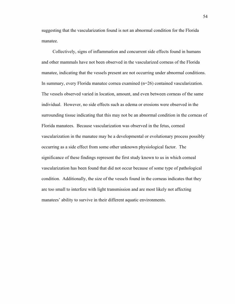
54
suggesting that the vascularization found is not an abnormal condition for the Florida
manatee.
Collectively, signs of inflammation and concurrent side effects found in humans
and other mammals have not been observed in the vascularized corneas of the Florida
manatee, indicating that the vessels present are not occurring under abnormal conditions.
In summary, every Florida manatee cornea examined (n=26) contained vascularization.
The vessels observed varied in location, amount, and even between corneas of the same
individual. However, no side effects such as edema or erosions were observed in the
surrounding tissue indicating that this may not be an abnormal condition in the corneas of
Florida manatees. Because vascularization was observed in the fetus, corneal
vascularization in the manatee may be a developmental or evolutionary process possibly
occurring as a side effect from some other unknown physiological factor. The
significance of these findings represent the first study known to us in which corneal
vascularization has been found that did not occur because of some type of pathological
condition. Additionally, the size of the vessels found in the corneas indicates that they
are too small to interfere with light transmission and are most likely not affecting
manatees’ ability to survive in their different aquatic environments.

55
Figure 2-1: Florida manatee eye with ocular tissue and fat roptic nerve. The eye is being compared to a dim
=100 mm
emoved revealing the eye and e for size comparison.

56
A
B
C
Figure 2-2: These histological pictures of MNW-9111 show (A) a long blood vessel in the stroma (blue) that approaches the anterior epithelium (purple) at 100X, (B) blood vessel penetration between the stroma and anterior epithelium at 200X, and (C) close detail of the vessels present in the stroma at 400X.

57
C
B
A
Figure 2-3: These pictures were taken of MSW-9114 of the same blood vessel at varying magnifications to show the penetration of this vessel into the anterior epithelium (purple): (A) 200X, (B) 400X, and (C) 1000X.

58
B
A
Figure 2-4: (A) The arrangement of smaller vessels near the anterior epithelium (purple)
from the larger trunk vessel in the stroma (blue) at 200X, specimen MSW-9114. (B) This picture of UCF-9114 shows the smaller vessels (see arrow) that were outlined within the stroma for three-dimensional reconstruction (200X).

59
B
A
Figure 2-5: Pictures from MNW-9016 showing (A) a blood vessel passing through the
stroma (blue) at 200X, and (B) red blood cells present in the vessel as it penetrates the anterior epithelium (purple) at 1000X.

60
B
A
Figure 2-6: These pictures were taken from the unknown specimen. (A) Shows the edge of the limbal vascular arrangement at the peripheral cornea (200X), and (B) larger vessels that go right up to the anterior epithelium at 200X.

61
D
FE
C
BA
Figure 2-7: Anatomical orientation of manatee eyes as they are described in three-dimensional reconstruction: (A) left eye orientation (face-on view), (B) right eye orientation (face-on view), (C) dorsal eye orientation, (D) ventral eye orientation, (E) lateral eye orientation (or horizontal view if left or right is unknown), and (F) medial eye orientation (or horizontal view if left or right is unknown).

62
Figure 2-8: MNW-9016, (A) face-on view of vessels and corneal tissue, (B) face-on
view of only the vessels, (C) dorsal view of vessels and corneal tissue, (D) dorsal view of only the vessels, (E) horizontal view of vessels and corneal tissue, and (F) horizontal view of only the vessels at 25X (arrow indicates anterior position).
E
D
F
C
BA

63
E
D
F
C
BA
Figure 2-9: MNW-9022(R), (A) face-on view of vessels and corneal tissue, (B) face-on view of only the vessels, (C) dorsal view of vessels and corneal tissue, (D) dorsal view of only the vessels, (E) lateral view of vessels and corneal tissue, and (F) lateral view of only the vessels at 25X (arrow indicates anterior position).

64
Figure 2-10: MNW-9111, (A) face-on view of vessels and corneal tissue, (B) face-on
view of only the vessels, (C) ventral view of vessels and corneal tissue, (D) ventral view of only the vessels, (E) medial view of vessels and corneal tissue, and (F) medial view of only the vessels at 25X (arrow indicates anterior position).
E
D
F
C
A B

65
Figure 2-11: MNW-9116, (A) face-on on view of vessels and corneal tissue, (B) face-on
view of only the vessels, (C) horizontal view of vessels and corneal tissue, and (D) horizontal view of only the vessels at 25X.
DC
BA

66
Figure 2-12: MSE-9101, (A) face-on view of vessels and corneal tissue, (B) face-on
view of only the vessels, (C) ventral view of vessels and corneal tissue, (D) ventral view of only the vessels, (E) horizontal view of vessels and corneal tissue, and (F) horizontal view of only the vessels at 25X (arrow indicates anterior position).
E
D
F
C
A B

67
Figure 2-13: MSE-9105, (A) face-on view of vessels and corneal tissue, (B) face-on
view of only the vessels, (C) ventral view of vessels and corneal tissue, (D) ventral view of only the vessels, (E) medial view of vessels and corneal tissue, and (F) medial view of only the vessels at 25X (arrow indicates anterior position).
E
D
F
C
BA

68
Figure 2-14: MSE-9107, (A) face-on view of vessels and corneal tissue, (B) face-on
view of only the vessels, (C) dorsal view of vessels and corneal tissue, (D) dorsal view of only the vessels, (E) horizontal view of vessels and corneal tissue, and (F) horizontal view of only the vessels at 25X (arrow indicates anterior position).
E
D
F
C
BA
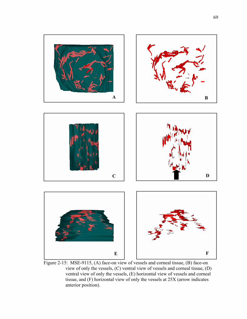
69
Figure 2-15: MSE-9115, (A) face-on view of vessels and corneal tissue, (B) face-on
view of only the vessels, (C) ventral view of vessels and corneal tissue, (D) ventral view of only the vessels, (E) horizontal view of vessels and corneal tissue, and (F) horizontal view of only the vessels at 25X (arrow indicates anterior position).
E
D
F
C
BA

70
Figure 2-16: MSE-9131, (A) face-on view of vessels and corneal tissue, (B) face-on
view of only the vessels, (C) dorsal view of vessels and corneal tissue, (D) dorsal view of only the vessels, (E) horizontal view of vessels and corneal tissue, and (F) horizontal view of only the vessels at 25X (arrow indicates anterior position).
E
D
F
C
BA

71
Figure 2-17: MSW-9114, (A) face-on on view of vessels and corneal tissue, (B) face-on
view of only the vessels, (C) ventral view of vessels and corneal tissue, (D) ventral view of only the vessels, (E) horizontal view of vessels and corneal tissue, and (F) horizontal view of only the vessels at 25X (arrow indicates anterior position).
E
D
F
C
BA

72
Figure 2-18: MSW-9131(R), (A) face-on view of vessels and corneal tissue, (B) face-on
view of only the vessels, (C) ventral view of vessels and corneal tissue, (D) ventral view of only the vessels, (E) lateral view of vessels and corneal tissue, and (F) lateral view of only the vessels at 25X (arrow indicates anterior position).
E
D
F
C
BA
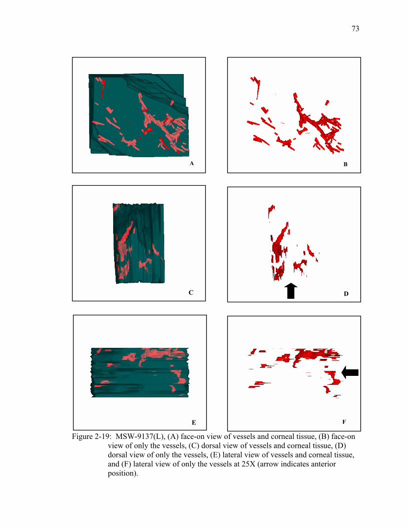
73
Figure 2-19: MSW-9137(L), (A) face-on view of vessels and corneal tissue, (B) face-on
view of only the vessels, (C) dorsal view of vessels and corneal tissue, (D) dorsal view of only the vessels, (E) lateral view of vessels and corneal tissue, and (F) lateral view of only the vessels at 25X (arrow indicates anterior position).
E
D
F
C
BA

74
Figure 2-20: MSW-9137(R), (A) face-on view of vessels and corneal tissue, (B) face-on
view of only the vessels, (C) dorsal view of vessels and corneal tissue, (D) dorsal view of only the vessels, (E) lateral view of vessels and corneal tissue, and (F) lateral view of only the vessels at 25X (arrow indicates anterior position).
E
D
F
C
BA

75
Figure 2-21: MSW-9141, (A) face-on view of vessels and corneal tissue, (B) face-on
view of only the vessels, (C) ventral view of vessels and corneal tissue, (D) ventral view of only the vessels, (E) horizontal view of vessels and corneal tissue, and (F) horizontal view of only the vessels at 25X (arrow indicates anterior position).
E
D
F
C
BA
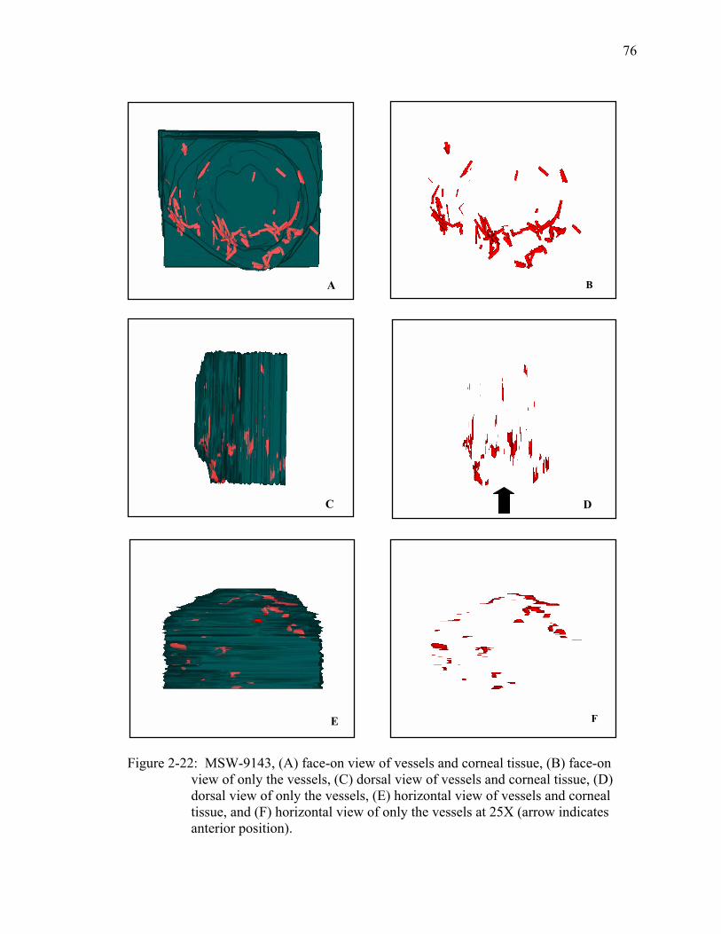
76
E
D
F
C
BA
Figure 2-22: MSW-9143, (A) face-on view of vessels and corneal tissue, (B) face-on view of only the vessels, (C) dorsal view of vessels and corneal tissue, (D) dorsal view of only the vessels, (E) horizontal view of vessels and corneal tissue, and (F) horizontal view of only the vessels at 25X (arrow indicates anterior position).
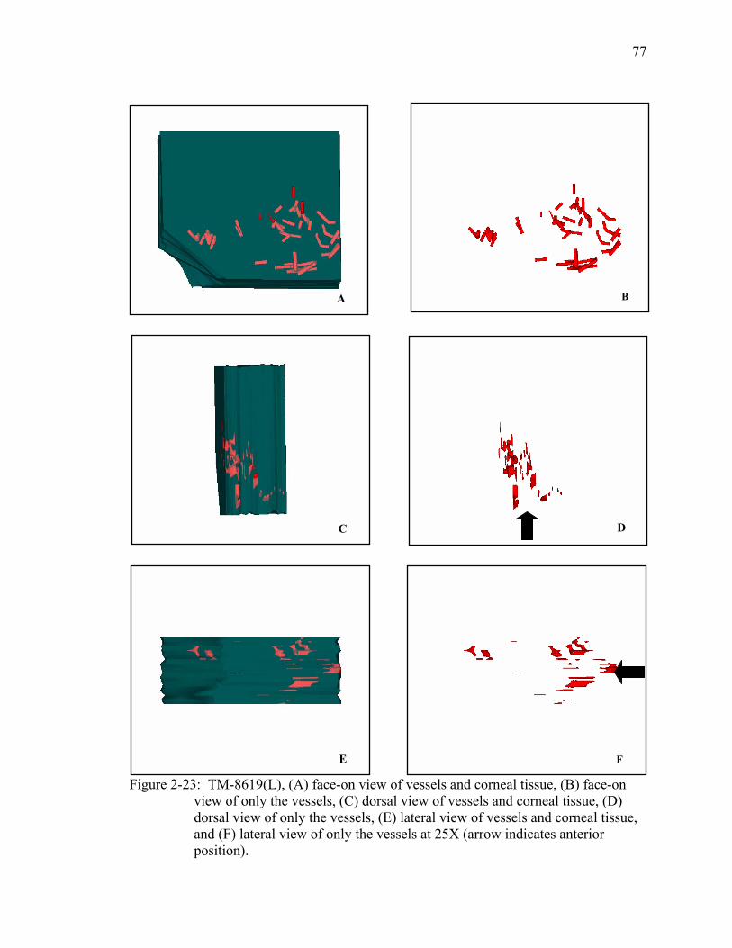
77
Figure 2-23: TM-8619(L), (A) face-on view of vessels and corneal tissue, (B) face-on
view of only the vessels, (C) dorsal view of vessels and corneal tissue, (D) dorsal view of only the vessels, (E) lateral view of vessels and corneal tissue, and (F) lateral view of only the vessels at 25X (arrow indicates anterior position).
E
D
F
C
BA

78
E
D
F
C
BA
Figure 2-24: TM-8619(R), (A) face-on view of vessels and corneal tissue, (B) face-on view of only the vessels, (C) dorsal view of vessels and corneal tissue, (D) dorsal view of only the vessels, (E) lateral view of vessels and corneal tissue, and (F) lateral view of only the vessels at 25X (arrow indicates anterior position).

79
Figure 2-25: TM-8630(L), (A) face-on view of vessels and corneal tissue, (B) face-on
view of only the vessels, (C) ventral view of vessels and corneal tissue, (D) ventral view of only the vessels, (E) medial view of vessels and corneal tissue, and (F) medial view of only the vessels at 25X (arrow indicates anterior position).
E
D
F
C
BA

80
Figure 2-26: TM-8630(R), (A) face-on view of vessels and corneal tissue, (B) face-on
view of only the vessels, (C) ventral view of vessels and corneal tissue, (D) ventral view of only the vessels, (E) medial view of vessels and corneal tissue, and (F) medial view of only the vessels at 25X (arrow indicates anterior position).
E
D
F
C
BA

81
Figure 2-27: UCF-9114, (A) face-on view of vessels and corneal tissue, (B) face-on view
of only the vessels, (C) dorsal view of vessels and corneal tissue, (D) dorsal view of only the vessels, (E) horizontal view of vessels and corneal tissue, and (F) horizontal view of only the vessels at 25X (arrow indicates anterior position).
E
D
F
C
BA

82
Figure 2-28: UCF-9137, (A) face-on view of vessels and corneal tissue, (B) face-on view
of only the vessels, (C) ventral view of vessels and corneal tissue, (D) ventral view of only the vessels, (E) horizontal view of vessels and corneal tissue, and (F) horizontal view of only the vessels at 25X (arrow indicates anterior position).
E
D
F
C
BA

83
Figure 2-29: UCF-9138, (A) face-on view of vessels and corneal tissue, (B) face-on view
of only the vessels, (C) ventral view of vessels and corneal tissue, (D) ventral view of only the vessels, (E) horizontal view of vessels and corneal tissue, and (F) horizontal view of only the vessels at 25X (arrow indicates anterior position).
E
D
F
C
BA

84
E
D
F
C
BA
Figure 2-30: UCF-9141, (A) face-on view of vessels and corneal tissue, (B) face-on view of only the vessels, (C) dorsal view of vessels and corneal tissue, (D) dorsal view of only the vessels, (E) horizontal view of vessels and corneal tissue, and (F) horizontal view of only the vessels at 25X (arrow indicates anterior position).
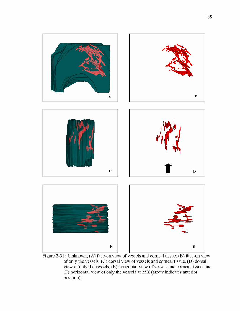
85
Figure 2-31: Unknown, (A) face-on view of vessels and corneal tissue, (B) face-on view
of only the vessels, (C) dorsal view of vessels and corneal tissue, (D) dorsal view of only the vessels, (E) horizontal view of vessels and corneal tissue, and (F) horizontal view of only the vessels at 25X (arrow indicates anterior position).
E
D
F
C
BA

86
Figure 2-32: Fetus(L), (A) face-on view of vessels and corneal tissue, (B) face-on view
of only the vessels, (C) dorsal view of vessels and corneal tissue, (D) dorsal view of only the vessels, (E) lateral view of vessels and corneal tissue, and (F) lateral view of only the vessels at 25X (arrow indicates anterior position).
E
D
F
C
BA

87
Figure 2-33: Fetus(R), (A) face-on view of vessels and corneal tissue, (B) face-on view
of only the vessels, (C) ventral view of vessels and corneal tissue, (D) ventral view of only the vessels, (E) lateral view of vessels and corneal tissue, and (F) lateral view of only the vessels at 25X (arrow indicates anterior position).
E
D
F
C
BA
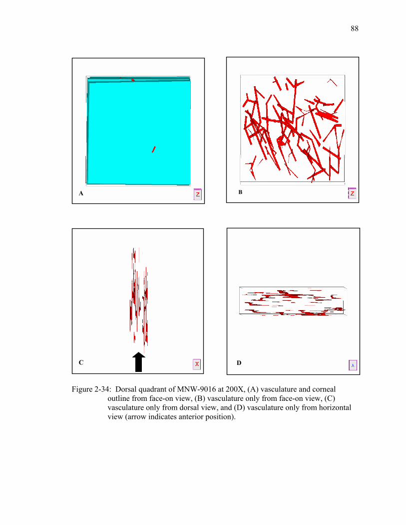
88
DC
BA
Figure 2-34: Dorsal quadrant of MNW-9016 at 200X, (A) vasculature and corneal outline from face-on view, (B) vasculature only from face-on view, (C) vasculature only from dorsal view, and (D) vasculature only from horizontal view (arrow indicates anterior position).
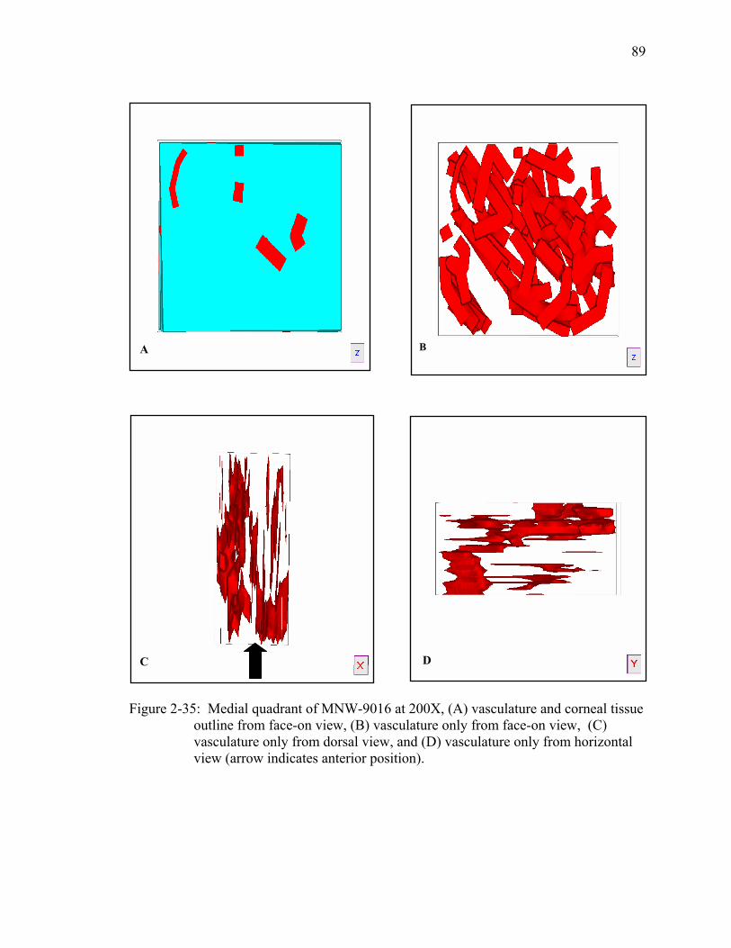
89
DC
BA
Figure 2-35: Medial quadrant of MNW-9016 at 200X, (A) vasculature and corneal tissue outline from face-on view, (B) vasculature only from face-on view, (C) vasculature only from dorsal view, and (D) vasculature only from horizontal view (arrow indicates anterior position).

90
DC
BA
Figure 2-36: Dorsal quadrant of MSW-9114 at 200X, (A) vasculature and corneal outline from face-on view, (B) vasculature only from face-on view, (C) vasculature only from dorsal view, and (D) vasculature only from horizontal view (arrow indicates anterior position).

91
DC
BA
Figure 2-37: Ventral quadrant of MSW-9114 at 200X, (A) vasculature and corneal tissue outline from face-on view, (B) vasculature only from face-on view, (C) vasculature only from dorsal view, and (D) vasculature only from horizontal view (arrow indicates anterior position).

92
DC
BA
Figure 2-38: Dorsal quadrant of TM-8619(L) at 200X, (A) vasculature and corneal outline from face-on view, (B) vasculature only from face-on view, (C) vasculature only from dorsal view, and (D) vasculature only from medial view (arrow indicates anterior position).

93
DC
BA
Figure 2-39: Medial quadrant of TM-8619(L) at 200X, (A) vasculature and corneal outline from face-on view, (B) vasculature only from face-on view, (C) vasculature only from dorsal view, and (D) vasculature only from medial view (arrow indicates anterior position).

94
DC
BA
Figure 2-40: Ventral quadrant of TM-8619(L) at 200X, (A) vasculature and corneal outline from face-on view, (B) vasculature only from face-on view, (C) vasculature only from dorsal view, and (D) vasculature only from medial view (arrow indicates anterior position).

95
DC
BA
Figure 2-41: Lateral quadrant of TM-8619(L) at 200X, (A) vasculature and corneal outline from face-on view, (B) vasculature only from face-on view, (C) vasculature only from dorsal view, and (D) vasculature only from medial view (arrow indicates anterior position).

CHAPTER 3 VASCULAR ENDOTHELIAL GROWTH FACTOR RECEPTOR-1 AND -2 IN THE
FLORIDA MANATEE CORNEA
Introduction
Cytokines are very small, nonstructural soluble proteins or glycoproteins made by
leukocytes and other cell types. Cytokines act as communicators between cells, and most
cytokines help regulate host responses to pathogens, disease, infection, or inflammation
(Fitzgerald et al. 2001; Dinarello 2000). Cytokines have often been compared to
hormones. However, this is not accurate because highly specialized tissues express
hormones and almost every tissue in the body makes cytokines. Hormones are also the
primary product produced by the cell, while cytokines account for only a small portion of
cell output (Dinarello 2000).
Cytokines bind to specific receptors located on the surface of target cells and they
display four key features: pleiotropy, redundancy, potency, and act as a network or
cascade. Because the receptors of cytokines are expressed on a variety of cell types and
their activated signaling pathways increase gene expression, they are pleiotropic.
Cytokines are redundant because there are many similarities in the amino acid sequences
of receptors. Cytokine receptors have a high affinity to ligands, which makes them
potent, act in synchronization, and counter-regulate inhibitory cytokines (Fitzgerald at al.
2001).
Environmental influences have the greatest effect on the function of cytokines,
being most commonly demonstrated with inflammation. A variety of cytokines and their
96

97
inhibitors can be detected at the area of inflammation. For example, anti-inflammatory
agent anti-TNF, the anticytokine that increases production of IL-1 receptor antagonist
(IL-1Ra), and the inhibitory cytokine IL-10 can be found at the same time and result in an
outcome that will be determined by this balancing of cytokines. There are cytokines that
act as inducers including IL-1, TNF, and IFNγ. Each of these cytokines can induce
cytokine gene expression and are called proinflammatory cytokines. Inhibitors of
cytokine gene expression also exist, such as the glucocorticoid hormones and synthetic
steroids, which are used as immunosuppressants and anti-inflammatory drugs. Cytokines
are also known to play important roles in blood vessel development (Fitzgerald et al.
2001).
Angiogenesis is the biological process that produces new blood vessels from
existing vasculature (Nicosia and Villaschi 1999; Battegay 1995; Folkman 1995; Schultz
and Grant 1991). Angiogenesis occurs in almost every tissue and is a necessary process
that is critical in normal conditions such as development, reproduction, healing of
wounds, and bone repair (Battegay 1995; Adamis et al. 1994). Angiogenesis can also
occur under pathological conditions such as cancer, tumor growth and metastasis,
rheumatoid arthritis, trauma, age-related macular degeneration, ischemic heart disease,
ischemic peripheral vascular disease, diabetic retinopathy or infection (Rosen 2002;
Schultz and Grant 1991).
The complex process of angiogenesis is highly regulated by pro-angiogenic and
anti-angiogenic growth factors. Several growth factors are very specific for the
endothelium (such as vascular endothelial growth factor, VEGF) and others carry out
several different functions (such as matrix metalloproteinases, MMPs) (Rosen 2002).

98
Endothelial cells separate and grow in an avascular tissue, and then unite to form a
primitive tubular network during early blood vessel formation. This early network of
vessels includes the aorta and major veins. Interconnected branching patterns of vessels
form through angiogenic remodeling that are characteristic of mature vasculature. At this
stage, endothelial cells join together securely with supporting cells to form mature vessel
walls (Yancopoulos et al. 2000).
The process of angiogenesis is a series of involved steps. Initially, diseased or
injured cells release pro-angiogenic growth factors into surrounding tissue, such as
vascular endothelial growth factor (VEGF), fibroblast growth factor (FGF), or platelet-
derived growth factor (PDGF). Angiogenic growth factors are then released into the
surrounding environment and bind to and activate existing endothelial cells of nearby
blood vessels. The newly activated endothelial cells signal the nucleus, which initiates
the production of enzymes such as MMPs. The extracellular matrix surrounding present
blood vessel is broken down by the enzymes, making it possible for endothelial cells to
enter the matrix and divide. These endothelial cells then travel towards the growth factor
stimulus, which is mediated by integrins (adhesion molecules) at the cell surface.
Endothelial cells create new blood vessels by forming hollow tubes. Blood begins to
circulate when individual vessels connect and form loops or networks, which can now
carry blood to the tissue that originally released the pro-angiogenic growth factors (Rosen
2002). These vessels sprout into previously avascular tissue (Yancopoulos et al. 2000) as
a result of endothelial cell migration and proliferation and further develop (Nicosia and
Villaschi 1999). This process is referred to as angiogenic sprouting and is responsible for

99
vascularizing some tissues throughout normal development (i.e., neural tube and retina),
as well as in pathological conditions (i.e., tumors) (Yancopoulos et al. 2000).
Perhaps the most important determinant in blood vessel formation during
angiogenesis is vascular endothelial growth factor, VEGF. It is distinguished for its
capacity to cause vascular leakage and permeability, in addition to its capacity to promote
vascular endothelial cell production (Yancopoulos et al. 2000). VEGF is a cytokine that
is an endothelial cell mitogen and a heparin-binding, dimeric protein (Miller et al. 1994;
Klagsbrun and D’Amore 1991) and is necessary for the initiation of immature vessel
formation during vasculogenesis or angiogenesis (Yancopoulos et al. 2000). VEGF is
able to stimulate endothelial migration, proliferation, and proteolytic activity and has a
peptide that is signaled through numerous pathways (Dunk and Ahmed 2001). VEGF is
made by a variety of cells, including endothelial cells, smooth muscle cells, and
fibroblasts (Nicosia and Villaschi 1999).
The VEGF gene is composed of eight exons that are separated by seven introns.
Alternative splicing of the VEGF gene produces four isoforms, including VEGF121,
VEGF189, VEGF206, and VEGF165 (Nicosia and Villaschi 1999). Five VEGF related
genes have been discovered (VEGF-B, VEGF-C, VEGF-D, VEGF-E, and placenta
growth factor) each producing glycosylated dimers that are secreted after cleavage of
their signal peptide (Rosen 2002). VEGF binds to two high affinity tyrosine kinase
receptors, VEGF receptor 1 (VEGFR-1/Flt) and VEGF receptor 2 (VEGFR-2/KDR)
(Beck and D’Amore 2001; Dunk and Ahmed 2001). Both VEGFR-1 and VEGFR-2 bind
VEGF with a high affinity and have seven immunoglobulin-like domains, one
transmembrane region, and a tyrosine kinase sequence that is interrupted by a kinase

100
insert domain. Expression of VEGFR-1 and 2 is restricted to the vascular endothelium.
It is currently believed that VEGFR-1 and 2 mediate all VEGF effects on vascular
endothelium. When VEGF binds to VEGFR-1 and 2, pro-angiogenic signals are
transmitted by the receptors to proteins located downstream and a signal cascade is
initiated (Rosen 2002). An additional receptor for VEGF is neuropilin-1. It modulates
the interaction between VEGF and VEGFR-2 (Ortega et al. 1999).
The expression of VEGFR-1 in naive cells mediates cell migration. While both
VEGF receptors are tyrosine phosphorylated, the phosphorylation of VEGFR-1 is not
required for function in vivo. The Src family of non-receptor tyrosine kinases comprises
several highly homologous kinases, including Fyn and Yes (Bevilagua et al. 2003), and
are phosphorylated in response to VEGF/VEGFR-1 but not to VEGF/VEGFR-2
interactions (Ortega et al. 1999). In contrast, phosphorylated VEGFR-2 links with Shc
(adaptors in tyrosine phosphorylation signal transduction pathways) (Lee et al. 2004),
Grb2 (domain-containing adapter proteins) (Schulze and Mann 2003), and Nck (a protein
required for translation and mediating signaling from the plasma membrane) (Kebache et
al. 2003), but also with phosphatases of SHP-1 (a protein-tyrosine phosphatase that is a
negative regulator of multiple signal transduction pathways) (Frank et al. 2003) and SHP-
2 (a protein-tyrosine phosphatase that plays an important role in intracellular signaling
brought forth by growth factors, hormones, and cytokines) (Tartaglia et al. 2004), and
initiates the phosphorylation of signaling molecules such as mitogen-activated protein
kinases (MAPKs), which transmit signals from the membrane to the nucleus in response
to growth factors and cellular stress (McMullen et al. 2003) through the stimulation of
Raf, a protein that responds to growth factor signals (Weber et al. 2003). VEGF has also

101
been found to induce the tyrosine phosphorylation and recruitment of focal adhesion
kinase (Ortega et al. 1999). Additionally, it has been shown that VEGFR-1 mediates
monocyte migration leading to the induction of nitric oxide (NO) production (Dunk and
Ahmed 2001).
Angiogenesis can occur in endothelial cells during such events as tumor
progression, diabetic retinopathy, and rheumatoid arthritis. Local angiogenesis may be
due to the release of soluble mediators by the involved tissues. At this point there is a
switch in the phenotype of the quiescent endothelial cell to an activated phenotype. The
endothelial cells are then able to respond to mitogenic signals (Ortega et al. 1999). There
is a larger increase in the upregulation of genes modulated by VEGFR-2, than with
VEGFR-1. These induced genes include growth factors, protease inhibitors, receptors,
transcription factors, and actin-binding proteins (Yang et al. 2002). At this point,
mitogenic growth factors are released and these change the activated phenotype to an
angiogenic phenotype. Interactions between angiogenic growth factors and their
receptors provide the signals for cell migration, proliferation, and differentiation to form
new vessels (Ortega et al. 1999).
Corneal angiogenesis is often used as a model to investigate other types of
vascularization. The causes of corneal vascularization are varied and in humans can
include infections (such as trachoma and herpes), immunologic processes, alkali burns,
toxic and nutritional deficiencies, and the wearing of contact lenses. In experimental
settings, corneal vascularization can be induced through chemical, microbiological, and
physical injuries, nutritional deficiencies, and immunological reactions (Klintworth
1991). Rabbits have most often been used to study experimentally induced corneal
102
vascularization. After an injury of some type has occurred in the cornea, cell migration
and mitosis are involved in healing of the epithelial corneal surface. If abrasion occurs,
cell migration takes place almost immediately. In order to return the epithelium to its
normal visual thickness, cell division follows. Cell migration can heal small injuries
within 24 hours. If a larger portion of the anterior epithelium is injured, the limbus is the
main source for production of epithelial stem cells. Endothelial cells are not able to
respond as quickly to cell loss. Endothelial cells can produce edema, which results in
corneal disease (Samuelson 1999).
The stromal edema that occurs with corneal vascularization aids in the growth of
blood vessels in the cornea and is usually an inflammatory process. There is an increase
in vascular permeability due to the inflammatory response as a result of endothelial cell
contractions between endothelial cell junctions. There are many inflammatory mediators
that are involved in this process including histamine, serotonin, ADP, prostaglandins,
many cytokines, free radicals, and immune aggregates. However, several factors can
help reverse the process including epinephrine, cortisone, and some drugs such as
antihistamines and glucocorticoids (Klintworth 1991). Injuries to the cornea result in
stromal edema with neutrophils moving to the source of the injury to release enzymes and
chemotaxic agents. Anteriorly, epithelial wound healing begins within one hour of the
injury and wing layer cells flatten and slide next to the injury site in order to cover the
area within 24 to 96 hours. The epithelium returns to its full thickness by the action of
basal stem cells of the limbus. Production of the stromal fibroblast takes place after 24
hours (Ahmadi and Jakobiec 2002).

103
Leukocytes move in the extravascular space within 24 hours and move through the
corneal stroma to the site of injury. The first new vessels appear after 27 hours.
Advancing new vessels remain permeable to fluid, plasma proteins, and smaller proteins.
Forming corneal blood vessels do not have firm intercellular complexes, whereas well
formed corneal vessels in advanced stages of wound healing do not leak. Once new
vessels have developed, the basal lamina breaks down, allowing the endothelial cells to
move into the extracellular matrix. Progression of these endothelial cells at the end of the
developing new blood vessels extends toward the damaged area (Klintworth 1991). The
movement of endothelial cells, which elongate and form solid sprouts developing lumen,
follows this. Pairs of sprouts form loops and blood flows, and the process is repeated
from the top of each loop (Pepose and Ubels 1992). New blood vessels can emerge
within 33 hours after injury. A network of vessels is formed that becomes linked and
these vessels then begin to pass materials between the new connections (Klintworth
1991).
Experimental induction of corneal neovascularization has been used for years to
describe vascularization induced by tumors, inflammatory and growth factors, and
corneal injuries (Ueda et al. 1997). There are two commonly used bioassays to measure
angiogenesis, the developing chick chorioallantoic membrane (CAM) and implantation in
the rabbit cornea. Angiogenic materials are modified during normal development of
vascular patterns in the CAM assay and with cornea implantation, the growth of blood
vessels is induced from existing normal vessels (Gaudric et al. 1992).
Many animal models demonstrate that cytokines and angiogenic factors are
involved in induced ocular neovascularization. The most commonly used animals are

104
non-human primates and small mammals such as rabbits and rats. VEGF has been shown
to increase vascular permeability and angiogenesis in the iris of cynomolgus monkeys
(Maccaca fascicularis). Induced ocular angiogenesis through laser photocoagulation of
retinal veins in cynomolgus monkeys showed a temporal association with changing
aqueous VEGF levels and iris neovascularization. No VEGF was detected prior to
photocoagulation, and afterwards levels of VEGF increased dramatically. These findings
showed a relationship between VEGF (a secreted angiogenic factor) and increased
development of ocular neovascularization (Miller et al. 1994). Not only has VEGF been
found to increase with iris vascularization, but also with retinal neovascularization.
Retinal ischemia produced by retinal vein occlusion in monkeys produces a large increase
of VEGF in the vitreous and anterior chamber of the eye (Folkman 1995).
Additional studies of the angiogenic cytokine factor VEGF, involved in induced
corneal vascularization, have shown interesting results. VEGF has been demonstrated to
advance a variety of steps in angiogenesis including proliferation, migration, proteolytic
activity, and capillary tube development of endothelial cells in human corneas. In normal
corneas, it was found that corneal endothelial cells and vascular endothelial cells of
limbal vessels made VEGF. In inflamed corneas, VEGF expression was much higher in
corneal endothelial cells, keratocytes, and vascular endothelial cells of new vessels.
These findings indicate that VEGF is expressed at higher levels in inflamed and
vascularized human corneas than in normal corneas and suggest that VEGF is involved in
the pathogenesis of corneal vascularization (Philipp et al. 2000). Differences seen in
VEGF levels before and after corneal injury have also been described in male Sprague-
Dawley rats. Induced corneal vascularization occurred in these rats by implantation of

105
anti-VEGF antibody or non-immune goat IgG pellets. Levels of VEGF in the rat corneas
increased 10-fold after being wounded. This information indicates that VEGF is needed
for wound and inflammation-related corneal vascularization (Amano et al. 1998).
Corneal vascularization induced in rats by cautery of silver nitrate shows
inflammation and vascularization that can be easily studied in order to investigate
inflammatory gene expression and the role of leukocytes in inflammatory angiogenesis.
Silver nitrate cautery of the rat cornea resulted in blood vessel growth from the limbus to
the lesion. Using the corneal micropocket assay, it was found that basic fibroblast growth
factor (bFGF) and VEGF did induce angiogenesis from the limbus to the implant. Basic
FGF was released after injury to promote healing and VEGF production was increased
subsequently. It was also found that leukocytes associated with inflammation were the
primary source of VEGF in the rat cornea after cautery. During inflammation, cytokines
helped regulate the expression of VEGF (Edelman et al. 1999).
Other cytokines besides VEGF have been shown to affect corneal angiogenesis
because a balance is needed between angiogenic factors and inhibitors (Yoshida et al.
1998). CAM assays were used to describe the effects of developing vascular patterns of
rabbits induced with corneal vascularization by implanted pellets containing bFGF.
Blood vessel growth was observed over progressive days after the implantation of the
pellet. However, inflammation in the cornea did not stimulate angiogenesis (as it might
with VEGF), but vascularization was induced by bFGF (D’Amato 1994). Interleukin
(IL)-8 appears to induce angiogenic factors in induced ocular vascularization. IL-8 is the
major regulator of a variety of inflammatory and proliferative response genes and is

106
found at higher levels in human patients with vascularization than without (Yoshida et al.
1998).
An interesting case of corneal vascularization was originally discovered by
Samuelson et al. (1997 and 1994) in which blood vessels were consistently present in the
anterior portion of the corneas in the Florida manatee. However, no signs of infection,
injury or other pathological conditions could be detected in the corneas. The reason for
the presence and function of the blood vessels is currently unknown. The objective of
this study is to investigate the presence or absence of vascular endothelial growth factor
receptors-1 and -2 in the corneas of the Florida manatee, Trichechus manatus latirostris,
through the use of immunohistochemistry. This will determine what role, if any,
VEGFR-1 and VEGFR-2 have in the angiogenic process occurring in the blood vessels
found in the cornea of these animals.
Materials and Methods
Specimen Collection
Florida manatee eyes were collected from Sea World of Florida in Orlando, Florida
within 24 hours after death. Each eye was labeled with an identification number given to
it by Sea World. Information including gender, size, and time when the animal died was
recorded. Manatee eyes from specimen TM-0310 (juvenile animal) were collected on
April 16, 2003 and eyes from specimen TM-0311 (fetal animal) were collected on April
23, 2003. Manatee eyes were placed in fresh 10% neutral buffered formalin solution
(Luna 1968).
Preparation of Eyes
All extraneous tissue surrounding the globe was removed using a scalpel. The
corneas were then removed from the globe by cutting 1 mm from the limbus into the

107
sclera with a sharp razor. A notch was cut into the limbus at 12:00 in the dorsal section
of each cornea in order to know orientation during histological examination.
Photographs were then taken of the corneas (see Figure 3-1). The remainder of the eye
was stored and the cornea was placed in a tissue cassette, labeled and then returned to the
10% neutral buffered formalin solution for a total of 24 hours. The tissue was changed to
phosphate buffered saline solution (PBS) for 12 hours and then processed.
Histology
In preparation for embedding, the corneas were dehydrated by placing them in
ascending concentrations of ethyl alcohol for an hour each: 70%, 80%, two changes of
95%, followed by three changes of 100%. The tissues were then cleared with two
changes of xylene and infiltrated with two changes of Fisher Tissue Prep at 57º Celsius.
The tissues were then embedded using Fisher Tissue Prep T565 in metal molds and
allowed to harden.
Using a Reichert-Jung 2030 microtome, the embedded corneas were cut in serial
dorsal/ventral sections at a thickness of 5 microns. The tissue ribbons were floated out on
a 60º Celsius water bath and placed on Fisherbrand Superfrost microscope slides (3 x 1”
x 1) with one section per slide. Each slide was labeled with the identification number of
the animal, order of section, and orientation of tissue. Twenty sections per eye where cut.
After cutting was completed, the slides were placed in a slide rack and into an oven at 60º
Celsius overnight to dry.
Immunohistochemistry
Sections were deparaffinized with three changes of xylene for five minutes each,
two changes of 100% ethanol alcohol, 95% ethanol alcohol, and distilled water for two
minutes each. The slides were rehydrated with Tris buffered saline (TBS) solution

108
(Humason 1972). The sections were then quenched in 3% hydrogen peroxide for 20
minutes. TBS was used to wash the slides three times for two minutes each. Blocking
was performed with 5% normal goat serum (53 µL of normal goat serum and 1000 µL of
Dako antibody diluent, no. S0809) (DakoCytomation, Carpinteria, California) for 20
minutes. The slides were then washed three times in TBS for two minutes each. A
primary antibody (Flt-1, polyclonal rabbit, catalog number SC316 or KDR, monoclonal
mouse, no. SC251, both from Santa Cruz Biotechnology, Santa Cruz, California) was
combined with the Dako antibody diluent at different concentrations of 1:50, 1:100, and
1:200. The Flt-1, polyclonal rabbit primary antibody was used for detection of VEGFR-1
and the KDR, monoclonal mouse primary antibody was used for VEGFR-2 detection.
The sections were incubated in a humidified chamber overnight at 10º Celsius. The
staining procedure was completed by using a detection kit from Dako (no. K0673). After
the sections were stained and rinsed twice with distilled water, twice with 95% ethanol
alcohol, and three times with xylene, they were coverslipped using Richard-Allan
Scientific mounting medium (Richard-Allan Scientific, Kalamazoo, Michigan).
Results
Vascular endothelial growth factor receptor-1 (VEGFR-1)
Both the juvenile (TM-0310) (see Figures 3-2, 3-3, and 3-4) and fetal (TM-0311)
(see Figures 3-6 and 3-7) animals had a positive reaction for VEGFR-1 in their corneal
tissue. The control samples (or negative control) had no positive reaction occur in them
(sees Figures 3-5 and 3-8). TM-0310 reacted positively in both corneas (left and right) at
1:50 and 1:100 concentrations of polyclonal rabbit primary antibody in Dako antibody
diluent. TM-0311 also showed positive reactions in both corneas at concentrations of
1:50 and 1:100, however; the positive reaction was more evident in the 1:100

109
concentration (Figure 3-6). Even though positive reactions for VEGFR-1 could be seen
in both individuals, the reaction was more evident in the juvenile manatee (TM-0310).
Preparation and fixation of corneal tissue with both animals was carried out in the same
way, so the difference seen in the degree of positive reaction between the animals was
most likely due to individual variation. An adult manatee cornea was not examined in
this study due to the difficulty in obtaining samples within 24 hours post mortem.
However, it would be beneficial to look at an adult cornea in the future.
With both the juvenile and fetal samples, the positive reaction was most apparent in
the stromal vessels and anterior epithelial vessels (Figures 3-3 and 3-6). Interestingly, no
positive reaction was observed in the limbal vessels of these animals (Figure 3-7). Only a
very slight positive reaction could be seen in the larger limbal vessels of the juvenile
animal, but it was minimal. It is possible that this may be an age characteristic. Again, it
would be beneficial to have an adult cornea for examination to investigate VEGFR-1
expression as a function of age.
Interestingly, in the juvenile sample (TM-0310), some larger vessels had no
positive reaction, while other larger vessels did (Figure 3-3). It is possible that the larger
vessels that did not exhibit a positive reaction were more established vessels and smaller
vessels were forming from these that were positive for VEGFR-1. Once again, it would
be beneficial to examine an adult cornea in order to understand what is occurring with
these larger vessels and if they are positive for VEGFR-1 or not.
Vascular endothelial growth factor receptor-2 (VEGFR-2)
With both the juvenile and fetal samples, no positive staining of VEGFR-2 was
observed in any of the immunohistological procedures carried out. The concentrations at
1:50, 1:100, and 1:200 of monoclonal mouse primary antibody to Dako antibody diluent

110
produced no reaction at all and the results were no different from that of the control
samples (negative control). The reaction was absent with respect to vessels in the cornea,
limbus, or conjunctiva. The experiment investigating VEGFR-2 was then carried out
using a positive control to check for possible defects in the monoclonal mouse primary
antibody or other substances used. Corneal tissue from a pitbull (canine) with glaucoma
and vascularization in the stroma and corneal tissue from a horse with keratitis and
vascularization throughout the entire cornea were used as positive controls. When the
immunohistochemical procedures were completed, these specimens did have a positive
reaction in their stromal vessels. This finding indicated that the procedure and substances
were not defective in some way. The experiment was then carried out a second time on
both of the manatee samples and again showed no positive reaction for VEGFR-2 (Figure
3-9).
Discussion
Of the two samples examined (TM-0310 and TM-0311), both had positive
reactions for VEGFR-1 in the corneal tissue at 1:50 and 1:100 concentrations of
polyclonal rabbit primary antibody in Dako antibody diluent. The juvenile sample (TM-
0310) contained more positive reactions than did the fetal sample (TM-0311). This may
be due to individual or age differences. To determine which, if not both are the case,
additional immunohistochemical procedures need to be carried out on more samples from
varying age groups. The difficulty with this goal, however, is obtaining samples from
manatees in different age groups within 24 hours after their death. In order for the
immunohistochemistry to be valid and work properly, the tissue samples must be
prepared and fixed immediately after death. It may be possible that an age pattern in

111
manatees could be discovered based on positive reactions of VEGFR-1 in corneal
vessels. If more positive reactions of VEGFR-1 are found in the cornea as the animal
ages, then some scale may be developed based on those results. Combined with the size
of their Descemet’s membrane and anterior lens capsule, it may be possible to determine
approximate age groups of manatees. Descemet’s membrane, the basement membrane of
the corneal endothelium, increases in thickness consistently over time with other
mammals and the anterior capsule of the lens, another basement membrane, also
increases in thickness with age. Even though these two ocular structures were not
examined in this study, it may be a possible path for investigation in the future for age
determination in the manatee.
With VEGFR-1, while positive reactions were found in the corneal tissue of both
samples including the stromal and anterior epithelial vessels and in the conjunctiva, no
comparable reactions of VEGFR-1 could be found in the limbal vessels. Even though we
had the advantage of examining the corneal, limbal, and associated conjunctival vessels,
which collectively comprise the entire vascular bed for the anterior-most portion of the
eye of the manatee and may be working in conjunction with each other to carry out
undetermined vascular functions within this region of the eye, we did not focus on the
conjunctiva. Since the conjunctiva and cornea are in such close proximity and can carry
out associated functions, it may be important to look at the conjunctiva more closely in
the future to determine whether these vessels play a similar role as the corneal vessels,
and what role that might be. By examining this entire vascular bed making up the
corneal, limbal, and conjunctival vessels, it may be inferred that this section of the
anterior globe is working together to facilitate some unknown component.

112
Neither the juvenile nor fetal sample examined demonstrated any positive reaction
with VEGFR-2 at any concentration of monoclonal mouse primary antibody. While the
immunohistochemical procedures carried out examining VEGFR-2 appeared to be no
different from the negative control samples, we are confident that the procedures were
carried out correctly because positive controls that were used did result in positive
reactions of VEGFR-2 in corneal vessels. These positive controls both suffered from
pathological problems resulting in vascularization in their corneas. Still, there is a
possibility that the antibody for VEGFR-2 was not sensitive to the manatee. Once again,
additional tissue would be very interesting to test in the future, especially samples that
vary in age.
If the VEGFR-2 immunohistochemical findings for the manatee are correct, the
presence or lack of these VEGF receptors is a significant finding. Since the two receptor
tyrosine kinases of vascular endothelial growth factor (VEGF) are VEGFR-1 and
VEGFR-2 and they start the signaling pathways during vasculogenesis and pathological
angiogenesis (Meyer and Rahimi 2003), the action of VEGF is mainly restricted to
endothelial cells to induce such responses as cell proliferation, migration, survival,
differentiation, and permeability to macromolecules (Kojima-Yuasa et al. 2003; Meyer
and Rahimi 2003). VEGF is able to stimulate endothelial cells by binding to and
activating VEGFR-1/Flt-1 and VEGFR-2/FLK-1 (Meyer and Rahimi 2003). When
VEGF binds to its receptors, dimerization and activation of the inherent kinase results.
The VEGF receptors will then autophosphorylate and ensuing signal transduction then
occurs (Kojima-Yuasa et al. 2003). Therefore, the effects of VEGF are highly mediated

113
by VEGFR-1 and VEGFR-2 (Suzuma et al. 1998) and it may be possible that all the
effects of VEGF are mediated by these receptors (Rosen 2002).
Even though all the specific roles of VEGFR-1 and 2 have not been defined in the
current literature and are still being investigated, it has been suggested that VEGFR-1 has
several roles including signal transduction, activation of unique signaling proteins found
in endothelial cells (Meyer and Rahimi 2003), and cell migration (Ortega et al. 1999) and
can be expressed in both endothelial cells and pericytes (Suzuma et al. 1998).
Additionally, VEGFR-1 is thought to be responsible for chemotaxis (McLeod et al.
2002), induction of nitric oxide (NO) production in trophoblasts, and stimulation of
matrix metalloproteinase expression in vascular smooth muscle cells (Dunk and Ahmed
2001). VEGFR-2 is thought to be responsible for migration of endothelial cells (Meyer
and Rahimi 2003; Ortega et al. 1999) and VEGFR-2 is expressed in microvascular
endothelial cells (Suzuma et al. 1998). It also is thought that VEGFR-2 is the primary
signal transduction pathway controlling angiogenesis (Rosen 2002) and may be
responsible for the initiation of mitogenesis (McLeod et al. 2002). Both receptors are
very different in their ability to go through autophosphorylation and kinase activation in
that VEGFR-2 has the ability to undergo ligand-dependent tyrosine phosphorylation and
kinase activation and VEGFR-1 cannot (Meyer and Rahimi 2003). Based on this
information and VEGFR-2 not being found in the manatee corneas examined, we
postulate that the process of angiogenesis occurring in this tissue is a non-pathological
and non-inflammatory response to currently unknown factors, and most likely due to
developmental or evolutionary influences.

114
Interestingly, it has been shown that VEGF is expressed in normal endothelial and
epithelial cells in the cornea, throughout the epithelium of the cornea, and by vascular
endothelial cells in limbal vessels that have no pathological or experimentally induced
conditions occurring in them, suggesting that VEGF may have a maintenance role in the
cornea (Philipp et al. 2002; Kvanta et al. 2000). This additional evidence further suggests
that the receptors for VEGF found in the manatee corneas examined are occurring there
normally and not under conditions brought on by such factors as trauma, injury, or
infection.
The presence of the blood vessels in the corneas of all manatees examined thus far
still remains enigmatic. The normal cornea in most mammalian species is a transparent
avascular layer of the eye that is able to refract and transmit light, as well as provide
support for the contents within the globe. The cornea is able to transmit light clearly in
part because it lacks blood vessels, has a keratinized surface epithelium, and is pigmented
(Muller et al. 2003). The avascularity found in the cornea is maintained through such
components as the major keratan sulfate proteoglycans (including lumican, keratocan,
and mimecan) of the stroma and interfibrillar proteins (including collagens type VI and
XII) (Kurpakus et al. 1999). The arrangement of collagen and proteoglycans in the
stroma allows light to pass without restriction through the extracellular matrix aiding in
corneal transparency (Twining et al. 1999). It has also been thought that additional
proteoglycans in the aqueous humor may help maintain corneal clarity. The aqueous
humor acts as a circulatory system in the eye by clearing it of harmful agents and waste
products from the surrounding environment. The replacement of aqueous humor
connected with heparan sulfate bind to and prevents growth factors from potentially

115
causing vascularity within the cornea (Fannon et al. 2003). Other factors such as
transketolase and aldehyde dehydrogenase 3 located in corneal cells are also important in
maintaining corneal clarity because these cells are able to respond quickly to injuries or
infections (Twining et al. 1999). It is possible that there is an abnormality occurring with
the proteoglycans in the manatee cornea or even within the corneal cells themselves.
This could help explain why these vessels are found.
There are other factors to consider that may be causing this rare phenomenon.
There is the possibility that an undiscovered genetic adaptation may be occurring in the
manatee. For example, a recently discovered low-density receptor-related lipoprotein
called Lrp5 has been found to provide dual function in very different organ systems when
there is a defect. The Lrp5 protein functions as a Wnt coreceptor for Wnt proteins, which
help control many developmental processes such as mesoderm induction, cell fate
determination, limb patterning, joint formation, and organogenesis. Specifically, Lrp5
binds to Wnt proteins in osteoblasts and Lrp5 is needed for normal deterioration of
embryonic vasculature in the eye. Lrp5 and Wnt proteins work in conjunction to regulate
osteoblast proliferation and function, and eye vascularization during late ocular
development. Recent studies have discovered that Lrp5 becomes inactivated in human
patients with osteoporosis-pseudoglioma syndrome and is mutated in patients that have
high bone mass syndrome. Mice were used to further investigate phenotypes that
occurred when the Lrp5 protein was deficient. It was found that they displayed two
phenotypes; a low bone mass density caused by a reduction in bone formation and the
retention of their hyaloid vasculature in the eye throughout life (Kato et al. 2002).
Currently, it is not known if manatees have a deficient Lrp5 protein or some other

116
comparable genetic mutation. However, further investigations into correlations between
the Lrp5 protein and other vascular components may prove to be very insightful. It
should be noted that no hyaloid vasculature has been observed in the adult manatee eye.
This may rule out the possibility of an Lrp5 deficiency, but there are other aspects of the
manatee genome that have not been explored which may be causing the vascularization
observed in the cornea.
Corneal vascularization in the Florida manatee may be caused by some other factor
such as ocular hypoxia. Angiogenesis often takes place when there is cell replication,
inflammation and tissue repair, and tissue hypoxia. When such an event occurs and
vascularization follows, large amounts of cells in the same region compete for oxygen
and existing nutrients. Metabolites accumulate, which can change the cellular makeup
and even cause some cells to release factors (such as cytokines) that stimulate vascular
growth and development. In some scenerios in which tissue hypoxia occurs which results
in angiogenesis, low oxygen tension is thought to be a major contributing factor in
vascularization. Additionally, the corneal epithelium relies on atmospheric oxygen for
anaerobic metabolism of carbohydrates to provide energy needs. If some barrier or other
obstacle blocks this oxygen supply from occurring, corneal vascularization often results
(Klintworth 1991).
Ocular hypoxia is also known to alter gene expression. For example, a recently
discovered protein called inhibitory PAS (Per/Arnt/Sim-IPAS) is linked with low levels
of VEGF expression in the cornea during hypoxic conditions. When an IPAS antisense
oligonucleotide was used in a mouse cornea to produce angiogenesis under normal
oxygen conditions, stimulation of hypoxia-dependent VEGF gene expression took place.

117
This finding showed that IPAS negatively regulates angiogenesis and produces an
avascular phenotype (Makino et al. 2001). It could be possible that some type of hypoxic
conditions may be taking place in the corneas of manatees. Perhaps some of the fresh
water areas where the manatees are found may be too slow moving and even depleted of
oxygen, therefore preventing proper oxygen exchange from occurring between the water
and the anterior portion of the cornea. If there is a lack of oxygen, this could lead to
hypoxic conditions where blood vessels may develop. This may also explain why some
vessels have been observed developing in the conjunctiva of the manatee eye. In the
future, oxygen exchange rates or possible oxygen depletion could be tested by
investigating oxygen levels found in some of the waters throughout the state of Florida
(salt, fresh, and brackish waters alike). However, this is an unlikely explanation because
vascularization was observed in all manatee specimens examined and not just a few from
a certain area where low oxygen levels are suspected. A more likely explanation for the
vascularization observed may be caused by the unusually thick tear film found in the
manatee and other marine mammals such as the bottlenose dolphin (Dawson, personal
communication).
Perhaps the vascular bed found in the manatee cornea is an adaptation for this
aquatic mammal that allows it to have better control in dealing with changes in osmotic
conditions as it travels from fresh to salt to brackish waters and back again. Whatever the
cause for this vascularization found in the cornea of manatees, it definitely is a unique
and interesting characteristic that requires further investigation to better understand the
mechanisms causing such an occurrence, be it environmental, genetic, or evolutionary.
Learning more about this condition in a non-inflammatory and non-pathological setting

118
could possibly lead to a better understanding of how to treat and provide increased care
for other species that suffer from corneal vascularization associated with trauma, injury,
or infection.
C
BA
Figure 3-1: Florida manatee eye (A) showing the left cornea only and all other ocular tissue removed, (B) showing the right cornea only with all other ocular tissue removed, and (C) ocular tissue and fat removed revealing the eye and optic nerve compared to a dime for size comparison.

119
A
B
Figure 3-2: Pictures taken from TM-0310 showing a positive reaction of VEGFR-1 at (A) 20X with a 1:50 concentration of polyclonal rabbit primary antibody in Dako antibody diluent nicely indicating where the vessels are present and (B) 100X at 1:50 showing location of basal cells which are positive for VEGFR-1.

120
C
B
D
A
Figure 3-3: VEGFR-1 positive reactions in TM-0310 (A) showing the prevalence of the positive reaction at 200X with 1:50, (B) a larger vessel at 400X with 1:50 most likely more established with no positive reaction in it (as seen in smaller vessels) but can see positive reaction in anterior epithelium along basal layer, (C) a larger vessel at 200X with 1:100 that did have a positive reaction unlike in B, and (D) a branching vessel at 400X with 1:100 showing a positive reaction.

121
C
BA
Figure 3-4: VEGFR-1 positive reactions in TM-0310 (A) at 400X with 1:100 showing a prevalent positive reaction around the large vessel indicating that growth may still be occurring, (B) 400X with 1:100 showing good localization of positive reaction, and (C) 200X with 1:100 of a good overview with more vessels forming around already existing vessels, possibly indicating the process of angiogenesis taking place.

122
B
A
Figure 3-5: VEGFR-1 reactions in TM-0310 of control samples (A) at 20X and (B) 400X showing no positive reaction in either sample.

123
DC
BA
Figure 3-6: Pictures taken from TM-0311 showing a positive reaction of VEFGR-1 at (A) 20X with 1:100 concentration, indicating a good overview but not as strong of a positive reaction on periphery as with TM-0310, (B) 100X with 1:100, indicating a positive reaction but still not as strong as with TM-0310, (C) 200X with 1:100, indicating a positive reaction in larger vessels, and (D) 200X with 1:100, also indicating a positive reaction.

124
C
B
D
A
Figure 3-7: VEGFR-1 positive reactions in TM-0311 (A) at 400X with 1:100, showing a positive reaction that is similar to those seen in TM-0310, (B) 200X with 1:100, showing no reaction in limbal vessels, indicating that the vessels are most likely already developed and are not forming any new vessels, (C) 400X with 1:50, showing several vessels but not as strong of a positive reaction a seen in TM-0310, indicating individual differences in vessel development, and (D) 400X with 1:50, with no reaction at all in the limbus (colored cells are pigment cells).

125
B
A
Figure 3-8: VEGFR-1 reactions in TM-0311 of control samples (A) at 200X and (B) at 400X, showing no positive reactions, but showing the pigmentary patterns of melanocytes in the limbus.

126
C
BA
Figure 3-9: Pictures taken of TM-0310 showing no positive reactions of VEGFR-2 that appears to be different from the control samples at (A) 20X with a 1:100 concentration of monoclonal mouse antibody to Dako antibody diluent, (B) 100X with 1:100, and (C) 200X with 1:100.

CHAPTER 4 ADDITIONAL STUDIES
Corneal Vascularization in the Antillean Manatee
Since corneal vascularization was found in every specimen of the Florida manatees
examined, investigation additional non-Florida manatees could determine if similar or
even the same unusual occurrence was taking place. Robert K. Bonde of the United
States Geological Service/Sirenia Project located in Gainesville, Florida was able to
collect an eye from an Antillean manatee that was found stranded in Puerto Rico on July
21, 2003. The eye was immediately placed in 10% neutral buffered formalin after
necropsy and upon receiving the manatee eye on August 25, 2003, it was examined and
photographed (see Figure 4-1). The Antillean manatee eye was very similar in size and
shape to that of the Florida manatee. However, this cornea was much more opaque and
cloudy in appearance than any observed with the Florida manatee specimens. After
photographs of the cornea were taken, it was then processed histologically using the same
methods as with the Florida manatee corneas (see Chapter 2).
The histological findings of this cornea were very interesting. It too was
vascularized, but much more extensively than that observed in the Florida manatee
corneas (see Figures 4-2 through 4-6). The vessels appeared to be arranged randomly as
with the Florida manatee, but showed a much greater density, having been observed
throughout the entire corneal tissue. Once again, vessels were observed extending from
the stroma penetrating the anterior epithelium (see Figures 4-5 and 4-6). Furthermore,
the stromal collagen was arranged in a wavy pattern that was not observed in the Florida
127

128
manatee (see Figure 4-3). Unusual projections were also seen in the posterior stroma and
endothelium that were not seen in the Florida manatee (see Figure 4-8). It is currently
not known what function these projections have and whether these projections are found
in all Antillean manatees.
An additional feature that was observed in the Antillean manatee cornea and not in
the Florida manatee cornea was a light yellow layer on the outside of the tissue
surrounding the anterior epithelium that looked similar to adipose tissue (see Figure 4-7).
Additional slides were stained with PAS to detect the presence of fungi that might have
caused such an occurrence. The stain indicated that the sections were PAS negative for
fungi (see Figure 4-9). Currently, it is believed that this layer is mucus and may be more
predominant in the Antillean manatee than the Florida manatee because of the primarily
salt water habitat in which the Antillean manatee lives. At this point it is not possible to
speculate whether morphological features seen in this individual occur in all Antillean
manatee corneas. It would be very interesting and informative to examine additional
corneas from Antillean manatees and perform three-dimensional reconstructions, which
were not carried out on this specimen. It would be interesting to determine if any
patterns do exist and how much actual blood vessel coverage there is.
The presence of corneal vascularization in an Antillean manatee helps further
supports the premise that this is a normal process taking place in these animals especially
in the absence of any signs of inflammation, infection, or injury.
Ophthalmic Examinations in Two Captive Florida Manatees
Two captive Florida manatees, Hugh and Buffett, at Mote Marine Laboratory in
Sarasota, Florida had been trained by Dr. Gordon Bauer of New College and Debi
Colbert and Joe Gaspard of Mote Marine Laboratory to receive ophthalmic examinations.

129
The animals were trained to place their heads on a platform located directly next to the
concrete edge of their enclosure approximately six inches above the water’s surface.
They were also trained to keep their eyes open for up to a duration of thirty seconds while
remaining stationary. Dr. Chris Murphy from University of Wisconsin’s College of
Veterinary Medicine performed streak retinoscopy, infrared photorefraction, and
keratometry on the manatees’ eyes while they had their heads stationed on the platform.
Additionally, refraction was carried out under water through the use of streak
retinoscopy. It was found that both manatees had poor reflexes that were most likely
caused by their vascularized corneas and ophthalmic lesions. Small central cataracts
were observed in both eyes of Buffett, whereas Hugh seemed to have a rather clear visual
axis in his left eye. It was noted that Hugh’s right eye did have an axial opacity.
Underwater examinations to determine reflexes could not be obtained because of
obstructions within the manatees’ enclosure (i.e., plexiglass).
Even though the ophthalmic examinations did not take place under ideal
conditions, the best images were obtained with Buffett, both above and below the water.
It appeared that underwater and above water Buffett was emmetropic and sometimes
slightly hyperopic by approximately one diopter. These findings suggest that the
manatees examined are able to change their focus by a very small amount in both the air
and water rapidly.

130
Figure 4-1: The cornea from an Antillean manatee eye. Notice the cornea is cloudy.

131
Figure 4-2: This corneal section was cut at 10 microns and stained using Masson’s
Trichrome stain. The red tissue is the anterior epithelium and the blue to purple is stroma. This corneal tissue from the Antillean manatee was taken at 25X.

132
Figure 4-3: These pictures show the unusually nonorganized, wavy stroma observed in
the Antillean manatee cornea at (A) 25X, (B) 100X, and (C) 200X.
C
BA

133
B
A
Figure 4-4: Extensive vasculature was observed in the (A) stroma of the Antillean manatee cornea at 100X and (B) as the stroma approaches the anterior epithelium at 100X.

134
Figure 4-5: These pictures illustrate the vessels in the anterior epithelium at (A) 200X,
(B) 400X, and (C) capillaries in the anterior epithelium at 400X.
C
BA

135
Figure 4-6: Vasculature can be observed (A) beneath the anterior epithelium at 200X and
(B) penetrating the anterior epithelium at 400X.
B
A

136
A
Figure 4-7: The unusual material thought to be mucus surrounding the anterior
epithelium at (A) 200X and (B) 400X.
B

137
A
Figure 4-8: Unusual projections were observed in the posterior stroma and adjacent
endothelium at (A) 25X and (B) 100X.
B

138
A
Figure 4-9: A PAS stain of the Antillean manatee cornea showing no reaction to a fungus
in the anterior epithelium at (A) 100X and (B) 200X.
B
139
CHAPTER 5 GENERAL CONCLUSIONS
All of the manatee corneas examined in this study (26 Florida manatee corneas and
one Antillean manatee cornea) were vascularized. The vessels observed varied in
location, amount, and even between corneas of the same individual. However, no
pathological signs such as edema or erosions were observed in the surrounding tissue,
indicating that this may not be an abnormal condition in the corneas of Florida manatees.
Similar characteristics were observed among the corneas, including irregular vessel
patterns, vessel penetration between the stroma and anterior epithelium, and no signs of
injury or trauma in the tissue. The significance of these findings is that they represent the
first study known to us in which corneal vascularization has been found that did not occur
because of an underlying abnormal or pathological condition.
The blood vessel coverage expressed as a percentage in the Florida manatees
ranged from 0.1% to 1.2%. This level of vascularization most likely does not interfere
with visual functions at a point that would cause significant visual loss. Additionally, the
size of the vessels found in the corneas indicates that they are too small to interfere with
light transmission and are most likely not affecting manatees’ ability to survive in their
different aquatic environments. Varying factors including gender, age, season, and
location along the coast where the animal was found were examined statistically based on
the percentage of blood vessel coverage in the corneas and were not found to be
significant, indicating that the vascularization occurs most likely at similar rates and
frequencies among the manatees sampled.
140
An unexpected aspect to this study was the unique opportunity to study the cornea
of a manatee fetus. Surprisingly, the irregular corneal vascular patterns observed in the
other manatees were also observed in the fetus. This finding suggests that the
vascularization is stimulated in the animal during development and not through
exogenous factors such as the environment, especially when this animal was never
exposed to an aquatic environment outside that of its mother’s uterus. Because
vascularization was observed in the fetal specimen, this lead us to believe that corneal
vascularization in the manatee may be a developmental and evolutionary process.
In the immunohistochemical procedures that were carried out on two manatee
samples investigating the presence or absence of vascular endothelial growth receptors
(VEGFR-1 and VEGFR-2), VEGFR-1 was found in both samples of corneal tissue,
whereas VEGFR-2 was not. This information re-enforces the postulation that the process
of angiogenesis occurring in this tissue is a non-pathological and non-inflammatory
response to currently unknown factors, being most likely developmental or evolutionary
influences. Interestingly, VEGF is expressed in normal endothelial and epithelial cells in
the cornea, throughout the epithelium of the cornea, and by vascular endothelial cells in
limbal vessels that have no pathological or experimentally induced conditions occurring
in them suggesting that VEGF may have a maintenance role in the cornea. This
additional evidence further suggests that the receptors for VEGF found in the manatee
corneas examined are occurring there normally and not under conditions brought on by
such factors as trauma, injury, or infection.
Perhaps the vascular bed found in the manatee cornea is an adaptation for this
aquatic mammal that allows it to compensate for control changes in osmotic conditions as

141
it travels from fresh to salt to brackish waters and back again. Whatever the cause for
this vascularization found in the cornea of manatees, it definitely is a unique and
interesting characteristic that requires further investigation to better understand the
mechanisms causing such an occurrence, be it environmental, genetic, or evolutionary.
Learning more about this condition in a non-inflammatory and non-pathological setting
could possibly lead to a better understanding of how to treat and provide increased care
for other species that suffer from corneal vascularization associated with trauma, injury,
or infection.

142
LIST OF REFERENCES
Adamis, A.P., J.W. Miller, M.T. Bernal, D.J. D’Amico, J. Folkman, T. Yeo, and K. Yeo. Increased vascular endothelial growth factor levels in the vitreous of eyeswith proliferative diabetic retinopathy. American Journal of Ophthalmology, 118: 445-450.
Ahmadi, A.J. and F. A. Jakobiec. 2002. Corneal wound healing: Cytokines and extracellular matrix proteins. International Ophthalmology Clinics, OcularTrauma, 42 (3); 13-22.
Agrawal, V.B. and R.J. Tsai. 2003. Corneal epithelial wound healing. Indian JournalOf Ophthalmology, 51 (1): 5-15.
Alm, A. 1992. Ocular Circulation. Pages 198-227. In W. Hart Jr., editor. Adler’s Physiology of the Eye, Ninth Edition. Mosby-Year Books, Inc., St. Louis, Missouri.
Amano, S., R. Rohan, M. Kuroki, M. Tolentino, and A.P. Adamis. 1998. Requirementfor vascular endothelial growth factor in wound- and inflammation-related corneal neovascularization. Investigative Ophthalmology and Visual Research,39 (1): 18-22.
Bachteler, D. and G. Dehnhardt. 1999. Active touch performance in the Antillean manatee: Evidence for a functional differentiation of facial tactile hairs. Zoology,102: 61-69.
Battegay, E.J. 1995. Angiogenesis: mechanistic insights, neovascular diseases, and therapeutic prospects. Journal of Molecular Medicine, 73: 333-346.
Bauer, G.B., D.E. Colbert, and W. Fellner. 1999. Underwater visual acuity of the Florida manatee, Trichechus manatus latirostris. Paper presented at the 13th Biennial Conference on the Biology of Marine Mammals. Maui, Hawaii, November 28- December 3.
Beck, C. and and N. Barros. 1991. The impact of debris on the Florida manatee. MarinePollution Bulletin, 22: 508-510.
Beck, L. and P.A. D’Amore. 1997. Vascular development: cellular and molecular regulation. FASEB Journal, 11: 365-373.

143
Bentley, E., G.A. Abrams, D. Covitz, C.S. Cook, C.A. Fischer, D. Hacker, C.M. Stuhr,T.W. Reid, and C.J. Murphy. 2001. Morphology and immunohistochemistry of spontaneous chronic corneal epithelial defects (SCCED) in dogs. Investigative Ophthalmology and Visual Science, 42 (10): 2262-2269.
Bevilaqua, L.R., J.I. Rossato, J.H. Medina, I. Izquierdo, and M. Cammarota. 2003. Src kinase activity is required for avoidance memory and formation and recall.Behavioral Pharmocology, 14 (8): 649-652.
Bhutto, I.A. and T. Amemiya. 2001. Microvascular architecture of the rat choroid:corrosion cast study. The Anatomical Record, 264: 63-71.
Bossart, G. 2001. Manatees. Pages 939-960. In L. Dierauf and F. Gulland, editors.Marine Mammal Medicine. CRC Press, Boca Raton, Florida.
Bossart, G., D. Baden, R. Ewing, B. Roberts, and S. Wright. 1998. Brevotoxicosis in manatees (Trichechus manatus latirostris) from the 1996 epizootic: Gross,histologic, and immunohistochemical features. Toxicologic Pathology, 26:276-282.
Bossart, G.D., R.A. Meisner, S.A. Rommel, S. Ghim, and A.B. Jenson. 2002. Pathological features of the Florida manatee cold stress syndrome. Aquatic Mammals, 29 (1): 9-17.
Braekevelt, C.R. 1983. Fine structure of the choriocapillaris, Bruch’s membrane, andretinal epithelium in the sheep. Anatomy and Embryology, 166 (3): 415-425.
Braekevelt, C.R. 1986. Fine structure of the tapetum cellulosum of the grey seal (Halichoerus grypus). Acta Anatomica, 127: 81-87.
Braekevelt, C.R. 1986b. Retinal epithelial fine structure in the grey seal (Halichoerus grypus). Acta Anatomica, 127: 255-261.
Brecht, M., B. Preilowski, and M.M. Merzenich. 1997. Functional architecture of the mystacial vibrissae. Behavioural Brain Research, 84: 81-97.
Buck, R.C. 1983. Ultrastructural characteristics associated with the anchoring of corneal epithelium in several classes of vertebrates. Journal of Anatomy, 137: 743-756.
Buggage, R.R., E. Torczynski, and H.E. Grossniklaus. 1996. Choroid and suprachoroid.Pages 1-33. In W. Tasmna and E.A. Jaeger, editors. Duane’s Flundations of Clinical Ophthalmology, Volume 1. Lippincott-Raven Publishers, Philadelphia.
Cao, J., S. McLeod, C.A. Merges, and G.A. Lutty. 1998. Choriocapillaris degenerationand related pathologic changes in human diabetic eyes. Arch Ophthalmology, 116: 589-597.

144
Cohen, J.L., G.S. Tucker, and D.K. Odell. 1982. The photoreceptors of the West Indianmanatee. Journal of Morphology, 173: 197-202.
Colbert, D.E. and G.B. Bauer. 1999. Basic husbandry training of two Florida manatees, Trichechus manatus. Soundings, 24 (3): 18-21.
Collin, S.P. and H.B. Collin. 2000. A comparative SEM study of the vertebrate cornealepithelium. Cornea, 19 (2): 218-230.
D’Amato, R.J., M.S. Loughnan, E. Flynn, and J. Folkman. 1994. Thalidomide is an inhibitor of angiogenesis. Proceedings of the National Academy of Sciences of the United States of America, 91 (9): 4082-4085.
Dawson, W.W., J.P. Schroeder, and S.N. Sharpe. 1987. Corneal surface properties of two marine mammal species. Marine Mammal Science, 3 (2): 186-197.
Dehnhardt, G., B. Mauck, W. Hanke, and H. Bleckmann. 2001. Hydrodynamic trail-following in Harbor seals (Phoca vitulina). Science, 293 (5527): 102-104.
Dinarello, C.A. 2000. Impact of basic research on tomorrow’s medicine. Chest, 118 (2):503-508.
Drohan, L., C.E. Colby, M.E. Brindle, S. Sanislo, and R.L. Ariagno. 2002. Candida (Amphotericin-Sensitive) lens abscess associated with decreasing arterial blood flow in a very low birth weight preterm infant. Pediatrics, 110 (5): 1-4.
Duignan, P., C. House, M. Walsh, T. Campbell, G. Bossart, N. Duffy, P. Fernandes, B.Rima, S. Wright, and J. Geraci. 1995. Conservation and Management of MarineMammals. Smithsonian Institution Press, Washington.
Dunk, C. and A. Ahmed. 2001. Vascular endothelial growth factor receptor-2-mediatedmitogenesis is negatively regulated by vascular endothelial growth factor receptor-1 in tumor epithelial cells. American Journal of Pathology, 158 (1): 265-273.
Dunlop, S.A., S.R. Moore, and L.D. Beazley. 1997. Changing patterns of vasculature in the developing amphibian retina. The Journal of Experimental Biology, 200: 2479-2492.
Ebara, S., K. Kumamoto, T. Matsuura, J.E. Mazurkiewicz, and F.L. Rice. 2002. Similarities and differences in the innervation of mystacial vibrissal follicle-sinus complexes in the rat and cat: A confocal microscopic study. The Journal ofComparative Neurology, 499: 103-119.
Edelman. J.L., M.R. Castro, and Y. Wen. 1999. Correlation of VEGF expression byleukocytes with the growth and regression of blood vessels in the rat cornea.Investigative Ophthalmology and Visual Research, 40 (6): 1112-1123.

145
Fannon, M., K. Forsten-Williams, C.J. Dowd, D.A. Freedman, J. Folkman, and M.A. Nugent. 2003. Binding inhibition of angiogenic factors by heparan sulfateproteoglycans in aqueous humor: potential mechanism for maintenance of an avascular cornea. FASEB Journal, 17: 902-904.
Fitzgerald, K.A., L.A.J. O’Neill, A.J.H. Gearing, and R.E. Callard. 2001. The CytokineFacts Book, Second Edition. Academic Press, San Diego. 515 pages.
Flower, R.W. 1993. Extraction of choriocapillaris hemodynamic data from ICGfluorescence angiograms. Investigative Ophthalmology and Visual Science, 34 (9): 2720-2729.
Folkman, J. 1995. Angiogenesis in cancer, vascular, rheumatoid and other disease. Nature Medicine, 1 (1): 27-31.
Frank, C., C. Burkhardt, D. Imhof, J. Ringel, O. Zschornig, K. Wieligmann, M. Zacharias, and F.D. Bohmer. 2003. Effective dephoshorylation of Src substrates by SHP-1. Journal of Biological Chemistry.
Freund, D.E., R.L. McCally, R.A. Farrell, S.M. Cristol, N.L. L’Hernault, and H.F.Edelhauser. 1995. Ultrastructure in anterior and posterior stroma of perfused human and rabbit corneas. Relation to transparency. Investigative Ophthalmology and Visual Science, 36 (8): 1508-1523.
Gallivan, G., R. Best, and J. Kanwisher. 1983. Temperature regulation in the Amazonian manatee, Trichechus inunguis. Physiological Zoology, 56: 255-262.
Gaudric, A., T. N’guyen, M. Moenner, A. Glacet-Bernard, and D. Barritault. 1992. Quantification of angiogenesis due to basic fibroblast growth factor in a modified rabbit corneal model. Ophthalmic Research, 24: 181-188.
Gerstein, E.R. 2002. Manatees, bioacoustics, and boats. American Scientist, 90: 154-163.
Gerstein, E.R., L. Gerstein, S.E. Forsythe, and J.E. Blue. 1999. The underwater audiogram of the West Indian manatee (Trichechus manatus). Journal of Acoustic Society of America, 105 (6): 3575-3583.
Griebel, U. and A. Schmid. 1996. Color vision in the Manatee (Trichechus manatus).Vision Research, 36 (17): 2747-2757.
Griebel, U. and A. Schmid. 1997. Brightness discrimination ability in the West Indianmanatee (Trichechus manatus). The Journal of Experimental Biology, 200: 1587-1592.
Hartman, D. 1979. Ecology and behavior of the manatee (Trichechus manatus) in Florida. Special publication number 5, American Society of Mammalogists.

146
Hayashi, S., T. Osawa, and K. Tohyama. 2002. Comparative observations on corneas, with special reference to Bowman’s layer and Descemet’s membrane in mammals and amphibians. Journal of Morphology, 254 (3): 247-258.
Hogan, M.J., J.A. Alvarado, and J.E. Weddell. 1983. Histology of the human eye. WBSaunders, Philadelphia.
Huang, Y., K.M. Meek, M. Ho, and C.A. Paterson. 2001. Analysis of birefringenceduring wound healing and remodeling following alkali burns in rabbit cornea.Experimental Eye Research, 73: 521-532.
Humason, G.L. 1972. Animal Tissue Techniques, Third Edition. W.H. Freemanand Company, San Francisco. 276 pages.
Ikebe, T., T. Shimada, K. Ina, H. Kitamura, and K. Nakatsuka. 2001. The three-dimensional architecture of retinal blood vessels in KK mice, with special reference to the smooth muscle cells and pericytes. Journal of Electron Microscopy, 50 (2): 125-132.
Irvine, A. 1983. Manatee metabolism and its influence on distribution in Florida. Biological Conservation, 25: 315-334.
Joyce, N.C. 2003. Proliferative capacity of the corneal endothelium. Progress in Retinal and Eye Research, 22: 359-389.
Kastelein, R.A., P. Bunskoek, M. Hagedoorn, W.W. Au, and D. de Haan. 2002b. Audiogram of a harbor porpoise (Phocoena phocoena) measured with narrow-band frequency-modulated signals. Journal of Acoustic Society of America, 112 (1): 334-344.
Kastelein, R.A., M. Hagedoorn, W.W. Au, and D. de Haan. 2003. Audiogram of a striped dolphin (Stenella coeruleoalba). Journal of Acoustic Society of America, 113 (2): 1130-1137.
Kastelein, R.A., P. Mosterd, B. van Santen, M. Hagedoorn, and D. de Haan. 2002a.Underwater audiogram of a Pacific walrus (Odobenus rosmarus divergens) measured with narrow-band frequency-modulated signals. Journal of Acoustic Society of America, 112 (1): 2173-2182.
Kato, M., M.S. Patel, R. Levasseur, I. Lobov, B.H.J. Chang, D.A. Glass II, C. Hartmann, L.Li, T. Hwang, C.F. Brayton, R.A. Lang, G. Karenty, and L. Chan. 2002.Cbfa1-independent decrease in osteoblast proliferation, osteopenia, and persistentembryonic eye vascularization in mice deficient in Lrp5, a Wnt coreceptor. Journal of Cell Biology, 157 (2): 303-314.
Kebache, S., E. Cardin, D.T. Nguyen, E. Chevet, and L. Larose. 2003. Nck-1 antagonizes endoplasmic reticulum stress-induced inhibition of translation. Journal of Biological Chemistry.
147
Ketten, D.R. 1991. The marine mammal ear: Specializations for aquatic audition and echolocation. Pages 717-754. In D. Webster, R. Fay, and A. Popper, editors. The Evolutionary Biology of Hearing. Springer-Verlag, New York.
Kinkel, M.D., J.G.M. Thewissen, and H.A. Oelschlager. 2001. Rotation of middle earossicles during cetacean development. Journal of Morphology, 249: 126-131.
Klagsbrun, M. and P.A. D’Amore. 1991. Regulators of angiogenesis. Annual Review of Physiology, 53: 217-239.
Klintworth, G.K. 1991. Corneal Angiogenesis: A Comprehensive Clinical Review. Pringer-Verlag Press, New York. 135 pages.
Kojima-Yuasa, A., J.J. Jua, D.O. Kennedy, and I. Matsui-Yuasa. 2003. Green tea extractinhibits angiogenesis of human umbilical vein endothelial cells through reductionof expression of VEGF receptors. Life Sciences, 73: 1299-1313.
Kurpakus, W.M., K.A. Kernacki, and L.D. Hazlett. 1999. Corneal cell proteins and ocular surface pathology. Biotechnology and Histochemistry, 74 (3): 146-159.
Kvanta, A., S. Sarman, P. Fagerholm, S. Seregard, and B. Steen. 2000. Expression of matrix metalloproteinases-2 (MMP-2) and vascular endothelial growth factor(VEGF) in inflammation-associated corneal neovascularization. ExperimentalEye Research, 70: 419-428.
Laing, R.A., M.M. Sanstrom, A.R. Berrospi, and H.M. Leibowitz. 1976. Changes in the corneal endothelium as a function of age. Experimental Eye Research, 22 (6): 587-594.
Lee, M.S., T. Igawa, S.J. Chen, D. Van Bemmel, J.S. Lin, F.F. Lin, S.L. Johansson,J.K. Christman, and M.K. Lin. 2004. p66Shc protein is upregulated by steroidhormones in hormone-sensitive cancer cells and in primary prostate carcinomas. International Journal of Cancer, 108 (5): 672-678.
Lee, P., C.C. Wang, and A.P. Adamis. 1998. Ocular neovascularization: Anepidemiologic review. Survey of Ophthalmology, 43 (3): 245-269.
Lefebvre, L. and T. O’Shea. 1996. Florida manatees. National Biological Service: OurLiving Resources-Coastal and Marine Ecosystems, 267-269.
Levin, M.J. and C.J. Pfeiffer. 2002. Gross and microscopic observations on the lingual Structure of the Florida manatee, Trichechus manatus latirostris. Anatomy, Histology, and Embryology, 31 (5): 278-285.
Los, L.I., M.J.A. Van Luyn, P.S. Eggli, F. Dijk, and P. Nieuwenhuis. 2000. Vascularremnants in the rabbit vitreous body II. Enzyme digestion and immunohisto-chemical studies. Experimental Eye Research, 71: 153-165.

148
Luna, L.G. 1968. Manual of Histologic Staining Methods of the Armed Forces Institute of Pathology, Third Edition. McGraw-Hill Book Company, New York. 258 pages.
Mackay-Sim, A., D. Duvall, and B.M. Graves. 1985. The West Indian manatee (Trichechus manatus) lacks a vomeronasal organ. Brain, Behavior, and Evolution, 27 (2-4): 186-194.
Madsen, C.J. and L.M. Herman. 1980. Social and ecological correlates of cetacean Vision and visual appearance. Pages 101-147. In L.M. Herman, editor.Cetacean Behavior: Mechanisms and Functions. John Wiley and Sons, New York.
Makino, Y., R. Cao, K. Svensson, G. Bertilsson, M. Asman, H. Tanaka, Y. Cao, A. Berkenstam, and L. Poellinger. 2001. Inhibitory PAS domain protein is a negative regulator of hypoxia-inducible gene expression. Nature, 414: 550-554.
Mallo, M., H. Schrewe, J.F. Martin, E.N. Olson, and S. Ohnemus. 2000. Assembling afunctional tympanic membrane: Signals from the external acoustic meatus coordinate development of the malleal manubrium. Development, 127: 4127-4136.
Marshall, C., L. Clark, and R. Reep. 1998a. The muscular hydrostat of the Floridamanatee (Trichechus manatus latirostris): A functional morphological modelof perioral bristle use. Marine Mammal Science, 14 (2): 290-303.
Marshall, C., G. Huth, V. Edmonds, D. Halin, and R. Reep. 1998b. Prehensile use of perioral bristles during feeding and associated behaviors of the Florida manatee (Trichechus manatus latirostris). Marine Mammal Science, 14 (2): 274-289.
Marshall, C., P. Kubilis, G. Huth, V. Edmonds, D. Halin, and R. Reep. 2000. Food-handling ability and feeding-cycle length of manatees feeding on several speciesof aquatic plants. Journal of Mammalogy, 81 (3): 649-658.
Mass, A.M., D.K. Odell, D.R. Ketten, and A.Y. Supin. 1997. Ganglion layer topographyand retinal resolution of the Caribbean manatee, Trichechus manatus. DokladyBiological Sciences, 355: 392-394.
Mass, A.M. and A.Y. Supin. 1995. Ganglion cell topography of the retina in the bottle-nosed dolphin, Tursiops truncates. Brain, Behavior, and Evolution, 45 (5): 257-265.
McMullen, M., R. Keller, M. Sussman, and K. Pumiglia. 2003. Vascular endothelialGrowth factor-mediated activation of p38 is dependent upon Src and RAFTK/Pyk2. Oncogene.
McLeod, D.S., M. Taomoto, J. Cao, Z. Zhu, L. Witte, and G.A. Lutty. 2002. Localization of VEGF receptor-2 (KDR/Flk-1) and effects of blocking it in oxygen-induced retinopathy. Investigative Ophthalmology and Visual Science,43 (2): 474-482.

149
Mead, A.W. 1976. Vascularity in the reptilian spectacle. Investigative Ophthalmology, 15 (7): 587-591.
Meyer, R.D. and N. Rahimi. 2003. Comparative structure-function analysis of VEGFR-1 and VEGFR-2. Annals of the New York Academy of Sciences,995: 200-207.
Miles, S.A. 2002. Angioarchitecture of the Ciliary Body in Four Cetacean Species. Master’s Thesis, University of Florida. Gainesville, Florida.
Miller, J.W., A.P. Adamis, D.T. Shima, P.A. D’Amore, R.S. Moulton, M.S. O’Reilly, J.Folkman, H.F. Dvorak, L.F. Brown, B. Berse, T. Yeo, and K. Yeo. 1994. Vascular endothelial growth factor/vascular permeability factor is temporally and spatially correlated with ocular angiogenesis in a primate model. AmericanJournal of Pathology, 145 (3): 574-584.
Morrison, J.C., M.P. DeFrank, and E.M. Van Buskirk. 1987. Comparative micro-vascular anatomy of mammalian ciliary processes. Investigative Ophthalmologyand Visual Science, 28 (8): 1325-1340.
Morrison, J.C. and E.M. Van Buskirk. 1984. Ciliary process microvasculature of the primate eye. American Journal of Ophthalmology, 97 (3): 372-383.
Muller, L.J., C.F. Marfurt, F. Kruse, and T.M.T. Tervo. 2003. Corneal nerves: Structure, contents, and function. Experimental Eye Research, 76: 521-542.
Murphy, C.J., R.W. Bellhorn, T. Williams, M.S. Burns, F. Schaeffel, and H.C. Howland. Refractive state, ocular anatomy, and accommodative range of the sea otter (Enhydra lutris). Vision Research, 30 (1): 23-32.
Natiello, M. 1999. The ciliary body and its vasculature in the West Indian manatee asdetermined by histological and three-dimensional reconstruction. Master’s thesis.University of Florida. Gainesville, Florida.
Nicosia, R.F. and S. Villaschi. 1999. Autoregulation of angiogenesis by cells of the vessel wall. Pages 1-43. In Kwang W. Jeon, editor. International Review of Cytology, A Survey of Cell Biology. Academic Press, London.
Ninomiya, H. and H. Kuno. 2001. Microvasculature of the rat eye: scanning electronmicroscopy of vascular corrosion casts. Veterinary Ophthalmology, 4: 55-59.
Nummela, S., T. Wagar, S. Hemila, and T. Reuter. 1999. Scaling of the cetacean middleear. Hearing Research, 133: 71-81.
Ortega, N., H. Hutchings, and J. Plouet. 1999. Signal relays in the VEGF system.Frontiers in Bioscience, 4: 141-152.

150
Pannarale, L., P. Onori, M. Ripani, and E. Gaudio. 1996. Precapillary patterns and perivascular cells in the retinal microvasculature. A scanning electron microscope study. Journal of Anatomy, 188 (3): 693-703.
Peichl, L., G. Behrmann, and R.H.H. Kroger. 2001. For whales and seals the ocean is not blue: A visual pigment loss in marine mammals. European JournalNeuroscience, 13: 1520-1528.
Pepose, J. and J. Ubels. 1992. The cornea. Chapter 2. Pages 29-70. In W. Hart Jr.,editor. Adler’s Physiology of the Eye, Ninth Edition. Mosby-Year Books, Inc., St. Louis, Missouri.
Philipp, W., L. Speicher, and C. Humpel. 2000. Expression of vascular endothelial growth factor and its receptors in inflamed and vascularized human corneas.Investigative Ophthalmology and Visual Research, 41 (9): 2514-2522.
Piggins, D., W.R.A. Muntz, and R.C. Best. 1983. Physical and morphological aspectsof the eye of the manatee Trichechus inunguis NATTERER 1883: (Sirenia:mammalia). Mar. Behav. Physiology, 9: 111-130.
Provancha, J. and C. Hall. 1991. Observations of associations between seagrass bedsand manatees in east central Florida. Florida Scientist, 54: 87-98.
Rasmussen, L.E.L. and B.L. Munger. 1996. The sensorineural specializations of the trunk tip (finger) of the Asian elephant, Elephas maximus. The AnatomicalRecord, 246: 127-134.
Rao, S.K., S. Krishnakumar, R.R. Sudhir, K.N. Sulochana, and P. Padmanabhan. 2002.Bilateral corneal fibrosis in homocystinuria. Cornea, 21 (7): 730-732.
Reep, R., C. Marshall, and M. Stoll. 2002. Tactile hairs on the postcranial body inFlorida manatees: A mammalian lateral line? Brain, Behavior, and Evolution, 59: 141-154.
Reep, R., C. Marshall, M. Stoll, and D. Whitaker. 1998. Distribution and innervation of facial bristles and hairs in the Florida manatee (Trichechus manatus latirostris).Marine Mammal Science, 14 (2): 257-273.
Reep, R., M. Stoll, C. Marshall, B. Homer, and D. Samuelson. 2001. Microanatomy of facial vibrissae in the Florida manatee: The basis for specialized sensory function and oripulation. Brain, Behavior, and Evolution, 58: 1-14.
Reynolds, J. 1999. Efforts to conserve the manatees. Pages 267-295. In J. Twiss and R.Reeves, editors. Conservation and Management of Marine Mammals. Smithsonian Institution Press, Washington.
Reynolds, J. and D. Odell. 1991. Manatees and Dugongs. Facts on File, Inc., New York.

151
Ridgway, S.H., D.A. Carder, T. kamolnick, R.R. Smith, C.E. Schlundt, and W.R. Elsberry. 2001. Hearing and whistling in the deep sea: Depth influences whistleSpectra but does not attenuate hearing by white whale (Delphinapterus leucas) (Odontoceti, Cetacea). The Journal of Experimental Biology, 204: 3829-3841.
Rosen, L.S. 2002. Clinical experience with angiogenesis signaling inhibitors: Focus on vascular endothelial growth factor (VEGF) blockers. Cancer Control, 9(2): 36-44.
Ruskell, G. 1998. The retinopial vein: A vein passing directly from the retina to thepia mater at the optic nerve head. British Journal of Ophthalmology, 82: 495-497.
Saari, J.C. 1992. The biochemistry of sensory transduction in vertebrate photoreceptors.Pages 460-484. In W.M. Hart, Jr., editor. Adler’s Physiology of the Eye, NinthEdition. Mosby-Year Book, Inc., St. Louis, Missouri.
Samuelson, D. 1999. Ophthalmic anatomy, chapter 1. Pages 31-150. In Kirk N. Gelatt, editor. Veterinary Ophthalmology. Lippincott Williams and Wilkins, Philadelphia.
Samuelson, D.A., P.A. Lewis, and M. Pinkwasser. 1997. Corneal vascularization in the West Indian manatee (Trichechus manatus). Investigative Ophthalmology and Visual Science (ARVO Supplement), 38: S517.
Samuelson, D.A., R.L. Reep, P.A. Lewis, and W.M. Chisholm. 1994. The ocular anatomy of the West Indian manatee. First International Manatee and DugongConference, 1: 33-34. Gainesville, Florida.
SAS/STAT. Guide for Personal Computers, Version 6th edition, SAS Institute, Inc.,Cary.
Schultz, G.S. and M.B. Grant. 1991. Neovascular growth factors. Eye, 5: 170-180.
Schulze, W.X. and M. Mann. 2003. A novel proteomic screen for peptide-proteininteractions. Journal of Biological Chemistry.
Sivak, J.G. 1977. The role of the spectacle in the visual optics of the snake eye. VisionResearch, 17: 293-298.
Sonoda, K., T. Sakamoto, H. Yoshikawa, S. Ashizuka, Y. Ohshima, K. Kishihara, K.Nomoto, Y. Ishibashi, and H. Inomata. 1998. Inhibition of corneal inflammationby the topical use of Ras farnesyltransferase inhibitors: Selective inhibitionof macrophage localization. Investigative Ophthalmology and Visual Science,39 (12): 2245-2251.
Steinle, J.J., D. Krizsan-Agbas, and P.G. Smith. 2000. Regional regulation of choroidal blood flow by autonomic innervation in the rat. American Journal of Physiological, Regulatory, and Integrative Comparative Physiology, 279: 202-209.

152
Strek, W., P. Strek, M. Nowogrodzka-Zagorska, J.A. Litwin, K. Pitynski, and A.J.Miodonski. 1993. Hyaloid vessels of the human fetal eye. A scanning electronmicroscopic study of corrosion casts. Archives of Ophthalmology, 111 (11): 1573-1577.
Supin, A.Y., V.V. Popov, and A.M. Mass. 2001. The Sensory Physiology of AquaticMammals. Kluwer Academic Publishers, Boston.
Suzuma, K., H. Takagi, A. Otani, I. Suzuma, and Y. Honda. 1998. Increased expressionof KDR/Flk-1 (VEGFR-2) in murine model of ischemia-induced retinalNeovascularization. Microvascular Research, 56: 183-191.
Tartaglia, M., C.M. Niemeyer, K.M. Shannon, and M.L. Loh. 2004. SHP-2 and myeloidmalignancies. Current Opinion in Hematology, 11 (1): 44-50.
Terry, M.A. and P.J. Ousley. 2003. Replacing the endothelium without corneal surfaceincisions or sutures: The first United States clinical series using the deep lamellarendothelial keratoplasty procedure. Ophthalmology, 110 (4): 755-764.
Timney, B. and K. Keil. 1992. Visual acuity in the horse. Vision Reseach, 32 (12): 2289-2293.
Twining, S.S., P.M. Wilson, and C. Ngamkitidechakul. 1999. Extrahepatic synthesis of Plasminogen in the human cornea is up-regulated by interleukins-1α and -1β.Biochemical Journal, 339: 705-712.
Ueda, T., T. Ueda, S. Fukuda, R. Browne, E. Jenis, R. Spengler, R. Chou, P. Buch, A.Aljada, P. Dandona, R. Sasisekharan, C.K. Dorey, and D. Armstrong. 1997.Lipid hydroperoxide-induced tumor necrosis factor (TNF)-α, vascular endothelial growth factor and neovascularization in the rabbit cornea: effect of TNF inhibition. Angiogenesis, 1 (2): 174-184.
United States Fish and Wildlife Service. 2001. Florida Manatee Recovery Plan. 3rdRevision, pages 1-109. United States Fish and Wildlife Service, Atlanta, Georgia.
United States Marine Mammal Commission. 2001. Annual Report to Congress, pages United States Marine Mammal Commission, Bethesda, Maryland.
Vemuganti, G.K., M.S. Sridhar, D.P. Edward, and S. Singh. 2002. Subepithelial amyloid deposits in congenital hereditary endothelial dystrophy: A histo-pathologic study of five cases. Cornea, 21 (5): 524-529.
Walls, G.L. 1942. The Vertebrate Eye and Its Adaptive Radiation. Hafner Press, New York, N.Y.

153
Wartzok, D. and D. Ketten. 1999. Marine mammal sensory systems. Pages 117-175.In J. Reynolds and S. Rommel, editors. Biology of Marine Mammals. Smithsonian Instiution Press, New York.
Watson, A.G. and R.K. Bonde. 1986. Congenital malformations of the flipper in three West Indian manatees, Trichechus manatus, and a proposed mechanism for development of ectrodactyly and cleft hand in mammals. Clinical Orthopaedics,202: 294-301.
Weber, A., U.R. Hengge, D. Urbanik, A. Markwart, A. Mirmohammadsaegh, W.B. Reicehl, C. Wittekind, P. Wiedemann, and A. Tannapfel. 2003. Absence ofmutations of the BRAF gene and constitutive activations of extracellular-regulated kinase in malignant melanomas of the uvea. Laboratory Investigation,12: 1771-1776.
West, J.A., J.G. Sivak, C.J. Murphy, and K.M. Kovaks. 1991. A comparative study ofthe anatomy of the iris and ciliary body in aquatic mammals. Canadian Journal of Zoology, 69: 2594-2607.
Westheimer, G. 1992. Visual acuity. Pages 531-547. In W.M. Hart, Jr., editor. Adler’s Physiology of the Eye, NinthEdition. Mosby-Year Book, Inc., St. Louis, Missouri.
Whittle, E.J., C. Bryans, and B. Timney. 1987. Visual acuity in esotropic cats followingOcclusion of the non-deviating eye. Behavioural Brain Research, 24: 101-109.
Yancopoulos, G.D., S. Davis, N.W. Gale, J.S. Rudge, S. J. Wiegand, and J. Holash. Vascular-specific growth factors and blood vessel formation. Nature, 407: 242-248.
Yang, S., K. Toy, G. Ingle, C. Zlot, P.M. Williams, G. Fuh, B. Li, A. Vos, and M.E. Gerritsen. 2002. Vascular endothelial growth factor-induced genes in human umbilical vein endothelial cells, relative roles of KDR and Flt-1 receptors.Arteriosclerosis, Thrombosis, and Vascular Biology, 22: 1797-1803.
Yazdanfar, S., A.M. Rollins, and J.A. Izatt. 2003. In vivo imaging of human retinal flow dynamics by color Doppler optical coherence tomography. Arch Ophthalmology, 121: 235-239.

154
BIOGRAPHICAL SKETCH
Jenny Harper was born in Georgia on February 3, 1978, and lived in Bainbridge,
Georgia, until she was 19. Jenny received an Associate of Arts degree from Bainbridge
College in May 1997 and a bachelor’s degree in biology from the University of Georgia
in May 1999. Jenny then did research at Georgia Southern University for her thesis
involving the captive behavior of Florida manatees. She graduated with her master’s
degree from Georgia Southern University in December 2001. Jenny arrived in
Gainesville to start her PhD at the University of Florida under Dr. Don Samuelson in
August 2001. After completion of her PhD, Jenny hopes to work in the field of wildlife
biology and also plans on enjoying time with her and her husband’s new baby.