corneal foreign body injury during overnight orthokeratology lens wear: a case report
TRANSCRIPT
www.elsevier.com/locate/clae
Available online at www.sciencedirect.com
Contact Lens & Anterior Eye 31 (2008) 158–160
Case report
Corneal foreign body injury during overnight orthokeratology
lens wear: A case report§
Larry Hou-Yan Ng *
The Hong Kong Polytechnic University, School of Optometry, Hung Hom, Kowloon, Hong Kong, China
Abstract
Purpose: A case of asymptomatic corneal foreign body injury during orthokeratology lens wear is reported.
Case report: An 8-year-old Chinese female myopic child with 21 months of overnight orthokeratology lens wear experienced a corneal
foreign body injury without symptoms. The foreign body was removed and the eye treated with prophylactic antibiotic and ocular lubricant.
Orthokeratology treatment was resumed 4 weeks after initial detection and management and a small residual corneal scar remained.
Discussion: The mechanisms, differential diagnoses, management and role of neural sensitivity in corneal foreign body injury during
orthokeratology lens wear are discussed. Clinicians should be aware that subtle corneal insult may be without symptoms during prolonged
overnight orthokeratology lens wear.
# 2008 British Contact Lens Association. Published by Elsevier Ltd. All rights reserved.
Keywords: Orthokeratology; Foreign body; Asymptomatic; Hypoaesthesia; Corneal sensitivity
1. Introduction
Epithelial abrasion and staining may result when debris
flows under a contact lens or a foreign body becomes trapped
between the lens and the cornea. The pattern and severity of
the injury would vary depending on the size, shape, duration
of injury, compression pressure, and flow pattern of the
foreign bodies [1]. Among the various types of foreign body,
metallic and organic materials are usually of the greatest
concern as they may produce an antigenic response or lead to
infection. Patient symptoms can range from none to severe
eye pain. Corneal insult by entrapment of a foreign body
under a contact lens is a possible consequence of the
extended wearing of rigid contact lenses or the overnight
lens wear [2] that is commonly carried out by the majority of
orthokeratology patients.
§ This case report has been presented as a poster in the British Contact
Lens Association 30th Clinical Conference and Exhibition, Birmingham,
UK, May 2006.
* Tel.: +852 27667927; fax: +852 23625440.
E-mail address: [email protected].
1367-0484/$ – see front matter # 2008 British Contact Lens Association. Publi
doi:10.1016/j.clae.2008.01.006
The following case describes an incident of asympto-
matic metallic corneal foreign body injury to an orthoker-
atology patient.
2. Case report
An 8-year-old myopic Chinese female, LKY, with 21
months of overnight orthokeratology lens-wear experienced
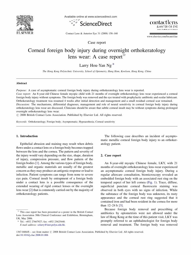
an asymptomatic corneal foreign body injury. During a
regular aftercare consultation, biomicroscopy revealed an
embedded foreign body with an associated rust ring on the
temporal aspect of her left cornea (Fig. 1). Trace, diffuse,
superficial punctate corneal fluorescein staining was
observed in both eyes with no signs of infection. While
the substance of the foreign body was unknown, its rusty
appearance and the corneal rust ring suggested that it
contained iron and had been resident in the cornea for more
than 12–24 h [3].
Because foreign body removal and prescribing of
antibiotics by optometrists were not allowed under the
law of Hong Kong at the time of this patient visit, LKY was
promptly referred to an ophthalmologist for foreign body
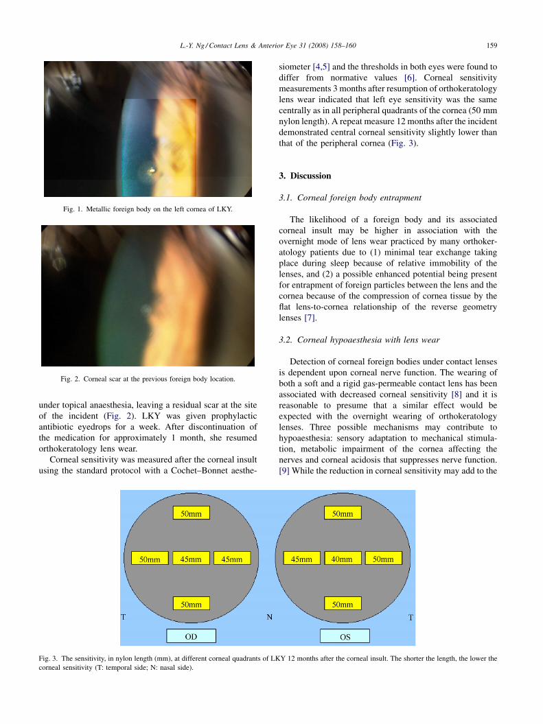
removal and treatment. The foreign body was removed
shed by Elsevier Ltd. All rights reserved.
L.-Y. Ng / Contact Lens & Anterior Eye 31 (2008) 158–160 159
Fig. 1. Metallic foreign body on the left cornea of LKY.
Fig. 2. Corneal scar at the previous foreign body location.
under topical anaesthesia, leaving a residual scar at the site
of the incident (Fig. 2). LKY was given prophylactic
antibiotic eyedrops for a week. After discontinuation of
the medication for approximately 1 month, she resumed
orthokeratology lens wear.
Corneal sensitivity was measured after the corneal insult
using the standard protocol with a Cochet–Bonnet aesthe-
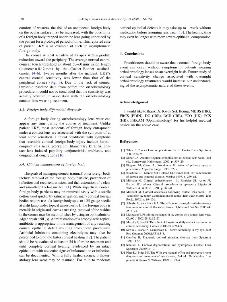
Fig. 3. The sensitivity, in nylon length (mm), at different corneal quadrants of LK
corneal sensitivity (T: temporal side; N: nasal side).
siometer [4,5] and the thresholds in both eyes were found to
differ from normative values [6]. Corneal sensitivity
measurements 3 months after resumption of orthokeratology
lens wear indicated that left eye sensitivity was the same
centrally as in all peripheral quadrants of the cornea (50 mm
nylon length). A repeat measure 12 months after the incident
demonstrated central corneal sensitivity slightly lower than
that of the peripheral cornea (Fig. 3).
3. Discussion
3.1. Corneal foreign body entrapment
The likelihood of a foreign body and its associated
corneal insult may be higher in association with the
overnight mode of lens wear practiced by many orthoker-
atology patients due to (1) minimal tear exchange taking
place during sleep because of relative immobility of the
lenses, and (2) a possible enhanced potential being present
for entrapment of foreign particles between the lens and the
cornea because of the compression of cornea tissue by the
flat lens-to-cornea relationship of the reverse geometry
lenses [7].
3.2. Corneal hypoaesthesia with lens wear
Detection of corneal foreign bodies under contact lenses
is dependent upon corneal nerve function. The wearing of
both a soft and a rigid gas-permeable contact lens has been
associated with decreased corneal sensitivity [8] and it is
reasonable to presume that a similar effect would be
expected with the overnight wearing of orthokeratology
lenses. Three possible mechanisms may contribute to
hypoaesthesia: sensory adaptation to mechanical stimula-
tion, metabolic impairment of the cornea affecting the
nerves and corneal acidosis that suppresses nerve function.
[9] While the reduction in corneal sensitivity may add to the
Y 12 months after the corneal insult. The shorter the length, the lower the
L.-Y. Ng / Contact Lens & Anterior Eye 31 (2008) 158–160160
comfort of wearers, the risk of an undetected foreign body
on the ocular surface may be increased, with the possibility
of a foreign body trapped under the lens going unnoticed by
the patient for a prolonged period of time. This reported case
of patient LKY is an example of such an asymptomatic
foreign body.
The cornea is most sensitive at its apex with a gradual
reduction toward the periphery. The average normal central
corneal touch threshold is about 50–60 mm nylon length
(diameter = 0.12 mm) by the Cochet–Bonnet aesthesi-
ometer [4–6]. Twelve months after the incident, LKY’s
central corneal sensitivity was lower than that of the
peripheral cornea (Fig. 3). Due to the lack of corneal
threshold baseline data from before the orthokeratology
procedure, it could not be concluded that the sensitivity was
actually lowered in association with the orthokeratology
contact lens-wearing treatment.
3.3. Foreign body differential diagnosis
A foreign body during orthokeratology lens wear can
appear any time during the course of treatment. Unlike
patient LKY, most incidents of foreign body entrapment
under a contact lens are associated with the symptom of at
least some sensation. Clinical conditions with symptoms
that resemble corneal foreign body injury include kerato-
conjunctivitis sicca, pterygium, filamentary keratitis, con-
tact lens induced papillary conjunctivitis, trichiasis, and
conjunctival concretions [10].
3.4. Clinical management of foreign body
The goals of managing corneal trauma from a foreign body
include removal of the foreign body particle, prevention of
infection and recurrent erosion, and the restoration of a clear
and smooth epithelial surface [11]. While superficial corneal
foreign body particles may be removed easily with a sterile
cotton wool spud or by saline irrigation, many corneal foreign
bodies require use of a foreign body spud or a 25-gauge needle
at a slit lamp under topical anaesthesia. If the foreign body is
metallic in origin and leaves a rust ring, removal of the residue
in the cornea may be accomplished by using an ophthalmic or
Alger brush drill (3). Administration of a prophylactic topical
antibiotic is appropriate in the management of any resulting
corneal epithelial defect resulting from these procedures.
Artificial lubricants containing electrolytes may also be
prescribed to promote faster corneal healing [12]. The patient
should be re-evaluated at least in 24 h after the treatment and
until complete corneal healing, evidenced by an intact
epithelium with no ocular signs of inflammation or infection,
can be documented. With a fully healed cornea, orthoker-
atology lens wear may be resumed. For mild to moderate
corneal epithelial defects it may take up to 1 week without
medication before resuming lens wear [13]. The healing time
may even be longer with more severe epithelial compromise.
4. Conclusions
Practitioners should be aware that a corneal foreign body
event can occur without symptoms in patients wearing
orthokeratology lenses on an overnight basis. Future study of
corneal sensitivity change associated with overnight
orthokeratology treatments would increase our understand-
ing of the asymptomatic nature of these events.
Acknowledgment
I would like to thank Dr. Kwok Sek Keung, MBBS (HK),
FRCS (EDIN), DO (IRE), DCH (IRE), FCO (HK), FCS
(HK), FHKAM (Ophthalmology) for his helpful medical
advice on the above case.
References
[1] White P. Contact lens complications. Part II. Contact Lens Spectrum
2000;2:34–9.
[2] Silbert JA. Anterior segment complication of contact lens wear, 2nd
ed., Butterworth Heinemann; 2000. p. 309–20.
[3] Fingeret M, Casser L, Woodcome H. Atlas of primary eyecare
procedures. Appleton Lange 1990;152–5.
[4] Krachmer JH, Mannis MJ, Holland EJ. Cornea (vol. 1): fundamentals
of cornea and external disease. Mosby; 1997, p. 279–81.
[5] Millodot M. Corneal esthesiometry. In: Eskridge JB, Amos JF,
Bartlett JD, editors. Clinical procedures in optometry. Lippincott
Williams & Wilkins; 1991. p. 371–8.
[6] Millodot M. Corneal anesthesia following contact lens wear. In:
Tomlinson A, editor. Complications of contact lens wear. Mosby-Year
Book; 1992. p. 89–101.
[7] Alharbi A, Swarbrick HA. The effects of overnight orthokeratology
lens wear on corneal thickness. Invest Ophthalmol Vis Sci 2003;44:
2518–23.
[8] Liesegang T. Physiologic changes of the cornea with contact lens wear.
CLAO J 2002;28(1):12–27.
[9] Murphy P, Patel S. The effect of long-term, daily contact lens wear on
corneal sensitivity. Cornea 2001;20(3):264–9.
[10] Sowka J, Kabat A, Lauderdale F. There’s something in my eye, doc!
Rev Optomet 2000;15(5):85–8.
[11] Onofrey B. Traumatic corneal abrasion. Contact Lens Spectrum
1998;11:50.
[12] Sclafani L. Corneal degenerations and dystrophies. Contact Lens
Spectrum 2003;8:34–9.
[13] Rhee DJ, Pyfer MF. The Wills eye manual: office and emergency room
diagnosis and treatment of eye disease, 3rd ed., Philadelphia: Lip-
pincott Williams & Wilkins; 1999. p. 23–4.