cornea dystrophy and degeneration -...
TRANSCRIPT

9/23/14
1
Corneal Dystrophy/Degeneration: What Every Optometrist Should Know
Marc R. Bloomenstein OD, FAAO Schwartz Laser Eye Center
Scottsdale, Arizona Email: [email protected]
Disclosure � Presenter is on speakers panel of Alcon, Allergan,
Abbott, Bausch + Lomb, Tear Lab, RPS and BVI
� Past-‐President of the Optometric Council on Refractive Technology (OCRT)
� OSSO Board Member
� Presenter has NO financial interest in any products mentioned
� Except he does have stock in a certain coffee company...

9/23/14
2
Corneal Dystrophies � Group of corneal diseases that are genetically determined
and have been traditionally classified with respect to the corneal layer affected
� Defined as a corneal opacity or alteration, which is most often bilateral and progressive and centrally located
� Tend to be avascular and involve all the areas of the cornea
� New Classification system describes old name, new name, defective gene, inheritance pattern, phenotype of disorder and typical complications.
Anterior Dystrophies
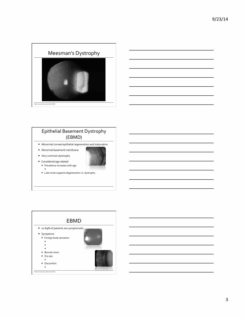
Meesman’s Dystrophy � Autosomal dominantly
� Symptoms � Foreign body sensation due to epithelial erosion � Decreased visual acuity is usually minimal
� Signs: � Myriads of tiny intraepithelial cysts that are most prominently
seen in the interpalpebral zone � Slowly progressive � Bilateral, symmetric � Develops in the first 1 or 2 years of life
� Treatment: � Superficial corneal debridement � PTK
9/23/14
3
Meesman’s Dystrophy
Photo Courtesy Tracy Swartz OD, FAAO
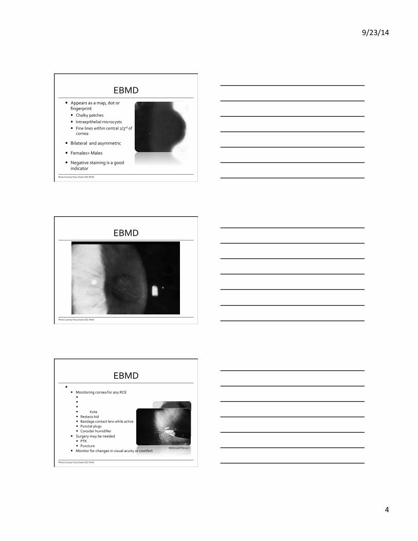
Epithelial Basement Dystrophy (EBMD)
� Abnormal corneal epithelial regeneration and maturation
� Abnormal basement membrane
� Very common dystrophy
� Considered age related � Prevalence increases with age
� Onset is around 40-‐70 y.o.
� Late onset supports degeneration vs. dystrophy
EBMD � 10-‐69% of patients are symptomatic
� Symptoms � Foreign body sensation
� Worse in dry weather
� Wind
� Air conditioning � Blurred vision � Dry eye
� Increased TBUT � Discomfort
� 10% experience RCE’s
Photo Courtesy Tracy Swartz OD, FAAO
9/23/14
4
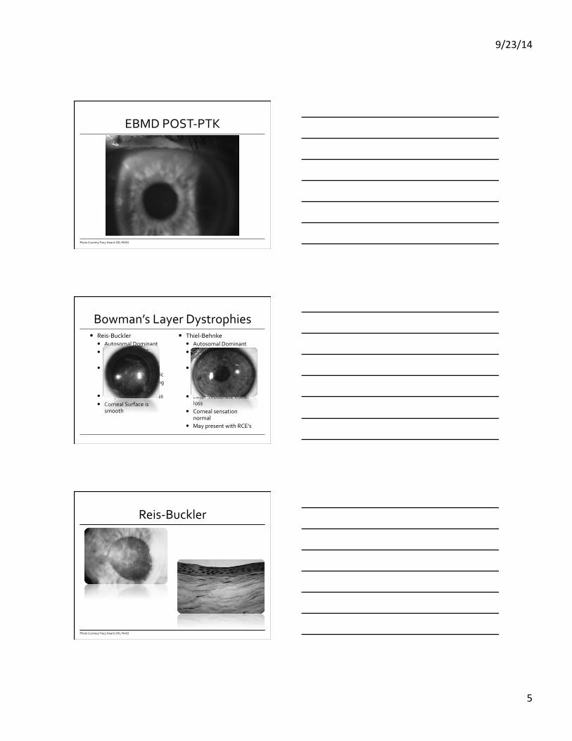
EBMD � Appears as a map, dot or
fingerprint � Chalky patches � Intraepithelial microcysts � Fine lines within central 2/3rd of
cornea
� Bilateral and asymmetric
� Females> Males
� Negative staining is a good indicator
Photo Courtesy Tracy Swartz OD, FAAO
EBMD
Photo Courtesy Tracy Swartz OD, FAAO
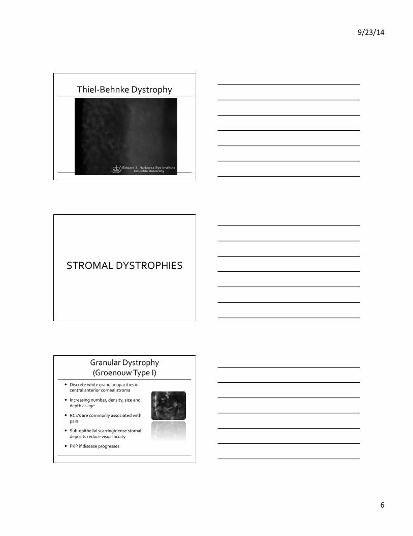
EBMD � Treatment of EBMD
� Monitoring cornea for any RCE � Azasite qhs or oral doxycycline (50 mg/QD) � Lotemax tid � Copious Artificial tears � Fresh Kote � Restasis bid � Bandage contact lens while active � Punctal plugs � Consider humidifier
� Surgery may be needed � PTK � Puncture
� Monitor for changes in visual acuity or comfort
Photo Courtesy Tracy Swartz OD, FAAO
Melton and Thomas
9/23/14
5
EBMD POST-‐PTK
Photo Courtesy Tracy Swartz OD, FAAO
Bowman’s Layer Dystrophies � Reis-‐Buckler
� Autosomal Dominant � Anterior Cornea
� Just under epithelium
� Honey Comb appearance-‐geographic � Scarring and thickening
in Bowman’s � Early marked visual loss � Corneal Surface is
smooth
� Thiel-‐Behnke � Autosomal Dominant � Anterior Cornea
� Just under epithelium
� Honey Comb appearance-‐more distinct � Variant of Reis-‐Buckler
� Later moderate visual loss
� Corneal sensation normal
� May present with RCE’s
Reis-‐Buckler
Photo Courtesy Tracy Swartz OD, FAAO
9/23/14
6
Thiel-‐Behnke Dystrophy
STROMAL DYSTROPHIES
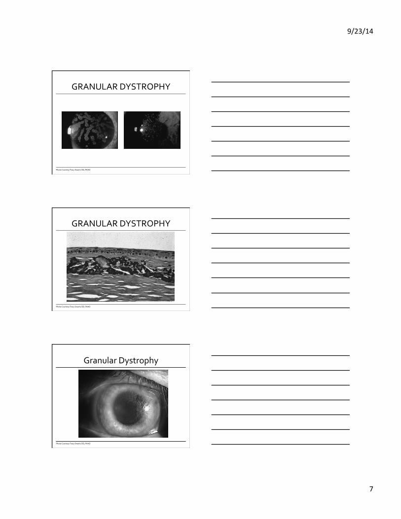
Granular Dystrophy (Groenouw Type I)
� Discrete white granular opacities in central anterior corneal stroma
� Increasing number, density, size and depth as age
� RCE’s are commonly associated with pain
� Sub-‐epithelial scarring/dense stomal deposits reduce visual acuity
� PKP if disease progresses
9/23/14
7
GRANULAR DYSTROPHY
Photo Courtesy Tracy Swartz OD, FAAO
GRANULAR DYSTROPHY
Photo Courtesy Tracy Swartz OD, FAAO
Granular Dystrophy
Photo Courtesy Tracy Swartz OD, FAAO
9/23/14
8
GRANULAR DYSTOPHY
Photo Courtesy Tracy Swartz OD, FAAO
GRANULAR DYSTROPHY
Photo Courtesy Tracy Swartz OD, FAAO
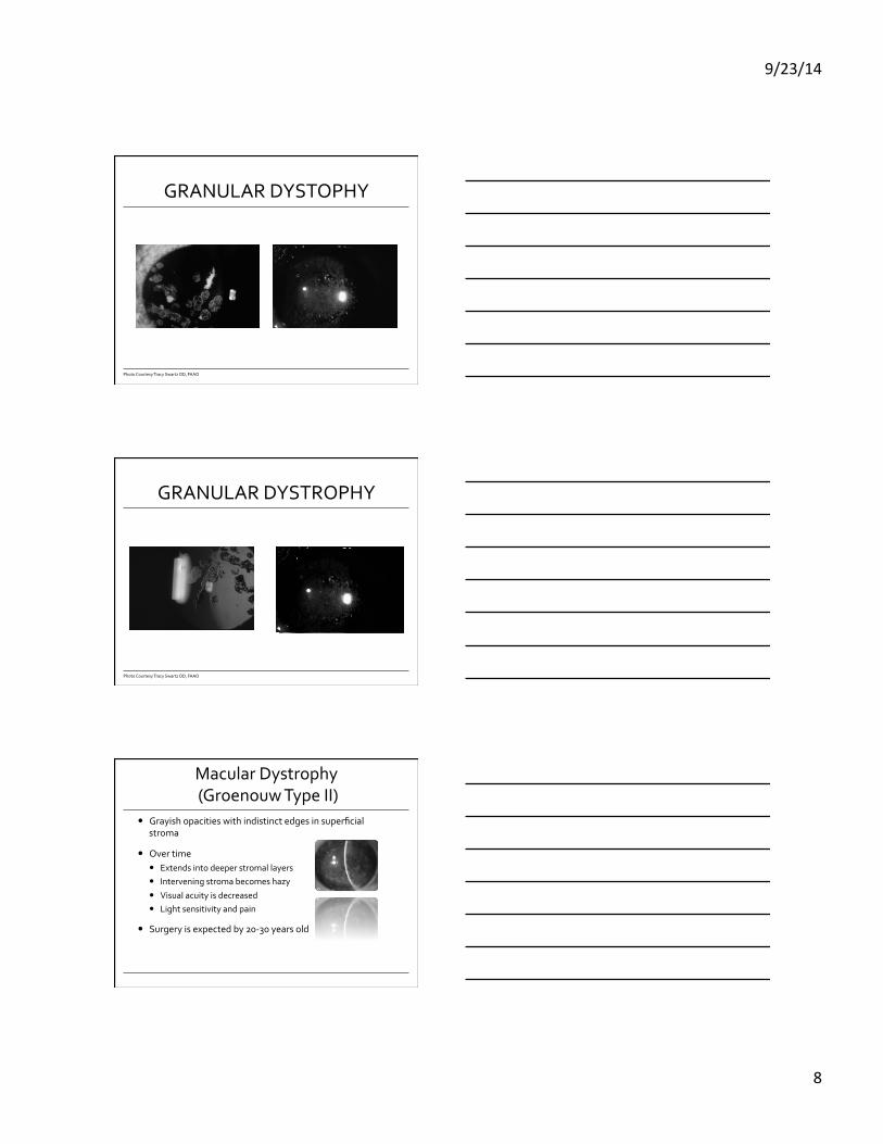
Macular Dystrophy (Groenouw Type II)
� Grayish opacities with indistinct edges in superficial stroma
� Over time � Extends into deeper stromal layers � Intervening stroma becomes hazy
� Visual acuity is decreased � Light sensitivity and pain
� Surgery is expected by 20-‐30 years old
9/23/14
9
Macular Dystrophy
Photo Courtesy Tracy Swartz OD, FAAO
Macular Dystrophy
Photo Courtesy Tracy Swartz OD, FAAO
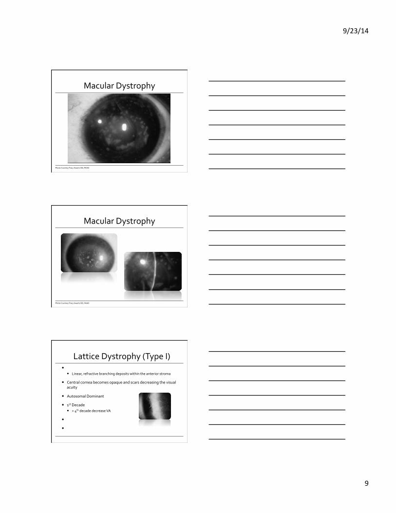
Lattice Dystrophy (Type I) � Clinically appears
� Linear, refractive branching deposits within the anterior stroma
� Central cornea becomes opaque and scars decreasing the visual acuity
� Autosomal Dominant
� 1st Decade � > 4th decade decrease VA
� RCE’s are associated with Lattice
� Surgical intervention recommended with decreased acuity

9/23/14
10
Lattice Dystrophy
Photo Courtesy Tracy Swartz OD, FAAO
Lattice Dystrophy
Photo Courtesy Tracy Swartz OD, FAAO
Central Crystalline Dystrophy of Schnyder
� Central discoid opacification posterior to Bowman's membrane in anterior stroma
� Opacities consist of: � Small needle shaped refractileals crystals
� White
� Polychromatic
� May extend into deeper stroma-‐avoiding epithelium
� Vision is relatively unaffected
� Associated with cholesterolimia
9/23/14
11
Schnyder’s Crystalline Dystrophy
Photo Courtesy Tracy Swartz OD, FAAO
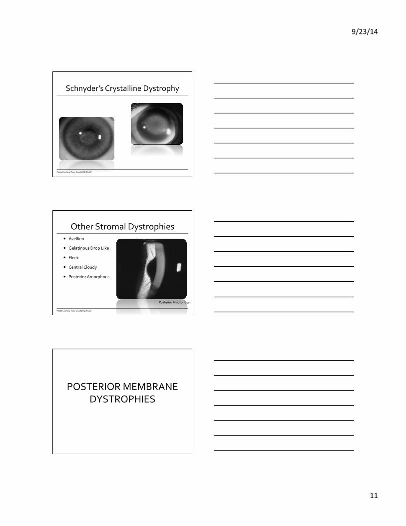
Other Stromal Dystrophies � Avellino
� Gelatinous Drop Like
� Fleck
� Central Cloudy
� Posterior Amorphous
Photo Courtesy Tracy Swartz OD, FAAO
Posterior Amorphous
POSTERIOR MEMBRANE DYSTROPHIES
9/23/14
12
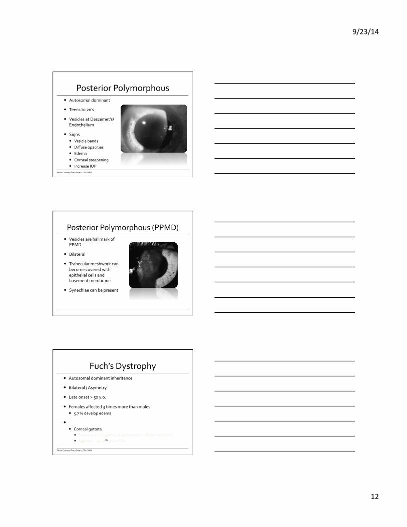
Posterior Polymorphous � Autosomal dominant
� Teens to 20’s
� Vesicles at Descemet’s/Endothelium
� Signs � Vesicle bands � Diffuse opacities � Edema
� Corneal steepening � Increase IOP
Photo Courtesy Tracy Swartz OD, FAAO
Posterior Polymorphous (PPMD) � Vesicles are hallmark of
PPMD
� Bilateral
� Trabecular meshwork can become covered with epithelial cells and basement membrane
� Synechiae can be present
Fuch’s Dystrophy � Autosomal dominant inheritance
� Bilateral / Asymetry
� Late onset > 50 y.o.
� Females affected 3 times more than males � 5.7 % develop edema
� Characterized � Corneal guttata
� Excessive accumulation of abnormal endothelial secretions
� Appears in 30-‐40th year of life
Photo Courtesy Tracy Swartz OD, FAAO
9/23/14
13
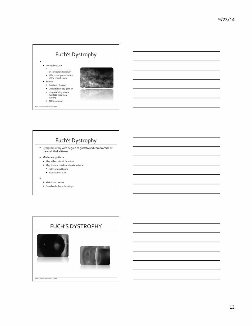
Fuch’s Dystrophy � Characterized
� Corneal Guttata � Small refractile “drops”
on corneal endothelium
� Affects the “pump” action of the endothelium
� Edema � Greater in the AM
� Desiccates as day goes on � Long standing edema
may lead to corneal scarring
� RCE’s common
Photo Courtesy Tracy Swartz OD, FAAO
Fuch’s Dystrophy � Symptoms vary with degree of guttata and compromise of
the endothelial tissue
� Moderate guttata � May affect visual function � May induce mild-‐moderate edema
� Halos around lights � Hazy vision > a.m.
� Severe guttata � Vision decreases � Possible bullous develops
FUCH’S DYSTROPHY
Photo Courtesy Tracy Swartz OD, FAAO
9/23/14
14
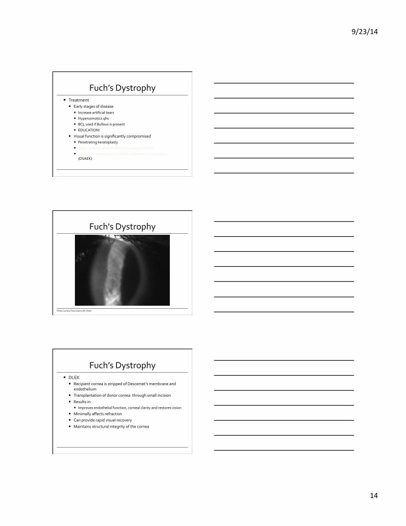
Fuch’s Dystrophy � Treatment
� Early stages of disease � Increase artificial tears � Hyperosmotics qhs
� BCL used if Bullous is present � EDUCATION!
� Visual function is significantly compromised � Penetrating keratoplasty � Deep Lamellar endothelial keratoplasty (DLEK)
� Descemet stripping automated endothelial keratoplasty (DSAEK)
Fuch's Dystrophy
Photo Courtesy Tracy Swartz OD, FAAO
Fuch’s Dystrophy � DLEK
� Recipient cornea is stripped of Descemet’s membrane and endothelium
� Transplantation of donor cornea through small incision
� Results in � Improves endothelial function, corneal clarity and restores vision
� Minimally affects refraction � Can provide rapid visual recovery � Maintains structural integrity of the cornea

9/23/14
15
Congenital Hereditary Endothelium Dystrophy (CHED)
� Rare congenital dystrophy � First weeks-‐6 months old
� Bilateral-‐symmetric
� Non-‐inflammatory clouding
� Signs � Opacification extending to limbus
with clear zones � Thickening � No neo/No extra tissue � No increase in IOP
Photo Courtesy Tracy Swartz OD, FAAO
Congenital Hereditary Endothelium Dystrophy
� Nystagmus is present
� VA can be as low as 20/100
� No neo/No extra tissue
� No increase in IOP
� Diagnosis of exclusion
Photo Courtesy Tracy Swartz OD, FAAO
Congenital Hereditary Endothelium Dystrophy
Photo Courtesy Tracy Swartz OD, FAAO
9/23/14
16
Iridocorneal Endothelial Syndrome
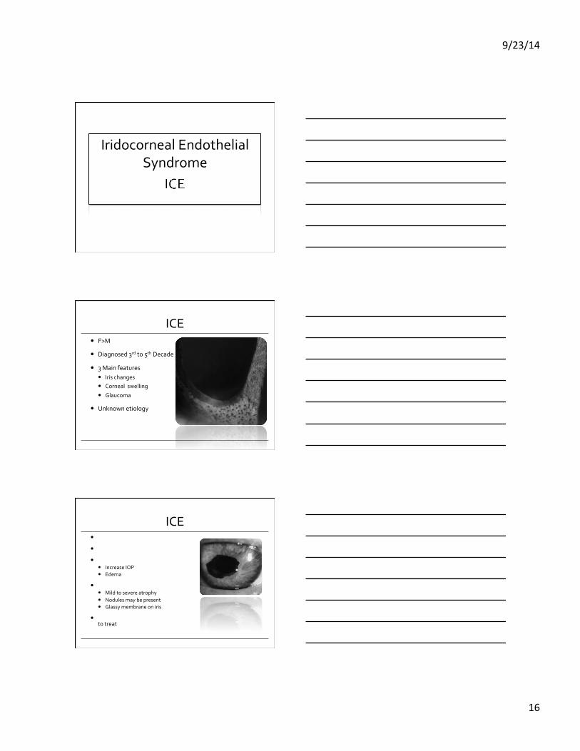
ICE � F>M
� Diagnosed 3rd to 5th Decade
� 3 Main features � Iris changes � Corneal swelling
� Glaucoma
� Unknown etiology
ICE � Abnormal endothelium
� Irido-‐corneal adhesions
� 80-‐100% develop glaucoma � Increase IOP � Edema
� Iris � Mild to severe atrophy � Nodules may be present � Glassy membrane on iris
� Condition can be relentless and difficult to treat
9/23/14
17
Corneal Degenerations
Corneal Degenerations � Defined as a deterioration or change from a higher to a
lower form, especially change of tissue to a lower or less functionally active
� Non-‐inherited
� Unilateral or bilateral
� Asymmetric
� Develop in later years
� Variable progression
� Systemic disease can be associated
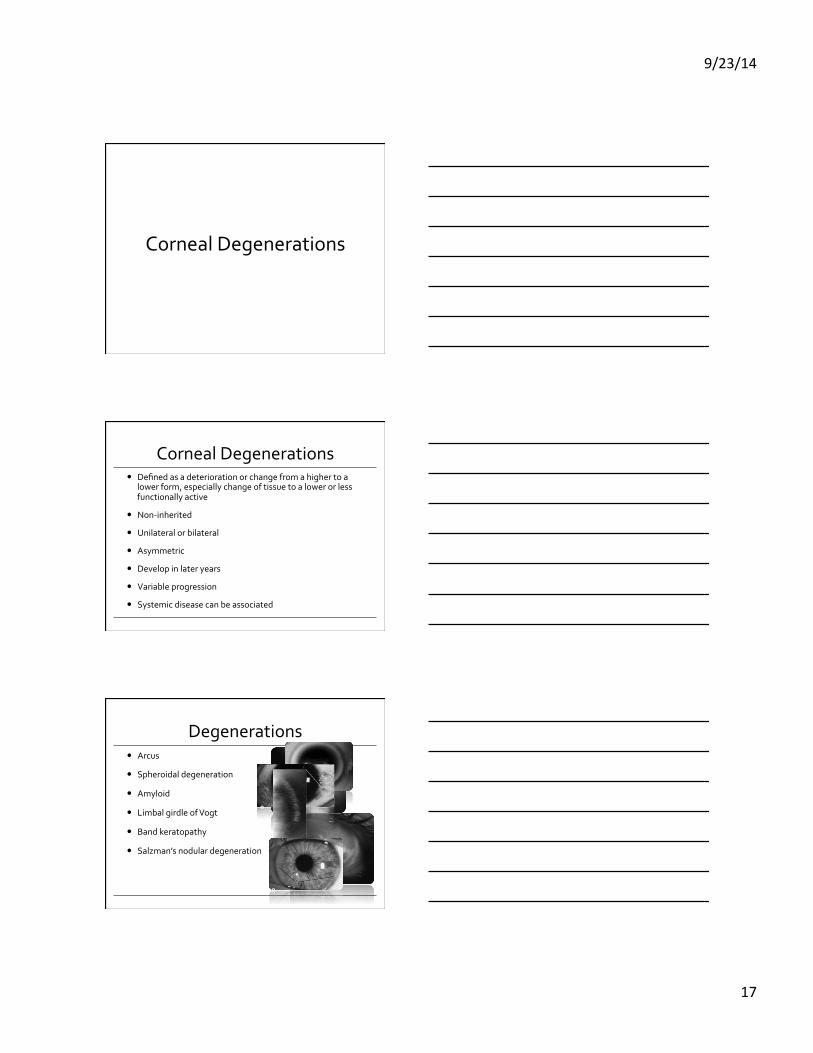
Degenerations � Arcus
� Spheroidal degeneration
� Amyloid
� Limbal girdle of Vogt
� Band keratopathy
� Salzman’s nodular degeneration
9/23/14
18
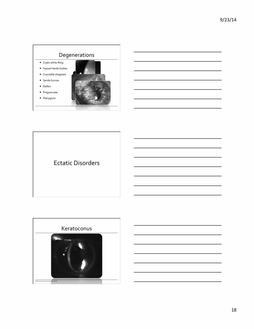
Degenerations � Coats white Ring
� Hassal-‐Henle bodies
� Crocodile shagreen
� Senile furrow
� Dellen
� Pingueculae
� Pterygium
Ectatic Disorders
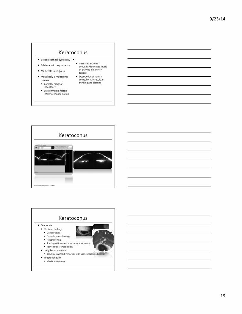
Keratoconus
Photo Courtesy Tracy Swartz OD, FAAO
9/23/14
19
Keratoconus � Ectatic corneal dystrophy
� Bilateral with asymmetry
� Manifests in 20-‐30’ss
� Most likely a multigenic disease � Complex mode of
inheritance � Environmental factors
influence manifestation
� Etiology � Increased enzyme
activities /decreased levels of enzyme inhibitors= toxicity
� Destruction of normal corneal matrix results in thinning and scarring
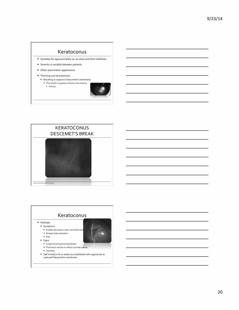
Keratoconus
Photo Courtesy Tracy Swartz OD, FAAO
Keratoconus � Diagnosis
� Slit lamp findings � Munson’s Sign
� Central corneal thinning � Fleischer’s ring � Scarring at Bowman’s layer or anterior stroma
� Vogt’s striae (vertical striae) � Irregular astigmatism
� Resulting in difficult refraction with both contacts and glasses
� Topographically � Inferior steepening
9/23/14
20
Keratoconus � Gestates for approximately 10-‐20 years and then stabilizes
� Severity is variable between patients
� Often asymmetric appearance
� Thinning can be extensive: � Resulting in rupture in Descemet’s membrane
� This results in aqueous infusion into stroma � Hydrops
KERATOCONUS DESCEMET’S BREAK
Photo Courtesy Tracy Swartz OD, FAAO
Keratoconus � Hydrops
� Symptoms � Sudden decrease in best corrected vision � Foreign body sensation � Pain
� Signs � Conjunctival hyperemia/redness
� Prominent central or inferior corneal edema
� Clouding � Self-‐limited in 8-‐10 weeks as endothelial cells regenerate at
ruptured Descemet’s membrane
9/23/14
21
Keratoconus � Treatment
� Hydrops � Hyperosmotics
� Antibiotics to avoid secondary infection � PKP
� RGP’s � Bi-‐Aspheric I-‐Kone Design Valley Contax � Hyrokone � SynergEyes High Dk Hybrid
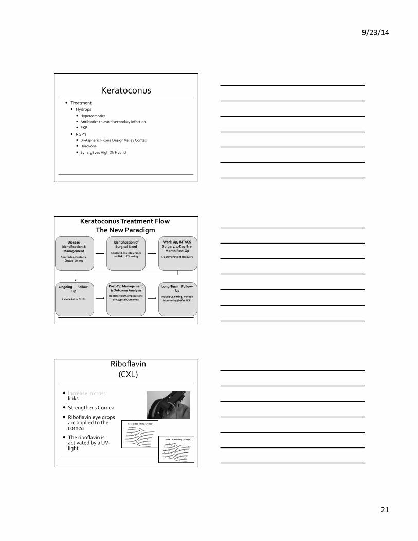
Disease Identification & Management
Spectacles, Contacts,
Custom Lenses
Identification of Surgical Need
Contact Lens Intolerance
or Risk of Scarring
Work-‐Up, INTACS Surgery, 1-‐Day & 3-‐Month Post-‐Op
1-‐2 Days Patient Recovery
Ongoing Follow-‐Up
Include Initial CL Fit
Post-‐Op Management & Outcome Analysis
Re-‐Referral if Complications or Atypical Outcomes
Long-‐Term Follow-‐Up
Include CL Fitting, Periodic Monitoring (Defer PKP)
Keratoconus Treatment Flow
The New Paradigm
Corneal Collagen Cross-‐Linking with Riboflavin
(CXL)
� Increase in cross links
� Strengthens Cornea � Riboflavin eye drops
are applied to the cornea
� The riboflavin is activated by a UV-‐light
9/23/14
22
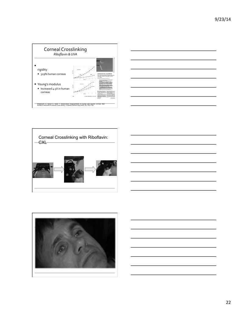
� Increase in corneal rigidity � 329% human corneas
� Young’s modulus � Increased 4.5X in human
corneas
Wollensack G, Spoerl E, Seiler T. Stress-strain measurements of human and porcine corneas after riboflavin-ultraviolet-A-induced cross-linking J Cataract Refract Surg 2003; 29:1780-1785
Corneal Crosslinking Riboflavin & UVA
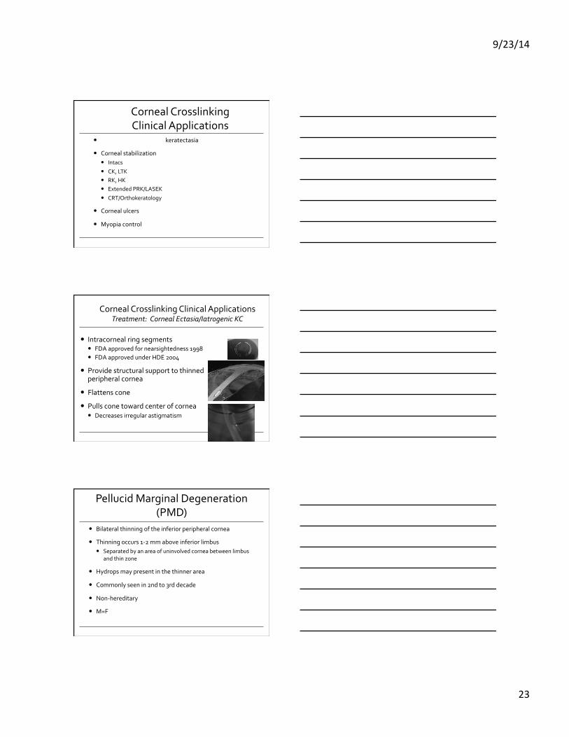
Corneal Crosslinking with Riboflavin: CXL
9/23/14
23
Corneal Crosslinking Clinical Applications
� Keratoconus/Iatrogenic keratectasia
� Corneal stabilization � Intacs � CK, LTK � RK, HK � Extended PRK/LASEK � CRT/Orthokeratology
� Corneal ulcers
� Myopia control
Corneal Crosslinking Clinical Applications Treatment: Corneal Ectasia/Iatrogenic KC
� Intracorneal ring segments � FDA approved for nearsightedness 1998 � FDA approved under HDE 2004
� Provide structural support to thinned peripheral cornea
� Flattens cone � Pulls cone toward center of cornea
� Decreases irregular astigmatism
Pellucid Marginal Degeneration (PMD)
� Bilateral thinning of the inferior peripheral cornea
� Thinning occurs 1-‐2 mm above inferior limbus � Separated by an area of uninvolved cornea between limbus
and thin zone
� Hydrops may present in the thinner area
� Commonly seen in 2nd to 3rd decade
� Non-‐hereditary
� M=F
9/23/14
24
Pellucid Marginal Degeneration
PMD � Subjective symptoms
� Increase in against-‐the-‐rule astigmatism
� Unexplained decrease in visual acuity
� Affected area is clear of lipids or vascularization
� Corneal topography has distinct inferior steepening � Crab claw � Kissing doves � Beard and mustache
PMD � Treatment
� Glasses � Traditionally may be sufficient with PMD
� Matching astigmatism
� Contact lens � Challenging fits with increase astigmatism (ATR)
� Asymmetrical astigmatism
� Surgical intervention � PK � Inferior lamellar patch graft
9/23/14
25
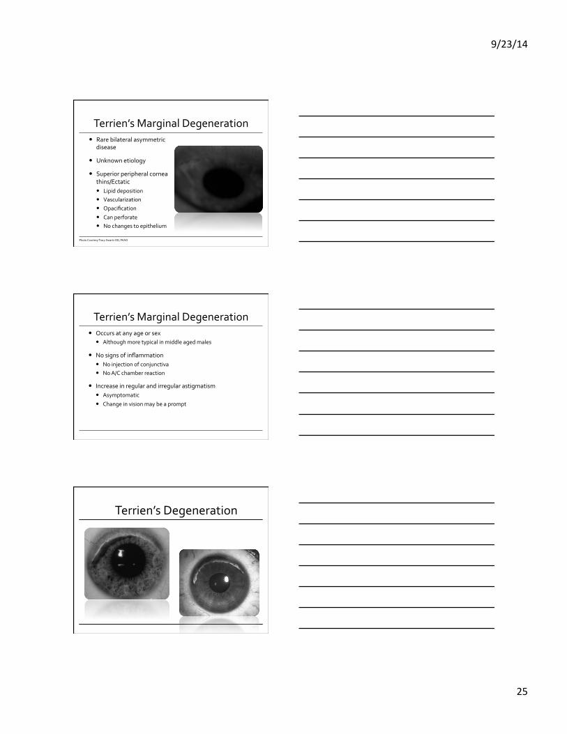
Terrien’s Marginal Degeneration � Rare bilateral asymmetric
disease
� Unknown etiology
� Superior peripheral cornea thins/Ectatic � Lipid deposition � Vascularization � Opacification
� Can perforate � No changes to epithelium
Photo Courtesy Tracy Swartz OD, FAAO
Terrien’s Marginal Degeneration � Occurs at any age or sex
� Although more typical in middle aged males
� No signs of inflammation � No injection of conjunctiva � No A/C chamber reaction
� Increase in regular and irregular astigmatism � Asymptomatic
� Change in vision may be a prompt
Terrien’s Degeneration
9/23/14
26
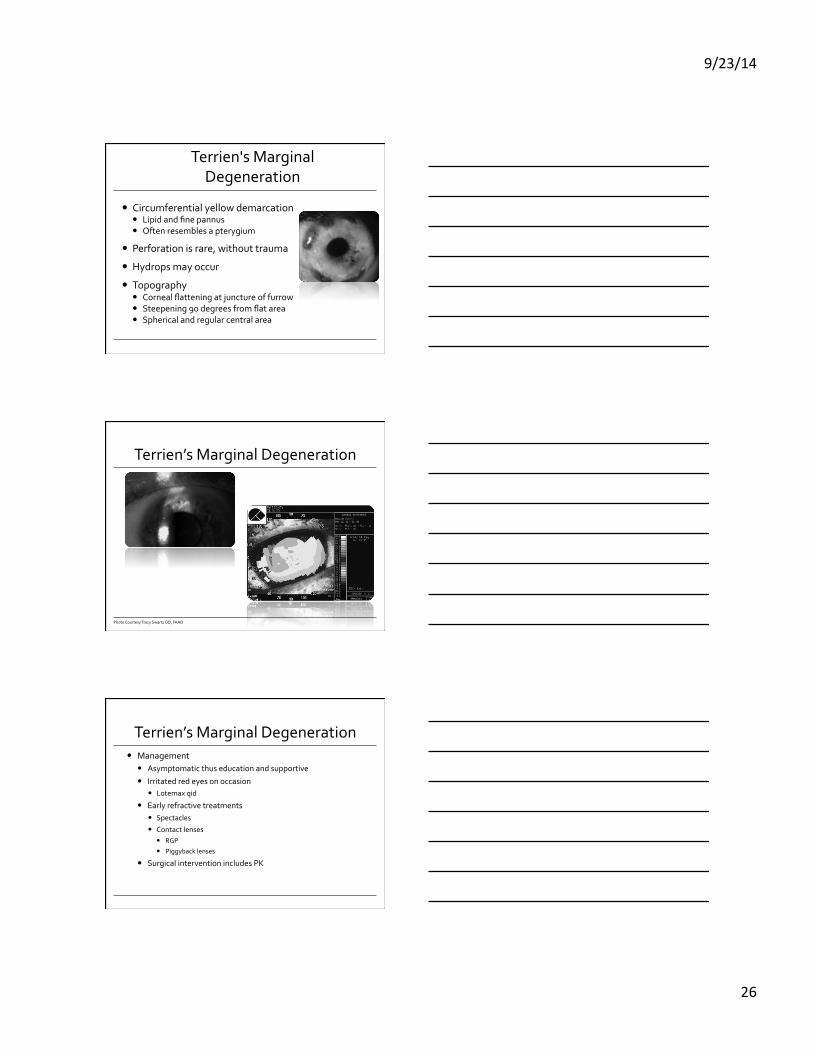
Terrien's Marginal Degeneration
� Circumferential yellow demarcation � Lipid and fine pannus � Often resembles a pterygium
� Perforation is rare, without trauma
� Hydrops may occur
� Topography � Corneal flattening at juncture of furrow � Steepening 90 degrees from flat area � Spherical and regular central area
Terrien’s Marginal Degeneration
Photo Courtesy Tracy Swartz OD, FAAO
Terrien’s Marginal Degeneration � Management
� Asymptomatic thus education and supportive
� Irritated red eyes on occasion � Lotemax qid
� Early refractive treatments � Spectacles � Contact lenses
� RGP
� Piggyback lenses
� Surgical intervention includes PK
9/23/14
27
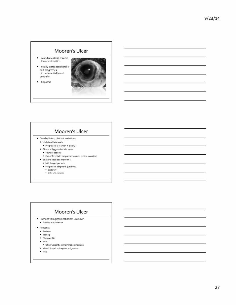
Mooren’s Ulcer � Painful relentless chronic
ulcerative keratitis
� Initially starts peripherally and progresses circumferentially and centrally
� Idiopathic
Mooren’s Ulcer � Divided into 3 distinct variations
� Unilateral Mooren’s � Progressive ulceration in elderly
� Bilateral Aggressive Mooren’s � Younger patients � Circumferentially progresses towards central ulceration
� Bilateral indolent Mooren’s � Middle aged patients
� Progressive peripheral guttering � Bilaterally
� Little inflammation
Mooren’s Ulcer � Pathophysilogical mechanism unknown
� Possibly autoimmune
� Presents � Redness
� Tearing
� Photophobia
� PAIN
� Often worse than inflammation indicates
� Visual disruption-‐irregular astigmatism
� Iritis

9/23/14
28
Mooren’s Ulcer � Treatment
� Steroids � Pred Forte q1h
� Cycloplegia � Topical antibiotic
� 4th generation floroquinolone
� Oral steroids � Conjunctival resection � Immunosuppressive therapy
Thank you