copy right- hongqi zhang-department of anatomy-fudan university systematic...
TRANSCRIPT

Copy Right- Hongqi ZHANG-Department of Anatomy-Fudan University
Copy Right- Hongqi ZHANG-Department of Anatomy-Fudan University1
Dr.Hongqi Zhang (张红旗)Email: [email protected]
Systematic Anatomy
Nervous system
The conducting pathway

Copy Right- Hongqi ZHANG-Department of Anatomy-Fudan University
Copy Right- Hongqi ZHANG-Department of Anatomy-Fudan University
Concept
Nervours pathway are the routes formed by chains of neurons,through which sensory awareness reaches the cerebral cortex and a motor response is initiated.
Category of nervous pathways:
The ascending (afferent or sensory) pathways
The descending (efferent or motor) pathways
Pyramidal system
Extrapyramidal system.
Each nervous pathway consists of a chain of tracts and associated nuclei.
General introduction about the nervous pathways

Copy Right- Hongqi ZHANG-Department of Anatomy-Fudan University
Copy Right- Hongqi ZHANG-Department of Anatomy-Fudan University
Ascending (afferent or sensory) pathwayDifferent types of receptors receive different types of stimuli from the external and internal environment and generate corresponding nervous impulse.which are conducted upward to the cerebral cortex centers to be processed to give rise to corresponding sensations.
Superficial sensation : touch,pain.temperature & two-point discrimination.Deep sensations : proprioception,and vibration sense Visceral sensations: hunger.nausea and visceral pain.Special sensations: smelling,sight,hearing.taste.

Copy Right- Hongqi ZHANG-Department of Anatomy-Fudan University
Copy Right- Hongqi ZHANG-Department of Anatomy-Fudan University
A chain of three long neurons and a number of interneurons conduct the nerve impulses generated in response to stimuli from an individual receptor to the somatosensory cortex
Generally,there are three grades neuron implicatedIn ascending pathway
First-order neuron: the cell bodies lie in a dorsal root ganglion or somatic afferent ganglia of cranial nn.
Second-order neurons: the cell bodies lie within the spinal cord or brain stem. such as dorsal column nuclei(sensation of trunk & limbs) or sensory nucleus of trigerminal nerve, their axons usually decussate, ending in the thalamus.
Third-order neurons: the cell bodies lie in the thalamus. project to the sensory cortex (center).
Copy Right- Hongqi ZHANG-Department of Anatomy-Fudan University
Copy Right- Hongqi ZHANG-Department of Anatomy-Fudan University
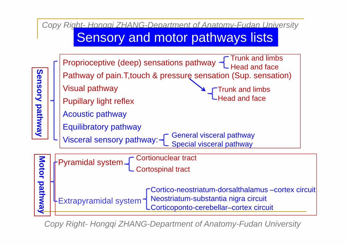
Proprioceptive (deep) sensations pathway Pathway of pain.T,touch & pressure sensation (Sup. sensation) Visual pathwayPupillary light reflex Acoustic pathwayEquilibratory pathwayVisceral sensory pathway:
Sensory pathway
General visceral pathwaySpecial visceral pathway
Motor pathw
ay
Pyramidal system
Extrapyramidal system
Cortionuclear tractCortospinal tract
Cortico-neostriatum-dorsalthalamus –cortex circuitNeostriatum-substantia nigra circuitCorticoponto-cerebellar–cortex circuit
Trunk and limbsHead and face
Sensory and motor pathways lists
Trunk and limbsHead and face

Conscious proprioceptiveand fine touch pathwayof trunk and limbs
Spinal ganglion1st neuron body
Fasciculus gracilis
Fasciculus cuneatus
Gracile and cuneate nuclei2nd neuron body
Decussation of medial lemniscus
Medial lemniscus
VPL3rd neuron body
Central thalamicradiation
T4
VPL-ventral posterolateral nucleus

Fasciculus gracilis Fasciculus cuneatus
Fasciculus gracilis
Fasciculus cuneatus
Spinal ganglion

Cell body position of 1st order neuron Spinal ganglion
Cell body position of 2nd order neuron Gracile and cuneate nuclei in medullaablongata
Cell body position of 3rd order neuron Ventral posterolateral nucleus (VPL) in thalamus
Position of decussation Upper portion of medulla ablongataPosition of fiber tract in spinal cord Post.funiculus of spinal cordName of fiber tract in brain stem Medial lemniscusPosition in internal capsule Posterior limb Projection area in cortex Upper 2/3 of postcentral gyrus and
ant.part of paracentral lobulaThe feature:(A)Fasciculus gracils – conduct sensory impulse above T 4;Fasciculus cuneatus-– conduct sensory impulse below T 5;(B)Fiber arrangement in post funiculus are fiber from sacral,lumbar,thoracic and cervical part from medial to lateral
Features of Conscious proprioceptive and fine touch pathway of trunk & limbs
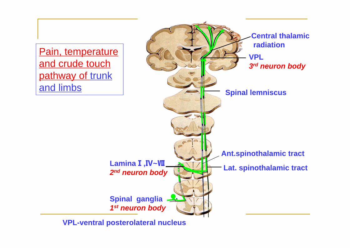
Spinal ganglia1st neuron body
LaminaⅠ,Ⅳ~Ⅶ2nd neuron body Lat. spinothalamic tract
Ant.spinothalamic tract
Spinal lemniscus
VPL3rd neuron body
Central thalamicradiation
Pain, temperature and crude touch pathway of trunk and limbs
VPL-ventral posterolateral nucleus

Spinothalamus tract
Spinal ganglion

Cell body position of 1st order neuron Spinal ganglion
Cell body position of 2nd order neuron Nucleus proprius of post.horn(LaminaⅠ,Ⅳ~Ⅶ) of spinal cord
Cell body position of 3rd order neuron Ventral posterolateral nucleus (VPL) in thalamus
Position of decussation Different spinal segment of spinal cordPosition of fiber tract in spinal cord Ant.& lat.funiculus of spinal cordName of fiber tract in brain stem spinal lemniscusPosition in internal capsule Posterior limbProjection area in cortex Upper 2/3 of postcentral gyrus and
post.part of paracentral lobulaThe other feature:(A) After central process of spinal ganglion enter the spinal cord,ascend 1-2 spinal segments then decussate to contralateral spinal cord.(B) The decussation in spinal cord is not at the same level,rather than at different segments.
Features of pain, temperature and crude touch pathway of trunk and limbs of trunk & limbs

Unconscious proprioceptivepathway of trunk and limbs
Spinal ganglion
Post. spinocerebellar tract
Ant. Spinocerebellartract
Lamina Ⅴ-Ⅶ
Inf. cerebellar peduncle
Sup. cerebellar peduncle
Nucleus thoracicus
In this conducting tract,only two-order neurons are involved.
1st neuron body 2nd neuron body
Learn it by yourself

Trigeminal ganglionGeniculate ganglion of facial n.Sup. ganglions of glossopharangeal n. and vagus n.1°neuron
Pontine nucleus of V2nd neuron body
Spinal nucleus of V1st neuron body
Trigeminal lemniscus Trigeminothalamic tract
Spinal tract of trigeminal n.
VPM 3rd neuron body
Central thalamicradiation
Pain,temperature and crude touch pathway of head and face
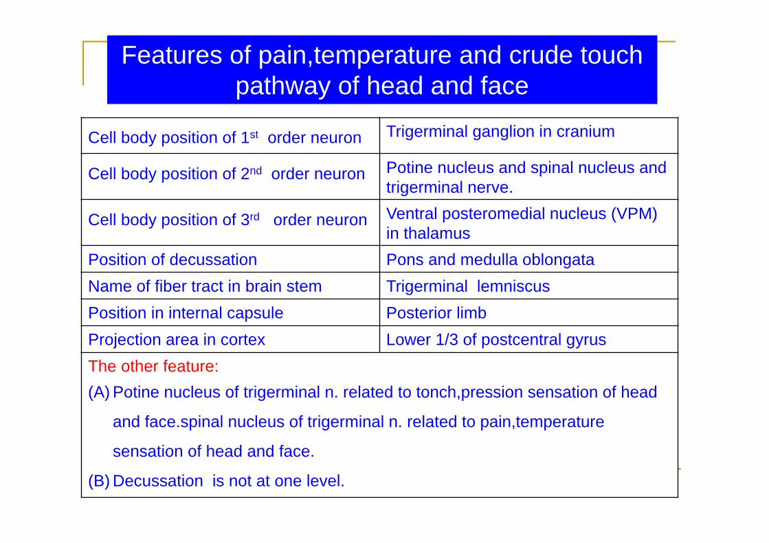
Cell body position of 1st order neuron Trigerminal ganglion in cranium
Cell body position of 2nd order neuron Potine nucleus and spinal nucleus and trigerminal nerve.
Cell body position of 3rd order neuron Ventral posteromedial nucleus (VPM) in thalamus
Position of decussation Pons and medulla oblongataName of fiber tract in brain stem Trigerminal lemniscusPosition in internal capsule Posterior limbProjection area in cortex Lower 1/3 of postcentral gyrusThe other feature:(A) Potine nucleus of trigerminal n. related to tonch,pression sensation of head
and face.spinal nucleus of trigerminal n. related to pain,temperature
sensation of head and face.
(B) Decussation is not at one level.
Features of pain,temperature and crude touch pathway of head and face

Copy Right- Hongqi ZHANG-Department of Anatomy-Fudan University
Copy Right- Hongqi ZHANG-Department of Anatomy-Fudan University
Review of the structure related to eye
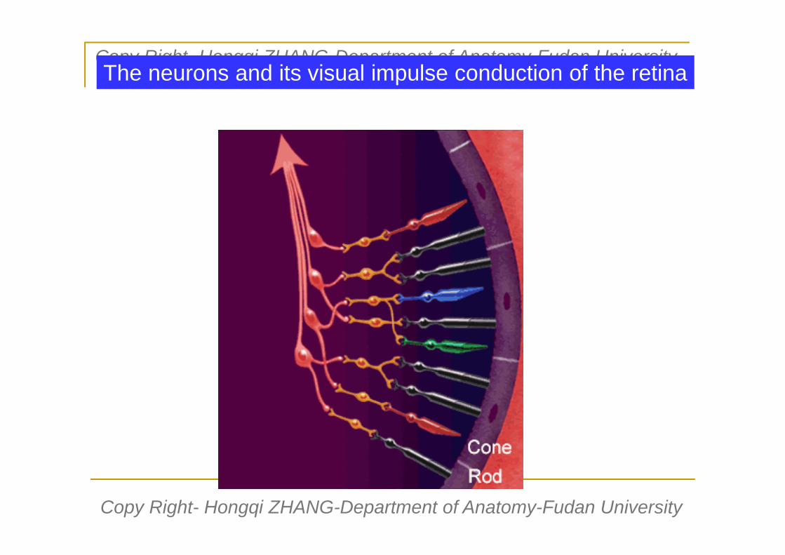
Rod cellCone cell
Bipolarcell
Ganglioncell
Pigmented cells
Copy Right- Hongqi ZHANG-Department of Anatomy-Fudan University
Copy Right- Hongqi ZHANG-Department of Anatomy-Fudan University
The neurons and its visual impulse conduction of the retina

Optic nerve
Optic chiasma
Optic tract
Lateral geniculate body
Optic radiation
Visual area
Visual pathway

Visual pathway
Optic nerve
Optic chiasma
Optic radiation
Lateral geniculate body
Visual area
Optic tract

Copy Right- Hongqi ZHANG-Department of Anatomy-Fudan University
Copy Right- Hongqi ZHANG-Department of Anatomy-Fudan University
A Ipsilateral complete blindness
B bitemporal hemianopia
CEF Contralateral homonymous hemianopia of both visual fields
D ipsilateral nasal hemianopia
D
A B
C
DE
F

Projection of optic nerve in occipital lobe cortex
Learn it by yourself

Pathway for papillary light reflex ?

Pathway for pupillary light reflex
Pretectal area
Accessory oculomotor nuclei
Occculomotor n.
Ciliary ganglia
Sphincter pupilCiliary muscle

Cell body position of 1st order neuron Bipolar neuron in retina
Cell body position of 2nd order neuron Ganglion cell in retina
Cell body position of 3rd order neuron Sup.cuniculus in midbrain
Cell body position of 4th order neuron Lateral geniculate body in diencephalon
Position of decussation Optic chiasma in diencephalonPosition in internal capsule Posterior limb (optic radiation)Projection area in cortex Upper and lower close to calcarine
sulcus in occipital lobe (Broadmen:area 17)
The other feature:(A) Four grade neurons are involved in optic pathway .(B) Decussation feature: fiber from nasal half retina decussate only(C) the fiber from upper half retina project to cortex above calcarine sulcus
the fiber from lower half retina project to cortex below calcarine sulcus(D)One eye visual impulse project to double cortex of occipital lobe
Features of optic pathway

Pretectal area
Accessory oculomotor nuclei
Occculomotor n.
Ciliary ganglia
Sphincter pupilCiliary muscle
Pathway for pupillary light reflex
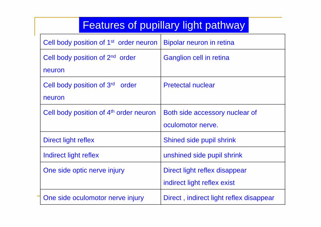
Cell body position of 1st order neuron Bipolar neuron in retina
Cell body position of 2nd order
neuron
Ganglion cell in retina
Cell body position of 3rd order
neuron
Pretectal nuclear
Cell body position of 4th order neuron Both side accessory nuclear of
oculomotor nerve.
Direct light reflex Shined side pupil shrink
Indirect light reflex unshined side pupil shrink
One side optic nerve injury Direct light reflex disappear
indirect light reflex exist
One side oculomotor nerve injury Direct , indirect light reflex disappear
Features of pupillary light pathway
Copy Right- Hongqi ZHANG-Department of Anatomy-Fudan University
Copy Right- Hongqi ZHANG-Department of Anatomy-Fudan University
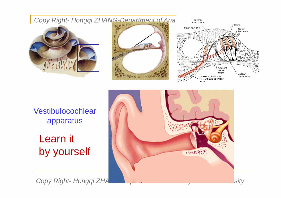
Vestibulocochlearapparatus
Learn it by yourself
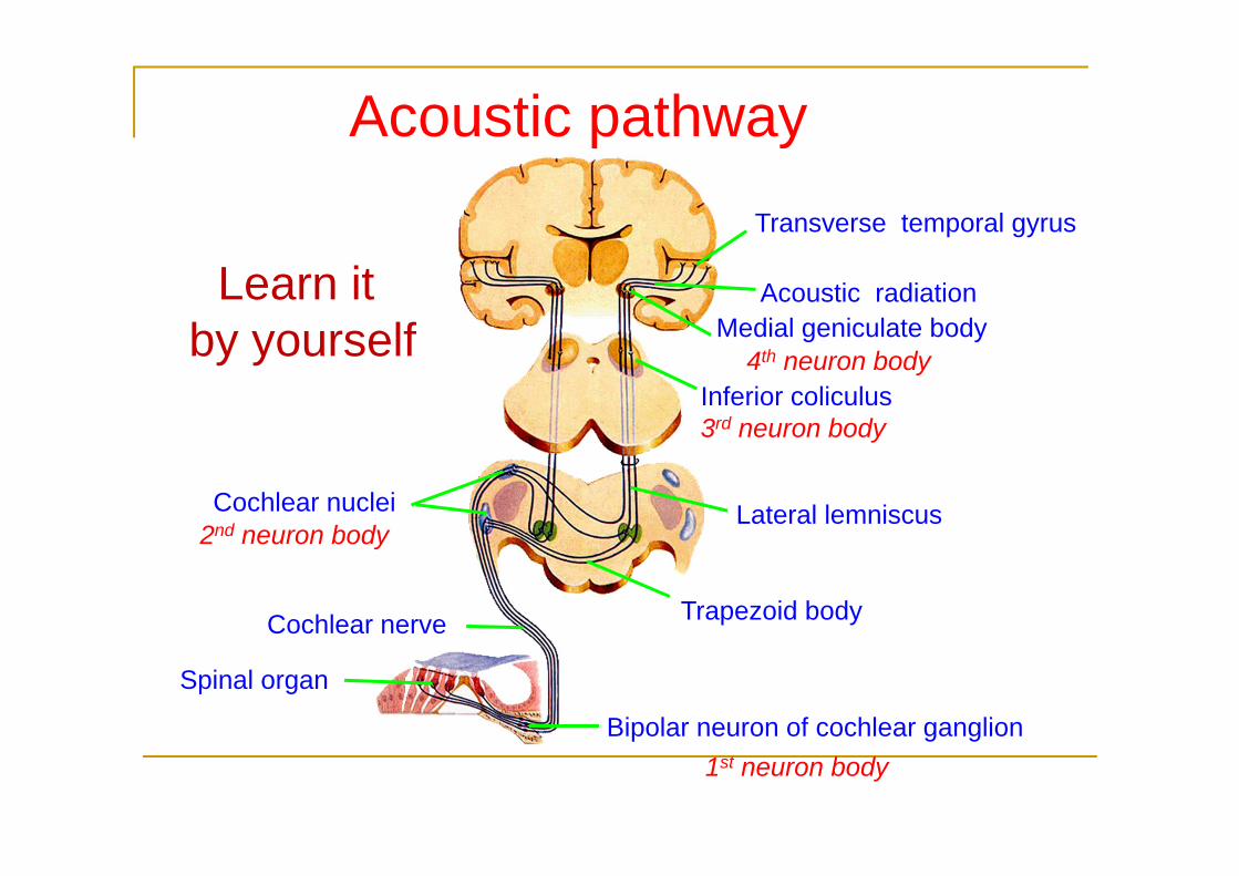
Acoustic pathway
Spinal organ
Bipolar neuron of cochlear ganglion
Cochlear nerve
Cochlear nuclei
Trapezoid body
Lateral lemniscus
Medial geniculate body Acoustic radiation
Transverse temporal gyrus
Inferior coliculus3rd neuron body
1st neuron body
2nd neuron body
4th neuron body
Learn it by yourself

Cell body position of 1st order neuron Spiral ganglioni
Cell body position of 2nd order neuron Cochlear nuclei in pons
Cell body position of 3rd order neuron Lower colicullus
Cell body position of 4th order neuron Medial geniculate bodyPosition of decussation Pons Position in internal capsule Posterior limb (acoustic radiation)Projection area in cortex Transverse temporary gyrus
(Broadmen:areas 41,42)
The other feature:
(A) One cerebral hearing center receive both hearing impulse. if one side
hearing cortex injury,no apparent effect to hearing sensation.
(B) The fiber from cochlear nucleus:about 1/3 ascending at same side.about
2/3 fibers firstly decussate to contralateral side then ascend.
Features of acoustic pathway

Copy Right- Hongqi ZHANG-Department of Anatomy-Fudan University
Copy Right- Hongqi ZHANG-Department of Anatomy-Fudan University
Descending (efferent or motor ) pathway
Pyramidal system and extrapyramidal systemGenerally,in pyramidal system,two order neurons are involved.The first-order neuron, the cell bodies lie in the motor area of cerebral cortex.The second-order neuron, the cell bodies lie in the somatic motor nuclei in the brain stem and somatic motor nuclei in the ant.horn of spinal cord. their axons innervate the skeletal muscle of the head,the trunk and the limbs.But in extrapyramidal system involved more than 2 neurons (3-6) are needed.
Copy Right- Hongqi ZHANG-Department of Anatomy-Fudan University
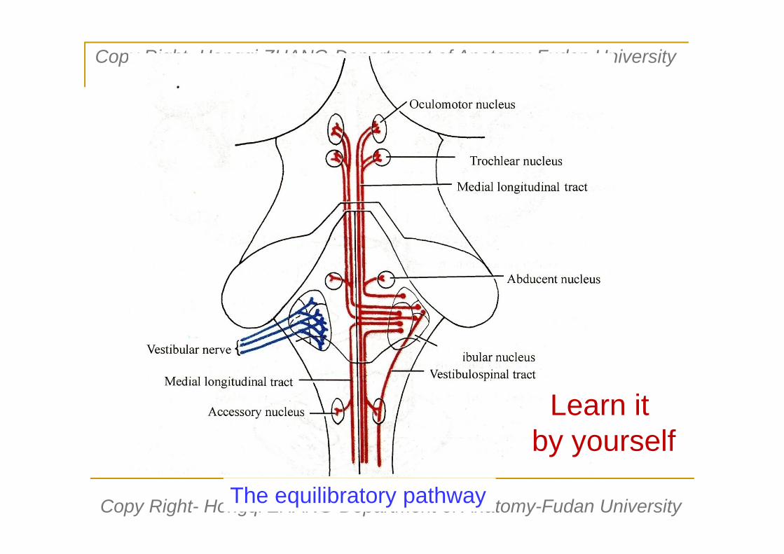
Copy Right- Hongqi ZHANG-Department of Anatomy-Fudan UniversityThe equilibratory pathway
Learn it by yourself

Copy Right- Hongqi ZHANG-Department of Anatomy-Fudan University
Copy Right- Hongqi ZHANG-Department of Anatomy-Fudan University
Descending (motor) conducting pathway
Pyramidal systemExtrapyramidal system

Corticospinal tract
Corticospinal tract
Decussation of pyramid
Lat. corticospinal tract
Ant. corticospinal tract
upper motor neuron
Motor neuron of ant.hornLower motor neuron
pyramidUncrossed fiber(2%)Decussation of
pyramidLat. corticospinal tract (90%)
Ant. corticospinal tract
Cortex motor area
Uncrossed fiber (8%)

Cell body position of 1st order neuron Cortex in upper 2/3 of precentral gyrus and ant,part of paracentral lobula
Cell body position of 2nd order neuron Motor neurons of ant. horn in spinal cord
Position passing internal capsule Posterior limb
Position of tract in brain stem Midbrain: cerebral pudenclePons: deep surface of basilar partMedulla: pyramid
Position of decussation Pyramidal decussation:in medulla
Name & position of tracts in spinal
cord
Ant,corticospinal tract (ant.funiculus)Lat.corticospinal tract (lat.funiculus)
Projection area in cortex Upper & lower cortex close to calcarinesulcus in occipital lobe (Broadmen:area 17)
The other feature:
(A) Lat.cordticospinal tract innervate same side skeletal mm. of limbs
(B) Ant.cordticospinal tract innervate both side skeletal mm. of trunk
Features of corticospinal tract

Copy Right- Hongqi ZHANG-Department of Anatomy-Fudan University
Copy Right- Hongqi ZHANG-Department of Anatomy-Fudan University
pyramidUncrossed fiber (2%)
Decussation ofpyramid
Lat. corticospinal tract ( 90%decussated fiber)
Ant. corticospinal tract
Lateral corticospinal tract of one sideInnervate the skeletal muscle of same side upper and lower limbsAnterior corticospinal tract of one sideInnervate the trunk skeletal muscle of both left and right sides
Uncrossed fiber (8%)
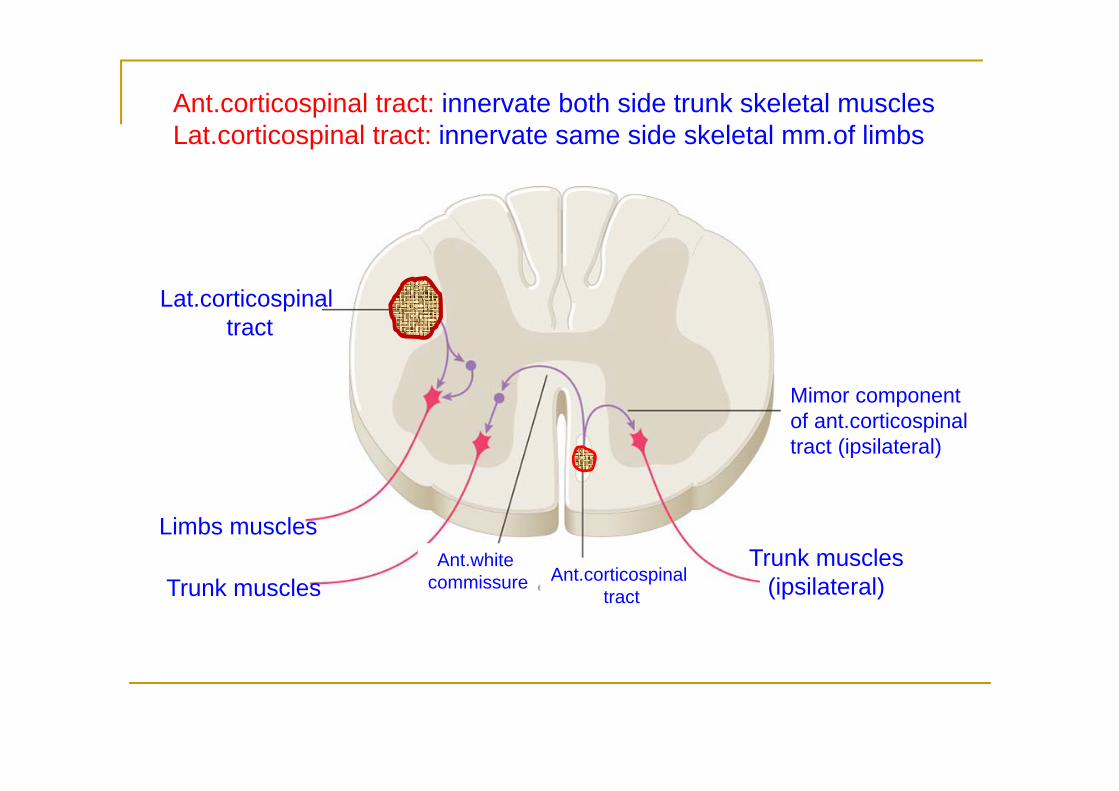
Lat.corticospinaltract
Limbs muscles
Trunk musclesAnt.white
commissure Ant.corticospinaltract
Mimor component of ant.corticospinal tract (ipsilateral)
Trunk muscles(ipsilateral)
Ant.corticospinal tract: innervate both side trunk skeletal musclesLat.corticospinal tract: innervate same side skeletal mm.of limbs
Copy Right- Hongqi ZHANG-Department of Anatomy-Fudan University
Copy Right- Hongqi ZHANG-Department of Anatomy-Fudan University
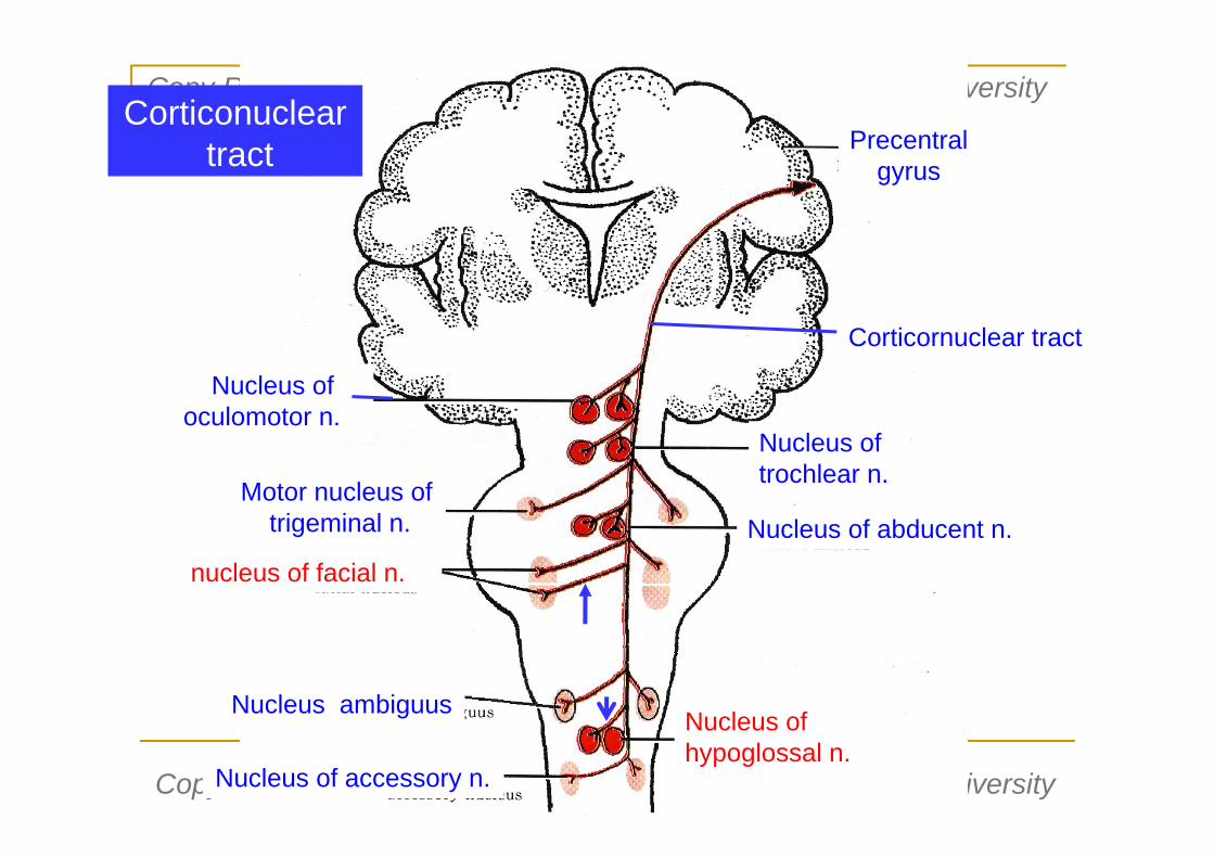
Nucleus of oculomotor n.
Nucleus of trochlear n.
Motor nucleus oftrigeminal n. Nucleus of abducent n.
nucleus of facial n.
Nucleus ambiguus
Nucleus of accessory n.
Nucleus of hypoglossal n.
Precentralgyrus
Corticornuclear tract
Nucleus of oculomotor n.
Nucleus of trochlear n.
Motor nucleus oftrigeminal n. Nucleus of abducent n.
nucleus of facial n.
Nucleus ambiguus
Nucleus of accessory n.
Nucleus of hypoglossal n.
Precentralgyrus
Corticornuclear tract
Corticonucleartract

Copy Right- Hongqi ZHANG-Department of Anatomy-Fudan University
Copy Right- Hongqi ZHANG-Department of Anatomy-Fudan University
Nucleus of oculomotor n.
trigeminal Motor nucleus
Facia nucleus (upper).
Nucleus ambiguusNucleus of accessory n.
Nucleus of trochlear n.
Nucleus of abducent n.
Nucleus of hypoglossal n.
Morter cortex
Facia nucleus (lower)
One side corticornuclear tract
Nuclear of hypoglossal nReceive fiber of contralateral corticonuclear tract only
Lower part of facial nucleusReceive the fiber of contralateral corticonuclear tract only

Copy Right- Hongqi ZHANG-Department of Anatomy-Fudan University
Copy Right- Hongqi ZHANG-Department of Anatomy-Fudan University
Nuclear of hypoglossal nReceive the fiber of contralateral
corticonuclear tract only
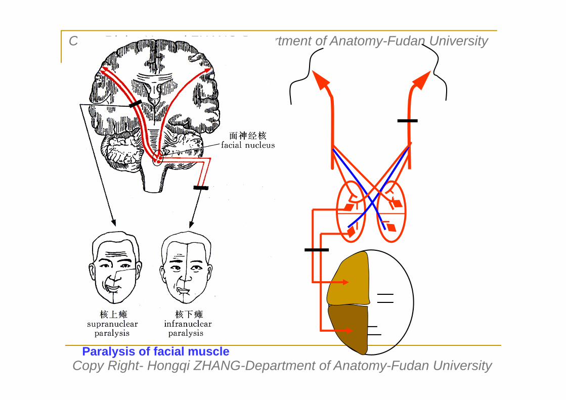
Lower part of facial nucleusReceive the fiber of contralateral corticonuclear tract only
Upper part of facial nucleusReceive the fiber of corticonuclear tract of both left and right side
Facial nuclear
corticonuclear tract
Facial nuclear
hypoglossal nuclear
Corticonuclear tract
Copy Right- Hongqi ZHANG-Department of Anatomy-Fudan University
Copy Right- Hongqi ZHANG-Department of Anatomy-Fudan University
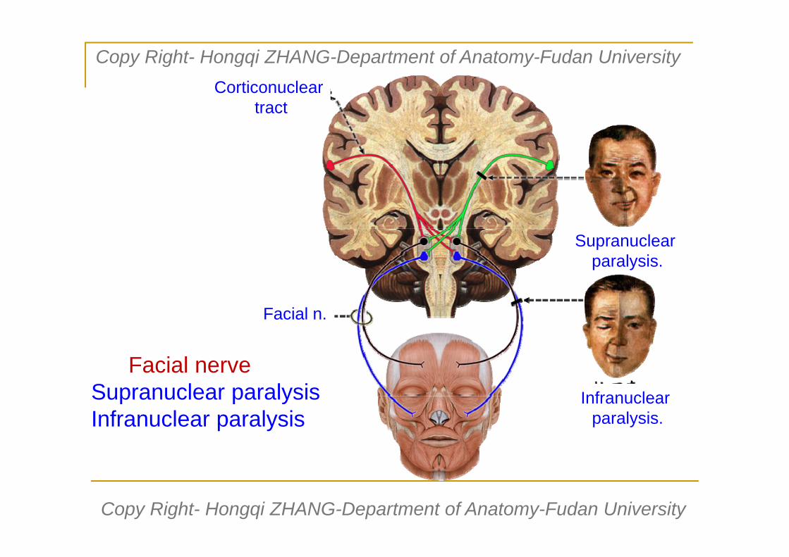
Corticonucleartract
Facial n.
Supranuclear paralysis.
Infranuclear paralysis.
Facial nerveSupranuclear paralysisInfranuclear paralysis

Copy Right- Hongqi ZHANG-Department of Anatomy-Fudan University
Copy Right- Hongqi ZHANG-Department of Anatomy-Fudan University
Corticonucleartract
Hypoglassol n.
Supranuclear paralysis.
Infranuclear paralysis.
Hypoglassol nerveSupranuclear paralysisInfranuclear paralysis

Cell body position of 1st order neuron
Cortex in upper 1/3 of precentral gyrus
Cell body position of 2nd order neuron
Somatic motor nuclei in brain stem
Position passing internal capsule GenuPosition of tract in brain stem Corticonuclear tract give rise to motor
nuclei at different level.Position of decussation Pyramidal decussation:in medullaName & position of tracts in spinal cord
Ant,corticospinal tract (ant.funiculus)Lat.corticospinal tract (lat.funiculus)
The other feature:(A) One side corticonuclear innervate both sides: oculomotor n,trochear n,motor
nuclear of trigerminal nerve,abducent nuclear,upper part of facial
nuclear,nuclear ambiguus,accessory nuclear .
(B) Lower part of facial nuclear and hypoglassol nuclear receive only the fiber of
contralateral corticonuclear tract.
Features of cortinuclear tract

The following contents
Learn by yourself

Copy Right- Hongqi ZHANG-Department of Anatomy-Fudan University
Copy Right- Hongqi ZHANG-Department of Anatomy-Fudan University
Descending pathway-Extrapyramidal system
It is the descending nervous pathway which influence and control the movement of body except the pyramidal system.
The system includes the corpus striatum.together with the subthalamic mucleus.substantia nigra.red nucleus and reticular formatin of brain stem. the main functions of the extrapyramidal system in man are to regrlate the tonicity of the muscles,maintain the normal body posture and produce habitual and rhythmic movements.

Copy Right- Hongqi ZHANG-Department of Anatomy-Fudan University
Copy Right- Hongqi ZHANG-Department of Anatomy-Fudan University
Concept: is a common name for the descending pathways besides the pyramidal system.
Function is to regulate the toniety of the muscles, coordinate the muscular activities,mainitain the normal body posture and produce habitual and rhythmic movements.
For example:…
The extrapyramidal system mainly include:
The Cortico-neostriatum-dorsal thalamus-cortex circuit.
The corticoponto-cerebellar-cortex circuit.
The neostriatum-substantia nigra circuit.
The globus palidus-subthalamus circuit.
Nervous pathway- Extrapyramidal system

Copy Right- Hongqi ZHANG-Department of Anatomy-Fudan University
Copy Right- Hongqi ZHANG-Department of Anatomy-Fudan UniversityCorticoponto-cerebellar-cortex circuit
Six orders neuronsRelay.Three decussation

Copy Right- Hongqi ZHANG-Department of Anatomy-Fudan University
Copy Right- Hongqi ZHANG-Department of Anatomy-Fudan University
Summary of nervous pathwaysAscending pathway consists of three order neurons
Descending pathway consists of two order neurons
General speaking,during their conducting course of both
ascending & descending pathways.There is a
decussation,that is,one side hemisphere receive
sensory impulse of contralateral half of the body.and
innervate the movement of contralateral half of the body.
One side visual area and auditory area receive visual
impulse and auditory impulse of both sides

Babinski reflex - an UMN sign Adult response - plantar flexion of the big toe and adduction of
the smaller toes Pathological (Infant) response - dorsoflexion (extension) of
the big toe and fanning of the other toes Indicative of upper motor neuron(UMN) damage

Copy Right- Hongqi ZHANG-Department of Anatomy-Fudan University
Copy Right- Hongqi ZHANG-Department of Anatomy-Fudan University
Clinical features of upper & lower motor neurons lesion
Upper motor neuron Lower motor neuronCell body location Cerebral cortex & their
axon (motor tract)Motor nuclei of cranial nerve. Motor neuron of Ant.horn of spinal cord
Muscle tone increased decreased
Deep reflex hyperactive Decreased or absent
Superficial reflex Decreased or absent absent
Pathological reflex
Existed (babinski sign) absent
Muscle size Slight atrophy Pronounced atrophy
Classical description
Spastic paralysis Flaccid paralysis

Copy Right- Hongqi ZHANG-Department of Anatomy-Fudan University
Copy Right- Hongqi ZHANG-Department of Anatomy-Fudan University

Copy Right- Hongqi ZHANG-Department of Anatomy-Fudan University
Copy Right- Hongqi ZHANG-Department of Anatomy-Fudan University

Copy Right- Hongqi ZHANG-Department of Anatomy-Fudan University
Copy Right- Hongqi ZHANG-Department of Anatomy-Fudan University
Corticospinal tract

Lat.corticospinal tract
Copy Right- Hongqi ZHANG-Department of Anatomy-Fudan University
Copy Right- Hongqi ZHANG-Department of Anatomy-Fudan UniversityParalysis of facial muscle

Copy Right- Hongqi ZHANG-Department of Anatomy-Fudan University
Copy Right- Hongqi ZHANG-Department of Anatomy-Fudan UniversityParalysis of tongue muscle

Copy Right- Hongqi ZHANG-Department of Anatomy-Fudan University
Copy Right- Hongqi ZHANG-Department of Anatomy-Fudan University