control of spoilage fungi by protective lactic acid bacteria displaying probiotic properties
TRANSCRIPT

Control of Spoilage Fungi by Protective Lactic AcidBacteria Displaying Probiotic Properties
Kontham Kulangara Varsha & Sulochana Priya &
Leena Devendra & Kesavan Madhavan Nampoothiri
Received: 19 December 2013 /Accepted: 3 February 2014# Springer Science+Business Media New York 2014
Abstract Thirty-six lactic acid bacteria belong to Lactococcus, Lactobacillus, Enterococcus,and Pediococcus were isolated, and the spectrum of antifungal activity was verified againstFusarium oxysporum (KACC 42109), Aspergillus niger (KACC 42589), Fusariummoniliforme (KACC 08141), Penicillium chrysogenum (NII 08137), and the yeast Candidaalbicans (MTCC 3017). Three isolates, identified as Pediococcus pentosaceus (TG2), Lacto-bacillus casei (DY2), and Lactococcus (BSN) were selected further, and their antifungalcompounds were identified by ESI-MS and HPLC analysis as a range of carboxylic acidsalong with some unidentified, higher molecular weight compounds. An attempt to check outthe shelf life extension of wheat bread without fungal spoilage was performed by fermentingthe dough with the Lactococcus isolate. Apart from growth in low pH and tolerance to bilesalts, probiotic potential of these three isolates was further substantiated by in vitro screeningmethods that include transit tolerance to the conditions in the upper human gastrointestinaltract and bacterial adhesion capacity to human intestinal cell lines.
Keywords Lactic acid bacteria . Antifungal . Protective culture . Bio preservation . Sourdough .
Probiotics
Introduction
Biological preservation of food has gained major attention during recent years where antag-onistic microorganisms or their antimicrobial metabolites can prevent the growth of pathogenicbacteria and fungus in food. Lactic acid bacteria (LAB) have a long tradition of being used infood fermentations and secured the position of food-grade bacteria due to their generallyregarded as safe status. LAB are established as probiotics and good candidate for biological
Appl Biochem BiotechnolDOI 10.1007/s12010-014-0779-4
K. K. Varsha : L. Devendra :K. M. Nampoothiri (*)Biotechnology Division, National Institute for Interdisciplinary Science and Technology (NIIST), CSIR,Trivandrum 695 019 Kerala, Indiae-mail: [email protected]
S. PriyaAgroprocessing Division, National Institute for Interdisciplinary Science and Technology (NIIST), CSIR,Trivandrum 695 019 Kerala, India

food preservation due to their wide spectrum antimicrobial activities. As food additives, LABcan perform as protective culture for improving microbiological safety of the product withoutchanging the sensory characteristics of the food. Fungal growth inhibition by LAB due to thesecretion of antagonistic compounds like organic acids [1, 2], reuterin [3], proteinaceouscompounds [4], and cyclic dipeptides [5] obtained foremost attention in the area ofbiopreservation.
Growth inhibitory action of Lactobacillus, Enterococcus, and Leuconostoc cultures werereported against varying fungal groups such as Debaryomyces hansenii, Saccharomycescerevisiae, and Penicillium spp. [6]. Application of Lactobacillus plantarum IMAU10014against Botrytis cinerea, Glomerella cingulate, Phytophthora drechsleri Tucker, Penicilliumcitrinum, and Fusarium oxysporum [7] has been investigated. Activity of Lactobacillus andWeisella against Aspergillus nigerMUCL 28699, Candida albicansMUCL 30112, Aspergillustubingensis MP1, and Penicillium crustosum MY1 [8] have also been reported.
Studies have been conducted regarding the application of LAB as biopreservative to controlfungal spoilage of bread [9]. LAB was effectively used to control the growth of Listeriamonocytogenes and Salmonella enteritidis in chicken meat [10] and growth of Salmonellatyphimurium, Escherichia coli, and L. monocytogenes in iceberg lettuce and golden deliciousapples [11, 12]. LAB was also used for the vacuum-packed storage of Dicentrarchus labrax[13] and smoked salmon [14]. Citrus fungal pathogens Penicillium digitatum and Geotrichumcitriaurantiiwere efficiently inhibited by LAB [15]. Production of the antagonistic compoundsby L. plantarum in grass silage was investigated and that led to the possibility of efficientlyusing LAB for silage preservation [16]. Microgard, a commercial food additive extractedfrom Propionibacterium freudenreichii can be termed as an example of protective culture.It is marketed as a food preservative for use in cottage cheese and fruit flavored yogurtwhere the shelf life of cottage cheese was extended 6–9 days [17]. The use of probioticstrains as protective cultures have several advantages when considering the fact that one ofthe major processes of food biopreservation is fermentation and the major carriers ofprobiotic cultures are fermented dairy products and sausages, the same cultures can beused for various purposes under different conditions by ensuring that the use is standard-ized and reproducible [18].
Our group is interested in new LAB isolates with probiotic properties for a range ofapplications that have been reported earlier [19-21]. This study focuses on the isolation ofantifungal LAB for food preservation.
Materials and Methods
Microorganisms and Growth Medium
Thirty-six antifungal LAB strains were isolated on MRS-CaCO3 plates from different sourceslike rotten jackfruit, guava and fecal samples of deer, Indian gaur, and tiger puke. The sampleswere collected from Silent Valley National Park which is a part of the Western Ghat’s naturalworld heritage site of India and one of the biodiversity hot spots in the Kerala belt. All isolateswere cultivated on de Man Rogosa Sharpe (MRS) agar or in MRS broth (Himedia, India) at30 °C for 24 h and maintained for longer storage at −20 °C in MRS supplemented with 20 %(v/v) glycerol.
The fungal strains, F. oxysporum (KACC 42109), A. niger (KACC 42589), Fusariummoniliforme (KACC 08141), Penicillium chrysogenum (NII 08137), and C. albicans (MTCC3017) were maintained on potato dextrose agar (Himedia, India) at 30 °C.
Appl Biochem Biotechnol
Antifungal Activity Assay
Twenty-four-hour-old cells (A600, 0.1) of new LAB isolates were used to inoculate MRSbroth and incubated at 30 °C under static condition for 48 h. After incubation, the cellswere removed by centrifugation at 8,590×g for 15 min, and the culture supernatants wereconcentrated 10-fold in a vacuum concentrator at 30 °C with respect to their initial volumeand used for agar well diffusion assay. For this, 100 μl of the above samples were added tothe wells (5 mm diameter) cut on potato dextrose agar which was previously spread platedwith 1×10⁴ spores of F. oxysporum and kept at 30 °C for 4 days. An inhibition ofF. oxysporum growth more than 8 mm diameter was looked further for growth inhibitionof other fungal cultures like A. niger, F. moniliforme, P. chrysogenum, and the yeastC. albicans.
Identification of the Strains and Their Tolerance to Gastric/Intestinal Inhibitory Substances
The selected isolates were identified by 16S rRNA sequencing method using the universalprimers 27F and 1492R. Ability of LAB isolates to tolerate gastric and intestinal conditionswere primarily checked by survival through adverse growth conditions such as presence ofphenol (0.3, 0.4, and 0.5 % (v/v)), bile salt (0.3, 0.5, and 0.8 % (w/v)) and initial medium pHadjusted to 2 and 2.5. Cells were incubated for 24 h under these varying conditions at 30 °C instatic condition, and the growth was monitored.
Effect of Temperature, pH, and Enzyme (Lipase and Proteolytic) Treatment on AntifungalActivity of the Culture Filtrates of Selected Strains
Antifungal activity of the culture filtrates of BSN, TG2, and DY2 (around pH 4.3) afterexposure to high temperature (100 °C for 30 min and at 121 °C for 15 min) and varying pH(6.0, 6.5, and 7 adjusted with 0.1 M NaOH) was determined by agar well diffusion assay. Theenzymes: lipase, proteinase k, trypsin, and α-chymotrypsin (Sigma-Aldrich, USA) wereprepared in appropriate buffers and treated with the culture supernatant at specific pH andtemperature. Final concentrations of the enzymes in culture filtrates were 1 mg/ml, andsamples without enzymes were kept as control. Activity was detected by the well diffusionmethod against F. oxysporum.
Preparation and Purification of Bioactive from Culture Supernatant
Ten liters of MRS medium was inoculated (1 % (v/v)) with 24-h-old culture and incubatedat 37 °C for 48 h at static condition. After incubation, the supernatant was collected bycentrifugation at 8,590×g for 15 min and extracted with double volume of hexane,chloroform, and ethyl acetate in a sequential order. The solvent was evaporated underreduced pressure in a rotavapour (Buchi) and the semi-viscous concentrated sampleobtained was loaded onto a SPE silica column (600×40 mm). The active compoundswere eluted using chloroform/methanol gradient. Growth inhibitory activity of all thefractions from three isolates was checked against F. oxysporum by agar well diffusionmethod. Active fractions of each isolate were purified further by preparative thin-layerchromatography. Fractions of BSN, TG2 and DY2 were separated by loading 200 μl ofeach sample on silica plate (Merck; 60F254, 0.5 mm). Compounds adsorbed on silica werescraped out and extracted by treating with chloroform/methanol (1:1, v/v) and subjected toESI-MS and HPLC analysis.
Appl Biochem Biotechnol

Identification of the Antifungal Compounds by ESI-MS and HPLC
Compounds separated by preparative TLC were identified by comparing data from ESI-MS and high-performance liquid chromatography with that of commercially availablecompounds that include acetic acid, stearic acid, palmitic acid, 3-phenyllactic acid,caproic acid, benzoic acid, salicylic acid, and propionic acid (Sigma, USA). Liquidchromatography–mass spectrometry (Thermo Orbitrap LC/MS) of separated compoundswas performed using electrospray ionization method. For chromatographic analysisLuna silica column (Phenomenex, 250×4.60 mm) was used with chloroform as mobilephase.
Prevention of Fungal Growth on Baked Sourdough Fermented with BSN
The sourdough was baked to check the efficiency of one of our isolates, the Lactococcus(BSN) on prevention of fungal growth. BSN cells were collected after 24 h of incubation inMRS broth, washed twice with sterile distilled water, and resuspended in the same. This cellsuspension (4×10⁹ cfu) was used along with commercially available yeast cells in sourdoughmanufacture. Two controls were kept during the study, the dough without BSN and the doughwithout yeast to test how the absence or presence of each would affect the fungal growthinhibiting activity of sourdough. The dough formulations (Table 1) were mixed thoroughly andwere placed in separate aluminum pans and fermented at 30 °C for 36 h. After fermentation,the sourdoughs were baked in a batch oven at 180 °C for 30 min and the bread loaves werecooled at room temperature for 90 min.
Antifungal potential of baked sourdough was checked against A. niger, F. oxysporum, andF. moniliforme. Fifty-microliter suspension of each fungus containing approximately 104
spores along with some mycelia were sprayed on both sides of the loaves and kept forincubation at 30 °C for 5 days under sterile conditions. The loaves were checked daily forvisible fungal outgrowth, and the shelf life was defined as the time for fungus to becomevisible on bread loaves.
Survival of LAB in Simulated Gastrointestinal Environment
Simulated gastric and intestinal juices were prepared as previously described [22, 23] withsome modifications. It was prepared by dissolving 13.3 mg/l pepsin from porcine gastricmucosa (Sigma, USA) in 0.5 % (w/v) sterile saline and adjusting the pH to 2.5. Simulatedsmall intestinal juice was prepared by dissolving 250 mg/l pancreatin from porcine pancreas(Sigma) and 500 mg/l porcine bile (Himedia) in 0.5 % (w/v) sterile saline and adjusting the pHto 7.5. Freshly prepared solutions were used for the experiments. Cultures of BSN, TG2 and
Table 1 Dough formulations
aYeast alonebYeast and BSN togethercBSN alone
Flour (g) Ia IIb IIIc
200 200 200
NaCl (g) 2 2 2
Glucose (g) 4 4 4
Yeast (g) 1 1 –
BSN (cfu) – 4×10⁹ 4×10⁹
Water (ml) 150 150 150
Appl Biochem Biotechnol
DY2 were grown overnight (18 h) in 5 ml MRS medium and centrifuged at 8,590×g for15 min to obtain the pellets. The pellets were then resuspended and washed twice in sterilesaline. After centrifugation, the pellets were resuspended in equal volume of simulated gastricjuice at 37 °C and incubated for 90 min with constant shaking. The initial number of viablecells resuspended in gastric juice was 1.4×10⁸ cells of BSN, 6×10⁷ cells of TG2, and 4×10⁷
cells of DY2/ml. Samples were retrieved after 90 min; serially diluted and appropriate dilutionswere plated on MRS medium and incubated at 37 °C for 48 h. Cells after simulated gastricenvironment treatment were collected by centrifugation at 8,590×g for 15 min and resuspend-ed in same amount of simulated intestinal juice and kept for 6 h incubation at 37 °C. Sampleswere retrieved at 0 and 6 h, and the appropriate dilutions were plated on MRS medium tocheck the viable number of bacteria.
In Vitro Bacterial Adhesion Capacity to Intestinal Epithelial Cell Line
A modified method [18] was adapted for checking the intestinal adhesion ability of selectedLAB isolates. Adhesion capacity of BSN, TG2 and DY2 to the human colon adenocarcinomacells HT-29 was investigated. The strain E. coli E1T was kept as negative control. HT-29 cellline was obtained from NCCS, Pune and maintained in Eagle’s minimum essential medium,supplemented with 10 % fetal bovine serum, 100 mg/l streptomycin, 100 U/l penicillin, and50 μg/ml gentamycin. The cells were grown at 37 °C in a humidified atmosphere of 5 % CO2
and 95 % air until a confluent monolayer was obtained. Monolayers of HT-29 cells wereseeded at a concentration of 2×10⁵ cells/ml and dispensed into each 200 mm2 well of a 24-well tissue culture plate. The 24-h-old bacterial cells washed twice in PBS and resuspended inthe HT-29 growth medium without gentamicin to a final concentration of approximately 3.5×10⁸ cfu/ml of BSN, 3.3×10⁶ cfu/ml of DY2, and 4.5×10⁸ cfu/ml of TG2. Viable number ofE. coli cells used was 9×10⁸ cfu/ml. This cell suspension (1 ml) was added to each well of thetissue culture plate. After 90 m of incubation with the isolates, the monolayers were washedtwo times with phosphate-buffered saline (PBS; pH 7.4) to remove nonadherent bacteria. TheHT-29 cells were lysed by addition of 0.1 % (v/v) Triton-X100 and the number of viableadherent bacteria was determined by plating serial dilutions onto MRS medium. Colony-forming units were enumerated after incubation for 48 h at 37 °C and the adhesion capacitywas described as the percentage of bacteria adhered to HT-29 cells in relation to the totalnumber of bacteria added. Each adhesion assay was conducted two times with duplicatedeterminations.
Results
Preliminary Screening and Characterization of LAB Isolates
Out of 36 isolates, 11 showed more than 8-mm diameter zone of growth inhibition againstF. oxysporum (Table 2). Isolates TG2 and DY2 showing maximum antifungal activity wererecognized as Pediococcus pentosaceus and Lactobacillus casei respectively by 16S rRNAsequencing (Table 2). Strain BSN showed maximum similarity to Lactococcus garvieae butdiffered from the type strain in phenotypic and genetic features (data not shown) and furtherexperiments on characterization of this isolate is going on. Isolates TG2 and DY2 showed awide spectrum of activity (+++) against all the tested fungi where BSN showed (+++) activityagainst F. moniliforme and C. albicans along with (++) activity against P. chrysogenum. Someof the probiotic properties of the isolates were also summarized in Table 2. Isolates BSN, TG2,
Appl Biochem Biotechnol

and SVG3 survived through pH 2 and all the isolates could tolerate 0.8 % bile and 0.4 %phenol in the medium.
Effect of Temperature, pH, Lipase and Proteolytic Enzymes on Antifungal Activityof the Culture Filtrates of Selected Strains
Antifungal activity of the active fraction from these three isolates was retained after exposureto 100 °C for 30 min and at 121 °C for 15 min. Antifungal activity decreased with an increasein pH, holding 40 % of the activity at pH 6.5 and 20 % activity at pH 7 in comparison with thecontrol which was an untreated sample (around pH 4.3). Treatment with proteolytic enzymesand lipase showed no effect on the growth-inhibiting activity of the fractions.
Purification and Identification of Antifungal Compounds
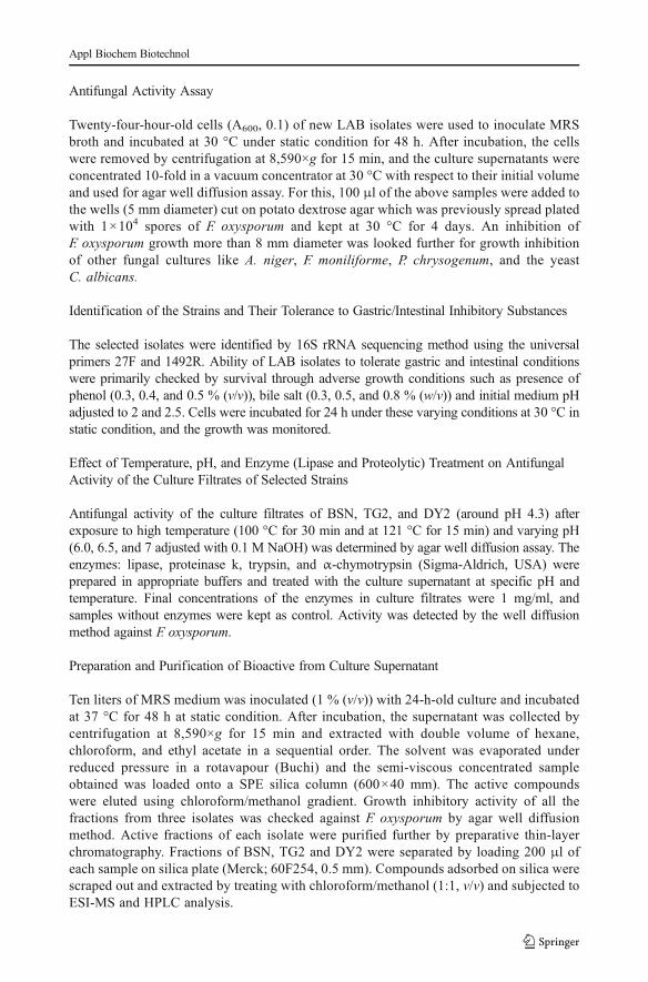
After sequential extraction with the solvents, it is found that ethyl acetate extract retained amajor portion of activity against F. oxysporum. Silica column-purified fractions were checkedfor growth inhibiting activity against F. oxysporum and the active fractions were furtherseparated on preparative TLC, and the activity was confirmed against the same pathogen(Fig. 1). From the visible zone of inhibition, it was obvious that, the fractions from BSN andTG2 are more effective in fungal growth inhibition. These partially purified compounds wereanalyzed by ESI-MS along with analytical HPLC and discovered that BSN producing a rangeof carboxylic acids including benzoic acid, caproic acid, salicylic acid, stearic acid, andpalmitic acid (Table 3). Similarly, TG2 and DY2produced stearic acid and salicylic acid.Though these acids contribute to the major portion of antifungal activity shown by the isolatessome compounds with higher molecular weight and showing antifungal activity remainedunidentified and characterization of these compounds are in progress.
Prevention of Fungal Growth on Baked Sourdough Fermented with BSN
Comparison with the spoilage rate in the control sourdough revealed that the dough treatedwith both BSN and yeast could delay the growth of A. niger, F. oxysporum, and prevented theattack of F. moniliforme (Fig. 2). Sporulation of the respective fungi occurred in the bread
Table 2 Identification and tolerance to growth inhibiting conditions of antifungal LAB
Strain pH Bile (%) Phenol (%) Antifungal activity Identification 16sRNA
2 2.5 0.5 0.8 0.4 0.5
BSN + + + + + − ++ Lactococcus
TG1 − + + + + − ++ Lactobacillus plantarum
TG2 + + + + + − +++ Pediococcus pentosaceus
DY1 − + + + + + ++ L. plantarum
DY2 − + + + + − +++ Lactobacillus casei
DR2 − + + + + − + Enterococcus faecalis
SVG3 + + + + + + ++ L. plantarum
RJF4 − + + + + − ++ L. plantarum
Calculation of antifungal activity: 8 mm diameter clearing zone (+), 10 mm diameter clearing zone (++), andmore than 10 mm diameter clearing zone (+++; against Fusarium oxysporum)
Appl Biochem Biotechnol
treated with yeast alone in 3 days. BSN and yeast treated sourdough loaves delayed the growthof A. niger and F. oxysporum and prevented the growth of F. moniliforme. Sporulation ofA. niger and visible mycelia growth of F. oxysporum occurred on the fifth day of incubation.However, the bread made without yeast lacked the normal texture and was brittle in nature.
Survival of LAB in Simulated Gastrointestinal Environment
After 90 min of incubation in the simulated gastric juice, approximately 3 log reduction of cellviability was occurred for BSN, less than 2 log reduction for TG2, and below 1 log reductionfor DY2 (Fig. 3). It revealed that Lactobacillus was more competent in artificial gastricconditions when comparing to the survival of Lactococcus and Pediococcus in the same.After 6 h of simulated intestinal juice treatment, about 2.5 log reduction of cell viabilityoccurred for both BSN and DY2 and less than 1 log decrease for TG2 when compared with theinitial number of cells inoculated to simulated intestinal juice.
In Vitro Bacterial Adhesion Capacity to Intestinal Epithelial Cell Line
Three isolates were examined for their ability to adhere to HT-29 cell line along with E. coli asnegative control. All the three isolates showed higher adhesion capacity than the E. coli straintested which was 0.005 % with a 4 log reduction of viable cells after binding. The isolate TG2hold high adhesion capacity of 15.2 % with around 1.5 log reduction from the initial cellnumber (see “In Vitro Bacterial Adhesion Capacity to Intestinal Epithelial Cell Line” undermaterials and methods). DY2 possessed adhesion capability of about 1 %, and the loweradhesion ability was exhibited by BSN, which was 0.3 % with around 2 log reduction in theviable cell number for both BSN and DY2 after binding.
Fig. 1 Antifungal activity of preparatory TLC-purified fractions against F. oxysporum. Activity of differentfractions from BSN (b), TG2 (t), and DY2 (d)
Table 3 ESI-MS results of the an-tifungal fractions of LAB isolates Acid Mass Molecular formula Isolate
Benzoic 122.12 C7H6O2 BSN
Palmitic 256.42 C16H32O2 BSN
Salicylic 138.12 C7H6O3 BSN, TG2, and DY2
Stearic 284.47 C18H36O2 BSN, TG2, and DY2
Caproic acid 116.15 C6H12O2 BSN
Appl Biochem Biotechnol

Fig. 2 Antifungal activity of bread fermented with Lactococcus (BSN) and yeast on fifth day againstF. moniliforme (a), A. niger (b), and F. oxysporum (c). a1, b1, c1 Bread treated with yeast alone; a2, b2, c2bread treated with yeast and BSN; a3, b3, c3 bread treated with BSN alone
Fig. 3 Survival of LAB isolates in simulated gastric juice (diamonds) along with efficiency to survive simulatedintestinal juice on the secondary Yaxis (squares). The initial numbers of viable cells resuspended in gastric juicewere 1.4×10⁸ cells of BSN, 6×10⁷ cells of TG2, and 4×10⁷ cells of DY2
Appl Biochem Biotechnol
Discussion
Fungal contamination is one of the major causes of spoilage in food and feed. Moreover,mycotoxins cause serious health problems in humans ranging from immune suppression todeath in severe cases. Aflatoxins are hepatocarcinogenic and fumonisins may cause neuraltube defects in maize-consuming communities through disruption of ceramide synthase andsphingolipid biosynthesis [24]. Contamination of feedstuffs with mycotoxins also create aserious threat to the health and productivity of animals [25]. Thus, food safety remains a majorconcern when addressing current health problems in developing countries, and pursuingnatural methods for food protection is gaining huge attention recently because of the con-sumers’ demand for chemical-free, safe, and mildly processed food with extended shelf life.Selected strains of LAB with potent antibacterial and antifungal properties can be used as“green preservatives” apart from their contribution to texture, flavor, and nutritional value tothe food or feed.
Thirty-six strains of LAB with growth-inhibiting activity against F. oxysporum was isolatedfrom various sources and found to be the members of various LAB genera like Lactobacillus,Lactococcus, Pediococcus, and Enterococcus. Three isolates from three different genera wereselected based on their activity against the above test organism. Spectrum of activity was alsochecked against three major plant pathogenic and mycotoxin-producing genera of fungi thatinclude A. niger, F. moniliforme, and P. chrysogenum associated with the growth in and damageto food even at refrigeration temperatures. Anti-mold activity was confirmed againstC. albicans which is a reference strain for antifungal susceptibility checking.
Antifungal activity of L. caseiwas reported previously as a synergistic effect of cyclo-(Leu-Pro), 2, 6-diphenyl-piperidine, and 5,10-diethoxy-2,3,7,8-tetrahydro-1H, 6H-dipyrrolo [1,2-a;1′,2′-d] pyrazine [26] and P. pentosaceus as a biopreservative in bakery products and tomatopuree by delaying the growth of Aspergillus oryzae, and A. niger was described by Muhialdinet al. [27]. Lactococcus, P. pentosaceus, and L. casei are reported to produce various antimi-crobial compounds like carboxylic acids, proteinaceous compounds, and cyclo-dipeptides[28]. In our study, an array of carboxylic acids was detected in some of the fractions fromLactococcus, including benzoic acid, caproic acid, salicylic acid, palmitic acid, and stearicacid. Stearic acid and salicylic acid were also produced by Pediococcus and Lactobacillus.The role of these acids toward fungal growth inhibition can be considered vital based onprevious studies by various research groups [28, 29]. Apart from these recognizedcompounds, the presence of certain unidentified compounds with higher molecular weightthan the reported ones direct us to think about the existence and involvement of a fewunknown antifungal compounds contributing to the antifungal activity (study is in prog-ress). When considering the role of carboxylic acids as precursors to a variety of com-pounds including esters, amides, and carboxylate salts, the possibility of formation ofvarious compounds are not to be kept aside.
The experiment where Lactococcus BSN strain was used in fermentation of sourdoughalong with commercially available yeast showed the potential of this new isolate to extend theshelf life of sourdough compared with the sourdough treated with yeast alone. Ryan et al. [30]reported that the antifungal activity of sourdough where the dough was treated with antibioticsto make sure that no viable bacteria remained in the dough. In our study, we baked thesourdough and checked the antifungal activity on the baked loaves. Ability of Lactobacillus toprevent fungal growth when present along with yeast is reported earlier by Lavermicocca et al.[31]. Inability of the sourdough loaves treated only with BSN, to prevent fungal growthdemonstrates the synergistic action of yeast and BSN that led to the extended shelf life ofsourdough. Much report are not available where lactococci have been used for preservation of
Appl Biochem Biotechnol
baked foods, and this experiment demonstrates the possibility of using the sourdough treatedwith BSN for manufacturing bread.
These new LAB isolates also hold substantial probiotic features. Enduring through thegastrointestinal conditions is an important characteristic of LAB strains, as they are a veryimportant group of intestinal microbiota, and colonization of which are possible only if theysubsist in the acidic gastric acid environment as well as exposure to bile and pancreatic juice inthe upper small intestine to exert its beneficial effects in the gut. All the isolates could survive0.4 % phenol and 0.8 % bile in the medium. Phenol resistance is considered an advantage ofprobiotics owing to the fact that it can be released to the body by deamination of aromaticamino acids [32]. Survival at 0.5 % bile is regarded as very good [33] and our isolates couldsurvive 0.8 % bile in the medium. Both Lactococcus and Pediococcus could survive pH 2 for24 h which substantiates their origin and establishment in animal intestine, and the Lactoba-cillus strain could tolerate a pH of 2.5. Survival at low pH was considered a very importantprobiotic characteristic, as the bacteria have to subsist the depleted pH of gastric juice whiletravelling from the mouth to the intestine. One of the indispensable qualities of probiotic is itsability to reach, survive, and persist in the environment where it is proposed to act, and toaffect its environment favorably, the number of probiotic bacteria should be preferablybetween 105 to 108 cfu/g of intestinal content [34], so the number of bacteria selected for thisstudy was confined to the above range.
To confirm the initial results from low pH and bile tolerance as well as to check the abilityof these isolates to survive gastrointestinal environment, survival in simulated gastric andintestinal juice was verified. In this model, digestive system Lactobacillus showed highestsurvival rate which is in accordance with earlier reports [35]. Only a few investigations showedthat lactococcal strains can tolerate artificial gastric juice (pH 2.5) and artificial bile [36]. Thelactococcal strain under study could tolerate the artificial gastrointestinal conditions and can beconsidered a candidate to be used in functional foods. Ability of Pediococcus as a probioticcandidate is well studied and reported up to 0.5 % bile (0.3 % is the mean concentration in theintestine) tolerance along with low pH tolerance at 3 and 58 % simulated gastric juice survival[37-39]. The Pediococcus strain reported in this article was capable of survival at pH 2 and inthe presence of 0.8 % bile along with a very low decrease in viability after treatment in theartificial gastrointestinal environment.
Even though it is difficult to conclude from in vitro cell adhesion experiments instead of thesituation in gastrointestinal tract, where the host defense systems, competition with the residentmicrobiota, mucosal shedding, and peristaltic flow are likely to modify the bacterial adhesion[40], the ability of bacteria to adhere to the intestinal cell lines are important to understand themechanisms of adhesion and provide important information on the differences among LABspecies. Cell adhesion property of diverse LAB strains to different cell lines varies from 1 to25 % [18, 23]. Adhesion capacity of LAB to mucus-producing HT 29 cell lines was exploredin our study. Adhesive properties of different bacteria depend on the origin and the initial dosesprovided and also when applied as a combination of probiotic bacteria, stronger adhesivecharacteristics were reported [41]. Investigations by Jankowska et al. [42] showed thatpathogenic bacteria adhered better to well-differentiated Caco-2 cells, whereas lactobacilliand lactococci displayed better adhesion to nondifferentiated Caco-2 cells which may givesupport to the competitive exclusion of pathogenic bacteria by LAB. Earlier studies [43]reported below 5 and 14.4 % adhesion ability of L. casei and very little binding abilities ofL. casei rhamnosus and P. pentosaceus [37] on Caco-2 cells.
In our study, TG2 revealed a higher amount of adhesion (15.2 %) than the previouslyreported P. pentosaceus Q3 that showed 6.2 % [23] and P. pentosaceus OZF strain, which was14.4 % on Caco-2 cells [44]. The new L. casei strain holds a binding ability of 1 %, and both
Appl Biochem Biotechnol

these values are higher when considering binding on HT-29 cells. Cell-binding capability ofLactococcus strain is lower (0.3 %) but still higher than the adhesion shown by E. coli. Thein vitro studies we followed to explore the probiotic potential of new LAB isolates revealedthat they can survive and adapt to the gastrointestinal conditions and be able to bind to theintestinal cell line. The in vitro probiotic characterization experiments performed revealed TG2hold the maximum of probiotic features in all the isolates checked and can be a potentialprobiotic strain.
Conclusions
Present study investigates the efficacy of lactic acid bacteria belonging to three different generasuch as Lactococcus, Pediococcus, and Lactobacillus to prevent the food spoilage fungalgrowth in vitro. Experiments revealed the presence of significant carboxylic acids as the majorgrowth-inhibiting factor apart from some unknown compounds that are yet to be identified.The cultures also displayed their potential to survive in gastric and intestinal environmentalong with adhesion capacity to the mucus-producing cell lines. Hence, they could be furtherdeveloped as probiotic and can prevent food spoilage as well if incorporated in foods.
Acknowledgments Research fellowship from Council of Scientific and Industrial Research, New Delhi, Indiawas greatly acknowledged. Research grant from the Department of Biotechnology (DBT), New Delhi to initiateprobiotic research and help from Kerala Forest Research Institute, Peechi for sample collection are greatlyappreciated.
References
1. Cabo, M. L., Braber, A. F., & Koenraad, P. M. F. J. (2002). Journal of Food Protection, 65, 1309–1316.2. Lind, H., Sjogren, J., Gohil, S., Kenne, L., Schnürer, J., & Broberg, A. (2007). FEMS Microbiology Letters,
271, 310–315.3. Talarico, T. L., Casas, I. A., Chung, T. C., & Dobrogosz, W. J. (1988). Antimicrobial Agents and
Chemotherapy, 32, 1854–1858.4. Magnusson, J., & Schnurer, J. (2001). Applied and Environmental Microbiology, 67, 1–5.5. Strom, K., Sjogren, J., Broberg, A., & Schnurer, J. (2002). Applied and Environmental Microbiology, 68,
4322–4327.6. Voulgari, K., Hatzikamari, M., Delepoglou, A., Georgakopoulos, P., Litopoulou-Tzanetaki, E., & Tzanetakis,
N. (2010). Food Control, 21, 136–142.7. Wang, H., Yan, Y., Wang, J., Zhang, H., & Qi, W. (2012). IMAU10014. PLoS ONE, 7(1), e 29452. doi:10.
1371/journal.pone.0029452.8. Ndagano, D., Lamoureux, T., Dortu, C., Vandermoten, S., & Thonart, P. (2011). Journal of Food Science, 76,
M305–M311.9. Ryan, L. A. M., Zannini, E., Dal Bello, F., Pawlowska, A., Koehler, P., & Arendt, E. K. (2011). International
Journal of Food Microbiology, 146, 276–283.10. Maragkoudakis, P. A., Mountzouris, K. C., Psyrras, D., Cremonese, S., Fischer, J., Cantor, M. D., et al.
(2009). International Journal of Food Microbiology, 130, 219–226.11. Randazzo, C. L., Pitino, I., Scifo, G. O., & Caggia, C. (2009). Food Control, 20, 756–763.12. Trias, R., Baneras, L., Badosa, E., & Montesinos, E. (2008). International Journal of Food Microbiology,
123, 50–60.13. El Bassi, L., Hassouna, M., Shinzato, N., & Matsui, T. (2009). Journal of Food Science, 74, M335–M339.14. Tome, E., Teixeira, P., & Gibbs, P. A. (2006). Food Microbiology, 23, 399–405.15. Gerez, C. L., Carbajo, M. S., Rollán, G., Torres Leal, G., & Font de Valdez, G. (2010). Journal of Food
Science, 75, M354–M359.16. Broberg, A., Jacobsson, K., Strom, K., & Schnurer, J. (2007). Applied and Environmental Microbiology, 73,
5547–5552.
Appl Biochem Biotechnol

17. Lund, B., Baird-Parker, T. C., & Gould, G. W. (2000). The microbiological safety and quality of food, vol. 1.Gaithersburg: Aspen.
18. Klingberg, T. D., Axelsson, L., Naterstad, K., Elsser, D., & Budde, B. B. (2005). International Journal ofFood Microbiology, 105, 419–431.
19. Aswathy, R. G., Ismail, B., John, R. P., & Nampoothiri, K. M. (2008). Applied Biochemistry andBiotechnology, 151, 244–255.
20. Gangadharan, D., Sivaramakrishnan, S., Pandey, A., & Nampoothiri, K. M. (2010). International Journal ofDairy Technology, 63, 339–348.
21. Divya, J. B., Varsha, K. K., & Nampoothiri, K. M. (2012). Applied Biochemistry and Biotechnology, 167,1314–1324.
22. Corcoran, B. M., Stanton, C., Fitzgerald, G. F., & Ross, R. P. (2005). Applied and EnvironmentalMicrobiology, 71, 3060–3067.
23. Jensen, H., Grimmer, S., Naterstad, K., & Axelsson, L. (2012). International Journal of Food Microbiology,153, 216–222.
24. Wild, C. P., & Gong, Y. Y. (2010). Carcinogenesis, 31, 71–82.25. Kabak, B., Dobson, A. D. W., & Var, I. (2006). Critical Reviews in Food Science and Nutrition, 46, 593–
619.26. Li, H., Liu, L., Zhang, S., Cui, W., & Lv, J. (2012). Current Microbiology, 65, 156–161.27. Muhialdin, B. J., Hassan, Z., & Sadon, S. K. (2011). Journal of Food Science, 76, M493–M499.28. Schnurer, J., & Magnusson, J. (2005). Trends in Food Science & Technology, 16, 70–78.29. Niku-Paavola, M. L., Laitila, A., Mattila-Sandholm, T., & Haikara, A. (1999). Journal of Applied
Microbiology, 86, 29–35.30. Ryan, L. A. M., Dal Bello, F., & Arendt, E. K. (2008). International Journal of Food Microbiology, 125,
274–278.31. Lavermicocca, P., Valerio, F., Evidente, A., Lazzaroni, S., Corsetti, A., & Gobbetti, M. (2000). Applied and
Environmental Microbiology, 66, 4084–4090.32. Suskovic, J., Brkic, B., Matosic, S., & Maric, V. (1997). Milchwissenschaft, 52, 430–435.33. Charteris, W. P., Kelly, P. M., Morelli, L., & Collins, J. K. (1998). Journal of Applied Microbiology, 84, 759–
768.34. Marteau, P., Pochart, P., Bouhnik, Y., & Rambaud, J. C. (1993).World Review of Nutrition and Dietetics, 74,
1–21.35. Faye, T., Tamburello, A., Vegarud, G. E., & Skeie, S. (2012). Journal of Dairy Science, 95, 558–566.36. Lee, N. K., Noh, G. E., Choi, G. H., Park, E. J., Chang, H. I., Yun, C. W., et al. (2007). Food Science and
Biotechnology, 16, 843–847.37. Ramirez-Chavarin, M. L., Wacher, C., Eslava-Campos, C. A., & Perez-Chabela, M. L. (2013). International
Food Research Journal, 20, 991–1000.38. Immerstrand, T., Catherine, P. J., Anna, R., Sahar, D., Book, M. O., Asa, L., et al. (2010). Journal of Food
Protection, 73, 960–966.39. Shukla, R., & Goyal, A. (2013). Probiotics and antimicrobial proteins, 1–11.40. Lebeer, S., Vanderleyden, J., & De Keersmaecker, S. C. J. (2008). Microbiology and Molecular Biology
Reviews, 72, 728–764.41. W, K., Stanislaw Bielecki, J.T., & Jacek, P. (2000). Functional foods with lactic acid bacteria: probiotics–
prebiotics–nutraceuticals. Progress in Biotechnology, vol. 17. Elsevier. 101–107.42. Jankowska, A., Wrzesinski, M., Laubitz, D., Kazimierczak, W., Skrzypek, H., Bardowski, J., et al. (2008).
Journal of Physiology and Pharmacology, 59, 795–810.43. Gu, R.-X., Yang, Z.-Q., Li, Z.-H., Chen, S.-L., & Luo, Z.-L. (2008). Anaerobe, 14, 313–317.44. Osmanagaoglu, O., Kiran, F., & Ataoglu, H. (2010). Probiotics and Antimicrobial Proteins, 2, 162–174.
Appl Biochem Biotechnol