congenital malformations in embryos of female mice exposed...
TRANSCRIPT

Original article
einstein. 2009; 7(1 Pt 1):52-7
Congenital malformations in embryos of female mice exposed to alcohol and nicotinamide
Malformações congênitas de embriões de camundongas expostas ao álcool e à nicotinamidaNatasha Soares Simões dos Santos1, Marjourie Dragoni de Arruda Biscaro2, Beatriz Christina Lorenzetti Santos3,
Suzana Guimarães Moraes4
aBStractObjective: To compare the incidence of congenital malformations among the offspring of female mice exposed to alcohol or alcohol plus nicotinamide. Methods: Three groups of pregnant C57BL/6J mice were studied; G1 received alcohol (5 g/kg) in saline solution (20% - vol/vol); G2 received nicotinamide, 50 mg/ml associated to alcohol; and G3, only saline solution; all by intraperitoneal injection on the seventh day of pregnancy. The animals were killed in a CO2 chamber on day 18 of pregnancy. The intrauterine content was assessed and the number of complete and reabsorbed fetuses was counted. The complete fetuses had their weight and crown-rump length measured and malformations were identified. results: G1 showed the highest number of malformations: micrognathia, low set ears, hypertrophic nose, scoliosis, and atrophy of the lower and upper limbs. Weight was significantly different among the groups (p = 0.0139), and in G1 it was below average as compared to G3 (p = 0.3133). As for length, the lowest values were found in G2 and G3 showed the highest ones. There was a significant difference among the groups (p = 0.0145). conclusions: Ethanol, when administered to pregnant mice was teratogenic. However, length of G1 fetuses was, in average, higher than that of other groups. Nicotinamide decreased the number of malformations and may be a possible protector against alcohol effects.
Keywords: Congenital abnormalities; Alcoholism; NAD
reSUMOObjetivo: Comparar a incidência de malformações congênitas entre a prole de camundongas expostas ao álcool ou a este associado à nicotinamida. Métodos: Utilizaram-se camundongas C57BL/6J, sendo divididas em três grupos (G1, G2 e G3). No G1 administrou-se álcool (5 g/kg) em soro fisiológico (20% - v/v); no G2, 50 mg/ml
de nicotinamida associada ao álcool e no G3, soro fisiológico, sendo em todos os grupos via intraperitoneal, no sétimo dia de prenhez. As camundongas prenhes foram sacrificadas em câmara de CO2 no 18º dia de gestação. Avaliou-se o conteúdo uterino, foram contabilizados o número de fetos reabsorvidos e íntegros. Estes foram pesados, seu comprimento crânio-caudal medido e foram identificadas as malformações congênitas. resultados: No G1 observou-se um maior número de malformações, sendo: micrognatia, implantação baixa de orelha, nariz hipertrófico, escoliose, atrofia de membro superior e inferior. Quanto ao estudo comparativo do peso encontrou-se uma diferença significativa entre os grupos (p = 0,0139), sendo que os de G1 possuíam um peso abaixo da média quando comparado aos de G3 (p = 0.3133). No estudo comparativo do comprimento, foram observados os menores valores no G2 e os maiores no G3. Nesta análise foi observado o contraste significativo entre os grupos (p = 0,0145). conclusões: O etanol administrado durante a prenhez causou, de modo geral, teratogenia na prole de camundongas. Contudo, o comprimento dos fetos do G1, em média, foi maior quando comparado à medida observada nos demais grupos. A nicotinamida diminuiu o número de malformações podendo ser um possível protetor para amenizar os efeitos do álcool.
Palavras-chave: Anormalidades congênitas; Alcoolismo; NAD
intrODUctiOnEthanol may interfere in the morphogenesis of several organs, incurring in permanent and irreversible physical, cognitive and behavioral changes, which characterize the Fetal Alcohol Syndrome (FAS)(1). Ethanol effects during pregnancy vary according to several factors, such as the top blood alcohol levels attained, pattern
Study carried out at Faculdade de Medicina de Jundiaí– FMJ, Jundiaí (SP), Brazil.1 Medical student at Faculdade de Medicina de Jundiaí – FMJ, Jundiaí (SP), Brazil.2 Medical student at Faculdade de Medicina de Jundiaí – FMJ, Jundiaí (SP), Brazil.3 Medical student at Faculdade de Medicina de Jundiaí – FMJ, Jundiaí (SP), Brazil.4 PhD, Adjunct Professor at Faculdade de Medicina de Jundiaí - FMJ, Jundiaí (SP), Brazil.
Corresponding author: Natasha Soares Simões dos Santos – Avenida Presidente Wilson, 23 – apto. 81 – Gonzaga – CEP 10065-200 – Santos (SP), Brasil – Tel.: 11 8868-7421 – e-mail: [email protected]
Received on Jan 9, 2009 – Accepted on Jan 18, 2009
einstein. 2009; 7(1 Pt 1):52-7
Congenital malformations in embryos of female mice exposed to alcohol and nicotinamide 53
of consumption, species and individual susceptibility, among others(2).
There is no cure for the FAS, but it can be fully prevented. Healthcare professionals discourage the use of alcoholic beverages by women intending to get pregnant and by those sexually active not using contraceptive methods. The reason for this is that some studies say that there is no safe alcohol dose(3). The incidence of the FAS in concepts of mothers who use alcohol vary from 30 to 50%, in different populations(4), with estimated 1-3:1000 live births in the United States of America and 1:600 in Sweden(6).
The lack of symptom specificity and the absence of definite diagnostic criteria hamper investigation and classification of this syndrome(7). According to the Research Society on Alcohol, three criteria must be considered during evaluation: a specific pattern of facial anomalies, which include small palpebral fissures, ptosis, flat hemiface, anteverted nostrils, smooth and thin philtrum, micrognathia; pre- or postnatal growth deficit; motor or behavioral evidence of central nervous system dysfunction(8-9).
Because of the great importance of this syndrome, investigators have been concentrating efforts in an attempt to elucidate the mechanisms of action of alcohol in the developing fetus. Much of what is known about FAS and on the effects of prenatal exposure to alcohol derives from animal studies. These models have contributed to understand the mechanisms leading to morphological and behavioral changes found in fetuses exposed to alcohol, and have also suggested some preventive actions for FAS. The most promising form of prevention is nicotinamide, used in the treatment of auto-immune disorders, recently described as a potential therapy to avoid the development of FAS(10-11).
Nicotinamide is a soluble B vitamin (vitamin B3); it prevents the nicotinamide adenine dinucleotide (NAD+) wasting and protects against the low protection of adenosine triphosphate (ATP), in addition of having neuroprotective action against injuries caused by neurochemical toxins in rodent brains. This medication was studied at Cornell University, in the United States of America; it was demonstrated that this drug could be used to reduce damage caused in children whose mothers drank alcohol during pregnancy(10-11).
OBJectiVeBecause FAS is a major public health problem and knowledge about its prevention is still limited, the objective of the present study was to compare the incidents of congenital malformations between the
offspring of pregnant mice exposed to alcohol, or to alcohol plus nicotinamide.
MetHODSSampleFor this study C57BL/6J mice from the animal center of Universidade Estadual de Campinas kept in the animal laboratory of Faculdade de Medicina de Jundiaí were used. Animals of both sexes, three to five month-old were kept in cages at controlled temperature (25 ± 2 ̊ C) and exposed to light for 12 hours (7 AM to 7 PM). Water and commercial chow were given ad libitum. The study was approved by the Ethics Committee of Faculdade de Medicina de Jundiaí. They were sorted into three groups: a Control Group (30 animals); one group receiving alcohol (35 animals); one group receiving alcohol + nicotinamide (13 animals).
MatingThe female C57BL/6J mice were placed with male mice of the same strain and the presence of vaginal tampon was observed every morning. The day the tampon was identified was considered as the first day of pregnancy.
alcohol and nicotinamide administrationThe pregnant female mice were sorted into the three study groups, as follows:
Group 1 (G1):• intraperitoneal alcohol injection, on the seventh day of pregnancy;Group 2 (G2): intraperitoneal alcohol and •nicotinamide injection, on the seventh day of pregnancy;Group 3 (G3):• Control; intraperitoneal saline injection, on the seventh day of pregnancy.
G1 was injected with a single dose of ethanol (5 g/kg) in saline solution (20% - vol/vol); the addition of nicotinamide 50 mg/ml to the alcohol dose was injected in G2. G3 was injected with saline solution alone (same volume as the other groups), simulating the injection stress only.
collection and analysis of the fetusesThe pregnant mice were killed in CO2 chamber on the 18th day of pregnancy (three days prior to term). The uterus was dissected and transferred to a glass dish containing saline solution. The uterine content was assessed and the number of complete
einstein. 2009; 7(1 Pt 1):52-7
54 Santos NSS, Biscaro MDA, Santos BCL
and reabsorbed fetuses was counted. The complete fetuses were weighed and their crown-rump length measured. Then the congenital malformations were identified, with emphasis in cranio-facial, limb and spinal anomalies.
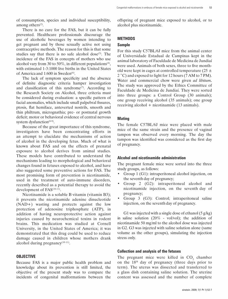
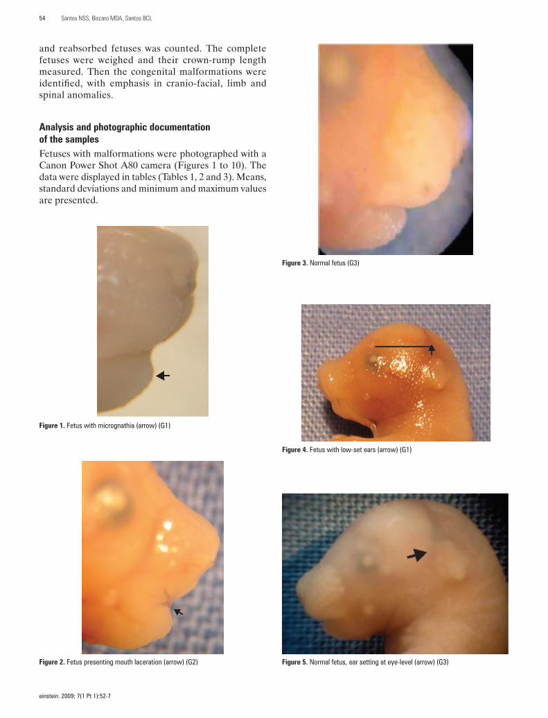
analysis and photographic documentation of the samples Fetuses with malformations were photographed with a Canon Power Shot A80 camera (Figures 1 to 10). The data were displayed in tables (Tables 1, 2 and 3). Means, standard deviations and minimum and maximum values are presented.
Figure 1. Fetus with micrognathia (arrow) (G1)
Figure 2. Fetus presenting mouth laceration (arrow) (G2)
Figure 3. Normal fetus (G3)
Figure 4. Fetus with low-set ears (arrow) (G1)
Figure 5. Normal fetus, ear setting at eye-level (arrow) (G3)

einstein. 2009; 7(1 Pt 1):52-7
Congenital malformations in embryos of female mice exposed to alcohol and nicotinamide 55
Figure 9. Fetus with nose hyperpigmentation (arrow) (G3)
Figure 10. Normal fetus (G3)Figure 7. Normal fetus (G3)
Figure 8. Fetus with pigmented paranasal region (arrow) (G2)
Figure 6. Fetus presenting scoliosis (G1)
Weights and lengths were compared among the groups using ANOVA, and since the results were significant, the Tukey test for multiple comparisons was applied. The SAS version 9.02 package was used and the level of significance adopted was 5% (p < 0.05).
reSUltSThe teratogenic effects of ethanol observed were micrognathia, low set ears, hypertrophic nose, atrophy of the upper and lower limbs and skeletal deformities (scoliosis). These deformities were less frequent in G2.
The highest proportion of mouth laceration and paranasal hyperpigmentation were observed in G2; in G3 nose hyperpigmentation was frequent and not

einstein. 2009; 7(1 Pt 1):52-7
56 Santos NSS, Biscaro MDA, Santos BCL
table 1. Description of absolute and relative frequencies of malformation in fetuses
Saline (control group) alcohol alcohol +
nicotinamide
n = 30 n = 35 n = 13
n % n % n %
Micrognathia 2 6.7 6 17.1 1 7.7
Low-set ears - - 7 20.0 2 15.4
Hypertrophic nose - - 1 2.9 - -
Upper limb atrophy - - 2 5.7 - -
Lower limb atrophy - - 1 2.9 - -
Skeletal deformities (scoliosis) - - 2 5.7 - -
Mouth laceration - - 1 2.9 1 7.7
Reabsorved fetus 1 3.3 1 2.9 3 23.1
Pigmented nose 5 16.7 - - - -
Pigmented paranasal region - - - - 1 7.7
Total number of fetuses 30 35 13
Total number of mice 4 5 3
Mean number of fetuses/mouse 7.5 7.0 4.3
found in the other groups. The highest proportion of reabsorbed fetuses occurred in G2.
Weight was significantly different among the groups (p = 0.0139). The Tukey test for multiple comparisons was then performed and revealed that G2 was statistically different from G3 (p = 0.102). G1 fetuses weighed less than the G3 mean weight (p = 0.3133).
As for length, the highest was in G1 and the lowest in G2. The two groups were significantly different (p = 0.0145). The Tukey test for multiple comparisons was then performed and it showed that G2 was statistically different than G1 (p = 0.0363) and that G1 was different
from G3, although the p value was close to 0.05, but below this value, therefore, significant.
Thus teratogenesis was lower, in general, in G2 than in the group exposed to alcohol.
DiScUSSiOnThere are many studies in the literature showing the effects of ethanol on the offspring of alcohol-treated female mice, clearly demonstrating the association between alcoholism during pregnancy and teratogenic defects found in the fetus. The C57B/6J female mice were chosen for the present study due to their small size, low cost and easy handling. The comparison between the findings in human and murine offsprings exposed to ethanol in uterus is quite consisted and justifies the choice of such species for FAS animal studies(12).
Skeletal teratogenesis by ethanol was found only in G1, in accordance with the literature(13), showing that alcohol consumption during pregnancy adversely affects fetal skeleton development. In this experimental group, a higher proportion of micrognathia was also found, as in other studies(14). It may be assumed that nicotinamide had a protective effect, because of the lower rate of this malformation.
The proportion of other teratogenic effects of alcohol, such as low set ears, hypertrophic nose, and atrophy of the upper and lower limbs was lower in the group receiving nicotinamide, suggesting the likely protective role of this vitamin. However, in the nicotinamide treated group, the highest proportions of mouth lacerating and paranasal hyperpigmentation were not observed in other groups. The scarcity of studies analyzing these parameters makes the comparison between the findings of the present study and the literature difficult; lacerations may be explained by an artifact originated at the time of collecting and handling of the fetuses.
table 2. Comparative study on fetal weight
groupsWeight (g) Statistical analysis
Mean Standard-deviation Minimum Median Maximum p-value
(anOVa) tukey test
Nicotinamide 0.7667 0.1111 0.5921 0.7553 0.9753 0.0139 Nicotinamide versus Saline solution 0.0102Saline solution 0.8517 0.0636 0.6988 0.8507 0.9629 Nicotinamide versus Alcohol 0.1345Alcohol 0.8206 0.091 0.6574 0.8195 0.9848 Alcohol versus Saline solution 0.3133
table 3. Comparative study on fetal length
groupslength (cm) Statistical analysis
Mean Standard-deviation Minimum Median Maximum p-value
(anOVa) tukey test
Nicotinamide 1.877 0.130 1.700 1.900 2.200 0.0145 Nicotinamide versus Saline solution 0.7821Saline solution 1.903 0.125 1.500 1.900 2.100 Nicotinamide versus Alcohol 0.0363Alcohol 1.974 0.109 1.800 2.000 2.200 Alcohol versus Saline solution 0.0490

einstein. 2009; 7(1 Pt 1):52-7
Congenital malformations in embryos of female mice exposed to alcohol and nicotinamide 57
As for the higher number of pups in G1, it does not agree with the literature findings(12). In addition, the weight of G1 fetuses was lower than the G3 mean weight, as also observed in a Brazilian study(12), although not found by other studies(14). G2 fetuses, unexpectedly, weighted less than those of the other groups, witch might be due to the small number of animals in this group.
Regarding the number of reabsorbed fetuses, the present study does not agree with the literature findings(14), which show reduced fetal viability and increased number of reabsorbed fetuses, while G3 in this study had a rate higher than G1. However, the highest proportion was found in G2.
Recording the length of the fetuses, G1 was significantly different from G3 and not similar to other studies(14). The shortest lengths were found in G2.
Lastly, it is important to stress that the objective of the present study was not to propose an alternative for alcohol intake during pregnancy, but to propose a possible strategy to reduce the effects of this drug on the offspring when information and medical follow-up are not sufficient to avoid the use of alcohol by the pregnant woman, as well as in chronic alcoholics.
cOnclUSiOnSNicotinamide reduced the number of malformations and it may be a protective factor for attenuating the effects of alcohol. Based on the results of this study, it may be said that the best proposal for preventing FAS is still to avoid the use of alcoholic beverages during pregnancy. Larger studies on the effects of nicotinamide are necessary for enhancing knowledge on the action of this vitamin.
acKnOWleDgeMentSThe authors are grateful to the staff of Faculdade de Medicina de Jundiaí, to Conselho Nacional de
Desenvolvimento Científico e Tecnológico (CNPq) and to Dr. Herman Grinfeld.
reFerenceS1. Cook JD. Biochemical markers of alcohol use in pregnant woman. Clin
Biochem. 2003;36(1):9-19.
2. Molina JC, Spear NE, Spear LP, Menella JA, Lewis MJ. International society for developmental psychobiology 39th annual meeting symposium: alcohol and development: beyond fetal alcohol syndrome. Dev Psychobiol. 2007;49(3):227-42.
3. Delgado AF, Cardieri JMA, Cristofani LM, Waksman RD. Síndrome de abstinência no recém-nascido. Pediatria (São Paulo). 1991;13(2):56-61.
4. Larroque B. Alcohol and the fetus. Int J Epidemiol. 1992;21 Suppl 1: S8-16.
5. Little BB, Snell LM, Rosenfeld CR, Gilstrap LC 3rd, Gant NF. Failure to recognize fetal alcohol syndrome in newborn infants. Am J Dis Child. 1990;144(10):1142-6.
6. Autti-Rämö L, Granström ML. The effect of intrauterine alcohol exposition in various durations on early cognitive development. Neuropediatrics. 1991;22(4):203-10.
7. Gahagan S, Sharpe TT, Brimacombe M, Fry-Johnson Y, Levine R, Mengel M, et al. Pediatricians’ knowledge, training, and experience in the care of children with fetal alcohol syndrome. Pediatrics. 2006;118(3):e657-68.
8. Rossett HL, Weiner L, Edelin KC. Strategies for prevention of fetal alcohol effects. Obstet Gynecol. 1981;57(1):1-7.
9. Streissguth AP, Barr HM, Sampson PD, Bookstein FL. Prenatal alcohol and offspring development: the first fourteen years. Drug Alcohol Depend. 1994;36(2):89-99.
10. Ieraci A, Herrera DG. Nicotinamide protects against ethanol-induced apoptotic neurodegeneration in the developing mouse brain. PLoS Med. 2006;3(4):e101.
11. Spong CY. Protection against prenatal alcohol-induced damage. PLoS Medicine. 2006;3(4):474-5.
12. Grinfeld H. Que efeitos podem ser esperados da exposição pré-natal ao etanol em camundongas prenhes e sua descendência? Einstein. 2004;2(3):187-92.
13. Mishra SR, Sahai A, Srivastava AK, Agrawal AK, Singh PJ, Mishra RK. Skeletal anomalies in fetal alcohol syndrome: a study on developing mice embryos. J Anat Soc India. 2003;52(1):51-4.
14. Oyedele OO, Kramer B. Acute ethanol administration causes malformations but does not affect cranial morphometry in neonatal mice. Alcohol. 2008;42(1):21-7.