computed tomography angiography versus computed … ct vs cta final.pdf · computed tomography...
TRANSCRIPT
TITLE: Computed Tomography Angiography versus Computed Tomography for the
Diagnosis and Management of Hyperacute Stroke: A Review of Comparative Clinical Evidence and Guidelines
DATE: 10 December 2013 CONTEXT AND POLICY ISSUES A stroke is a sudden loss of brain function caused by the interruption of flow of blood to the brain which may result in death of neurons in the affected area and it is characterized by rapidly developing clinical symptoms lasting over 24 hours or leading to death, with no apparent cause other than that of vascular origin.1,2 Ischemic stroke is stroke caused by vascular insufficiency (such as cerebrovascular thromboembolism) rather than by hemorrhage, while hemorrhagic stroke is the consequence of ruptured blood vessels in the brain.1,2 A discussion of stroke often includes transient ischemic attack (TIA), which is a brief episode of neurological dysfunction caused by focal brain or retinal ischemia.3 Though similar to that of stroke, clinical symptoms of TIA typically last less than one hour without objective evidence of acute infarction.3 Approximately 15% to 30% of debilitating strokes are heralded by TIAs, normally within 7 days preceding the stroke.4 Additionally, there is an estimated 10% risk of recurrent stroke within 90 days after a TIA or minor stroke, with the majority occurring within 48 hours of the TIA.4 Bader and Palmer have described hyperacute stroke patients as those presenting within 6 hours of stroke onset.5 Hyperacute stroke is a medical emergency requiring hyperacute care to optimize prognosis and ensure improved patient outcomes. Hyperacute care refers to the key interventions involved in the assessment, stabilization and treatment in the first hours after stroke onset. Broadly speaking it refers to care offered in the first 24 hours after stroke (ischemic and hemorrhagic) and the first 48 hours after TIA.6 The principal aim of this phase of care is to diagnose the stroke type, and to coordinate and execute the treatment plan as rapidly as possible.6 Interventions for hyperacute ischemic stroke mainly involve thrombolysis to remove intravascular thrombi with tissue-plasminogen activator (tPA) or physical thrombectomy. Acute interventions such as thrombolytic therapy are time-sensitive and are not applicable to all patients.7 The guidelines for using tPA for the treatment of acute ischemic stroke for instance, require intravenous drug administration within 4.5 hours of stroke onset; preceded by a computed tomographic (CT) scan to exclude the presence of Disclaimer: The Rapid Response Service is an information service for those involved in planning and providing health care in Canada. Rapid responses are based on a limited literature search and are not comprehensive, systematic reviews. The intent is to provide a list of sources of the best evidence on the topic that CADTH could identify using all reasonable efforts within the time allowed. Rapid responses should be considered along with other types of information and health care considerations. The information included in this response is not intended to replace professional medical advice, nor should it be construed as a recommendation for or against the use of a particular health technology. Readers are also cautioned that a lack of good quality evidence does not necessarily mean a lack of effectiveness particularly in the case of new and emerging health technologies, for which little information can be found, but which may in future prove to be effective. While CADTH has taken care in the preparation of the report to ensure that its contents are accurate, complete and up to date, CADTH does not make any guarantee to that effect. CADTH is not liable for any loss or damages resulting from use of the information in the report. Copyright: This report contains CADTH copyright material and may contain material in which a third party owns copyright. This report may be used for the purposes of research or private study only. It may not be copied, posted on a web site, redistributed by email or stored on an electronic system without the prior written permission of CADTH or applicable copyright owner. Links: This report may contain links to other information available on the websites of third parties on the Internet. CADTH does not have control over the content of such sites. Use of third party sites is governed by the owners’ own terms and conditions.
hemorrhage, which is a contraindication to the use of the drug.7 These guidelines did not provide evidence-based strategies or recommendations for hyperacute intervention of TIA besides imaging. However, since it has been recommended that all patients with suspected transient ischemic attack undergo brain imaging immediately (CT, or MRI if urgently available) and vascular imaging of the brain and neck arteries within 24 hours,6 it is expected that patients will be triaged for appropriate intervention based on findings from imaging. Hemorrhagic stroke may be caused by primary or secondary intracranial hemorrhage (ICH), Primary ICH is often associated with hypertension or cerebral amyloid angiopathy (CAA); and secondary ICH may be caused by aneurysms, anticoagulation, or hemorrhage due to neoplasm, infarction, or sinus thrombosis.8 Hyperacute stroke interventions include intensive care unit admission, blood pressure control, correction of coagulopathy, and neurosurgical consultation.8 Though non-enhanced CT is a widely used screening technique in distinguishing ischemic from hemorrhagic acute stroke,8 it is not very sensitive at determining the underlying structural vascular abnormality in hemorrhagic stroke, which is critical for instituting the appropriate treatment.9 The reference standard for diagnosing vascular lesions is digital subtraction angiography (DSA).10 It is an invasive procedure and may not readily be available for critically ill patients, and it has other limitations including being an expensive and resource-intensive procedure, as well as being associated with 0.9% and 0.5% risk of transient and permanent neurologic deficits, respectively. Computed tomography angiography (CTA) provides a low risk, lower cost, and readily accessible alternative to DSA to detect underlying structural vascular abnormality in a non-invasive way and to predict the risk of hematoma growth and guides the use of hemostatic drugs. However, CTA cannot clearly display blood stream and blood supply for vascular abnormalities.9 Imaging modalities such as CT and computed tomography angiography (CTA) provide caregivers access to knowledge about the presence of hemorrhage or an intravascular thrombus that can be treated with thrombolysis or thrombectomy. Imaging also allows the detection and estimation of the size of a core of irreversibly infarcted tissue and/or hypoperfused tissue at risk for subsequent infarction unless adequate perfusion is restored.7 Such information guides treatment decisions such as selection of patients, based on the merit of each case, for best available intervention to ensure optimal care and better patient outcomes in a timely manner. This report aims to provide evidenced-based comparative information to facilitate decision making on the use of the CT and/or CTA imaging modalities for the diagnosis and management of hyperacute stroke. RESEARCH QUESTIONS 1. What is the clinical evidence regarding the comparative effectiveness of computed
tomography angiography versus computed tomography for the diagnosis of hyperacute stroke?
2. What are the evidence-based guidelines for the optimal imaging of patients presenting
with signs of hyperacute stroke?
CTA versus CT for the Diagnosis and Management of Hyperacute Stroke 2
KEY FINDINGS Non-enhanced CT (NECT) scanning of the head can be performed in a matter of seconds to evaluate hemorrhage and other insults to the brain. Computed tomography angiography (CTA) and its source images (CTA-SI) can provide a qualitative cerebral blood volume (CBV) map that is more able, compared with non-enhanced CT, to distinguish between brain tissue that is irreversibly infarcted and that which is potentially salvageable; thereby allowing selection of patients who are likely to benefit from therapy. Patients who have immediate access to services that offer diagnostic testing such as imaging (CT/CTA, or MRI/MRA if urgently available)6 achieve better outcomes owing to timely initiation of appropriate prophylactic medication which results in fewer recurrent strokes and fewer adverse events compared to patients who had a lengthier delay in receiving this care. Another CT modality is CT perfusion (CTP) imaging which can be performed during a CTA examination, but with a separate contrast bolus. A multimodal evaluation that includes CTA and CTP may permit assessment of the site of vascular occlusion, infarct core, salvageable brain tissue and degree of collateral circulation. METHODS Literature Search Strategy A limited literature search was conducted on key resources including PubMed, The Cochrane Library (2013, Issue 10), University of York Centre for Reviews and Dissemination (CRD) databases, Canadian and major international health technology agencies, as well as a focused Internet search. To address question one, methodological filters were applied to limit retrieval to health technology assessments, systematic reviews, meta-analyses, randomized controlled trials and non-randomized studies. To address question two, methodological filters were applied to limit retrieval to guidelines. Where possible, retrieval was limited to the human population. The search was also limited to English language documents published between January 1, 2008 and November 12, 2013. Rapid Response reports are organized so that the evidence for each research question is presented separately. Selection Criteria and Methods One researcher screened retrieved citations and abstracts to select titles for full-text article review. Full-text publications deemed to meet the selection criteria outlined in Table 1 were included this review. Table 1: Selection Criteria Population
Adults presenting with sings of early/hyperacute stroke
Intervention
Q1 Computed Tomography Angiography Q2 CTA, CT, other imaging
Comparator
Q1 Computed Tomography
Outcomes
Q1 Comparative effectiveness for the diagnosis of acute stroke Accuracy of early diagnosis resulting in optimal treatment Q2 Optimal diagnosis option
Study Designs
Health Technology Assessments/ Systematic review/Meta-analysis; Randomized controlled trials (RCTs); Non-randomized studies; and Guidelines
CTA versus CT for the Diagnosis and Management of Hyperacute Stroke 3
Exclusion Criteria Articles were excluded if they did not meet the selection criteria in Table 1. Thus, studies that did not cover CT or CTA were excluded. Studies were also excluded if they were published prior to 2008, if they did not have a comparator group, if they were duplicate publications of a study already selected, or already included in at least one of the selected HTAs or systematic reviews. Critical Appraisal of Individual Studies Studies included in this review were appraised for quality using the Down and Black checklist.11 Evidence-based guidelines were appraised using the new Appraisal of Guidelines for Research and Evaluation instrument (AGREE II). The strengths and limitations of individual studies were summarized and presented instead of calculated numerical scores. SUMMARY OF EVIDENCE Quantity of Research Available The search identified 250 citations and abstracts, from which 19 potentially relevant articles were selected for further evaluation. Three potentially relevant articles were added from grey literature. After examining the full-text articles, one was exclude because it was in a language other than English and 17 more were excluded for irrelevance with regards to population, interventions, comparators or outcomes; leaving three retrospective studies and one Scientific Statement that were used to prepare this report. The latter provides a set of recommendations for imaging interventions in acute stroke care. The PRISMA flow diagram in in Appendix 1 gives an overview of the study selection process. Summary of Study Characteristics Characteristics of included studies have been provided in Appendix 2. Country of origin
Two studies4,12 were conducted in Canada and a third study,3 was done in Sweden. The Scientific Statement7 was published by the American Heart Association (AHA). It has 395 references from many different countries covering stroke; including application of imaging techniques in acute stroke care. The document did not lay out study setting, patient population, interventions, comparators, and outcomes. Study characteristics of the Scientific Statement have been summarized separately in Table 3 of Appendix 2.
Study setting
All the studies examined data from stroke units in various institutional settings. Two studies4,12 included imaging data for patients from a tertiary care referral center with an acute stroke team. A third study3 only mentioned that the imaging data were from patients admitted to the acute stroke unit, without further information about the institution(s).
Patient population
One study4 used data from 510 patients (median age 69 years; range 27-99 years) who had consecutive transient ischemic attack and minor stroke indicated by a National Institute of Health Stroke Scale (NIHSS) score of ≤3, and underwent CT/CTA and subsequent MRI. A second study12 included imaging data from 457 patients who underwent acute CT head, CTA of
CTA versus CT for the Diagnosis and Management of Hyperacute Stroke 4
the Circle of Willis (COW) and neck for assessment of possible mild ischemic stroke or TIA. Data for a third study3 were from fifty patients; aged 61± 15years with acute ischemic stroke admitted to the acute stroke unit. They were categorized into 3 groups namely; patients with repeat TIAs (Group I); patients not eligible for thrombolysis treatment (Group II); and patients who were non-responders to thrombolysis treatment using tPA (Group III). Interventions and comparators
Non-enhanced CT and CTA, as well as MRI were used as interventions and compared to each other in one study.4 In a second study,12 National Institute of Health Stroke Scale (NIHSS) was the intervention and non-enhanced CT and CTA were the comparator. Non-enhanced CT was the intervention in a third study3 with CTP and CTA as comparators.
Outcome measure
The outcome of interest for one study,4 was recurrent stroke, defined as a functional deterioration in neurological status of vascular origin lasting 24 hours or a new sudden focal neurological deficit of vascular origin lasting at least 24 hours (that was not felt to be secondary to other nonvascular factors: drugs, fever, infection) within 90 days. Association between clinical or imaging features and functional impairment on the modified Rankin Scale (mRS≥2) at 90 days was the outcome for a second study.12 The outcome measure of interest in a third study3 was added utility; defined as the number of missed or unrecognized abnormalities, indicated by additional symptom-related clinical information not revealed in the initial assessment with non-enhanced CT. The Scientific Statement provided different classes of recommendations for the application of imaging modalities (including CT and CTA) in acute stroke care based on stated levels of evidence. Summary of Critical Appraisal Three cohort studies of patients who presented with acute stroke or TIA were found through the literature search for this report. Information about the included Scientific Statement has been summarized in Table 3 of Appendix 2. Further details of summary of critical appraisal of individual studies are provided in Appendix 3. Reporting
For each study, the objectives, main study outcomes to be measured, characteristics of patients included and interventions of interest have been clearly described. In addition, the main study findings of each of the studies were clearly described. None of the studies reported adverse events that may have been a consequence of the applied interventions, though one study4 reported that 5 patients (1%) died during the 90-day follow-up period. Two studies4,12 reported actual probability values for the main outcomes where the P-value was not less than 0.001. External validity
The participants in the three studies3,4,12 were representative of patients presenting with acute stroke or TIA. The various periods within which imaging interventions were reported to have taken place conformed to the definition of hyperacute care period (within 24 hours of stroke onset). Two studies4,12 were conducted at a tertiary care referral centre with an acute stroke team all of whom have subspecialty training in Stroke Neurology. Given the seeming specialized settings and the level of expertise of the medical staff involved, it is uncertain how the findings from these studies are generalizable to other facilities that cater to acute stroke patients. A third
CTA versus CT for the Diagnosis and Management of Hyperacute Stroke 5

study3 was reported to have been conducted in acute stroke unit but the nature of the institution was not described.
Internal validity
Two studies3,12 were of retrospective cohort design. One of them12 analyzed CT and CTA imaging data of 457 acute stroke patients while the other3 involved similar data for 50 patients. Another study4 was a prospective cohort study involving 510 patients who received both CT and CTA as part of assessment of acute stroke. All the studies reported using data from patients consecutively admitted for treatment. Consecutive enrollment reduces selection bias. These patients received both CT and CTA on the same machines as part of a same-day evaluation during the same assessment period, minimizing the risk of changes in condition between scans, or differences in standard of care during separate assessment periods. None of the studies reported any evaluation of power to detect clinically significant differences. There is no report of patient blinding in any of the studies to the kind of imaging procedure undertaken. However, such blinding would be inconsequential since it could not have influenced the outcome. In one study,4 a panel of 3 physicians, which included 2 stroke neurologists and a neuroradiologist, reviewed and adjudicated the imaging and clinical information on any patient with a recurrent stroke. A Cox proportional hazards model was used for analysis of the primary outcome (recurrent stroke), while a secondary analysis to compare the accuracy of prediction of recurrent stroke with CT/CTA and DWI MRI was completed using receiver operator characteristic curves. Another study12 used a three-reader consensus approach (requiring two out of three in agreement) to rate the presence of acute ischemia on non-contrast CT (NCT). According to the authors, readers were aware of minimal clinical data, including symptoms or the presence of aphasia.12 The primary outcome was functional impairment (defined as mRS≥2) at 90 days after symptom onset. Fisher’s exact test was used in two-sided tests for comparison, and considered statistically significant at P<0.05 for all tests. In a third study,3 two neuroradiologists evaluated the images in a consensus pattern. However, it was not reported whether they were blinded to the clinical information of the patients which could bias their evaluation. Outcome measures (additional clinical information) were clearly described and Fisher’s exact test was used to assess the significance of difference in outcome.3 The recommendations for imaging of acute ischemic stroke7 described the overall objectives and clinical questions that have been covered, as well as the patients to whom the recommendations are to apply. The document was prepared by stakeholders including neuroscientists, neuroradiologists, and other clinicians as an improvement on what the authors described as a consensus roadmap for the use of a variety of imaging techniques produced by imagers and clinicians from many subspecialties within the neurosciences.7 It was not stated whether patients views and preferences were sought or incorporated. According to the authors, all relevant articles in English literature were included, but with a focus on literature from 2000 to 2006. The quality of each article was assessed for its level of evidence using criteria provided in Table 2, which conform to the American Heart Association’s practice guidelines classification scheme. The American Heart Association states that Scientific Statements generally include a review of data available on a specific subject, an evaluation on the relationship to overall cardiovascular disease science, and often an AHA/ASA position on the basis of that evaluation, and the statements are peer reviewed. The recommendations for using specific imaging modalities for specific conditions have been listed clearly after a general discussion and summary of merits of available options. Applicability of the imaging techniques, including
CTA versus CT for the Diagnosis and Management of Hyperacute Stroke 6
methods of data collection, advantages, and limitations were provided. However, cost implications and a procedure for updating the recommendations were not discussed. Summary of Finding Further details on individual study findings and authors’ conclusions have been provided in Appendix 4.
1. What is the clinical evidence regarding the comparative effectiveness of computed tomography angiography versus computed tomography for the diagnosis of hyperacute stroke?
In one study which evaluated added utility,3 CTP and CTA provided additional information not revealed on NCT in groups of patients with different presentations of acute stroke or TIA. In a group of patients (Group II) not eligible for thrombolysis treatment either because they were examined after the window prescribed for such therapy (3 hours at time of study) had closed, or they had contraindications to tPA, additional clinical information was provided by CTP and CTA (compared to NCT alone) for all patients, compared to 34% of patients in Group I (patients who had suffered multiple TIA in the previous few days) and 33% of patients in Group III (patients who did not show clinical improvement after receiving tPA or presenting recurrent stroke symptoms within a day of the onset of the initial stroke). The difference between Group II and Groups I and III was statistically significant (P < 0.001). There was no statistically significant difference between Groups I and III in the percentage of patients for whom CTP and CTA provided additional clinical utility (P < 0.957). In Group I, the added utility of CTA over CT was 21%, and 80% in Group I and Group II, respectively. In Group III, CTA provided added utility in two out of six patients. It is worth adding that CTP and combined CTP and CTA provided even more utility benefit as detailed in Appendix 4. Using this multimodal approach the final diagnosis of seven patients in Group I was found to be something other than the initial TIA. In another study,4 the presence of a symptomatic intracranial or extracranial severe arterial stenosis or occlusion detected on CTA but not CT was found to be predictive of recurrent stroke. Through a receiver operating characteristic analysis, bimodal CT/CTA was found to be not significantly different from MRI in discriminative value in predicting recurrent stroke (0.67; 95% confidence interval [CI], 0.59 to 0.76 versus 0.59; 95% CI, 0.52 to 0.67; P=0.09).4 Viewed in the light that MRI is reported to greatly exceed NCT in detecting the ischemic tissue to ensure diagnosis while excluding mimics such as tumour and subdural hematoma,7 this finding is important given the higher cost and the relative sparse availability of MRI services. In another study,12 24% of patients with a positive composite CT metric (CT/CTA) had functional impairment vs. 8% in the patients without the CT imaging abnormalities [relative risk (RR) 2.92, 95% CI 1.8 to 4.71]. Poor outcomes trended positively with increasing numbers of CT/CTA abnormalities: 8% in patients with no CT/CTA abnormalities (n=330); 19% in patients with one CT/CTA abnormality (n=91); 34% in patients with two CT/CTA abnormalities (n=35); and 100% when there were three CT/CTA abnormalities (n=1) [Chi-square test for trend (P = 0.0001)]. The presence of an abnormality on the CT metric among patients with baseline NIHSS score of 0, was associated with a higher rate of functional impairment from 2.4% without an abnormality to 20% with an abnormality (RR 8.2; 95% CI 2.54 to 26.5; P=0.0006).12
CTA versus CT for the Diagnosis and Management of Hyperacute Stroke 7
2. What are the evidence-based guidelines for the optimal imaging of patients presenting with signs of hyperacute stroke?
Highlights of imaging recommendations for acute stroke care available in the Scientific Statement7 published by the American Heart Association (AHA) is provided below. Definitions of levels of evidence (LOE) and class of recommendation is given in Appendix 2, Table 2. Recommendations for Detecting Cerebral Ischemia and Excluding Mimics
1. For a patient within a 3-hour time period from onset of symptoms, either non-enhanced CT (NECT) or MRI is recommended before intravenous tPA administration to exclude intracranial hemorrhage (ICH) (absolute contraindication). Frank hypointensity on CT is a strong contraindication to treatment. Early signs of infarct on CT, regardless of their extent, are not a contraindication to treatment. (Class I, level of evidence [LOE]: A).
2. For a patient within 3 hours of onset of symptoms, there is a suboptimal detection rate of ischemic changes with NECT alone, and a more definitive diagnosis will be obtained with MR-DWI or CTA-SI, if this does not unduly delay the administration of intravenous tPA. (Class IIa, LOE: B).
3. For patients beyond 3 hours from onset of symptoms, either MR-DWI or CTA-SI should be performed along with vascular imaging and perfusion studies, particularly if mechanical thrombectomy or intra-arterial thrombolytic therapy is contemplated (Class I, LOE: A).
Recommendations for Imaging the Cerebral Vasculature
1. Acute large-vessel intracranial thrombus is very accurately detected by CTA, digital subtraction angiography (DSA), and magnetic resonance angiography (MRA). Each of these modalities far surpasses the sensitivity of nonvascular studies such as NECT, FLAIR, or gradient-echo MRI, and they are all recommended (Class I, LOE: A).
2. A vascular study is probably indicated during the initial imaging evaluation of the acute stroke patient within 3 hours of ictus, if such an evaluation does not unduly delay the administration of intravenous tPA, and only if an endovascular team is available to undertake intra-arterial therapy if that is contemplated on the basis of the findings (Class IIa, LOE: B).
3. A vascular study is strongly recommended during the initial imaging evaluation of the acute stroke patient who presents more than 3 hours after ictus, especially if either intra-arterial thrombolysis or mechanical thrombectomy is contemplated for management (Class I, LOE: A).
Clinical Role of Perfusion Imaging
1. The admission volumes of infarct core and ischemic penumbra may be significant predictors of clinical outcome, possibly exceeding the prognostic value of admission NIHSS score (Class IIb, LOE: B).
2. There is increasing but as yet indirect evidence that even relatively imprecise measures of core/penumbra mismatch may be used to select patients for treatment beyond a strict 3-hour time window for intravenous thrombolysis. Together with vascular imaging, these approaches may determine suitability for other therapies such as mechanical clot retrieval and intra-arterial thrombolysis, as well as provide a surrogate marker for treatment efficacy (Class IIb, LOE: B).
CTA versus CT for the Diagnosis and Management of Hyperacute Stroke 8
Limitations Retrospective studies are regarded as having lower quality of evidence than randomized controlled studies. They are prone to selection bias which could result in sensitivities not attainable in actual practice. One study3 had only participants that were sub-divided into three groups. The small sample size makes it difficult to detect all potentially clinically significant differences; which is exacerbated by the smaller size of the subgroups. Within this study, 14 out of the 29 patients with TIA were examined during the TIA episode. Though the remaining 15 patients were examined within 24 hours of onset, this could distort outcomes of the imaging technique used, producing higher sensitivity for NCT and some underestimation of the beneficial role of the CTP to detect small changes.
In another study,4 multiple data imputation based on a previously published meta-analysis was done as surrogate for missing MRI results in order to allow direct comparison of CT/CTA and DWI MRI for secondary analysis. Therefore, the precision of the point estimates derived in this way is uncertain. In the same study,4 patients were included very early into their presentation so distinctions of TIA or minor stroke could not be made. Therefore, it is uncertain how the findings of this study will compare with finding from cohorts examined later in the course of their disease.
A third study12 reported that depending on data from preexisting charts, the ability to information on stroke recurrence rates was limited and symptom progression could not be deduced. It is uncertain whether all the patients who had a NIHSS rating of 0 were completely resolved on first assessment. In cases where NIHSS and mRS scores were unavailable, they were derived retrospectively. Thus it is uncertain to what extent these derivations might have influenced the reported findings. In 30% of patients in this study,12 discharge destination other than home was used as surrogate for poor functional outcome. Since subsequent problems among patients at home would not have been captured, the possibility remains that important outcome data could have been missed. CONCLUSIONS AND IMPLICATIONS FOR DECISION OR POLICY MAKING Two of the included studies4,12 showed that assessment of the intracranial and extracranial vasculature using CT/CTA predicts recurrent stroke and clinical outcome in patients with TIA and minor stroke and helps to identify high-risk group suitable for aggressive acute stroke prevention treatment. The third study3 indicated that CTA and CT perfusion can provide additional, symptom-related information not revealed by non-enhanced CT and should be regarded as valuable tools in the management of acute stroke. The scientific statement recommends that for patients within 3 hours of onset of symptoms a more definitive diagnosis can be obtained CTA-SI (if administration of intravenous tPA will not be unduly delayed) since non-enhanced CT has suboptimal detection rate of ischemic changes in such early periods of disease onset. Non-enhanced CT has been the modality of choice for a long time in the initial workup of acute stroke. It has the advantage of being able to scan the head in a matter of seconds to evaluate hemorrhage and other insults to the brain. However, more modern techniques such as CTA can provide a qualitative cerebral blood volume (CBV) map that detects the core of infarction and improves the identification of the tissue at risk for infarction compared with NCT. Further investigation using higher quality randomized controlled trials may provide more conclusive information regarding the best imaging options for hyperacute stoke care.
CTA versus CT for the Diagnosis and Management of Hyperacute Stroke 9
PREPARED BY: Canadian Agency for Drugs and Technologies in Health Tel: 1-866-898-8439 www.cadth.ca
CTA versus CT for the Diagnosis and Management of Hyperacute Stroke 10
REFERENCES 1. Heart and Stroke Foundation of Canada [Internet]. Ottawa (ON): Heart and Stroke
Foundation; 2013. What is a stroke?; 2012 Feb [cited 2013 Nov 29]. Available from: http://www.heartandstroke.com/site/c.ikIQLcMWJtE/b.3483935/k.736A/Stroke__What_is_Stroke.htm
2. Alawneh JA, Clatworthy PL, Morris RS, Warburton E. Stroke management. Clin Evid. 2011;6:201.
3. Abul-Kasim K, Brizzi M, Petersson J, Sundgren PC. Added diagnostic utility of CT perfusion and CT angiography in acute ischemic stroke. Evaluation of three different patient categories. Funct Neurol. 2009 Apr;24(2):93-8.
4. Coutts SB, Modi J, Patel SK, Demchuk AM, Goyal M, Hill MD, et al. CT/CT angiography and MRI findings predict recurrent stroke after transient ischemic attack and minor stroke: results of the prospective CATCH study. Stroke. 2012 Apr;43(4):1013-7.
5. Bader MK, Palmer S. What's the "hyper" in hyperacute stroke? Strategies to improve outcomes in ischemic stroke patients presenting within 6 hours. AACN Adv Crit Care. 2006 Jun;17(2):194-214.
6. Lindsay MP, Gubitz G, Bayley M, Hill MD, Davies-Schnickel C, Singh S, et al. Canadian best practice recommendations for stroke care [Internet]. 4th ed. Ottawa (ON): Canadian Stroke Network; 2012. [cited 2013 Nov 29]. Available from: http://www.strokebestpractices.ca/
7. Latchaw RE, Alberts MJ, Lev MH, Connors JJ, Harbaugh RE, Higashida RT, et al. Recommendations for imaging of acute ischemic stroke: a scientific statement from the American Heart Association. Stroke. 2009 Nov;40(11):3646-78.
8. Khosravani H, Mayer SA, Demchuk A, Jahromi BS, Gladstone DJ, Flaherty M, et al. Emergency noninvasive angiography for acute intracerebral hemorrhage. AJNR Am J Neuroradiol. 2013 Aug;34(8):1481-7.
9. Ma J, Li H, You C, Huang S, Ma L, Ieong C. Accuracy of computed tomography angiography in detecting the underlying vascular abnormalities for spontaneous intracerebral hemorrhage: a comparative study and meta-analysis. Neurol India. 2012 May;60(3):299-303.
10. Matias-Guiu JA, Serna-Candel C, Espejo-Dominguez JM, Fernandez-Matarrubia M, Simal P, Matias-Guiu J. Large artery occlusion diagnosed by computed tomography angiography in acute ischaemic stroke: Frequency, predictive factors, and safety. Neurologia. 2013 Sep 26. Epub ahead of print.
11. Downs SH, Black N. The feasibility of creating a checklist for the assessment of the methodological quality both of randomised and non-randomised studies of health care interventions. J Epidemiol Community Health [Internet]. 1998 Jun [cited 2013 Nov 5];52(6):377-84. Available from: http://www.ncbi.nlm.nih.gov/pmc/articles/PMC1756728/pdf/v052p00377.pdf
CTA versus CT for the Diagnosis and Management of Hyperacute Stroke 11
12. Coutts SB, O'Reilly C, Hill MD, Steffenhagen N, Poppe AY, Boyko MJ, et al. Computed tomography and computed tomography angiography findings predict functional impairment in patients with minor stroke and transient ischaemic attack. Int J Stroke. 2009 Dec;4(6):448-53.
CTA versus CT for the Diagnosis and Management of Hyperacute Stroke 12
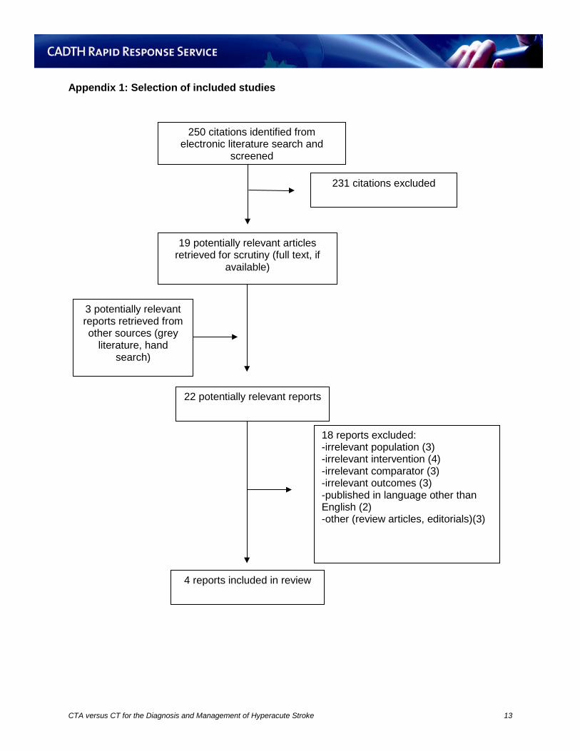
Appendix 1: Selection of included studies
231 citations excluded
19 potentially relevant articles retrieved for scrutiny (full text, if
available)
3 potentially relevant reports retrieved from other sources (grey
literature, hand search)
22 potentially relevant reports
18 reports excluded: -irrelevant population (3) -irrelevant intervention (4) -irrelevant comparator (3) -irrelevant outcomes (3) -published in language other than English (2) -other (review articles, editorials)(3)
4 reports included in review
250 citations identified from electronic literature search and
screened
CTA versus CT for the Diagnosis and Management of Hyperacute Stroke 13
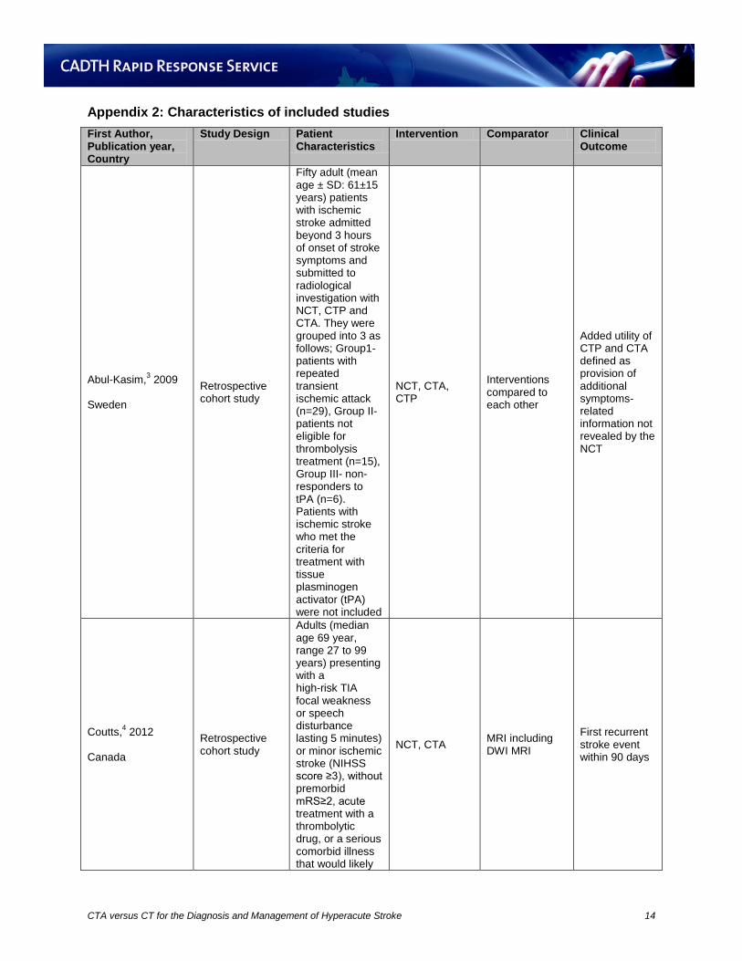
Appendix 2: Characteristics of included studies First Author, Publication year, Country
Study Design Patient Characteristics
Intervention Comparator Clinical Outcome
Abul-Kasim,3 2009 Sweden
Retrospective cohort study
Fifty adult (mean age ± SD: 61±15 years) patients with ischemic stroke admitted beyond 3 hours of onset of stroke symptoms and submitted to radiological investigation with NCT, CTP and CTA. They were grouped into 3 as follows; Group1- patients with repeated transient ischemic attack (n=29), Group II- patients not eligible for thrombolysis treatment (n=15), Group III- non-responders to tPA (n=6). Patients with ischemic stroke who met the criteria for treatment with tissue plasminogen activator (tPA) were not included
NCT, CTA, CTP
Interventions compared to each other
Added utility of CTP and CTA defined as provision of additional symptoms-related information not revealed by the NCT
Coutts,4 2012 Canada
Retrospective cohort study
Adults (median age 69 year, range 27 to 99 years) presenting with a high-risk TIA focal weakness or speech disturbance lasting 5 minutes) or minor ischemic stroke (NIHSS score ≥3), without premorbid mRS≥2, acute treatment with a thrombolytic drug, or a serious comorbid illness that would likely
NCT, CTA MRI including DWI MRI
First recurrent stroke event within 90 days
CTA versus CT for the Diagnosis and Management of Hyperacute Stroke 14
First Author, Publication year, Country
Study Design Patient Characteristics
Intervention Comparator Clinical Outcome
result in death within 3 months
Coutts,12 2009 Canada
Retrospective cohort study
Four hundred and fifty-seven patients referred to the acute stroke tertiary care referral centre with baseline NIHSS score ≤3, and pre-morbid mRS≤1, who underwent CTA imaging within 24 h of symptom onset; including high-risk TIA patients who had motor or speech symptoms lasting more than 5 minutes but not TIA patients with isolated sensory symptoms.
NCT, CTA
Clinical assessment with NIHSS and 90-mRS
Functional impairment on the modified Rankin Scale (mRS≥2) at 90 days
Latchaw,7 2009 USA
Scientific Statement See Appendix 2, Table 3: a
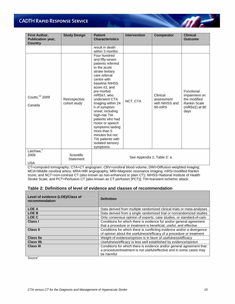
CT=computed tomography; CTA=CT angiogram; CBV=cerebral blood volume; DWI=Diffusion-weighted imaging; MCA=Middle cerebral artery; MRA=MR angiography; MRI=Magnetic resonance imaging; mRS=modified Rankin score; and NCT=non-contrast CT (also known as non-enhanced or plain CT); NIHSS=National Institute of Health Stroke Scale; and PCT=Perfusion CT [also known as CT perfusion (PCT)]; TIA=transient ischemic attack. Table 2: Definitions of level of evidence and classes of recommendation Level of evidence (LOE)/Class of recommendation
Definition
LOE A Data derived from multiple randomized clinical trials or meta-analyses LOE B Data derived from a single randomized trial or nonrandomized studies LOE C Only consensus opinion of experts, case studies, or standard-of-care Class I Conditions for which there is evidence for and/or general agreement
that a procedure or treatment is beneficial, useful, and effective Class II Conditions for which there is conflicting evidence and/or a divergence
of opinion about the usefulness/efficacy of a procedure or treatment Class IIa Weight of evidence/opinion is in favor of usefulness/efficacy Class IIb Usefulness/efficacy is less well established by evidence/opinion Class III Conditions for which there is evidence and/or general agreement that
a procedure/treatment is not useful/effective and in some cases may be harmful
Source7
CTA versus CT for the Diagnosis and Management of Hyperacute Stroke 15
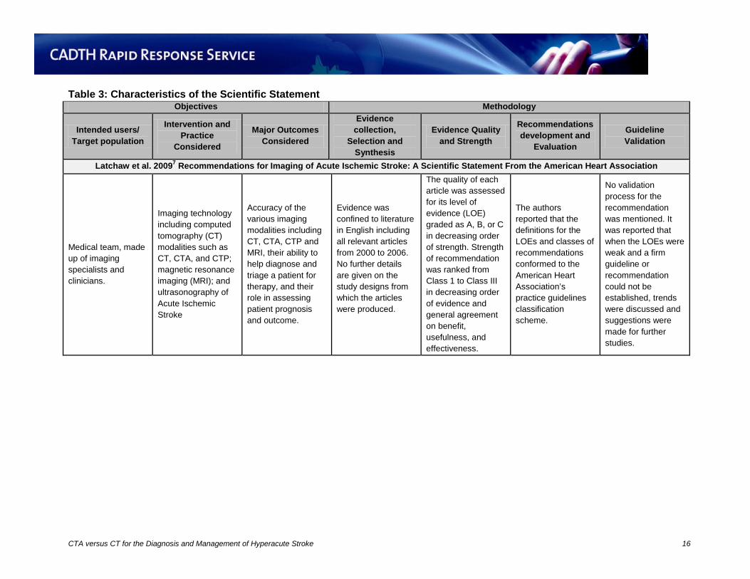
Table 3: Characteristics of the Scientific Statement Objectives Methodology
Intended users/ Target population
Intervention and Practice
Considered
Major Outcomes Considered
Evidence collection,
Selection and Synthesis
Evidence Quality and Strength
Recommendations development and
Evaluation
Guideline Validation
Latchaw et al. 20097 Recommendations for Imaging of Acute Ischemic Stroke: A Scientific Statement From the American Heart Association
Medical team, made up of imaging specialists and clinicians.
Imaging technology including computed tomography (CT) modalities such as CT, CTA, and CTP; magnetic resonance imaging (MRI); and ultrasonography of Acute Ischemic Stroke
Accuracy of the various imaging modalities including CT, CTA, CTP and MRI, their ability to help diagnose and triage a patient for therapy, and their role in assessing patient prognosis and outcome.
Evidence was confined to literature in English including all relevant articles from 2000 to 2006. No further details are given on the study designs from which the articles were produced.
The quality of each article was assessed for its level of evidence (LOE) graded as A, B, or C in decreasing order of strength. Strength of recommendation was ranked from Class 1 to Class III in decreasing order of evidence and general agreement on benefit, usefulness, and effectiveness.
The authors reported that the definitions for the LOEs and classes of recommendations conformed to the American Heart Association’s practice guidelines classification scheme.
No validation process for the recommendation was mentioned. It was reported that when the LOEs were weak and a firm guideline or recommendation could not be established, trends were discussed and suggestions were made for further studies.
CTA versus CT for the Diagnosis and Management of Hyperacute Stroke 16
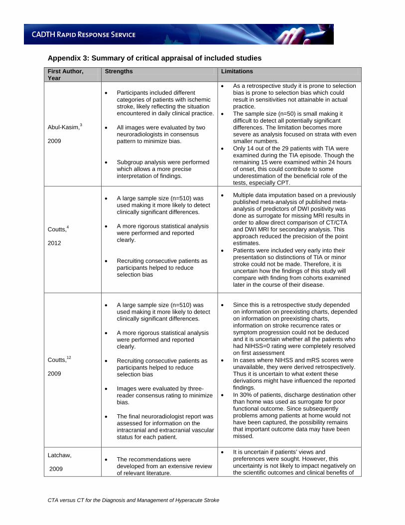
Appendix 3: Summary of critical appraisal of included studies First Author, Year
Strengths Limitations
Abul-Kasim,3 2009
• Participants included different categories of patients with ischemic stroke, likely reflecting the situation encountered in daily clinical practice.
• All images were evaluated by two
neuroradiologists in consensus pattern to minimize bias.
• Subgroup analysis were performed which allows a more precise interpretation of findings.
• As a retrospective study it is prone to selection bias is prone to selection bias which could result in sensitivities not attainable in actual practice.
• The sample size (n=50) is small making it difficult to detect all potentially significant differences. The limitation becomes more severe as analysis focused on strata with even smaller numbers.
• Only 14 out of the 29 patients with TIA were examined during the TIA episode. Though the remaining 15 were examined within 24 hours of onset, this could contribute to some underestimation of the beneficial role of the tests, especially CPT.
Coutts,4 2012
• A large sample size (n=510) was used making it more likely to detect clinically significant differences.
• A more rigorous statistical analysis
were performed and reported clearly.
• Recruiting consecutive patients as participants helped to reduce selection bias
• Multiple data imputation based on a previously published meta-analysis of published meta-analysis of predictors of DWI positivity was done as surrogate for missing MRI results in order to allow direct comparison of CT/CTA and DWI MRI for secondary analysis. This approach reduced the precision of the point estimates.
• Patients were included very early into their presentation so distinctions of TIA or minor stroke could not be made. Therefore, it is uncertain how the findings of this study will compare with finding from cohorts examined later in the course of their disease.
Coutts,12 2009
• A large sample size (n=510) was used making it more likely to detect clinically significant differences.
• A more rigorous statistical analysis
were performed and reported clearly.
• Recruiting consecutive patients as
participants helped to reduce selection bias
• Images were evaluated by three-reader consensus rating to minimize bias.
• The final neuroradiologist report was
assessed for information on the intracranial and extracranial vascular status for each patient.
• Since this is a retrospective study depended on information on preexisting charts, depended on information on preexisting charts, information on stroke recurrence rates or symptom progression could not be deduced and it is uncertain whether all the patients who had NIHSS=0 rating were completely resolved on first assessment
• In cases where NIHSS and mRS scores were unavailable, they were derived retrospectively. Thus it is uncertain to what extent these derivations might have influenced the reported findings.
• In 30% of patients, discharge destination other than home was used as surrogate for poor functional outcome. Since subsequently problems among patients at home would not have been captured, the possibility remains that important outcome data may have been missed.
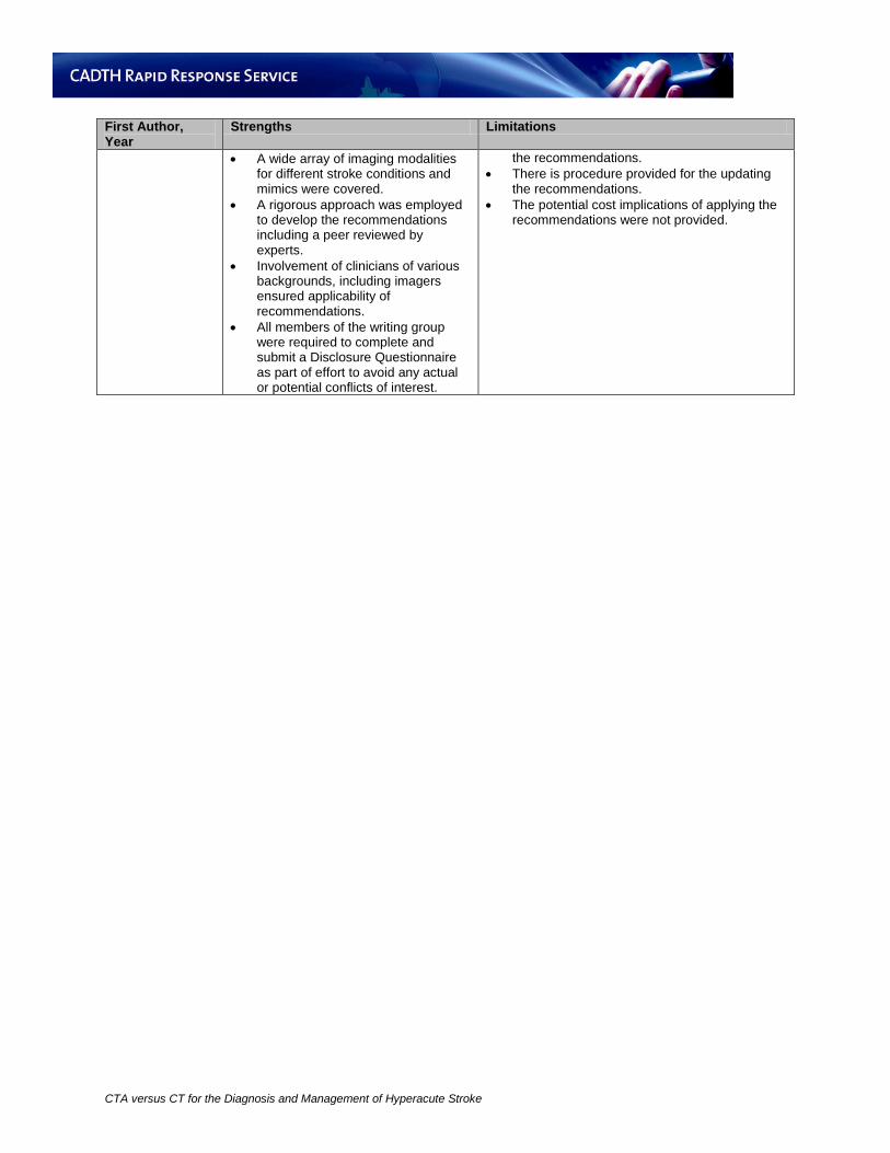
Latchaw, 2009
• The recommendations were developed from an extensive review of relevant literature.
• It is uncertain if patients’ views and preferences were sought. However, this uncertainty is not likely to impact negatively on the scientific outcomes and clinical benefits of
CTA versus CT for the Diagnosis and Management of Hyperacute Stroke
First Author, Year
Strengths Limitations
• A wide array of imaging modalities for different stroke conditions and mimics were covered.
• A rigorous approach was employed to develop the recommendations including a peer reviewed by experts.
• Involvement of clinicians of various backgrounds, including imagers ensured applicability of recommendations.
• All members of the writing group were required to complete and submit a Disclosure Questionnaire as part of effort to avoid any actual or potential conflicts of interest.
the recommendations. • There is procedure provided for the updating
the recommendations. • The potential cost implications of applying the
recommendations were not provided.
CTA versus CT for the Diagnosis and Management of Hyperacute Stroke
Appendix 4: Main study findings and authors’ conclusions First Author, Publication Year
Main Study Findings Authors’ Conclusions
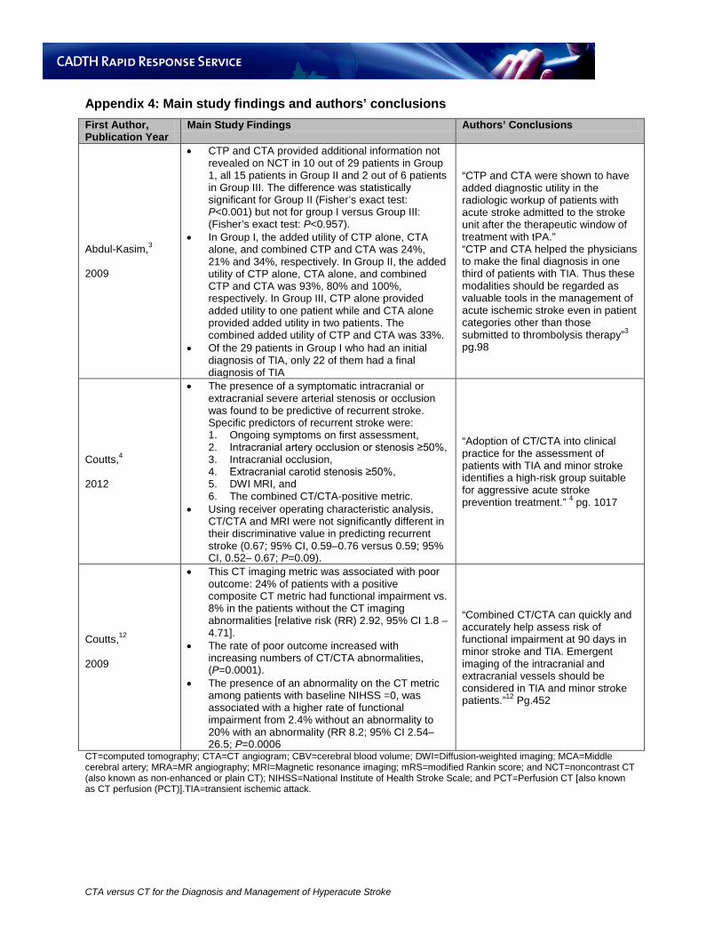
Abdul-Kasim,3 2009
• CTP and CTA provided additional information not revealed on NCT in 10 out of 29 patients in Group 1, all 15 patients in Group II and 2 out of 6 patients in Group III. The difference was statistically significant for Group II (Fisher’s exact test: P<0.001) but not for group I versus Group III: (Fisher’s exact test: P<0.957).
• In Group I, the added utility of CTP alone, CTA alone, and combined CTP and CTA was 24%, 21% and 34%, respectively. In Group II, the added utility of CTP alone, CTA alone, and combined CTP and CTA was 93%, 80% and 100%, respectively. In Group III, CTP alone provided added utility to one patient while and CTA alone provided added utility in two patients. The combined added utility of CTP and CTA was 33%.
• Of the 29 patients in Group I who had an initial diagnosis of TIA, only 22 of them had a final diagnosis of TIA
“CTP and CTA were shown to have added diagnostic utility in the radiologic workup of patients with acute stroke admitted to the stroke unit after the therapeutic window of treatment with tPA.” “CTP and CTA helped the physicians to make the final diagnosis in one third of patients with TIA. Thus these modalities should be regarded as valuable tools in the management of acute ischemic stroke even in patient categories other than those submitted to thrombolysis therapy”3 pg.98
Coutts,4 2012
• The presence of a symptomatic intracranial or extracranial severe arterial stenosis or occlusion was found to be predictive of recurrent stroke. Specific predictors of recurrent stroke were: 1. Ongoing symptoms on first assessment, 2. Intracranial artery occlusion or stenosis ≥50%, 3. Intracranial occlusion, 4. Extracranial carotid stenosis ≥50%, 5. DWI MRI, and 6. The combined CT/CTA-positive metric.
• Using receiver operating characteristic analysis, CT/CTA and MRI were not significantly different in their discriminative value in predicting recurrent stroke (0.67; 95% CI, 0.59–0.76 versus 0.59; 95% CI, 0.52– 0.67; P=0.09).
“Adoption of CT/CTA into clinical practice for the assessment of patients with TIA and minor stroke identifies a high-risk group suitable for aggressive acute stroke prevention treatment.” 4 pg. 1017
Coutts,12 2009
• This CT imaging metric was associated with poor outcome: 24% of patients with a positive composite CT metric had functional impairment vs. 8% in the patients without the CT imaging abnormalities [relative risk (RR) 2.92, 95% CI 1.8 – 4.71].
• The rate of poor outcome increased with increasing numbers of CT/CTA abnormalities, (P=0.0001).
• The presence of an abnormality on the CT metric among patients with baseline NIHSS =0, was associated with a higher rate of functional impairment from 2.4% without an abnormality to 20% with an abnormality (RR 8.2; 95% CI 2.54–26.5; P=0.0006
“Combined CT/CTA can quickly and accurately help assess risk of functional impairment at 90 days in minor stroke and TIA. Emergent imaging of the intracranial and extracranial vessels should be considered in TIA and minor stroke patients.”12 Pg.452
CT=computed tomography; CTA=CT angiogram; CBV=cerebral blood volume; DWI=Diffusion-weighted imaging; MCA=Middle cerebral artery; MRA=MR angiography; MRI=Magnetic resonance imaging; mRS=modified Rankin score; and NCT=noncontrast CT (also known as non-enhanced or plain CT); NIHSS=National Institute of Health Stroke Scale; and PCT=Perfusion CT [also known as CT perfusion (PCT)].TIA=transient ischemic attack.
CTA versus CT for the Diagnosis and Management of Hyperacute Stroke