comprehensive physiology || g protein-coupled receptors and the g protein family
TRANSCRIPT
6. G Protein-coupled G protein family
A L F R E D 0 U L L O A - A G U I R R E
P. M I C H A E L C O N N 1
C H A P T E R C O N T E N T S
Structure of G Protein-Coupled Receptors General features Ligand-binding domain G protein-coupling domain
General features G protein-regulatory cycle Structural and functional relationships of Ga-subunit GPy structure and function
Mechanisms that regulate receptor function
The Heterotrimeric G-Protein Family
Regulatory Mechanisms
Receptor conformation and signaling Receptor desensitization
G protein-mediated regulatory mechanisms
receptors and the
Department of Reproductive Biology, lnstituto Nacional de la Nutricion "Salvador Zubiran, " Mexico City, D. F., Mexico, and Divisions of Neuroscience and Reproductive Sciences, Oregon Regional Primate Research Center, Beaverton, Oregon
Divisions of Neuroscience and Reproductive Sciences, Oregon Regional Primate Research Center, Beaverton, Oregon, and Department of Physiology and Pharmacology, Oregon Health Sciences University, Portland, Oregon
CELLS COMMUNICATE WITH EACH OTHER and respond to their environment through chemical signals. Signal- ing molecules bind to specific receptors located in the target cell. For water-soluble signaling molecules, the complementary receptors are localized usually on the surface membrane of the target cell they influence. Upon binding, the signaling molecules activate receptor proteins, which act as signal transducers for specific extracellular messengers. An intracellular signal is then generated, initiating a cascade of amplified events that eventually culminates in a particular biological re- sponse (402). Thus, the primary function of cell-surface receptors is to discriminate the specific signaling mole- cule or ligand from among a large array of chemically diverse extracellular substances and to activate an ef- fector system that triggers an intracellular signal and eventually a biological effect.
There are three main classes of cell-surface receptor: ion channel-linked receptors, enzyme-coupled recep-
tors, and G protein-coupled receptors (GPCRs) (1 3, 105, 114,283,450,483). G protein-linked cell-surface receptors mediate their intracellular actions through the activation of one or more guanine nucleotide- binding signal-transducing proteins (G proteins) (27, 154, 184, 322, 344, 430, 441). G proteins are hetero- trimeric molecules composed of three subunits; in the presence of a specific agonist, receptors activate specific G proteins by catalyzing the exchange of the a-subunit- bound guanosine-diphosphate (GDP) for guanosine-5'- triphosphate (GTP) (27, 31, 32, 66, 403). This ex- change leads to dissociaton of the subunit complex into a py-dimer and the free a-subunit (184,322,403, 430). Although initially the GTP-activated Ga-subunit alone was considered to be the main promoter of effector activation (for example, the membrane- associated enzymes adenylyl cyclase and phospholipase C p or the Ca2+ channels) (24-26, 81), the py-dimer also may play a major role in signal transduction (62, 77, 117, 208, 209, 516).
Hundreds of receptors signal through G proteins. These receptors, which consist of a single polypeptide chain of variable length that threads back and forth across the lipid bilayer seven times to form characteris- tic transmembrane helices, form a large and function- ally diverse superfamily (13, 105,450). Based on nucle- otide and amino-acid sequence similarities, these protein receptor molecules can be separated further into three main families. The large rhodopsidp- adrenergic group, to which the majority of GPCRs identified to date belong (13, 105, 383, 450), is the best studied from structural and functional points of view; it is comprised of receptors that respond to a large variety of stimuli, including photons (341, 342) and odorants (365, 396), as well as hormones and neurotransmitter agonists of variable molecular struc-
87
88 HANDBOOK OF PHYSIOLOGY-CELLULAR ENDOCRINOLOGY
ture, ranging from small biogenic amines [catechola- mines and histamine (100, 110, 147, 155, 252)] to peptides [substance P and gonadotropin-releasing hor- mone (GnRH) (186, 232, 233, 397)] and complex glycoproteins [such as the gonadotropic hormones, lu- teinizing hormone (LH) and follicle-stimulating hor- mone (FSH) (159, 182,259,298, 315,325,422,481, 482); Fig. 6.11. Because of the large variability in the
structure of the ligands that bind these receptors, both the NH,- and the COOH-termini, but not the trans- membrane domains, may be highly variable in length (Fig. 6.1). The metabotropic glutamate receptor family, which is comprised of at least six closely related sub- types, binds glutamate, the major excitatory neuro- transmitter in the central nervous system; all receptor subtypes exhibit long NH,-terminal domains but
Carboxl- terminal tail
NH, C
/--S
0 Ligand binding ancVor specificity A Intramolecular signal transduction 0 Residues involved in G protein interaction 0 Sites lor mutations involved in constitutive activation + Potential sites lor phosphorylation by PKA W
PKC. A casein kinase and 0 receptor kinases C4 Residues involved in receptor sequestration
A++ Palmitoylation 2 Loops involved in G protein interaction 7 Regions involved in ligand binding
HO
D
FIG. 6.1. Putative membrane topography of G protein-coupled re- ceptors (GPCRs). A: Proposed seven-transmembrane-spanning do- mains of a prototypic GPCR belonging to the rhodopsidp-adrenergic family, showing some structural characteristics, including putative glycosylation sites (brunched-like structures) as well as some amino- acid residues involved in signal transduction, receptor phosphoryla- tion, sequestration, and palmitoylation. The structure also shows the location of some spontaneously occurring mutations leading to con- stitutive activation of the receptor (for example, thyrotropin, melanocyte-stimulating hormone, luteinizing hormone, rhodopsin, and adrenergic receptors) (250,263, 281, 370, 398,409,427, 492). A region in the NH,-terminal end of the first intracellular loop (Zi) in-
volved in effector activation by the human calcitonin receptor also is shown (355). Inset: Proposed arrangement for the transmembrane helices of the &-adrenergic receptor, depicting the binding site for epi- nephrine. Note the proximity between helices 2 and 7, which is char- acteristic of this family of GPCRs. [Reproduced from Ostrowski et al. (363) with permission]. B, C, D: Putative membrane topographies of the rat gonadotropin-releasing hormone receptor (232), the p- adrenergic receptor (363), and the luteinizing hormonekhoriogonad- otropin receptor (421), respectively, showing some particular struc- ture-function relationships (11,13a, 21,33,153,175,176, 193,405, 420,421,473, SOla, 538-540). PKA, protein kinase A; PKC, protein kinase C.

CHAPTER 6: G PROTEIN-COUPLED RECEPTORS 8 9
COOH-terminal domains of variable length (306, 338, 466). Although the calcium-sensing receptor exhibits only modest identity in its amino-acid sequence with the metabotropic glutamate receptor (18%-24%), it shares striking topological similarity and, thus, may be included as a member of this restricted GPCR family (42, 54). Finally, the secretinhasointestinal peptide (VIP) class-which binds several neuropeptides, includ- ing vasointestinal polypeptide (212) and pituitary ade- nylyl cyclase-activating peptide (392, 438), as well as several peptide hormones such as calcitonin (287), parathyroid hormone (3, 230, 414), secretin (211, 371), glucagon (220), glucagon-like peptide 1 (477, 514), growth hormone-releasing hormone (308), and corticotropin-releasing hormone (57) receptors-ex- hibits none of the fingerprint residues characteristic of the rhodopsidP-adrenergic receptor family, with the exception of the putative disulfide bridge between the third transmembrane domain (or the COOH-terminal end of the first extracellular loop) and the second extracellular loop (see STRUCTURE OF G PROTEIN- COUPLED RECEPTORS, below). These receptors are char- acterized by a large NH,-terminus with at least six highly conserved Cys residues conceivably involved in ligand binding (450).
Even though the receptor-(; protein system is a highly efficient means through which the cell responds to a wide variety of extracellular stimuli, in some abnormal conditions “loss-of-function’’ or “gain-of- function” mutations in the G proteins or the receptor molecule may modify the activity of the signal- transduction pathways and lead to altered cell function, including abnormal growth and tumorigenesis (75, 97, 111, 263, 273, 281, 370, 439, 486).
STRUCTURE OF G PROTEINXOUPLED RECEPTORS
General Features
Within each GPCR subfamily there is considerable structural homology. Cloning and sequence determina- tion as well as hydrophobic analysis of the primary sequences of at least 300 members of the main family of receptor proteins show the characteristic existence of seven stretches of 20-25 predominantly hydrophobic amino acids [Fig. 6.1; (13, 105, 136, 383, 451)]. By extrapolation from the structure of bacteriorhodopsin [a seven-transmembrane-domain protein from Halo- bacterium halobium which is not coupled to G proteins (1 83)], these stretches are predicted to form a-helical membrane-spanning domains, connected by alternat- ing extracellular and intracellular loops (which are predicted to be between ten and 40 amino acids in length, with the exception of the third intracellular loop, which may exhibit more than 150 residues) ori-
ented to form a ligand-binding pocket (12, 13, 136, 413, 451). The seven transmembrane domains are thought to form a barrel shape, oriented roughly per- pendicular to the plane of the membrane, the extracel- lular NH,- and the intracellular COOH-termini, and the three extracellular and intracellular connecting loops (Fig. 6.1A). Most of the primary sequence homol- ogy among the different subclasses of this type of protein receptor is contained within the hydrophobic transmembrane domain (100, 105, 244, 252, 363, 454). Residues which are highly conserved among the members of this receptor family (and probably among members of other families) apparently represent essen- tial determinants of receptor structure and function. For example, in the majority of the receptors belonging to the rhodopsinlP-adrenergic family, two highly con- served residues are an Asp in transmembrane domain 2 and an Asn in transmembrane domain 7 (22, 200, 224, 349, 420, 451, 456, 500); the exact locations of and interaction between these residues seem to be essential to keep helices 2 and 7 in close proximity and to allow receptor activation and signal transduction [Fig. 6.1A; (22,61,200,224,349,363,420,463,500, 537)]. On the other hand, binding specificity requires differences in residues between receptor subclasses but a high degree of conservation for subtypes of receptors with related ligands (137, 155, 451, 454). This is the case of several receptors, such as the muscarinic (45, 454, 508), adrenergic (98,453,454,456), dopaminer- gic (79), and serotonin (198) receptors, which exhibit a highly conserved third transmembrane domain Asp residue that facilitates the formation of an ionic interac- tion with the corresponding ligands (Fig. 6.2). Alter- nately, the hydrophilic loops connecting the transmem- brane segments as well as the NH,- and COOH-termini may vary substantially. Thus, the NH,-terminus may be formed by a relatively short peptide chain, such as in the photoreceptor rhodopsin (244, 341) and p2- adrenergic receptors (100, 110, 155); by medium- length chains, as in the members of the secretidVIP receptor subfamily (211, 212, 371); or by a very long chain, such as the gonadotropic hormone receptors (1 82, 259, 482), the metabotropic glutamate receptors (306, 338,466), and the calcium-sensing receptor [Fig. 6.1; (42,54)]. Although most GPCRs exhibit a COOH- terminal domain rich in Ser and Thr residues (which are potential sites for phosphorylation by kinases that induce GPCR desensitization), in some rare instances, such as in the GnRH receptor, there is a complete absence of the intracellular COOH-terminal domain in several species [Fig. 6.1B; (232, 233, 397)].
The general structural homology (primary and ter- tiary) shared by the large family of receptors belonging to the rhodopsidp-adrenergic class reflects their com- mon mechanism of action (100, 105, 136, 383, 450).
90 HANDBOOK OF PHYSIOLOGY-CELLULAR ENDOCRINOLOGY
MAR, SHT-R
..... :.:_i.-.-.. - OH I
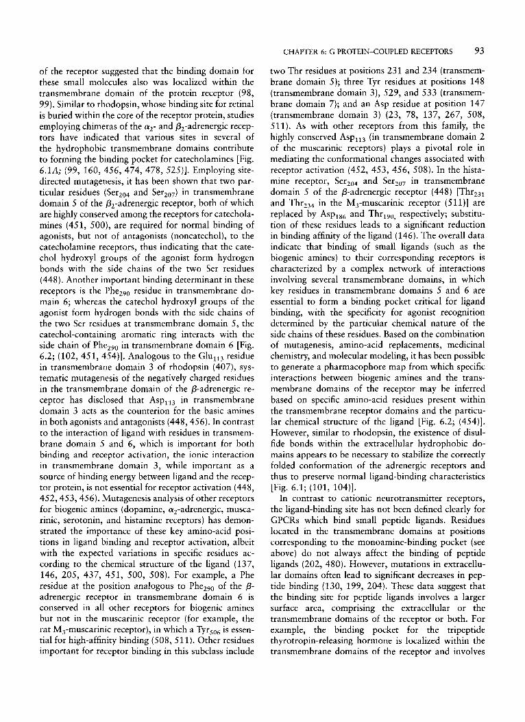
FIG. 6.2. Pharmacophore map of the catecholamine-binding site of the P-adrenergic receptor. A catecholamine ligand is shown in a hypothetical binding site intercalated among the transmembrane helices of the receptor. Each of the large semicircles represents a transmembrane helix of the receptor, inscribed with the type of binding interaction expected. Other G protein-coupled receptors expected to have similar interactions with their specific ligands are designated in boxes next to each helix. aAR, a- adrenergic receptor; PAR, P-adrenergic receptor; DAR, dopaminergic receptor; MAR, muscarinic acetylcholine receptor; SHT-R, 5-hydroxytryptamine (serotonin) receptor. Inset: Model for the ligand- binding site of the P-adrenergic receptor showing key interactions with the agonist isoproterenol. [Reproduced from Strader et al. (450, 454) with permission.]
For example, the rod photoreceptor cell bears the pho- tosensitive rhodopsin receptor molecule (1 15, 244, 458). Upon activation by photons of light, its cova- lently linked ligand, all-&-retinal, isomerizes to the all-trans isomer; this structural change in retinal forces a series of conformational modifications in rhodopsin, which in turn binds the trimeric retinal G protein transducin (G,) (244, 267, 354, 391, 435). The a- subunit of rhodopsin-activated transducin activates the effector enzyme cyclic guanosine monophosphate (cGMP) phosphodiesterase, which hydrolyzes GMP, with a consequent decrease in intracellular GMP levels, closure of GMP-gated membrane Na+-specific chan- nels, hyperpolarization of the plasma membrane, and reduction of the rate of neurotransmitter release from the synaptic region; this sequence of intracellular events allows light to free the neurons from neurotransmitter
inhibition and excite them (144,238,266,458,513). A similar pathway of GPCR-mediated signal transduction has been characterized for the adrenergic receptors (87, 105,45 1). These relatively widely distributed receptors interact with the endogenous catecholamines epineph- rine and norepinephrine, as well as with a large number of synthetic agonists and antagonists (450, 454). Upon specific agonist binding, P-adrenergic receptors (includ- ing subtypes PI, p2, and p3) activate a stimulatory G protein (GJ, which in turn activates the effector ade- nylyl cyclase (16, 26, 118); this enzyme is responsible for cyclic adenosine 3’,5’-monophosphate (AMP) for- mation, one of the main intracellular mediators of extracellular signals (417, 468).
G protein+oupled receptors contain a variable num- ber of Cys residues. Conserved Cys residues in the NH,-terminus and the extracellular loops (particularly
CHAPTER 6: G PROTEIN-COUPLED RECEPTORS 9 1
in the first and second) may form disulfide bonds that stabilize the structure of the functional protein (Fig. 6.1 ). Employing site-directed mutagenesis, substitution of either of the Cys residues present in the first (Cys,,,) and the second (CYS,,~) extracellular loops of the &- adrenergic receptor resulted in destabilization of the tertiary structure of the protein and alterations in the binding properties of the receptor (101, 104); similar results were obtained with substitutions performed in Cys,,, and Cys,,, of rhodopsin (235). For receptors in which the principal determinants of ligand binding reside within a large extracellular NH,-terminal do- main (see Ligund-Binding Domain, below), such as the calcium-sensing receptor and the metabotropic gluta- mate receptors, the numerous conserved Cys residues located within the extracellular domain may help to organize this region into a binding pocket appropriate for interaction with small charged ligands, such as glutamate or Ca2+ (42, 54, 306, 338). Most GPCRs exhibit one or two Cys residues in the membrane- proximal portion of the COOH-terminus; these resi- dues are particularly important because they are sus- ceptible to palmitoylation, that is, reversible thioesteri- fication of the C,, fatty acid palmitate (34) (Fig. 6.1). Palmitoylation of a number of GPCRs has been docu- mented. These include rhodopsin (the first GPCR shown to be modified by palmitoylation) (234, 364), &- and a,,-adrenergic (242, 356), D,-dopaminergic (351), 5-HT,,-serotonergic (350), and luteinizing hor- monekhoriogonadotropin (239, 540) receptors. All of these receptors have in common the presence of one or two Cys residues in the membrane-proximal domain of the COOH-terminus. Since fatty acylation of several soluble proteins promotes their association with the inner surface of the plasma membrane, it is thought that palmitoylation of Cys residues in the membrane- proximal portion of the COOH-terminus facilitates the formation of an anchoring that creates a fourth intracellular loop (Fig. 6.1A). Although a strict consen- sus sequence that determines the occurrence of covalent S-palmitoylation of the Cys residues present in the proximal segment of the COOH-terminus tail of these receptors has not yet been identified, the consensus sequence FN(X)n,B(X)n,CP . . . (where B is a hy- drophobic residue, n, and n, are residues that may vary between between 0 and 4, and Cp is the palmitoy- lated Cys), based on the primary sequence of the palmi- toylated receptors mentioned above (34), has been proposed. Modulation of receptor palmitoylation by agonist has been documented for the p2- and a,,- adrenergic receptors and for the D ,-dopamine receptor (241, 329, 351); these studies have suggested that stimulation of a receptor by its specific signaling mole- cule affects the turnover rate of the palmitate moiety
linked to the receptor by increasing its relative rate of depalmitoylation (34, 329). The physiological signifi- cance of receptor palmitoylation remains to be fully understood; however, mutations that prevent palmitoy- lation of the &-adrenergic receptor promote the func- tional uncoupling of the receptor and, consequently, a loss of G protein activation (356). Palmitoylation increases the hydrophobicity of a protein and may favor redistribution to the membrane compartment. Since prevention of palmitoylation of the P,-adrenergic receptor is accompanied by an increased protein kinase A (PKA)-dependent phosphorylation of the receptor (326, 329), it has been proposed that in receptors harboring phosphorylation sites near potentially palmi- toylated Cys residues, there is a close relationship be- tween palmitoylation, phosphorylation, and receptor desensitization (34) (see REGULATORY MECHANISMS, be- low). Although uncoupling of G proteins after muta- tion of a palmitoylated Cys residue also has been observed for the p opiate receptor (227), for other receptors, such as rhodopsin (234,328), a,,-adrenergic (242), M, muscarinic (489), and luteinizing hormone/ choriogonadotropin (540), abolition of palmitoylation has no effect on G protein and effector activation. Nevertheless, the fact that the presence of Cys residues susceptible to palmitoylation is highly conserved among this GPCR subfamily strongly suggests that this posttranslational modification is important for the function of these receptors.
Another posttranslational modification found in most GPCRs is the presence of one or more consensus sequences ( Asn-Xaa-SerlThr) for N-linked glycosyla- tion (17, 29, 103, 150, 173, 180, 418, 445, 535). Heterogeneity in the glycosylation pattern contributes to the anomalous migration of these glycoproteins on gel electrophoresis (82). The N-linked oligosaccharides usually are located near the NH,-terminus of the recep- tor molecule, though for some receptors there may be glycosylation sites in the first and second extracellular loops as well (232) (Fig. 6.1). For some receptors (for example, P-adrenergic receptor, rhodopsin, luteinizing hormonekhoriogonadotropin, and GnRH receptors), prevention of glycosylation, either by inhibiting post- translational glycosylation or by site-directed mutagen- esis, led to decreased cell-surface receptor expression without any significant change in binding or functional activity (83, 106, 150, 225, 294, 389, 469), whereas incorporation of an additional glycosylation site on the human GnRH receptor resulted in an enhancement of both binding affinity and the level of cell-surface expression of the receptor (82). Thus, it seems that for this type of receptor glycosylation is functionally important for membrane insertion and expression. GPCRs may exhibit a certain degree of heterogeneity
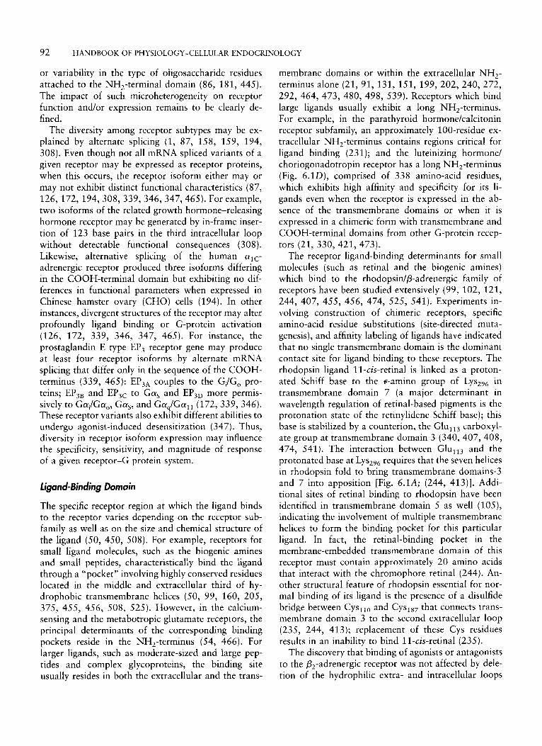
92 HANDBOOK OF PHYSIOLOGY-CELLULAR ENDOCRINOLOGY
or variability in the type of oiigosaccharide residues attached to the NH,-terminal domain (86, 181, 445). The impact of such microheterogeneity on receptor function and/or expression remains to be clearly de- fined.
The diversity among receptor subtypes may be ex- plained by alternate splicing (1, 87, 158, 159, 194, 308). Even though not all mRNA spliced variants of a given receptor may be expressed as receptor proteins, when this occurs, the receptor isoform either may or may not exhibit distinct functional characteristics (87, 126, 172, 194,308, 339,346,347,465). For example, two isoforms of the related growth hormone-releasing hormone receptor may be generated by in-frame inser- tion of 123 base pairs in the third intracellular loop without detectable functional consequences (308). Likewise, alternative splicing of the human alc- adrenergic receptor produced three isoforms differing in the COOH-terminal domain but exhibiting no dif- ferences in functional parameters when expressed in Chinese hamster ovary (CHO) cells (194). In other instances, divergent structures of the receptor may alter profoundly ligand binding or G-protein activation (126, 172, 339, 346, 347, 465). For instance, the prostaglandin E type EP, receptor gene may produce at least four receptor isoforms by alternate mRNA splicing that differ only in the sequence of the COOH- terminus (339, 465): EP,, couples to the GJG, pro- teins; EP,, and EP,, to Gas and EP,, more permis- sively to Gai/Ga,, Gas, and Gaq/Gal, (172, 339, 346). These receptor variants also exhibit different abilities to undergo agonist-induced desensitization (347). Thus, diversity in receptor isoform expression may influence the specificity, sensitivity, and magnitude of response of a given receptor-G protein system.
Ligand-Binding Domain
The specific receptor region at which the ligand binds to the receptor varies depending on the receptor sub- family as well as on the size and chemical structure of the ligand (50, 450, 508). For example, receptors for small ligand molecules, such as the biogenic amines and small peptides, characteristically bind the ligand through a “pocket” involving highly conserved residues located in the middle and extracellular third of hy- drophobic transmembrane helices (50, 99, 160, 205, 375, 455, 456, 508, 525). However, in the calcium- sensing and the metabotropic glutamate receptors, the principal determinants of the corresponding binding pockets reside in the NH,-terminus (54, 466). For larger ligands, such as moderate-sized and large pep- tides and complex glycoproteins, the binding site usually resides in both the extracellular and the trans-
membrane domains or within the extracellular NH2- terminus alone (21, 91, 131, 151, 199, 202, 240, 272, 292, 464, 473, 480, 498, 539). Receptors which bind large ligands usually exhibit a long NH,-terminus. For example, in the parathyroid hormone/calcitonin receptor subfamily, an approximately 1 OO-residue ex- tracellular NH2-terminus contains regions critical for ligand binding (23 1 ); and the luteinizing hormone/ choriogonadotropin receptor has a long NH,-terminus (Fig. 6.1D), comprised of 338 amino-acid residues, which exhibits high affinity and specificity for its li- gands even when the receptor is expressed in the ab- sence of the transmembrane domains or when it is expressed in a chimeric form with transmembrane and COOH-terminal domains from other G-protein recep- tors (21, 330,421, 473).
The receptor ligand-binding determinants for small molecules (such as retinal and the biogenic amines) which bind to the rhodopsinlp-adrenergic family of receptors have been studied extensively (99, 102, 121, 244, 407, 455, 456, 474, 525, 541). Experiments in- volving construction of chimeric receptors, specific amino-acid residue substitutions (site-directed muta- genesis), and affinity labeling of ligands have indicated that no single transmembrane domain is the dominant contact site for ligand binding to these receptors. The rhodopsin ligand ll-czs-retinal is linked as a proton- ated Schiff base to the €-amino group of Lys,,, in transmembrane domain 7 (a major determinant in wavelength regulation of retinal-based pigments is the protonation state of the retinylidene Schiff base); this base is stabilized by a counterion, the Glul1, carboxyl- ate group at transmembrane domain 3 (340, 407, 408, 474, 541). The interaction between GlulI3 and the protonated base at LYS,~, requires that the seven helices in rhodopsin fold to bring transmembrane domains-3 and 7 into apposition [Fig. 6.1A; (244, 413)]. Addi- tional sites of retinal binding to rhodopsin have been identified in transmembrane domain 5 as well (105), indicating the involvement of multiple transmembrane helices to form the binding pocket for this particular ligand. In fact, the retinal-binding pocket in the membrane-embedded transmembrane domain of this receptor must contain approximately 20 amino acids that interact with the chromophore retinal (244). An- other structural feature of rhodopsin essential for nor- mal binding of its ligand is the presence of a disulfide bridge between Cys,,, and CysI8, that connects trans- membrane domain 3 to the second extracellular loop (235, 244, 413); replacement of these Cys residues results in an inability to bind ll-czs-retinal (235).
The discovery that binding of agonists or antagonists to the P,-adrenergic receptor was not affected by dele- tion of the hydrophilic extra- and intracellular loops
CHAPTER 6: G PROTEIN-COUPLED RECEPTORS 93
of the receptor suggested that the binding domain for these small molecules also was localized within the transmembrane domain of the protein receptor (98, 99). Similar to rhodopsin, whose binding site for retinal is buried within the core of the receptor protein, studies employing chimeras of the a,- and P,-adrenergic recep- tors have indicated that various sites in several of the hydrophobic transmembrane domains contribute to forming the binding pocket for catecholamines [Fig. 6.1A; (99, 160, 456, 474, 478, 525)l. Employing site- directed mutagenesis, it has been shown that two par- ticular residues ( Ser,04 and ser,,,) in transmembrane domain 5 of the P,-adrenergic receptor, both of which are highly conserved among the receptors for catechola- mines (451, 500), are required for normal binding of agonists, but not of antagonists (noncatechol), to the catecholamine receptors, thus indicating that the cate- chol hydroxyl groups of the agonist form hydrogen bonds with the side chains of the two Ser residues (448). Another important binding determinant in these receptors is the Phe,,, residue in transmembrane do- main 6; whereas the catechol hydroxyl groups of the agonist form hydrogen bonds with the side chains of the two Ser residues at transmembrane domain 5 , the catechol-containing aromatic ring interacts with the side chain of Phe,,, in transmembrane domain 6 [Fig. 6.2; (102, 451, 454)]. Analogous to the Glu,,, residue in transmembrane domain 3 of rhodopsin (407), sys- tematic mutagenesis of the negatively charged residues in the transmembrane domain of the P-adrenergic re- ceptor has disclosed that Asp, 13 in transmembrane domain 3 acts as the counterion for the basic amines in both agonists and antagonists (448,456). In contrast to the interaction of ligand with residues in transmem- brane domain 5 and 6, which is important for both binding and receptor activation, the ionic interaction in transmembrane domain 3 , while important as a source of binding energy between ligand and the recep- tor protein, is not essential for receptor activation (448, 452,453,456). Mutagenesis analysis of other receptors for biogenic amines (dopamine, a,-adrenergic, musca- rinic, serotonin, and histamine receptors) has demon- strated the importance of these key amino-acid posi- tions in ligand binding and receptor activation, albeit with the expected variations in specific residues ac- cording to the chemical structure of the ligand (137, 146, 205, 437, 451, 500, 508). For example, a Phe residue at the position analogous to Phe,,, of the 0- adrenergic receptor in transmembrane domain 6 is conserved in all other receptors for biogenic amines but not in the muscarinic receptor (for example, the rat M3-muscarinic receptor), in which a Tyrso6 is ' essen- tial for high-affinity binding (508, 511). Other residues important for receptor binding in this subclass include
two Thr residues at positions 231 and 234 (transmem- brane domain 5); three Tyr residues at positions 148 (transmembrane domain 3), 529, and 533 (transmem- brane domain 7); and an Asp residue at position 147 (transmembrane domain 3) (23, 78, 137, 267, 508, 511). As with other receptors from this family, the highly conserved Asp,,, (in transmembrane domain 2 of the muscarinic receptors) plays a pivotal role in mediating the conformational changes associated with receptor activation (452, 453, 456, 508). In the hista- mine receptor, Ser,,, and Ser,,, in transmembrane domain 5 of the P-adrenergic receptor (448) [Thr,,, and Thr23, in the M,-muscarinic receptor (511)] are replaced by Asp,,, and Thr,,,, respectively; substitu- tion of these residues leads to a significant reduction in binding affinity of the ligand (146). The overall data indicate that binding of small ligands (such as the biogenic amines) to their corresponding receptors is characterized by a complex network of interactions involving several transmembrane domains, in which key residues in transmembrane domains 5 and 6 are essential to form a binding pocket critical for ligand binding, with the specificity for agonist recognition determined by the particular chemical nature of the side chains of these residues. Based on the combination of mutagenesis, amino-acid replacements, medicinal chemistry, and molecular modeling, it has been possible to generate a pharmacophore map from which specific interactions between biogenic amines and the trans- membrane domains of the receptor may be inferred based on specific amino-acid residues present within the transmembrane receptor domains and the particu- lar chemical structure of the ligand [Fig. 6.2; (454)]. However, similar to rhodopsin, the existence of disul- fide bonds within the extracellular hydrophobic do- mains appears to be necessary to stabilize the correctly folded conformation of the adrenergic receptors and thus to preserve normal ligand-binding characteristics [Fig. 6.1; (101, 104)].
In contrast to cationic neurotransmitter receptors, the ligand-binding site has not been defined clearly for GPCRs which bind small peptide ligands. Residues located in the transmembrane domains at positions corresponding to the monoamine-binding pocket (see above) do not always affect the binding of peptide ligands (202, 480). However, mutations in extracellu- lar domains often lead to significant decreases in pep- tide binding (130, 199, 204). These data suggest that the binding site for peptide ligands involves a larger surface area, comprising the extracellular or the transmembrane domains of the receptor or both. For example, the binding pocket for the tripeptide thyrotropin-releasing hormone is localized within the transmembrane domains of the receptor and involves
94 HANDBOOK OF PHYSIOLOGY-CELLULAR ENDOCRINOLOGY
specific residues in transmembrane domains 3 (Tyr,,,, AsnlIo), 6 (Arg,,,), and 7 AT^,,,^) (375). Binding of the decapeptide GnRH requires, in addition to the proximity of helices 2 and 7, an ionic interaction between Argg of the decapeptide and the carboxylate side chain of Glu,,, in the third extracellular loop (122, 538, 539). These two transmembrane domains possess two highly conserved residues; however, whereas most GPCRs exhibit an Asp or Glu residue at a conserved position in domain 2 and a conserved Asn residue in domain 7, in the GnRH receptor these residues are exchanged so that Asn,, is located in helix 2 and in helix 7 (232, 233, 538). Mutating the Asn,, in domain 2 to an Asp abolished agonist binding, whereas a second mutation in domain 7, replacing
by Asn, led to restoration of high-affinity ligand binding (420, 538). Further, when these mutations were introduced in a pituitary-derived lactotrope cell line expressing the rat GnRH receptor, they abolished the initial down-regulation of the receptor and im- paired its interaction with the G proteins involved in inositol phosphate production ( 1 1). Site-directed mutagenesis on the neurokinin-1 (NK,) receptor, which binds substance P, has shown that three residues in the first extracellular segment (Asn,,, Gln,,, and Phe,,) and two in the second (Asp,, and Hisl0,) are required particularly for ligand binding (1 30); several residues in the second and the seventh transmembrane domains of this receptor (Asn,,, Asn,,, Tyr9,, and Asn,, in domain 2 and T Y ~ , ~ , in domain 7) are also important in determining the affinity of the receptor for its ligand (128, 533). The residues in domain 2 are predicted to be positioned on the same face of the a-helical region, thus indicating that their side chains may form a hydrogen-bonding surface projecting into the ligand- binding site. In other peptide-binding receptors, such as the angiotensin, vasopressin, oxytocin, and neuro- peptide Y receptors, the ligands also bind to determi- nants located in both the first extracellular loop and several transmembrane domains (domains 2-7) (480), emphasizing the importance of the transmembrane do- mains in the binding of peptide ligands to GPCRs.
In many cases, the sites required for high-affinity binding of antagonists of these peptide ligands may be distinct from those of the naturally occurring ligands. This is exemplified by the tachykinin NK, receptor, in which nonpeptide antagonists do not interact with the extracellular domain but rather with residues located in transmembrane domains4 ( G ~ I I ~ ~ ~ ) , 5 (Hisl9,), 6 ( T Y ~ , ~ , and Hisz6,), and 7 (Tyre,,,), some of which do not participate in binding of the natural peptide ligand (127, 129, 202, 203) (see above); these antagonists may exhibit up to a 100-fold difference in affinity between species homologous to the NK, receptor, in
contrast to the similar affinities of the endogenous ligands (129). Another example is the GnRH receptor. Molecular modeling of this receptor predicts a Lys,,, residue in transmembrane domain 3 [positioned at a locus that corresponds to the conserved Asp, ,, of the cationic amine receptors (448, 456)] that contributes to the ligand-binding pocket (Fig. 6.1B); substitution of this charge-strengthened hydrogen bond donor by Gln resulted in considerable loss of agonist binding without any significant effect in antagonist binding (539). Employing photoaffinity-labeling techniques and enzymatic digestion of the labeled GnRH receptor, it has been demonstrated that agonists and antagonists bind to different regions of the receptor and, therefore, are likely to be oriented differently with respect to the receptor (219).
The NH,-terminus of receptors for other relatively larger peptide ligands and glycoprotein molecules plays a major role in determining high-affinity binding to the corresponding ligands. In the secretinNIP receptor subfamily, there is a sequence conservation in the NH2- terminus that strongly suggests a role of this region in mediating receptor-ligand interactions (199, 212, 231, 287, 414). For example, it has been shown that the NH,-terminal extracellular regions of the parathor- mone and secretin receptors contain domains that largely determine the binding affinity of these hor- mones (199, 231). Nevertheless, some of these ligands also may interact with other extracellular domains; although the first ten residues of the NH2-terminus of the secretin receptor are critical for ligand binding, other extracellular domains located at the COOH- terminus of the first extracellular loop as well as at the NH,-terminal half of the second loop also provide critical determinants (199). Other receptors in which the NH,-terminal region appears to be critical in de- termining ligand binding include the interleukin-8 (272, 464), the C5a (91), the glycoprotein hormone [luteinizing hormonekhoriogonadotropin, FSH, and thyroid-stimulating hormone (TSH) (21, 473, 498)], and the thrombin (151, 292) receptors. Glycoprotein receptors have large extracellular domains (300-400 amino acids in length) and bind complex ligands with molecular weights ranging from 28 to 38 kd. These receptors exhibit an extraordinary amino-acid homol- ogy (30%-5O%) in the extracellular domains, where multiple discontinuous segments spanning the entire domain contribute to the binding site and ligand speci- ficity (5, 21, 336, 337, 473, 498). The extracellular domain of the luteinizing hormonekhoriogonadotro- pin receptor exhibits multiple ( n = 14) Leu-rich repeats (421, 473); in particular, composite regions encom- passed by Leu-rich repeats 1-6 are involved in high- affinity binding and binding specificity (473). Some
CHAPTER 6: G PROTEIN-COUPLED RECEPTORS 95
studies employing mutant receptors have pointed to- ward the existence of an additional site important for the binding affinity of luteinizing hormone/choriogo- nadotropin located in the second transmembrane do- main (Asp,,,) as well as a low-affinity ligand-binding site in a specific region of the third extracellular con- necting loop (LyS,,,) [Fig. 6.1D; (153, 405)]. Appar- ently, this binding site is critical for activating specific transmembrane conductors that eventually lead to the generation of particular intracellular signals, such as cAMP (153). Finally, the thrombin receptor exhibits in its NH,-terminus a unique mechanism for ligand bind- ing and receptor activation. In this receptor, the ligand is a protease and its extracellular NH,-terminus con- tains both a ligand-specific anion-binding site and a specific proteolytic cleavage substrate site; it is activated by binding its tethered ligand domain, which is un- masked upon receptor cleavage by thrombin (151, 292).
G Protein4oupling Domain
G protein-coupled receptors characteristically bind G proteins, which in turn act as mediators of receptor- induced effector activation (120, 184, 344, 430). The nature of the second-messenger pathways activated in response to agonist binding to a GPCR essentially is determined by the type of G protein(s) coupled to each particular receptor. As can be expected from both the general topography of these receptors and the fact that GPCRs couple and interact with a number of distinct G proteins (at least 17 different species of Ga genes coding for four main classes and their numerous corres- ponding subtypes have been identified, see THE HETER- OTRIMERIC G-PROTEIN FAMILY, below), the G protein- coupling domains lie within the divergent sequences of the intracytoplasmic domains of the receptor (59, 80, 195, 258, 301, 355, 357, 510). When a particular ligand binds to its receptor, the molecular perturbation is relayed from the membrane-embedded helices to the cytoplasmic face of the protein molecule (291). These intracellular domains, particularly the regions closest to the plasma membrane, as well as some specific regions located in the intracellular ends of the trans- membrane domain helices and in the membrane- proximal portion of the COOH-terminus, are im- portant for receptor-G protein coupling, interaction, and specificity determination [Fig. 6.1; (11, 60, 137, 253, 357, 449)]. Segments of the intracellular loops are important not only in determining the selectivity and relative affinity of the receptor to different G proteins but also in activating the receptor and trans- mitting the signal generated following agonist binding (see REGULATORY MECHANISMS, below).
Within members of the various GPCR families there are some receptor classes with corresponding subtypes that may show particular preference or specificity to- ward a particular G-protein class [G,, G,/G,, G,, or G12, though specificity of the receptor for this latter class remains to be determined fully (96, 344)] (20, 369, 433, 523). This is the case, for example, of the muscarinic receptor class, in which the M, and M, receptors preferentially couple to proteins of the GJG, class, which are sensitive to pertussis toxin (see THE HETEROTRIMERIC G-PROTEIN FAMILY, below), whereas the M,, M,, and M5 receptors predominantly stimulate pertussis toxin-insensitive proteins of the G,/G, class (20, 369, 433). The different subclasses of the seroto- ninergic (5-HT) receptors (5-HTl-5-HT,) with their corresponding subtypes (for example, 5-HT,,, 5-HT,,, etc.) also show particular preferences: 5-HT, receptors bind preferentially to G,/G, proteins, whereas the 5- HT, and 5-HT4 and the 5-HT, and 5-HT, subtypes are coupled to members of the G,/G,, and G, protein classes, respectively (116, 331, 376, 395). In the meta- botropic glutamate receptor (mGluR) family, the mGluR, (including its splice variants mGluR,,, mGluRlp, and mGluR,,) and mGluR, types are associ- ated with effects most likely mediated by the G,/Gll class (185, 5341, whereas the mGluR2, mGluR,, mGluR,, and mGluR, classes exhibit some preference toward the G,/G, class (378, 537). An additional level of complexity in receptor-G protein coupling is the fact that certain receptors in a given family may show a strong preference for a particular member of a G- protein class; for example, the C5A receptor prefers GI, to other members of the G, class (277). Finally, there are receptors that couple normally to different G-protein classes. The receptors for glycoprotein hor- mones (luteinizing hormone/choriogonadotropin, TSH, and FSH) stimulate (albeit not to the same extent) both cAMP and inositol phosphate production throughout the pathways defined by G, and G, proteins, respec- tively (195, 260, 386, 490).
Experiments employing specifically directed muta- genesis, chimeric receptors, peptides corresponding to different positions of the receptor sequence, and anti- bodies against various cytoplasmic domains of specific G proteins have been used to characterize the sites of contact as well as some of the sequences involved in activation and specificity determination (59, 60, 80, 133,195,258,301,355,363,524). These studies have led to the recognition of the importance of the second and third intracellular loops as well as the membrane- proximal region of the COOH-terminus of several GPCRs in G-protein coupling. This issue has been explored deeply in some members of the rhodopsidp- adrenergic family. In rhodopsin, the second and third
96 HANDBOOK OF PHYSIOLOGY-CELLULAR ENDOCRINOLOGY
intracellular loops, as well as the fourth loop created by palmitoylation of Cys residues in the membrane- proximal portion of the COOH-terminus, are involved in the interaction between the receptor and G,; syn- thetic peptides derived from these loops were able to interact, either alone or in several combinations, with transducin (133). However, at least two sites, one in the second and another in the third intracellular loop, are required for activation of bound transducin (133), which indicates that determinants present in several intracellular domains are important for receptor-G protein interaction. In the muscarinic and catechola- mine receptors (and presumably in other receptors for biogenic amines and peptide hormones), both the NH,- and the COOH-terminal portions of the third intracel- lular loop appear to be critical determinants of G- protein binding and activation (74,178,265,268,275, 303, 425, 509, 510). Data from studies of deletions in limited regions of the NH2-terminal (residues 222- 229) and the COOH-terminal (residues 258-270) por- tions of the third intracellular loop in the @,-adrenergic receptor (which is coupled to the G, protein) as well as construction of chimeric a,/p,-adrenergic receptors and several P,-adrenergic receptor mutants have indi- cated that these particular regions are necessary for the @,-adrenergic receptor-mediated activation of adenylyl cyclase [Fig. 6.1C; (74, 253, 286, 357, 449)]. Appar- ently, in the P-adrenergic receptor, the COOH-terminal region of this loop is more important for G-protein coupling specificity than the opposite NH,-terminal region of the same loop since substitution of residues 216-237 in the latter with the corresponding residues from the qadrenergic receptor (which is coupled to the Gq/G,, class) failed to affect the ability of the @, receptor to couple G,, whereas substitution of residues 263-274 corresponding to the COOH-terminal region of the loop resulted in a severe impairment of coupling (286). Similar results have been found in the DIA- dopamine and the a,-adrenergic receptors (60, 73, 301). In these receptors, co-expression of the entire third intracellular loop of the al , receptor or domains involving both restricted NH,- and COOH-terminal regions of this loop with its parent receptor signifi- cantly decreased inositol phosphate production, pre- sumably through specific competitive inhibition of G- protein (Gq/Gl1) binding (60, 301). However, some chimeric studies also have indicated that a restricted region, composed of 27 amino-acid residues of the NH2-terminal region from the third intracellular loop of the &,,-receptor, plays a major role in determining the selectivity for receptor-Gq/G, , protein coupling (74). In all of these receptors, both specific amino- acid residues from the NH,- and the COOH-terminal
portions of the third intracellular loop and the second- ary structure of these regions, which form amphipathic helical extensions of the adjacent transmembrane do- mains, play crucial roles in determining coupling speci- ficity, selectivity, and efficiency to activate a particular class of G protein (Gi, G,, or G,) (59, 60, 188, 253).
For several GPCRs, the importance of the second intracellular loop in G-protein coupling and specificity has been well documented (74,80,244,314,357,360, 361). The amino-acid sequence of this loop is among the most highly conserved in the GPCR superfamily, and substitutions in some of its highly conserved resi- dues may severely impair G-protein coupling. The sec- ond intracellular loop is essential for maintaining the configuration of the G-protein binding site and, thus, for the normal interaction between the @,-adrenergic receptor and G, (357, 524). Construction of a chimeric a,,/@,-adrenergic receptor, in which the third intracel- lular loop of the a,-adrenergic receptor replaced the corresponding loop in the P2 receptor, resulted in a chimeric receptor capable of activating both the G,/ GI, and the G, pathways (74), thus suggesting the existence of determinants for G, specificity in regions other than the third intracellular loop, probably in the second loop. Similar findings have been reported for the M,-muscarinic receptor, which normally couples to the Gi protein class; replacement of the third intracel- lular loop of this receptor with the corresponding loop of the @,-adrenergic receptor resulted in a chimeric receptor species coupled to both Gi and G, (314). Other lines of evidence strongly suggest that the second intracellular loop is involved in G-protein coupling and selectivity (135, 361, 500, 508). Mutational analysis of the MI-muscarinic receptor has shown that a highly conserved Asp residue located at the beginning of the second intracellular loop is important for efficient G- protein coupling (508). In addition, specific regions located in the COOH-terminal region of this loop in the M2- and M,-muscarinic receptors are involved in selection and activation of G, (314, 361). The impor- tance of the second intracellular loop in receptor inter- action with G proteins also is exemplified in rhodopsin (134, 244, 258), the angiotensin I1 receptor (360), and the interleukin-8 receptor (80). In this latter receptor, four specific residues in the second intracellular loop ( T Y ~ , ~ ~ , Leul3,, Ile,39, and Val,4o) are critical for cou- pling to the Gi protein (80).
The COOH-terminal domain of GPCRs may be in- volved in G-protein coupling and activation (195, 258, 357). In rhodopsin, a synthetic polypeptide from the fourth intracellular loop is capable of interacting with G, (258). In the P,-adrenergic receptor, substitution of residues forming the fourth intracellular loop with
CHAPTER 6: G PROTEIN-COUPLED RECEPTORS 97
the corresponding residues from the a, receptor has resulted in a reduced efficiency of coupling to the corresponding G protein (357). In this receptor, substi- tution of the conserved Cys residue (Cys,,,) within the NH,-terminal segment of the cytoplasmic tail produces a significant reduction in its ability to stimulate ade- nylyl cyclase (356); as mentioned above, palmitoylation of this residue may induce anchoring of this portion of the COOH-terminus to the cell membrane, leading to formation of a short fourth intracellular loop, which in turn may facilitate receptor-G protein coupling. For other receptors of the same family, this structural feature might not be important for G-protein coupling. Mutations of this highly conserved Cys residue in a,- adrenergic receptors (241,242), as well as in the lutein- izing hormone/choriogonadotropin (540) and MI - and M,-muscarinic (489) receptors, had no effect on agonist-mediated activation of the corresponding sec- ond messengers. Similarly, mutations in the Cys3,, and/ or Cys,,, residues of bovine opsin which resulted in complete abolition of palmitoylation did not affect activation of G, (234). These data indicate that this shared structural motif plays differing roles in different receptor-(; protein interactions.
Although there is little evidence indicating that the first intracellular loop is involved in G-protein binding, one study employing chimeric calcitonin receptors has shown that substitution with the first intracellular loop of a particular isoform of the human calcitonin recep- tor, containing a unique insertion of 16 amino acids in this loop, completely abolished production of inositol phosphates while allowing stimulation of the CAMP pathway by the host porcine receptor (355).
The overall data indicate that many intracellular domains are involved in receptor-G protein coupling. The most important domains lie within the membrane- proximal regions of the second and third intracellular loops. Although these domains are crucial for high- affinity binding, coupling specificity, and activation of specific G proteins, other regions or particular residues located in the transmembrane helices or even in some extracellular loops play major roles in signal transduc- tion and G-protein activation [Fig. 6.1A, C; (11, 137, 153, 263, 273, 405)].
THE HETEROTRIMERIC G-PROTEIN FAMILY
General Features
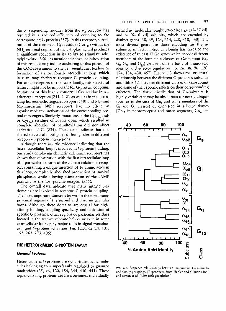
Heterotrimeric G proteins are signal-transducing mole- cules belonging to a superfamily regulated by guanine nucleotides (25, 96, 120, 184, 344, 430, 441). These signal-carrying proteins are heterotrimers, individually
termed a- (molecular weight 39-52 kd), p- (35-37 kd), and y- (6-10 kd) subunits, which are encoded by distinct genes (38, 39, 124, 214, 228, 318, 430). The most diverse genes are those encoding for the a- subunits; in fact, molecular cloning has revealed the existence of at least 17 G a genes which encode different members of the four main classes of Ga-subunit (Gs, Gi, G,, and GI,) grouped on the basis of amino-acid identity and effector regulation (15, 26, 38, 96, 120, 174, 184, 430, 457). Figure 6.3 shows the structural relationship between the different G-protein a-subunits and Table 6.1 lists the different classes of Ga-subunit and some of their specific effects on their corresponding effectors. The tissue distribution of Ga-subunits is highly variable; it may be ubiquitous (or nearly ubiqui- tous, as in the case of Gas and some members of the G, and G, classes) or expressed in selected tissues [Gatl in photoreceptor rod outer segments, GaOlf in
40 60 80 100 d d
,-+= 3 1
4
Gi
rn id ~ 0
- 2 Yo Amino Acid Identity - 0 3
CI)
FIG. 6.3. Sequence relationships between mammalian Ga-subunits and family groupings. [Reproduced from Hepler and Gilman (184) and Simon et al. (430) with permission.]
98 HANDBOOK OF PHYSIOLOGY-CELLULAR ENDOCRINOLOGY
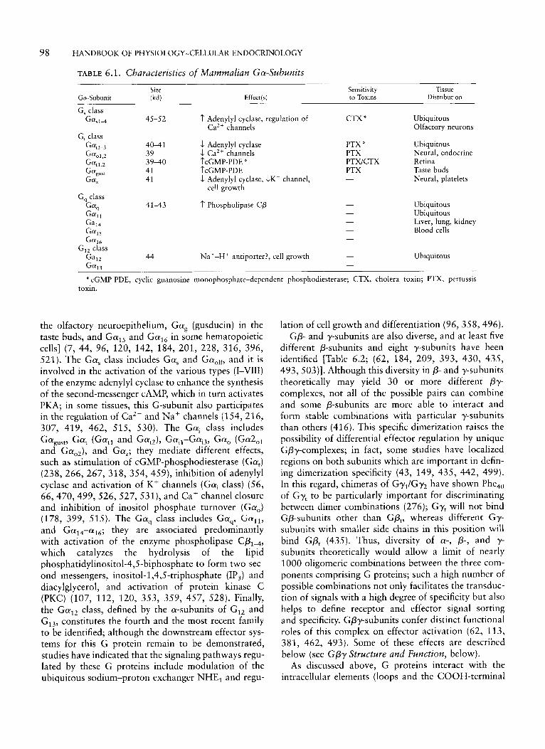
TABLE 6.1. Characteristics of Mammalian Ga-Subunits Size
Ga-Suhunit ikd) Effectlsl Sensitivity Tissue to Toxins Distribution
G, class G f f M 45-52 1' Adenylyl cyclase, regulation of CTX * Ubiquitous
Ca2+ channels Olfactory neurons G, class
Gff,l-3 40-41 J Adenylyl cyclase PTX * Ubiquitous Gff"l.2 39 1 Ca2+ channels PTX Neural, endocrine G"tl,2 39-40 ~'CGMP-PDE * PTWCTX Retina
41 I'CGMP-PDE PTX Taste buds 41 5 Adenylyl cyclase, J K + channel, - Neural, platelets
Gag,,,
cell growth
41-43 1' Phospholipase C p
44 Na+-H+ antiporter?, cell growth - -
Ubiquitous Ubiquitous Liver, lung, kidney Blood cells
Ubiquitous
* cGMP-PDE, cyclic guanosine monophosphate-dependent phosphodiesterase; CTX, cholera toxin; PTX, pertussis toxin.
the olfactory neuroepithelium, Gag (gusducin) in the taste buds, and Gals and Gal6 in some hematopoietic cells] (7, 44, 96, 120, 142, 184, 201, 228, 316, 396, 521). The Gas class includes Gas and Gaol*, and it is involved in the activation of the various types (I-VIII) of the enzyme adenylyl cyclase to enhance the synthesis of the second-messenger CAMP, which in turn activates PKA; in some tissues, this G-subunit also participates in the regulation of Ca2+ and Na+ channels (154,216, 307, 419, 462, 515, 530). The Gai class includes
and GaO2), and GaZ; they mediate different effects, such as stimulation of cGMP-phosphodiesterase (Gat) (238, 266, 267, 318, 354, 459), inhibition of adenylyl cyclase and activation of K+ channels (Gal class) (56, 66,470,499, 526, 527, 531), and Ca+ channel closure and inhibition of inositol phosphate turnover (Ga,) (178, 399, 515). The Gaq class includes Gaq, Gal l , and Ga,,-aI6; they are associated predominantly with activation of the enzyme phospholipase CP,,, which catalyzes the hydrolysis of the lipid phosphatidylinositol-4,5-biphosphate to form two sec- ond messengers, inositol-1,4,5-triphosphate (IP,) and diacylglycerol, and activation of protein kinase C (PKC) (107, 112, 120, 353, 359, 457, 528). Finally, the Ga12 class, defined by the a-subunits of GI, and G13, constitutes the fourth and the most recent family to be identified; although the downstream effector sys- tems for this G protein remain to be demonstrated, studies have indicated that the signaling pathways regu- lated by these G proteins include modulation of the ubiquitous sodium-proton exchanger NHE, and regu-
Gagust, Gat (Gat1 and GatZ), GaiI-Gai3, Gao (Ga'o,
lation of cell growth and differentiation (96, 358,496). GP- and y-subunits are also diverse, and at least five
different P-subunits and eight y-subunits have been identified [Table 6.2; (62, 184, 209, 393, 430, 435, 493,503)]. Although this diversity in P- and y-subunits theoretically may yield 30 or more different Py- complexes, not all of the possible pairs can combine and some P-subunits are more able to interact and form stable combinations with particular y-subunits than others (416). This specific dimerization raises the possibility of differential effector regulation by unique Gpy-complexes; in fact, some studies have localized regions on both subunits which are important in defin- ing dimerization specificity (43, 149, 435, 442, 499). In this regard, chimeras of Gy,lGy2 have shown Phe,, of Gy, to be particularly important for discriminating between dimer combinations (276); Gy, will not bind GP-subunits other than GP,, whereas different Gy- subunits with smaller side chains in this position will bind GPt (435). Thus, diversity of a-, P-, and y- subunits theoretically would allow a limit of nearly 1000 oligomeric combinations between the three com- ponents comprising G proteins; such a high number of possible combinations not only facilitates the transduc- tion of signals with a high degree of specificity but also helps to define receptor and effector signal sorting and specificity. Gpy-subunits confer distinct functional roles of this complex on effector activation (62, 113, 381, 462, 493). Some of these effects are described below (see GPy Structure and Function, below).
As discussed above, G proteins interact with the intracellular elements (loops and the COOH-terminal
CHAPTER 6: G PROTEIN-COUPLED RECEPTORS 99
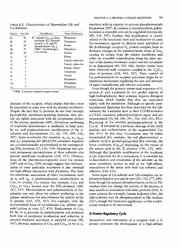
TABLE 6.2. Characteristics of Manzmalian GP- and G y-Subunits Subunit Size (kd) Gpy Effectors Tissue Distribution
P I 36 K' channel (IKACh); phos- Ubiquitous 35 pholipase A2; Adenylyl Ubiquitous
P2
P 3 36 phospholipase CP1-3; Retina P4 37 GRK * (P-adrenergic); Ubiquitous Ps 39 c-Src kinase Neural Y1 8 Retina Y2 6 Neural, endocrine Y3 7 Neural, endocrine Y4 7 Kidney, retina?
cyclases I, 11, and IV;
Y.5 7 Y6 7
Ubiquitous Neural
Y? 7 Ubiquitous YlO 7 Ubiquitous Yl 1 7 Ubiquitous
* GRK, G protein<oupled receptor kinase.
domain) of the receptor, which implies that they must be associated in some way with the plasma membrane. Despite the fact that G proteins do not bear strongly hydrophobic membrane-spanning domains, they usu- ally are tightly associated with the cytoplasmic.surface of the plasma membrane (440). Membrane anchoring of G-protein heterotrimers is mediated, in large part, by co- and posttranslational modifications of the a- subunits and &complexes (51, 62, 191, 209, 256, 507). Some a-subunits (for example, at, ai, ao, agust, and az) possessing the MGXXXS consensus sequence are co-translationally myristoylated at the correspond- ing NH2-terminus (27, 156, 518). Mutations that pre- vent permanent myristoylation of these subunits also prevent membrane localization (229, 333). Observa- tions of the pheromone-responsive yeast G a protein (447) and of Ga, (289) strongly suggest that myristoy- lation of some particular a-subunits is also essential for high-affinity interaction with By-dimers. The basis for membrane association of other Ga-subunits, such as a, and a,, is posttranslational palmitoylation at Cys, or another Cys residue (for example, Cys, and Cys,, in Ga,) located near the NH,-terminus (288, 367, 507). Myristoylation and palmitoylation of Ga- subunits not only allow for subunit anchoring to the plasma membrane but also influence the activity of the G protein (145, 470, 507). For example, only the myristoylated form of recombinant Gai inhibits ade- nylyl cyclase in vitro (27, 470). Replacement of Cys, by Ser in a, prevents its palmitoylation and produces both loss of membrane localization and reduction in receptor-mediated activation of adenylyl cyclase (90, 507); likewise, mutation of Cys, and Cys,, to Ser in a,
interferes with its capacity to activate phosphoinositide breakdown (507). In contrast to myristoylation, palmi- toylation is reversible and can be regulated dynamically (89, 334, 507). Further, this modification is closely related to the functional state and localization of some G a monomers; agonist or cholera toxin activation of the P-adrenergic receptor-G, protein complex leads to dramatic changes in the palmitoylation status of Gas, causing its release from the plasma membrane and either its reversible redistribution along the inner sur- face of the plasma membrane and/or into the cytoplasm or its degradation (89, 505, 506). Similar results have been observed with receptors coupled to the G,/G,, class of proteins (218, 443, 507). Thus, control of G a palmitoylation by receptor activation might be an additional mechanism regulating the rate and intensity of signal transduction and effector activation.
Even though the primary amino-acid sequences of G protein P- and y-subunits do not predict regions of high hydrophobicity, their isolation requires detergent extraction, which implies that they are associated tightly with the membrane. Although no specific post- translational lipidation has been described for the GP- subunits, the y-subunits have at their COOH-terminus a CAAX consensus polyisoprenylation signal and are isoprenylated (51, 64, 141, 191, 332, 410, 411, 431). Processing of Gy involves polyisoprenylation at the Cys at -4 followed by a protease cleavage of the AAX residues and carboxylation of the isoprenylated Cys (64, 411). In this way, Gy-subunits may be either farnesylated [for example, the rod photoreceptor y- subunit ( yl)] or geranyl-geranylated [for example, the brain y-subunits (Gy2J] depending on the nature of the amino acid in the X position (141, 191, 440). Although this lipophilic modification in the y-subunit is not important for /3-y association, it is essential for co-localization and orientation of the subunits on the inner membrane surface as well as for high-affinity interactions of the dimer with both G a subunits and effectors (210, 431).
Some types of Ga-subunit and Gpy-complex can be phosphorylated in vivo and in vitro (81,120,177,299). Even though Ga-subunit phosphorylation on Ser or Thr residues does not change the activity of the protein, it may modify its association with other proteins (254). In some systems (for example, human leukemia cells), the Gpy-subunits may be phosphorylated on His residues (517), though the functional significance of this modifi- cation remains to be ascertained.
G Protein-Regulatory Cycle
Association and interaction of a receptor with a G protein promotes the development of a high-affinity
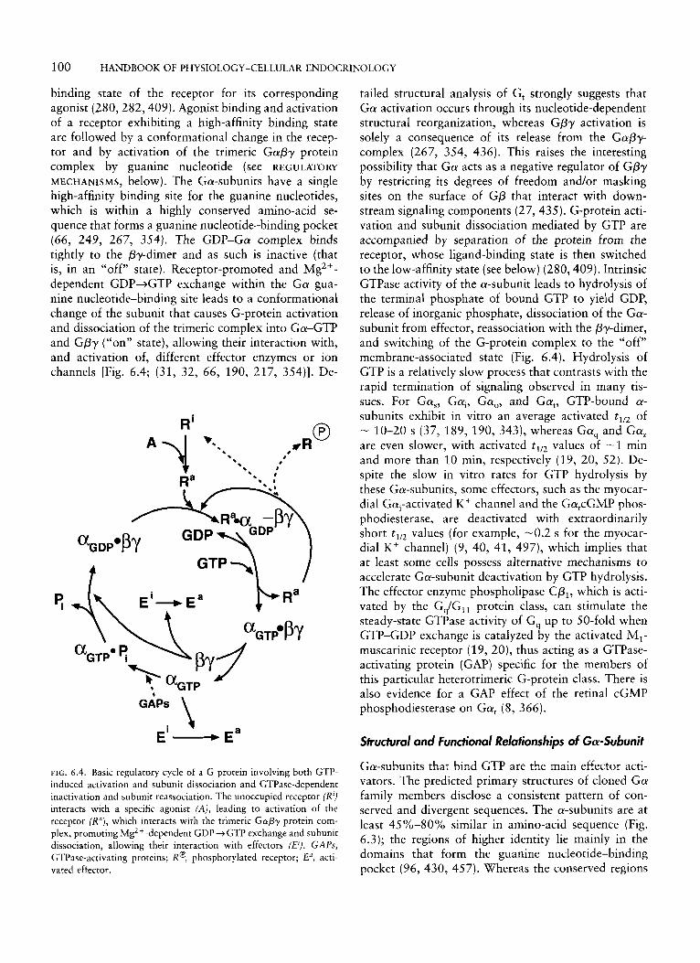
100 HANDBOOK OF PHYSIOLOGY-CELLULAR ENDOCRINOLOGY
binding state of the receptor for its corresponding agonist (280,282,409). Agonist binding and activation of a receptor exhibiting a high-affinity binding state are followed by a conformational change in the recep- tor and by activation of the trimeric GaPy protein complex by guanine nucleotide (see REGULATORY MECHANISMS, below). The Ga-subunits have a single high-affinity binding site for the guanine nucleotides, which is within a highly conserved amino-acid se- quence that forms a guanine nucleotide-binding pocket (66, 249, 267, 354). The GDP-Ga complex binds tightly to the py-dimer and as such is inactive (that is, in an “off” state). Receptor-promoted and Mg2+- dependent GDP+GTP exchange within the G a gua- nine nucleotide-binding site leads to a conformational change of the subunit that causes G-protein activation and dissociation of the trimeric complex into Ga-GTP and GPy (“on” state), allowing their interaction with, and activation of, different effector enzymes or ion channels [Fig. 6.4; (31, 32, 66, 190, 217, 354)J. De-
GAPs \
FIG. 6.4. Basic regulatory cycle of a G protein involving both GTP- induced activation and subunit dissociation and GTPase-dependent inactivation and subunit reassociation. The unoccupied receptor (R‘) interacts with a specific agonist (A), leading to activation of the receptor (Re), which interacts with the trimeric GmPy protein com- plex, promoting MgZ + -dependent GDP 3 GTP exchange and subunit dissociation, allowing their interaction with effectors (El). GAPs, GTPase-activating proteins; Re, phosphorylated receptor; E“, acti- vated effector.
tailed structural analysis of G, strongly suggests that G a activation occurs through its nucleotide-dependent structural reorganization, whereas Gpy activation is solely a consequence of its release from the GaPy- complex (267, 354, 436). This raises the interesting possibility that G a acts as a negative regulator of GPy by restricting its degrees of freedom and/or masking sites on the surface of GP that interact with down- stream signaling components (27,435). G-protein acti- vation and subunit dissociation mediated by GTP are accompanied by separation of the protein from the receptor, whose ligand-binding state is then switched to the low-affinity state (see below) (280,409). Intrinsic GTPase activity of the a-subunit leads to hydrolysis of the terminal phosphate of bound GTP to yield GDP, release of inorganic phosphate, dissociation of the Ga- subunit from effector, reassociation with the py-dimer, and switching of the G-protein complex to the “off” membrane-associated state (Fig. 6.4). Hydrolysis of GTP is a relatively slow process that contrasts with the rapid termination of signaling observed in many tis- sues. For Gas, G q , Ga,, and Gat, GTP-bound a- subunits exhibit in vitro an average activated tlI2 of - 10-20 s (37, 189, 190, 343), whereas Ga, and Gaz are even slower, with activated t,, values of -1 min and more than 10 min, respectively (19, 20, 52). De- spite the slow in vitro rates for GTP hydrolysis by these Ga-subunits, some effectors, such as the myocar- dial Gal-activated K+ channel and the GatcGMP phos- phodiesterase, are deactivated with extraordinarily short tIl2 values (for example, -0.2 s for the myocar- dial K+ channel) (9, 40, 41, 497), which implies that a t least some cells possess alternative mechanisms to accelerate Ga-subunit deactivation by GTP hydrolysis. The effector enzyme phospholipase Cp,, which is acti- vated by the Gq/G,, protein class, can stimulate the steady-state GTPase activity of G, up to 50-fold when GTP-GDP exchange is catalyzed by the activated MI- muscarinic receptor (19, 20), thus acting as a GTPase- activating protein (GAP) specific for the members of this particular heterotrimeric G-protein class. There is also evidence for a GAP effect of the retinal cGMP phosphodiesterase on Gat (8, 366).
Structural and Functional Relationships of Ga-Subunit
Ga-subunits that bind GTP are the main effector acti- vators. The predicted primary structures of cloned G a family members disclose a consistent pattern of con- served and divergent sequences. The a-subunits are at least 45%-80% similar in amino-acid sequence (Fig. 6.3); the regions of higher identity lie mainly in the domains that form the guanine nucleotide-binding pocket (96, 430, 457). Whereas the conserved regions
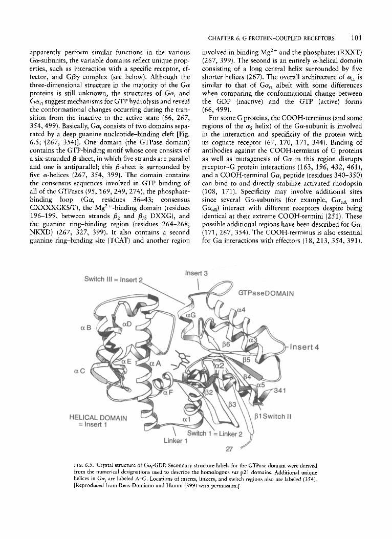
CHAPTER 6: G PROTEIN-COUPLED RECEPTORS 101
apparently perform similar functions in the various Ga-subunits, the variable domains reflect unique prop- erties, such as interaction with a specific receptor, ef- fector, and GPy complex (see below). Although the three-dimensional structure in the majority of the Ga proteins is still unknown, the structures of Gat and Gail suggest mechanisms for GTP hydrolysis and reveal the conformational changes occurring during the tran- sition from the inactive to the active state (66, 267, 354, 499). Basically, Gat consists of two domains sepa- rated by a deep guanine nucleotide-binding cleft [Fig. 6.5; (267, 354)]. One domain (the GTPase domain) contains the GTP-binding motif whose core consists of a six-stranded P-sheet, in which five strands are parallel and one is antiparallel; this p-sheet is surrounded by five a-helices (267, 354, 399). The domain contains the consensus sequences involved in GTP binding of all of the GTPases (95, 169,249,274), the phosphate- binding loop (Gat residues 36-43; consensus GXXXXGKSm), the Mg2 + -binding domain (residues 196-199, between strands p2 and p3; DXXG), and the guanine ring-binding region (residues 264-268; NKXD) (267, 327, 399). It also contains a second guanine ring-binding site (TCAT) and another region
involved in binding Mg2+ and the phosphates (RXXT) (267, 399). The second is an entirely a-helical domain consisting of a long central helix surrounded by five shorter helices (267). The overall architecture of ail is similar to that of Gat, albeit with some differences when comparing the conformational change between the GDP (inactive) and the GTP (active) forms (66, 499).
For some G proteins, the COOH-terminus (and some regions of the a5 helix) of the Ga-subunit is involved in the interaction and specificity of the protein with its cognate receptor (67, 170, 171, 344). Binding of antibodies against the COOH-terminus of G proteins as well as mutagenesis of Ga in this region disrupts receptor-G protein interactions (163, 196, 432, 461), and a COOH-terminal Gat peptide (residues 340-350) can bind to and directly stabilize activated rhodopsin (108, 171). Specificity may involve additional sites since several Ga-subunits (for example, GaOA and Ga,,) interact with different receptors despite being identical at their extreme COOH-termini (251). These possible additional regions have been described for Gat (171, 267, 354). The COOH-terminus is also essential for Ga interactions with effectors (18, 213, 354, 391).
Insert 3
isert 4
FIG. 6.5. Crystal structure of Gat-GDP. Secondary structure labels for the GTPase domain were derived from the numerical designations used to describe the homologous ras p21 domains. Additional unique helices in Gat are labeled A-G. Locations of inserts, linkers, and switch regions also are labeled (354). [Reproduced from Rens-Domiano and Hamm (399) with permission.]
102 HANDBOOK OF PHYSIOLOGY-CELLULAR ENDOCRINOLOGY
Construction of Gai/Gas chimeras and mutational studies have demonstrated that the adenylyl cyclase- activating region is contained in the COOH-terminus of Gas, corresponding to residues 235-356 (213, 362). For Gat, studies employing synthetic peptides have indicated that residues 293-314, which are adjacent to the receptor-activation domain, are important for Gat- regulated cGMP-phosphodiesterase activation (391 ); another region, corresponding to the phosphate- binding loop, undergoes a significant conformational change between the active (GTP-bound) and inactive (GDP-bound) crystal structures and, thus, also may be important for effector activation (267, 354). The crys- tal structure of Gai2 suggests the existence of an addi- tional region located in its a-helical domain which is also important for its interaction with the effector (66, 499). Finally, the NH,-terminus of the Ga-subunit, particularly its first 60 amino-acid residues, appears to be one of the sites of interaction with the Gpy-dimer (67, 93, 143, 399). Apparently, the Ga-subunit is able to interact with both the p- and the y-subunits (267). Monoclonal antibody 4A directed against the NH,- terminus of Gat causes dissociation from the py-dimer (309, 310); likewise, limited proteolysis and expression of NH,-terminally truncated Ga-subunits indicate that the myristoylated NH,-terminus and a region within the first 25 residues of the Ga-subunit are essential for interaction with GPy (93, 143, 399). Another possible site of Ga-subunit interaction with the Gay-dimer lies within the switch regions [that is, regions that undergo nucleotide-dependent conformational changes (Fig. 6.5) of the subunit], specifically in the switch I1 region (amino-acid residues 199-216 in Gail and 195-215 in Gat) (267, 499). The Gpy-dimer may bind to a hydrophobic pocket present in Ga-GDP, but it may be closed by the GTP-dependent conformational change (267, 399); this pocket is made up of the switch I1 region and the P-sheet hydrophobic core (267). A Cys in this switch region (Cys,,, in Ga,, cognate to Cys,,, in Gat) can be chemically cross-linked to Py (475), and the Gly,,,-to-Ala mutation in Gas that impairs the GTP-induced conformational changes in the switch I1 region also prevents dissociation of the Ga-subunit from the py-dimer (279, 320).
Certain subclasses of the Ga-subunit are adenosine diphosphate (ADP)-ribosylated by bacterial toxins from Bordetella pertusis (pertussis toxin or PTX) and Vibrio cholerae (cholera toxin or CTX) (2, 53, 488, 512). Cholera toxin catalyzes ADP-ribosylation of an Arg residue present within the GTP-binding domain of Gas and GaOlf in a ligand-independent fashion and of Gat and Gai in a ligand-dependent manner (2, 53, 488). This CTX-induced modification of the Ga- subunit considerably decreases the intrinsic GTPase
activity, causing constitutive activation of the protein. Mutation of the same Arg residue also inhibits GTPase activity, leading to a comparable constitutive activation (139). Gai, Gat, Gagust, and Ga, can be ADP- ribosylated by PTX on the fourth Cys residue from the COOH-terminus (335, 512); this modification, which is enhanced by the presence of the Gapy-dimer (that is, the presence of the G-protein heterotrimeric state), leads to uncoupling of the G protein from the receptor without modifying other functions, such as guanine nucleotide exchange or GTPase activity (67, 388).
GPy Structure and Function
Gpy-subunits are bound tightly to each other through noncovalent hydrophobic interactions (62, 209, 435); the complex is resistant to tryptic cleavage and can be dissociated only by treatment with denaturing agents. Separate expression of these subunits leads to unstable GP-subunits and unfolded Gy-subunits (191). The GP- subunits display 58%-90% identity, whereas the Gy- subunits are more diverse (430, 503). This probably accounts for the high degree of specificity for subunit association; GP, can associate with Gy, or Gy,, whereas GP2 can associate with Gy, but not with Gy,, and GP3 does not associate with either Gy, or Gy, (384, 416). Primary structures of the cloned GP- subunits as well as the crystal structure of the hetero- trimeric Gai& y, protein and the transducin py-dimer indicate the existence of two distinct domains. The relatively conserved NH,-terminal domain forms an amphipathic a-helical coiled coil that lies closely paral- lel to, and interacts with, the NH,-terminal helix of Gy (300); the existence of this proximity is supported by the fact that Cys residues located in both subunits (Cys,, of GP, and Cys,,-,, of Gyl) can be chemically cross-linked and Glu,, of G p is necessary for Gpy dimerization (43, 149, 435, 499). The second domain of G p consists of seven 40 to 43-residue repetitive segments that contain a characteristic tryptophan- aspartic acid pair, termed WD40, which may be found in a variety of apparently functionally unrelated pro- teins engaged in a variety of functions, including signal transduction, cell division, transcription, cytoskeletal assembly, and vesicle function (62,430, 435). The WD repeat regions in GP may be involved in the regulation of selectivity of the P-subunit to dimerize with a partic- ular y-subunit as well as in the capacity of the p- subunits to adopt multiple conformations, which in turn could greatly expand the number of protein- protein interactions (for example, with the P-adrenergic receptor kinase; see REGULATORY MECHANISMS, below) and signaling pathways (62, 345, 501). However, Gy- subunits are largely a-helical, and their selectivity for
CHAPTER 6: G PROTEIN-COUPLED RECEPTORS 103
The activity of the other adenylyl cyclase types is not altered by the Gpy-complex (462). The Gpy-dimer also activates isoforms 1-3 of phospholipase Cp (28, 36,47,48, 161,236,290); the stimulation is, however, subtype-specific, with a rank order of effect of phos- pholipase Cp3 2 p2 > > pl, which is different from that of Gaq,ll (27, 223, 368, 434, 528). GPy does not modify the activity of phospholipase Cp, (278). An- other important role of the Gpy-complex is regulation of the muscarine-gated K+ channels; after years of controversy over the role of the Ga-subunit vs. the Gpy-dimer in cardiac muscarinic K+ channel activa- tion, the bulk of evidence strongly indicates that the Gpy-dimer activates this particular channel more effec- tively than Ga, (295, 296, 516). Gpy-subunits also activate phospholipase A,, the enzyme responsible for arachidonic acid formation (22 1, 245); although ara- chidonic acid and its metabolites have been implicated in the activation of muscarine-dependent cardiac K+ channels (264), their effect is weaker than that elicited by the Gpy-complex (62). In contrast to the members of the Ga, family, the dimer does not influence signifi- cantly the activity of the cardiac ATP-sensitive K+ channel (215). The Gpy-dimer produces a 10-fold in- crease in the agonist-dependent phosphorylation of purified P,-adrenergic receptor (207, 379); this in- crease, which leads to receptor desensitization, occurs through a mechanism that involves interaction of the dimer with a specific region of the pleckstrin homology domain of the P-adrenergic receptor kinase (208, 382) and translocation of the kinase from the cytosol to the plasma membrane by the prenylated Gpy-subunit, allowing the enzyme to recognize and interact with those intracellular elements of the receptor susceptible to modification by phosphorylation (see Receptor De- sensitization, below). In this vein, it is interesting that rhodopsin kinase (the analog of the P-adrenergic recep- tor kinase in the retina) is farnesylated at its COOH- terminus and does not require GPy to phosphorylate the receptor (138).
In addition to the actions of the Ga-subunit and the Gpy-dimer described above, all classes of G protein may mediate long-term effects on gene expression and cell growth induced by mitogenic hormones that bind to GPCRs (10, 30, 96, 97, 117, 208, 381, 486). The best established pathway by which heterotrimeric G proteins transduce signals destined to phosphorylate nuclear transcription factors and thus promote the transcription of genes controlling cellular growth is the G protein-activatedlRas-regulated MAPK cascade, a series of protein-protein interactions and phosphoryla- tions (84, 352, 374, 381). Ras proteins are low- molecular-weight GTPases involved in mitogenesis and differentiation triggered by activated enzyme (tyrosine
the various @subunits is determined by a stretch of 14 amino acids located in the middle of the y-subunit (300, 435, 442); this region contains the Cys residue that is susceptible to cross-linking with the P-subunit (see above) (43, 149). In some systems, this subunit appears to be critical for determining the function of the Gpy-dimer. For example, the function of GP, y1 (localized in the retina) is quite different from that of GPly2 (found in the brain and other tissues); since the GP-subunits are the same, the functional differences between the two dimers are probably due to the differ- ent y-subunits (62). On the contrary, in other systems, the GP-subunits appear to determine effector selectivity (519, 536); this effect of GP has been demonstrated by in vitro studies, showing that while both Gp, and Gp, were able to activate phospholipase-C/3, in a y2- dependent manner, GP,, unlike pl, did not stimulate the mitogen-activated protein kinase (MAPK) or the cJun NH,-terminal kinase (JNK) pathways (536).
The association of the Gpy-dimer with G a is the best defined protein-protein interaction of the dimer. The Gpy-dimer is associated with the a-subunit through direct interactions between the prenylated form of the Gy-subunit and Ga(387, 532); the GP- and Ga-subunits are in close proximity, but apparently they do not interact independently of the Gy-subunit (267, 435, 532). The association of Gpy with G a increases the affinity of the latter subunit for GDP by about 100-fold, stabilizing the inactive state of the protein ( 190). However, the Gpy-complex associates with its cognate receptor through the COOH-terminus of the y-subunit, which becomes physically available for such interaction during the course of receptor- Gapy-complex coupling (62, 247, 248, 377). Thus, it seems that the Gy-subunit plays a key role in conferring the specificity of the G-protein interaction with recep- tors. The presence of the Gpy-dimer is a prerequisite for effective interaction of the G, protein with rhodop- sin and, hence, for receptor-catalyzed nucleotide ex- change (62, 123, 248, 428).
The Gpy-dimer is required for efficient association of the Ga-subunit with the plasma membrane and for an effective interaction of the Ga-subunit with the receptor (see above) (197, 247, 248, 377). In addition to this well-known effect, the Gpy-complex regulates the activity of many effectors (62, 161, 209, 290, 296, 467, 529, 536). It is involved in adenylyl cyclase types I1 and IV activation and type I inhibition (27, 119, 414, 467, 471). The extent of activation of these iso- forms may vary depending on the specific py-dimer involved; the type I1 isoform is activated effectively by GP1 y2, GP2y2, and Gp2~3, whereas the activation induced by transducin GP,y, is about one-tenth as effective as that mediated by the other dimers (210).
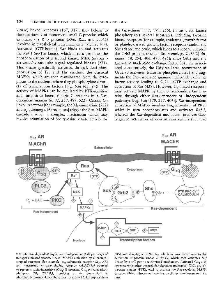
104 HANDBOOK OF PHYSIOLOGY-CELLULAR ENDOCRINOLOGY
kinase)-linked receptors (167, 317); they belong to the superfamily of monomeric small G proteins which embraces the Rho proteins (Rho, Rac, and cdc42) involved in cytoskeletal rearrangements (31, 32, 168). Activated (GTP-bound) Ras binds to and activates the Raf-I SerKhr kinase, which in turn promotes the phosphorylation of a second kinase, MEK (mitogen- activated/extracellular signal-regulated kinase) (271 ). This kinase specifically activates, through dual phos- phorylation of Tyr and Thr residues, the classical MAPKs, which are then translocated from the cyto- plasm to the nucleus, where they phosphorylate a vari- ety of transcription factors [Fig. 6.6; (65, 84)]. The activity of MAPKs can be regulated by PTX-sensitive and -insensitive heterotrimeric G proteins in a Ras- dependent manner (6, 92, 269, 487, 522). Certain Gi- linked receptors [for example, the M,-muscarinic (522) and qadrenergic (6) receptors] trigger the Ras-MAPK cascade through a complex mechanism which may involve stimulation of Src tyrosine kinase activity by
a l B AR M,AChR
the Gpy-dimer (117, 179, 255). In turn, Src kinase phosphorylates several substrates, including tyrosine kinase receptors (for example, epidermal growth factor or platelet-derived growth factor receptors) and/or the Shc adapter molecule, which binds to a second adapter, the Grb2 protein, through Src-homology 2 (SH2) do- mains (58, 254, 406, 479, 485); since Grb2 and the guanosine nucleotide exchange factor Sosl are associ- ated constitutively, the Gpy-mediated recruitment of Grb2 to activated (tyrosine-phosphorylated) Shc aug- ments the Shc-associated guanine nucleotide exchange factor activity, leading to GDP+GTP exchange and activation of Ras (429). However, G,-linked receptors may activate MAPK by their corresponding Ga pro- teins through either Ras-dependent or -independent pathways [Fig. 6.6; (179, 257, 406)]. Ras-independent activation of MAPKs involves Gaq activation of PKC, which in turn phosphorylates and activates Raf-I, whereas the Ras-dependent mechanism involves Gaq- triggered activation of downstream signals that lead
Extracellular
Ras-independent
Transcription factors . FIG. 6.6. Ras-dependent (right) and -independent (left) pathways of mitogen-activated protein kinase (MAPK) activation by G protein- coupled receptors [for example, a,,-adrenergic receptor (alB AR) and rnuscarinic MI-acetylcholine receptor JM,AChR)] coupled to pertussis toxin-insensitive (GaJ G proteins. Gaq activates phos- pholipase Cp, (PLCP,), resulting in the conversion of phosphatidylinositol-4,s-biphosphate to inositol-1,4,S-triphosphate
(IP,) and diacylglycerol (DAG), which in turn contributes to the activation of protein kinase C (PKC), which then activates Raf kinase by a still poorly understood mechanism. Activated Gaq also interacts with other intracellular signaling molecules [PKC, protein tyrosine kinases (PTK), etc.] to activate the Ras-regulated MAPK cascade. M E K , mitogen-activatecUextracelIular signal-regulated ki- nase.
CHAPTER 6: G PROTEIN-COUPLED RECEPTORS 105
to formation of the Shc-Grb2-Sos signaling complex [probably through a PKC- and/or a Ca2+-activated, PYK2-mediated Shc phosphorylation mechanism (284)] and the subsequent activation of Ras. Other studies suggest that MAPK activation by G, proteins may be modulated further by the contribution of their Gpy-subunits (77, 385). Another effector pathway whereby heterotrimeric G proteins may transduce sig- nals to the nucleus involves activation of JNK, a kinase related to MAPK but differentially regulated (70, 85, 96). Activation of JNK proceeds through a cascade of protein-protein interactions, triggered by the low- molecular-weight cdc42 and Rac G proteins, that in- volve the activation of several kinases, including the p21-activated kinase (PAK), MEK kinase (MEKK-I), and JNK kinase (JNKK) (71, 94, 271, 324); JNK, like MAPK, phosphorylates and increases the activity of several transcription factors (162, 324). Activation of Ml-muscarinic receptors as well as expression of con- stitutively active Gaq, G q 2 , or Gal, result in increased JNK activity (70, 96, 491). Finally, although cAMP is not mitogenic in most cell types, G, activation may induce growth responses in some in vitro and in vivo systems (117, 140, 270, 302, 484). Constitutively ac- tive mutants of the G,-linked TSH and growth hor- mone receptors have been isolated from thyroid and pituitary tumors (270,302). In other systems, however, G, appears to be involved in inhibition of nuclear transduction signals (69, 76, 423). In isoproterenol- stimulated COS-7 cells, G,-coupled receptors may me- diate simultaneous but opposing effects (76), with the Gpy-subunits inducing MAPK activation and the cAMP generated through Ga-stimulated adenylyl cy- clase inhibiting such effects. Thus, it appears that the intensity of the response in these P-adrenergic stimu- lated cells is determined by the balance between the two differing signals.
REGULATORY MECHANISMS
From this discussion, it is clear that the function of the GPCRs involves the concerted action of several control mechanisms operating at different levels, including the receptor molecule, the G protein, and the effector lev- els. However, our current understanding of the exact mechanisms that allow for a highly specific and equili- brated function of the receptor-G protein-effector sys- tem is still limited and further complicated by the existence of several factors and potential down- and upstream pathways or loops that may influence recep- tor availability and rates of activation and deactivation, the specificity of receptor-G protein interaction and signal transduction, and the intensity of G protein-
mediated activation of a given effector (394). Some of these mechanisms are discussed briefly in this section.
Mechanisms hut Regulate Receptor Function
Several feedback mechanisms that control the func- tional status of GPCRs have been explored extensively. Among these, three are worthy of particular consider- ation; the first mechanism involves changes in the dy- namic state of the receptor triggered by conformational changes in the receptor molecule and its interaction with its coupled G proteins, whereas the other two are involved with desensitization.
Receptor Conformation and Signaling. The GPCRs ex- ist in several dynamic states, as disclosed by marked shifts in agonist affinity (125, 280, 282, 409). Recep- tors are assumed to exist in an equilibrium between an inactive (R) and an active (R’) conformation. Binding of agonists exhibiting a preferentially higher affinity for R‘ promotes an isomerization step that stabilizes the receptor in a “relaxed” state and shifts the equilib- rium toward the R’ conformation, promoting its inter- action with G proteins and allowing the formation of a highly efficient ternary complex (agonist-R*-G protein) that stabilizes the R“ state and triggers G- protein activation (88). The requirement of such an isomerization step in the receptor molecule to switch into the R“ conformation was unmasked by the partic- ular behavior exhibited by a series of mutant adrenergic receptors, disclosing that certain intracellular regions of the receptor are critical for maintaining the receptor in a relatively constrained, “unrelaxed” state in the absence of agonist (282, 409). Mutations in specific amino-acid residues in the COOH-terminus of the third intracellular loop of the alB- and a,,-adrenergic recep- tors (coupled to the G,-phospholipase C p and the Gi- adenylyl cyclase pathways, respectively), as well as in the same loop of its relative, the G,-coupled p2.- adrenoreceptor, resulted, in all instances, in elevated basal, agonist-independent signaling activity, as well as in a supersensitivity to agonists [Fig. 6.1C; (250, 282, 398, 409)]. Thus, the mutations introduced allowed for conformational changes that led to a high level of constitutive receptor activation and interaction with the G proteins in the unliganded state, recreating the R“ state promoted in intact wild receptors only by agonist binding (152). Although the exact nature of the conformational changes that lead to the R‘ state are still unknown, analysis of several spontaneously occurring gain-of-function mutations that induce con- stitutive receptor hyperactivity suggests that multiple regions of the receptor molecule, including the extracel- Mar, intracellular, and transmembrane domains, are
106 HANDBOOK OF PHYSIOLOGY-CELLULAR ENDOCRINOLOGY
involved in the molecular mechanisms that constrain the G-protein coupling of the receptor, a constraint normally relieved upon agonist occupancy [Fig. 6.1 A, C; (250, 263, 281, 370, 390, 401, 409, 427, 492)]. Several physiological implications arise from this mech- anism of receptor activation. The equilibrium between R and R* implies that at any time a certain proportion of receptors resides in a constitutively active state even in the absence of an activating ligand. This, in fact, occurs, and some endogenous GPCRs exhibit demon- strable activity even in the absence of agonist (72,409). Thus, different receptor isoforms that bind the same ligand and activate the same effector may vary in their ability to spontaneously isomerize into the active state and, therefore, in their affinity for endogenous agonists. In this setting, it is expected that agonists acting through receptor variants with an elevated constitutive isomerization rate will have a higher potency, whereas those acting through variants with a low isomerization rate will provoke a more graded range of responses (409). Such differences in intrinsic capacity to isomerize could be accounted for by alterations of one or more critically placed amino-acid residues and could reason- ably explain the existence of distinct receptor subtypes for a single ligand (282). In addition, the existence of naturally occurring, constitutively active GPCRs may allow for certain antagonistic drugs and endogenous ligands to elicit negative activity or inverse agonism (that is, lowering the basal activity); as opposed to agonists, which are presumed to have a high affinity for R“, inverse agonists exhibit a high affinity for R and, therefore, decrease the R”/R ratio (323).
Receptor Desensitization. Continuous or prolonged stimulation of a cell generally results in progressively at- tenuated responses to subsequent stimulation by the same agonist. This decrease in cellular sensitivity to fur- ther stimulation, or desensitization, may be considered one of the most important mechanisms to protect the cell from excessive stimulation, particularly under con- ditions of high agonist exposure. Phosphorylation- mediated deactivation of rhodopsin exemplifies the par- amount importance of this protective mechanism; in this system, signaling is inactivated rapidly (high milli- second-second time scale) following exposure to light, thereby preventing a brief flash of light from being per- ceived as continuous illumination (35, 476, 520). Transgenic mice bearing a mutation in rhodopsin that prevents short-term desensitization exhibit a marked prolongation of light-evoked electrical responses from single rods, leading to retinal degeneration (55) . Thus, for many cells bearing GPCRs the functional status of such receptors is determined by the phosphorylation state (33,175,176,192,351,386,400,519).
Early receptor desensitization occurs rapidly, in sec- onds to minutes of agonist exposure, and involves phosphorylation of intracellular domains of the stimu- lated receptors by second messenger-dependent acti- vated kinases (PKA or PKC) (138, 243) and/or by a special class of Sermhr-specific kinases called G pro- tein-coupled receptor kinases (GRK1-6) (382), with the subsequent uncoupling of the receptors from their respective G proteins and loss of downstream signaling events (1 3 8). Homologous short-term desensitization occurs very rapidly, with a half-life from milliseconds to a few seconds, and is observed when the cell’s response decreases after high receptor occupancy by a specific agonist (138, 176, 192, 386, 404, 415); heterologous desensitization (nonagonist-specific) is observed after several minutes of low amounts of ago- nist exposure and usually occurs when a cell’s response to various agonists acting through different receptors decreases after receptor activation by a particular ago- nist (63, 132, 138). Whereas homologous desensitiza- tion may be mediated through receptor phosphoryla- tion triggered by both second messenger-activated protein kinases and GRKs, heterologous desensitiza- tion generally involves phosphorylation by second mes- senger-dependent kinases (63, 138, 192, 193, 386).
Several GPCRs desensitize through phosphorylation (176, 192, 386, 400, 476). The molecular basis of agonist-induced phosphorylation has been studied par- ticularly in the biogenic amine receptors (for example, the p-adrenergic receptor) and rhodopsin (132, 176, 519, 520). In these receptors, GRK-mediated Ser/Thr phosphorylation and subsequent G-protein uncoupling involve several steps. In unstimulated cells, GRKs are located primarily in the cytoplasm; upon agonist- induced receptor activation, GRKs translocate to the plasma membrane, a process that involves receptor- GRK interactions as well as isoprenylation [for example, farnesylation of rhodopsin kinase (206, 207)], palmitoylation [as in GRK4 and GRK6 (446)], or interaction of the GRK with the membrane- anchored Gpy-complex and phosphatidylinositol-4,5- biphosphate through its COOH-terminal pleckstrin ho- mology domain [such as the p-adrenergic receptor ki- nase (382)] (208). Once the receptor is phosphorylated near the sites required for G-protein interaction [for example, in at least seven and eleven sites located within 10- and 59-amino-acid stretches of the COOH- terminal domains of rhodopsin and the p,-adrenergic receptor, respectively (33, 175, 520); Fig. 6.1C], the subsequent binding of a group of inhibitory proteins, the arrestins, amplifies the desensitization process initi- ated by phosphorylation and turns off the receptor by impeding its coupling to G proteins (297, 444). Al- though the available studies do not allow unambiguous
CHAPTER 6: G PROTEIN-COUPLED RECEPTORS 107
action of GAPS or RGS (regulators of G-protein signal- ing) proteins (104a, 109, 348, 504), decreased func- tional activity of the Ca2+ ion channel, and reduced efficiency of mobilization by IP, of Ca2+ from the intracellular pool (3 13).
clarification of the in vivo specificity of the several GRK and arrestin isoforms, it is possible that, with few exceptions, short-term desensitization of GPCRs is regulated by more than one GRK or arrestin isoform.
It has been shown that short-term desensitized recep- tors are resensitized by a complex process that involves arrestin-mediated receptor sequestration into endo- somes, where they become dephosphorylated by a par- ticular group of membrane-associated receptor Ser/Thr phosphatases (380, 494, 495). For the p,-adrenergic receptor and the NK, receptor, resensitization requires vesicular acidification and subsequent changes in recep- tor conformation to become accessible to the Ser/Thr phosphatases (148, 157, 261). Whether this tightly regulated resensitization mechanism is applicable to other members of the GPCR superfamily remains to be investigated.
A more profound deactivation process, long-term desensitization, is observed after hours or days of high levels of agonist exposure and involves a decrease in the net complement of receptors specific for a particular agonist, without detectable changes in receptor affinity (46, 68, 164, 176, 226, 305, 312, 426, 460). This type of cellular desensitization is subserved by several biochemically distinct mechanisms, including receptor down-regulation (that is, loss in total cellular content of functional receptors) and internalization (loss of surface receptors) (68, 176, 222, 305, 312). Although this type of desensitization does not require previous phosphorylation of the receptor, in some systems it amplifies the loss of functional receptors initiated by short-term deactivation (176,502). For certain GPCRs, the net loss in receptor number andlor function in- volves the occurrence of various receptor and postre- ceptor events, including internalization, sequestration, and degradation of the receptor (222, 261, 305, 319), as well as changes in the rate of receptor synthesis by alterations in receptor mRNA levels or function (164- 166). In others, down-regulation does not require im- mediate changes in protein synthesis and decreased receptor mRNA levels are observed only after pro- longed exposure to agonist (68). The overall process leads to a significant reduction in the number of func- tionally available receptors on the cell surface, where they could interact with agonist and activate signaling. Finally, in certain cells bearing GPCRs, loss of ability to respond to prolonged agonist stimulation also may be achieved by mechanisms related not to the receptor itself but to downstream and negative feedback mecha- nisms involving inactivation of Ga-subunits or loss of functional activity of effectors. An example of these postreceptor mechanisms is the GnRH-responsive cell, in which loss of responsiveness is due, on the one hand, to receptor loss (68, 222) and, on the other, to the
G Protein-Mediated Regulatory Mechanisms
As discussed above, G proteins are the molecules that transduce the signal triggered by receptor activation (see THE HETEROTRIMERIC G-PROTEIN FAMILY, above). In addition to the particular characteristics of the indi- vidual a- and &-subunits that allow for some degree of selectivity for interaction with each other, with the receptor, and with their corresponding effectors, there are other mechanisms that tightly regulate the selectiv- ity and intensity of the signal transduced by these molecules. Thus, in some systems, activation of particu- lar effectors by the Gpy-complex is conditioned by G a priming [for example, stimulation of adenylyl cyclase types 11 and IV (1 19, 467)], whereas in others it seems to be independent [for example, activation of phospho- lipase Cp,-, (62, 368, 434)]. However, whereas in certain systems the effects mediated by the Gpy-dimer are the opposite of those triggered by Ga [for example, activation of type I adenylyl cyclase by the latter and inhibition by the former (119, 467, 470, 472, 527)], in others stimulation of the same effector (for example, activation of the acetylcholine-regulated cardiac K+ channels) occurs as a result of the activation of conver- gent pathways triggered independently by both ele- ments (295, 296, 515, 516, 531). Activation of diver- gent G protein-dependent pathways leading to stimulation of different effectors also may occur, as exemplified by phospholipase C p activation by PTX- sensitive G proteins (Gi and Go) through their corres- ponding Gpy-complexes (28, 113, 236). Thus, the existence of convergent, divergent, and opposite path- ways for effector activation, as well as of differences in selectivity for protein-protein interactions [activa- tion and selectivity being accomplished by differences in affinity or efficacy of interaction of the G-protein subunits with the receptor, effector, and themselves (20, 74, 116,253,277, 384, 416,433, 534); compart- mentation of particular activators and effectors (49, 412); and tissue distribution and expression of the different classes and subclasses of each G protein and effector (142, 228, 237, 316, 393, 459)] among the G proteins, allows for the different, fine-tuning mecha- nisms through which the cells may regulate the speci- ficity of the signal conveyed by these effector-activating protein molecules.
Cells also limit their extent of response to external stimuli by decreasing the levels of specific G proteins
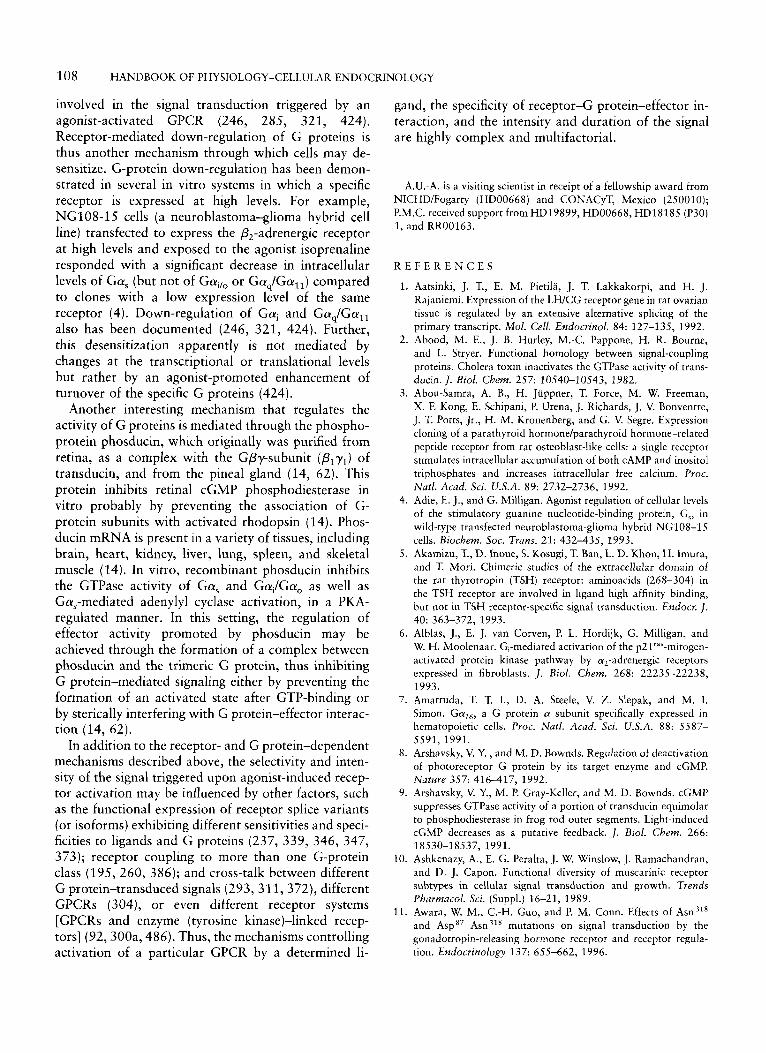
108 HANDBOOK OF PHYSIOLOGY-CELLULAR ENDOCRINOLOGY
involved in the signal transduction triggered by an agonist-activated GPCR (246, 285, 321, 424). Receptor-mediated down-regulation of G proteins is thus another mechanism through which cells may de- sensitize. G-protein down-regulation has been demon- strated in several in vitro systems in which a specific receptor is expressed at high levels. For example, NG108-15 cells (a neuroblastoma-glioma hybrid cell line) transfected to express the &-adrenergic receptor at high levels and exposed to the agonist isoprenaline responded with a significant decrease in intracellular levels of Gas (but not of Gado or GadGaI1) compared to clones with a low expression level of the same receptor (4). Down-regulation of Gai and Gaq/Gall also has been documented (246, 321, 424). Further, this desensitization apparently is not mediated by changes at the transcriptional or translational levels but rather by an agonist-promoted enhancement of turnover of the specific G proteins (424).
Another interesting mechanism that regulates the activity of G proteins is mediated through the phospho- protein phosducin, which originally was purified from retina, as a complex with the Gpy-subunit (Plyl) of transducin, and from the pineal gland (14, 62). This protein inhibits retinal cGMP phosphodiesterase in vitro probably by preventing the association of G- protein subunits with activated rhodopsin (14). Phos- ducin mRNA is present in a variety of tissues, including brain, heart, kidney, liver, lung, spleen, and skeletal muscle (14). In vitro, recombinant phosducin inhibits the GTPase activity of Gas and Gai/Gao as well as Gas-mediated adenylyl cyclase activation, in a PKA- regulated manner. In this setting, the regulation of effector activity promoted by phosducin may be achieved through the formation of a complex between phosducin and the trimeric G protein, thus inhibiting G protein-mediated signaling either by preventing the formation of an activated state after GTP-binding or by sterically interfering with G protein-effector interac- tion (14, 62).
In addition to the receptor- and G protein-dependent mechanisms described above, the selectivity and inten- sity of the signal triggered upon agonist-induced recep- tor activation may be influenced by other factors, such as the functional expression of receptor splice variants (or isoforms) exhibiting different sensitivities and speci- ficities to ligands and G proteins (237, 339, 346, 347, 373); receptor coupling to more than one G-protein class (195, 260, 386); and cross-talk between different G protein-transduced signals (293, 311, 372), different GPCRs (304), or even different receptor systems [GPCRs and enzyme (tyrosine kinase)-linked recep- tors] (92,30Oa, 486). Thus, the mechanisms controlling activation of a particular GPCR by a determined li-
gand, the specificity of receptor-G protein-effector in- teraction, and the intensity and duration of the signal are highly complex and multifactorial.
A.U.-A. is a visiting scientist in receipt of a fellowship award from NICHD/Fogarty (HD00668) and CONACyT, Mexico (250010); P.M.C. received support from HD19899, HD00668, HD18185 (P30) 1. and RR00163.
R E F E R E N C E S
1.
2.
3.
4.
5.
6.
7.
8.
9.
10.
11.
Aatsinki, J. T., E. M. Pietila, J. T. Lakkakorpi, and H. J. Rajaniemi. Expression of the LWCG receptor gene in rat ovarian tissue is regulated by an extensive alternative splicing of the primary transcript. Mol. Cell. Endocrinol. 84: 127-135, 1992. Abood, M. E., J. B. Hurley, M.-C. Pappone, H. R. Bourne, and L. Stryer. Functional homology between signal-coupling proteins. Cholera toxin inactivates the GTPase activity of trans- ducin. /. Biol. Cbem. 257: 10540-10543, 1982. Abou-Samra, A. B., H. Juppner, T. Force, M. W. Freeman, X. F. Kong, E. Schipani, P. Urena, J. Richards, J. V. Bonventre, J. T. Potts, Jr., H. M. Kronenberg, and G. V. Segre. Expression cloning of a parathyroid hormone/parathyroid hormone-related peptide receptor from rat osteoblast-like cells: a single receptor stimulates intracellular accumulation of both CAMP and inositol triphosphates and increases intracellular free calcium. Proc. Natl. Acad. Sci. U.S.A. 89: 2732-2736, 1992. Adie, E. J., and G. Milligan. Agonist regulation of cellular levels of the stimulatory guanine nucleotide-binding protein, G,, in wild-type transfected neuroblastoma-glioma hybrid NGI08-15 cells. Biochem. SOC. Trans. 21: 432-435, 1993. Akamizu, T., D. lnoue, S. Kosugi, T. Ban, L. D. Khon, H. Imura, and T. Mori. Chimeric studies of the extracellular domain of the rat thyrotropin (TSH) receptor: aminoacids (268-304) in the TSH receptor are involved in ligand high affinity binding, but not in TSH receptor-specific signal transduction. Endocr. 1.
Alblas, J., E. J. van Corven, P. L. Hordijk, G. Milligan, and W. H. Moolenaar. G;mediated activation of the p21'""-mitogen- activated protein kinase pathway by a,-adrenergic receptors expressed in fibroblasts. J . Biol. Chem. 268: 22235-22238, 1993. Amatruda, T. T. I., D. A. Steele, V. Z. Slepak, and M. I. Simon. Ga,6r a G protein (Y subunit specifically expressed in hematopoietic cells. Proc. Natl. Acad. Sci. U.S.A. 88: 5587- 5591, 1991. Arshavsky, V. Y. , and M. D. Bownds. Regulation of deactivation of photoreceptor G protein by its target enzyme and cGMP. Nature 357: 416417 , 1992. Arshavsky, V. Y., M. P. Gray-Keller, and M. D. Bownds. cGMP suppresses GTPase activity of a portion of transducin equimolar to phosphodiesterase in frog rod outer segments. Light-induced cGMP decreases as a putative feedback. 1. Biol. Chem. 266:
Ashkenazy, A., E. G. Peralta, J. W. Winslow, J. Ramachandran, and D. J. Capon. Functional diversity of muscarinic receptor subtypes in cellular signal transduction and growth. Trends Pharmacol. Sci. (Suppl.) 16-21, 1989. Awara, W. M., C.-H. Guo, and P. M. Conn. Effects of Asn318 and Asp" Asn318 mutations on signal transduction by the gonadotropin-releasing hormone receptor and receptor regula- tion. Endocrinology 137: 655-662, 1996.
40: 363-372, 1993.
18530-18537, 1991.
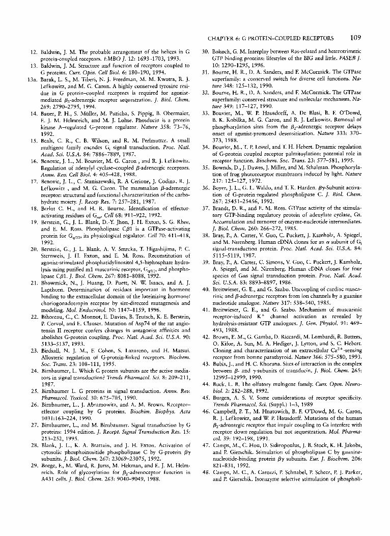
CHAPTER 6: G PROTEIN-COUPLED RECEPTORS 109
30. Bokoch, G. M. Interplay between Ras-related and heterotrimeric GTP binding proteins: lifestyles of the BIG and little. FASEB I.
31. Bourne, H. R., D. A. Sanders, and F. McCormick. The GTPase superfamily: a conserved switch for diverse cell functions. Na- ture 348: 125-132, 1990.
32. Bourne, H. R., D. A. Sanders, and F. McCormick. The GTPase superfamily: conserved structure and molecular mechanism. Na- ture 349: 117-127, 1990.
33. Bouvier, M., W. P. Hausdorff, A. De Blasi, B. F. O’Dowd, B. K. Kobilka, M. G. Caron, and R. J. Lefkowitz. Removal of phosphorylation sites from the P,-adrenergic receptor delays onset of agonist-promoted desensitization. Nature 333: 370- 373, 1988.
34. Bouvier, M., T. P. Loisel, and T. H. Hebert. Dynamic regulation of G-protein coupled receptor palmitoylation: potential role in receptor function. Biocbem. Soc. Trans. 23: 577-581, 1995.
35. Bownds, D., J. Dawes, J. Miller, and M. Sthalman. Phosphoryla- tion of frog photoreceptor membranes induced by light. Nature 237: 125-127, 1972.
36. Boyer, J. L., G. L. Waldo, and T. K. Harden. py-Subunit activa- tion of G-protein-regulated phospholipase C. J. Biol. Chem.
37. Brandt, D. R., and E. M. Ross. GTPase activity of the stimula- tory GTP-binding regulatory protein of adenylate cyclase, Gs. Accumulation and turnover of enzyme-nucleotide intermediates. J. Biol. Chem. 260: 266-272, 1985.
38. Bray, P., A. Carter, V. Guo, C. Puckett, J. Kamholz, A. Spiegel, and M. Nirenberg. Human cDNA clones for an a subunit of Gi signal-transduction protein. Proc. Natl. Acad. Sci. U.S.A. 84:
39. Bray, P., A. Carter, C. Simons, V. Guo, C. Puckett, J. Kamholz, A. Spiegel, and M. Nirenberg. Human cDNA clones for four species of Gas signal transduction protein. Proc. Natl. Acad. Sci. U.S.A. 83: 8893-8897, 1986.
40. Breitwieser, G. E., and G. Szabo. Uncoupling of cardiac musca- rinic and P-adrenergic receptors from ion channels by a guanine nucleotide analogue. Nature 317: 538-540, 1985.
41. Breitwieser, G. E., and G. Szabo. Mechanism of muscarinic receptor-induced K + channel activation as revealed by hydrolysis-resistant GTP analogues. J. Gen. Physiol. 91: 469- 493, 1988.
42. Brown, E. M., G. Gamba, D. Riccardi, M. Lombardi, R. Butters, 0. Kifor, A. Sun, M. A. Hediger, J. Lytton, and S. C. Hebert. Cloning and characterization of an extracellular Ca2 + -sensing receptor from bovine parathyroid. Nature 366: 575-580, 1993.
43. Bubis, J., and H. C. Khorana. Sites of interaction in the complex between P- and y-subunits of transducin. J. Biol. Chem. 265:
44. Buck, L. B. The olfatory multigene family. Curr. Op in . Neuro- biol. 2: 282-288, 1992.
45. Burgen, A. S. V. Some considerations of receptor specificity. Trends Pharmacol. Sci. (Suppl.) 1-3, 1989
46. Campbell, P. T., M. Hnatowich, B. F. O’Dowd, M. G. Caron, R. J. Lefkowitz, and W. P. Hausdorff. Mutations of the human &-adrenergic receptor that impair coupling to Gs interfere with receptor down-regulation but not sequestration. Mol. Pharma-
47. Camps, M., C. Hou, D. Sidiropoulus, J. B. Stock, K. H. Jakobs, and P. Gierschik. Stimulation of phospholipase C by guanine- nucleotide-binding protein Py subunits. Eur. ]. Biochem. 206:
48. Camps, M. C., A. Carozzi, P. Schnabel, P. Scheer, P. J. Parker, and P. Gierschik. Isoenzyrne selective stimulation of phospholi-
10: 1290-1295, 1996.
267: 25451-25456, 1992.
5115-5119, 1987.
12995-12999,1990.
C O ~ . 39: 192-198, 1991.
821-831, 1992.
12. Baldwin, J. M. The probable arrangement of the helices in G protein-coupled receptors. EMBO J. 12: 1693-1703, 1993.
13. Baldwin, J. M. Structure and function of receptors coupled to G proteins. Curr. Opin. Cell Biol. 6: 180-190, 1994.
13a. Barak, L. S., M. Tiberi, N. J. Freedman, M. M. Kwatra, R. J. Lefkowitz, and M. G. Caron. A highly conserved tyrosine resi- due in G protein-coupled receptors is required for agonist- mediated P,-adrenergic receptor sequestration. J. Biol. Chem.
14. Bauer, P. H., S . Miiller, M. Puzicha, S. Pippig, B. Obermaier, E. J. M. Helmreich, and M. J. Lohse. Phosducin is a protein kinase A-regulated G-protein regulator. Nature 358: 73-76, 1992.
15. Beak, C. R., C. B. Wilson, and R. M. Perlmutter. A small multigene family encodes G, signal transduction. Proc. Natl. Acad. Sci. U.S.A. 84: 7886-7889, 1987.
16. Benovic, J. L., M. Bouvier, M. G. Caron , and R. J. Lefkowitz. Regulation of adenylyl cyclase-coupled P-adrenergic receptors. Annu. Rev. Cell Biol. 4: 405428, 1988.
17. Benovic, J. L., C. Staniszewski, R. A Cerione, J. Codina, R. J. Lefkowitz , and M. G. Caron. The mammalian P-adrenergic receptor: structural and functional characterization of the carbo- hydrate moiety. J. Recep Res. 7: 257-281, 1987.
18. Berlot C. H., and H. R. Bourne. Identification of effector- activating residues of Gsa. Cell 68: 911-922, 1992.
19. Berstein, G., J. L. Blank, D.-Y. Jhon, J. H. Exton, S. G. Rhee, and E. M. Ross. Phospholipase CPl is a GTPase-activating protein for G,,l,, its physiological regulator. Cell 70: 411418, 1992.
20. Berstein, G., J. L. Blank, A. V. Smrcka, T. Higashijima, P. C. Sternweis, J. H. Exton, and E. M. Ross. Reconstitution of agonist-stimulated phosphatidylinositol4,S-biphosphate hydro- lysis using purified m l muscarinic receptor, G,,,,, and phospho- lipase CP1. 1. Bioi. Chem. 267: 8081-8088, 1992.
21. Bhowmick, N., J. Huang, D. Puett, N. W. Isaacs, and A. J. Lapthorn. Determination of residues important in hormone binding to the extracellular domain of the luteinizing hormone/ choriogonadotropin receptor by site-directed mutagenesis and modeling. Mol. Endocrinol. 10: 1147-1159, 1996.
22. Bihoreau, C., C. Monnot, E. Davies, B. Teutsch, K. E. Berstein, P. Corvol, and E. Clauser. Mutation of Asp74 of the rat angio- tensin I1 receptor confers changes in antagonist affinities and abolishes G-protein coupling. Proc. Natl. Acad. Sci. U.S.A. 90:
23. Birdsall, N. J. M., F. Cohen, S. Lazareno, and H. Matsui. Allosteric regulation of G-protein-linked receptors. Biochem. SOC. Trans. 23: 108-111, 1995.
24. Birnbaumer, L. Which G protein subunits are the active media- tors in signal transduction? Trends Pharmacol. Sci. 8 : 209-211, 1987.
25. Birnbaumer L. G proteins in signal transduction. Annu. Rev. Pharmacol. Toxicol. 30: 675-705, 1990.
26. Birnbaumer, L., J. Abramowitz, and A. M. Brown. Receptor- effector coupling by G proteins. Biochim. Biophys. Acta
27. Birnbaumer, L., and M. Birnbaumer. Signal transduction by G proteins: 1994 edition. J. Recept. Signal Transduction Res. 15:
28. Blank, J. L., K. A. Brattain, and J. H. Exton. Activation of cytosolic phosphoinositide phospholipase C by G-protein Py subunits. J. Biol. Chem. 267: 23069-23075, 1992.
29. Boege, F., M. Ward, R. Jurss, M. Hekman, and E. J. M. Helm- reich. Role of glycosylation for P,-adrenoceptor function in A431 cells. J . Biol. Chem. 263: 9040-9049, 1988.
269: 2790-2795, 1994.
5133-5137, 1993.
103 1: 163-224, 1990.
213-252, 1995.
11 0 HANDBOOK OF PHYSIOLOGY-CELLULAR ENDOCRINOLOGY
pase C-p2 by G protein py subunits. Nature 360: 684-686, 1992.
49. Carrasco, M. A., J. Sierralta, and P. DeMazancourt. Character- ization and subcellular distribution of G-proteins in highly puri- fied skeletal muscle fractions from rabbit and frog. Arch. Bio- chem. Biophys. 310: 76-81, 1994.
50. Cascieri, M. A., T. Ming Gong, and C. D. Strader. Molecular characterization of a common binding site for small molecules within the transmembrane domain of G-protein coupled recep- tors. J . Pharmacol. Toxicol. Methods 33: 179-185, 1995.
51. Casey, P. J. Lipid modifications of G proteins. Curr. Opin. Cell Biol. 6: 219-225, 1994.
52. Casey, P. J., H. K. W. Fong, M. I. Simon, and A. G. Gilman. Gz, a guanine nucleotide-binding protein with unique biochemical properties. J . Biol. Chem. 265: 2383-2390, 1990.
53. Cassel, D., and Z. Selinger. Mechanism of adenylate cyclase activation by cholera toxin. Inhibition of GTP hydrolysis a t the regulatory site. Proc. Natl. Acad. Sci. U.S.A. 74: 3307-3311, 1977.
54. Chattopadhyay, N., A. Mithal, and E. M. Brown. The calcium- sensing receptor: a window into the physiology and pathophysi- ology of mineral ion metabolism. Endocr. Rev. 17: 289-307, 1996.
55 . Chen, C. K., J. Inglese, R. J. Lefkowitz, and J. B. Hurley. Ca’+-dependent interaction of recoverin with rhodopsin. J. Biol. Chem. 270: 18060-18066, 1995.
56. Chen, J. Q., and R. Iyengar. Inhibition of cloned adenylyl cyclases by mutant-activated GI, and specific suppression of type-2 adenylyl cyclase inhibition by phorbol ester treatment. J. Biol. Chem. 268: 12253-12256, 1993.
57. Chen, R., K. A. Lewis, M. H . Perrin, and W. W. Vale. Expression cloning of a human corticotropin-releasing-factor receptor. Proc. Natl. Acad. Sci. U.S.A. 90: 8967-8971, 1993.
58. Chen, Y., D. Gral, A. E. Salcini, P. G. Pelicci, J. Pouyssegur, and E. Van Obberghen-Schilling. Shc adaptor proteins are key transducers of mitogenic signaling mediated by the G protein- coupled thrombin receptor. EMBO J. 15: 1037-1044, 1996.
59. Cheung, A. H., R. R. Huang, M. P. Graziano, and C. D. Strader. Specific activation of Gs by synthetic peptides corresponding to an intracellular loop of the P-adrenergic receptor. FEBS Lett.
60. Cheung, A. H., R. R. Huang, and C. D. Strader. Involvement of specific hydrophobic, but not hydrophilic amino acids in the third intracellular loop of the P-adrenergic receptor in the activation of G,. Mol. Pharmacol. 41: 1061-1065, 1992.
61. Chung, F.-Z., C.-D. Wang, P. C. Potter, J. C. Venter, and C. M. Fraser. Site-directed mutagenesis and continuous expres- sion of human P-adrenergic receptors. 1. Biol. Chem. 263: 4052- 4055, 1988.
62. Clapham, D. E., and E. J. Neer. New roles for G-protein py-dimers in transmembrane signalling. Nature 365: 403406, 1993
63. Clark, R. B., J. Friedman, R. A. Dixon, and C. D. Strader. Identification of a specific site required for rapid heterologous desensitization of the p-adrenergic receptor by CAMP-dependent protein kinase. Mol. Pharmacol. 36: 343-348, 1989.
64. Clarke, S. Protein isoprenylation and methylation at carboxy- terminal cysteine residues. Annu. Rev. Biochem. 61: 355-386, 1992,
65. Cobb, M. H., and E. J. Goldsmith. H o w MAP kinases are regulated. J . Biol. Chem. 270: 14843-14846, 1995.
66. Coleman, D. E., A. M. Berghuis, E. Lee, M. E. Linder, A. G. Gilman, and S. R. Sprang. Structures of active conformations of G,,, and the mechanism of GTP hydrolysis. Science 265:
279: 277-280, 1991.
1405-1412, 1994.
67. Conklin, B. R., and H. R. Bourne. Structural elements of Ga subunits that interact with GPy, receptors and effectors. Cell
68. Conn, P. M., D. C. Rogers, and S. G. Seay. Biphasic regulation of the gonadotropin-releasing hormone receptor by receptor microaggregation and intracelullar CaZ+ levels. Mol. Pharmacol.
69. Cook, S. J., and F. McCormick. Inhibition by CAMP of Ras- dependent activation of Raf. Science 262: 1069-1072, 1993.
70. Coso, 0. A,, M. Chiariello, G. Kalinec, L. M. Kyriakis, J. Woodgett, and J. S. Gutkind. Transforming G protein-coupled receptors potently activate JNK (SAPK). Evidence for a diver- gence from the tyrosine kinase signaling pathway. J. Biol. Chem.
71. Coso, 0. A., M. Chiariello, J. C. Yu, H. Teramoto, P. Crespo, N. Xu, T. Miki, and J. S. Gutkind. The small GTP binding proteins Racl and Cdc42 regulate the activity of the JNWSAPK signaling pathway. Cell 81: 1137-1146, 1995.
72. Costa, T., Y. Ogino, P. J. Munson, H. 0. Onaran, and D. Rodbard. Drug efficacy a t guanine nucleotide-binding regulatory protein-linked receptors: thermodynamic interpretation of nega- tive antagonism and of receptor activity in the absence of ligand. Mol. Pharmacol. 41: 549-560, 1992.
73. Cotecchia, S., S. Exum, M. G. Caron, and R. J. Lefkowitz. Regions of the a,-adrenergic receptor involved in coupling to phosphatidylinositol hydrolysis and enhanced sensitivity of bio- logical function. Proc. Natl. Acad. Sci. U.S.A. 87: 2896-2900, 1990.
74. Cotecchia, S., J. Ostrowski, M. A. Kjelsberg, M. G. Caron, and R. J. Lefkowitz. Discrete amino acid sequences of the a1- adrenergic receptor determine the selectivity of coupling to phos- phatidylinositol hydrolysis. J. Biol. Chem. 267: 1633-1 639, 1992.
75. Coughlin, S. R. Expanding horizons for receptors coupled to G proteins: diversity and disease. Curr. Opin. Cell Biol. 6: 191- 197, 1994
76. Crespo, P., T. G. Cachero, N. Xu, and J. S. Gutkind. Dual effect of P-adrenergic receptors on mitogen-activated protein kinase. Evidence for a py-dependent activation and a Gas-cAMP- mediated inhibition. J . Biol. Chem. 270: 25259-25265, 1995.
77. Crespo, P., W. F. Simonds, and J. S. Gutkind. Ras-dependent activation of MAP kinase pathway mediated by G-protein py subunits. Nature 369: 418420, 1994.
78. Curtis C. A. M., M. Wheatley, S. Bansal, N. J. Birdsall, P. Eveleigh, E. K. Pedder, D. Poyner, and E. C. Hulme. Propylben- zilylcholine mustard labels an acidic residue in transmembrane helix 3 of the muscarinic receptor. J. Biol. Chem. 264: 489- 495, 1989.
79. Dahl, S. G., 0. Edvardsen, and I . Sylte. Molecular dynamics of dopamine at the D, receptor. Proc. Natl. Acad. Sci. U.S.A. 88:
80. Damaj, B. B., S. R. McColl, K. Neote, N. Songqing, K. T. Ogborn, C. A. Hkbert, and P. H. Naccache. Identification of G- protein binding sites of the human interleukin-8 receptors by functional mapping of the intracellular loops. FASEB J . 10:
81. Daniel-Issakani, S., A. M. Spiegel, and B. Strulovici. Lipopoly- saccharide response is linked to the GTP binding domain, G,.,, in the promonocytic cell line U937. J . Biol. Cbem. 264: 20240- 20247, 1989.
82. Davidson, J. S., C. A. Flanagan, P. D. Davies, J. Hapgood, D. Myburgh, R. Elario, R. P. Millar, W. Forrest-Owen, and C. A. McArdle. Incorporation of an additional glycosylation site enhances expression of functional human gonadotropin- releasing hormone receptor. Endocrine 4: 207-212, 1996.
73: 631-641, 1993.
25: 51-55, 1984.
270: 5620-5624,1995.
81 11-81 15, 1991.
1426-1434, 1996.
CHAPTER 6: G PROTEIN-COUPLED RECEPTORS 11 1
83. Davidson, J. S., C. A. Flanagan, W. Zhou, I. I. Becker, R. Elario, W. Emeran, S. C. Seaflon, and R. P. Millar. Identification of N-glycosylation sites in the gonadotropin-releasing hormone receptor: role in receptor expression but not ligand binding. Mol. Cell. Endocrinol. 107: 241-245, 1995.
84. Davis, R. J. The mitogen-activated protein kinase signal trans- duction pathway. ]. B J O ~ . Chem. 268: 14553-14556, 1993.
85. Davis, R. J. MAPKs: new JNK expands the group. Trends Biochem. Sci. 19: 470473, 1994.
86. De Almeida Catanho, M.-T. J., A. Berault, M. Theoleyre, and M. Jutisz. Solubilization and partial purification of the high- affinity gonadoliberin receptor from the bovine pituitary gland. Arch. Biochem. Biophys. 225: 535-542, 1983.
87. De la Pefia, P., L. M. Delgado, D. Del Camino, and F. Barros. Two isoforms of the thyrotropin-releasing hormone receptor generated by alternative splicing have indistinguishable func- tional properties. J. Biol. Chem. 267: 25703-25708, 1992.
88. De Lean, A., J. M. Stadel, and R. J. Lefkowitz. A ternary complex model explains the agonist-specific binding properties of the adenylate cyclase-coupled P-adrenergic receptor. 1. Biol. Chem. 255: 7108-7117,1980.
89. Degtyarev, M. Y., A. M. Spiegel, and T. L. Z. Jones. Increased palmitoylation of the G, protein a subunit after activation by the P-adrenergic receptor or cholera toxin. J. Biol. Chern. 268:
90. Degtyarev, M. Y., A. M. Spiegel, and T. L. Z. Jones. The G protein a, subunit incorporates [ 3H]-palmitic acid and muta- tion of cysteine-3 prevents this modification. Biochemistry 32:
91. DeMartino, J. A., G. V. Riper, S. J. Siciliano, C. J. Molineaux, Z. D. Konteatis, H. Rosen, and M. S. Springer. The amino terminus of the human C5a receptor is required for high affinity C5a binding and for receptor activation by C5a but not C5a analogs. I . Biol. Chem. 269: 14446-14450, 1994.
92. Denhardt, D. T. Signal-transducing protein phosphorylation cascades mediated by Ras/Rho proteins in the mammalian cell: the potential for multiplex signalling. Biochem J. 318: 729- 747, 1996.
93. Denker, B. M., E. J. Neer, and C. J. Schmidt. Mutagenesis of the amino terminus of the a subunit of the G protein, Go: in vitro characterization of a,& interactions. J. Biol. Chem. 267:
94. Derijard, B., M. Hibi, I. Wu, T. Barret, B. Su, T. Deng, M. Karin, and R. J. Davis. JNK1: a protein kinase stimulated by W light and Ha-Ras that binds and phosphorylates the c-Jun activation domain. Cell 76: 1025-1037, 1994.
95. Dever, T. E., M. J. Glynias, and W. C. Merrick. The GTP- binding domain: three consensus sequence elements with dis- tinct spacing. Proc. Natl. Acad. Sci. U.S.A. 84: 1814-1818, 1987.
96. Dhanasekaran, N., and J. M. Dermott. Signaling by the GI, class of G proteins. Cell. Signal. 8: 235-245, 1996.
97. Dhanasekaran, N., I. E. Heasley, and G. L. Johnson. G protein- coupled receptor systems involved in cell growth and oncogene- sis. Endocr. Rev. 16: 259-270, 1995.
98. Dixon, R. A., I. S. Sigal, M. R. Candelore, R. B. Register, E. Rands, and C. D. Strader. Structural features required for ligand binding to the P-adrenergic receptor. EMBOJ. 6: 3269- 3275, 1987.
99. Dixon, R. A., I. S. Sigal, E. Rands, R. B. Register, M. R. Candelore, A. D. Blake, and S. D. Strader. Ligand binding to the P-adrenergic receptor involves its rhodopsin-like core. Nature 326: 73-77, 1987.
100. Dixon, R. A. F., B. K. Kobilka, D. J. Strader, J. L. Benovic, H. G. Dohlman, T. Frielle, M. A. Bolanowsky, C. D. Bennet,
23769-23772, 1993.
8057-8061, 1993.
6272-6277, 1992.
E. Rands, R. E. Diehl, R. A. Mumford, E. E. Slater, I. S. Sigal, M. G. Caron, R. J. Lef kowitz, and C. D. Strader. Cloning the gene and cDNA for mammalian P-adrenergic receptor and homology with rhodopsin. Nature 321: 75-79, 1986.
101. Dixon, R. A. F., I. S. Sigal, M. R. Candelore, R. B. Register, W. Scattergood, E. Rands, and C. D. Strader. Structural features required for ligand binding to the P-adrenergic receptor.
102. Dixon, R. A. F,, I. S. Sigal, and C. D. Strader. Structure- function analysis of the P-adrenergic receptor. Cold Spring Harb. Symp. Quant. Biol. 53: 487497, 1988.
103. Dohlman, H. G., M. Bouvier, J. L. Benovic, M. G. Caron, and R. J. Lef kowitz. The multiple membrane spanning topography of the 0,-adrenergic receptor. Localization of the sites of bind- ing, glycosylation and regulatory phosphorylation by limited proteolysis. 1. Biol. Chern. 262: 14282-14288, 1987.
104. Dohlman, H. G., M. G. Caron, A. DeBlasi, T. Frielle, and R. J. Lef kowitz. Role of extracellular disulfide-bonded cyste- ines in the ligand binding function of the P,-adrenergic recep- tor. Biochemistry 29: 2335-2342, 1990.
104a. Dohlman, H. G., and J. Thorner. RGS proteins and signaling by hetrotrimeric G proteins. J. Biol. Chem. 272: 3871-3874, 1997.
105. Dohlman, H. G., J. Thorner, M. G. Caron, and R. J. Lefkowitz. Model systems for thc study of seven-transmembrane-segment receptors. Annu. Rev. Biochem. 60: 653-688, 1991.
106. Doss, R. C., N. R. Kramarcy, T. K. Harden, and J. P. Perkins. Effects of tunicamycin on the expression of beta-adrenergic receptors in human astrocytoma cells during growth and recov- ery from agonist-induced down-regulation. Mol. Pharmacol.
107. Downes, C. P. G protein-dependent regulation of phospholi- pase C. Trends Pharmacol. Sci. (Suppl.): 39-42, 1989.
108. Dratz, E. D., J. E. Fursteneau, C. G. Lambert, D. L. Thireault, H. Rarick, T. Schepers, S. Pakhlevaniants, and H. E. Hamm. NMR structure of receptor-bound G-protein peptide. Nature
109. Druey, K. M., K. J. Blumer, V. H. Kang, and J. H. Kehrl. Inhibition of G-protein-mediated MAP kinase activation by a new mammalian gene family. Nature 379: 742-746, 1996.
110. Emorine, L. J., S. Maurullo, C. Delavier-Klutchko, S . V. Kaveri, 0. Durieu-Trautmann, and A. D. Strosberg. Structure of the gene encoding for human P,-adrenergic receptor: expression and promoter characterization. Proc. Natl. Acad. Sci. U.S.A.
111. Esapa, C., S. Foster, S. Johnson, J. L. Jameson, l? Kendall- Taylor, and P. E. Harris. G protein and thyrotropin receptor mutations in thyroid neoplasia. J. Clin. Endocrinol. Metab. 82: 493-496, 1997.
112. Exton, J. H. Phosphoinositide phospholipases and G proteins in hormone action. Annu. Rev. Physiol. 56: 349-369, 1994.
113. Exton, J. H. Regulation of phosphoinositide phospholipases by hormones, neurotransmitters, and other agonists linked to G proteins. Annu. Rev. Pharmacol. Toxicol. 36: 481-509, 1996.
114. F a d , W. J., D. E. Johnson, and L. T. Williams. Signaling by receptor tyrosine kinases. Annu. Rev. Biocbem. 62: 453481, 1993.
115. Farahbakhsh, Z. T., K. Hideg, and W. L. Hubbell. Photoacti- vated conformational changes in rhodopsin: a time-resolved spin label study. Science 262: 1416-1419, 1993.
116. Fargin, A., J. R. Raymond, J. W. Regan, S. Cotecchia, R. J. Lef kowitz, and M. G. Caron. Effector coupling mechanisms of the cloned 5-HTIA receptor. J. Biol. Chem. 264: 14848- 14852, 1989.
EMBOJ. 11: 3269-3275, 1987.
27: 507-516, 1985.
363: 276-280, 1993.
84: 6995-6999,1987.
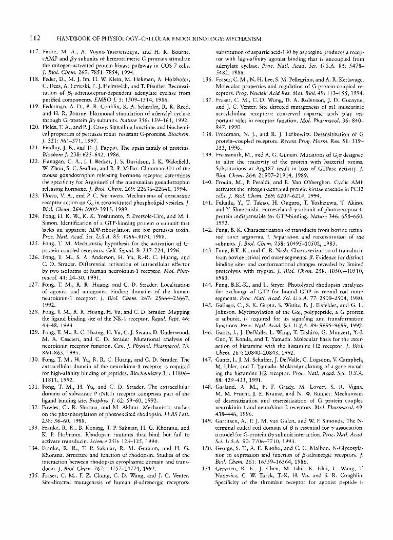
1 12 HANDBOOK OF PHYSIOLOGY-CELLULAR ENDOCRINOLOGY: MECHANISM
117. Faure, M. A., A. Voyno-Yasenetskaya, and H. R. Bourne. CAMP and Py subunits of heterotrimeric G proteins stimulate the mitogen-activated protein kinase pathway in COS-7 cells. J . Biol. Chem. 269: 7851-7854, 1994.
118. Feder, D., M. J. Im, H. W. Klein, M. Hekman, A. Holzhofer, C. Dees, A. Levitzki, E. J. Helmreich, and T. Pfeuffer. Reconsti- tution of PI-adrenoceptor-dependent adenylate cyclase from purified components. EMBO J. 5: 1509-1514, 1986.
119. Federman, A. D., B. R. Conklin, K. A. Schrader, R. R. Reed, and H. R. Bourne. Hormonal stimulation of adenylyl cyclase through G,-protein Py subunits. Nature 356: 159-161, 1992.
120. Fields, T. A., and P. J. Casey. Signalling functions and biochemi- cal properties of pertussis toxin-resistant G-proteins. Biochem.
121. Findlay, J. B., and D. J. Pappin. The opsin family of proteins. Biochem J. 238: 625-642, 1986.
122. Flanagan, C. A,, I. I. Becker, J. S. Davidson, I. K. Wakefield, W. Zhou, S. C. Seaflon, and R. P. Millar. Glutamate301 of the mouse gonadotrophin releasing hormone receptor determines the specificity for Arginine8 of the mammalian gonadotrophin releasing hormone. J. Biol. Chem. 269: 22636-22641, 1994.
123. Florio, V. A., and P. C. Sternweis. Mechanisms of muscarinic receptor action on Go in reconstituted phospholipid vesicles. J . Biol. Chem. 264: 3909-3915, 1989.
124. Fong, H. K. W., K. K. Yoshimoto, P. Eversole-Cire, and M. I. Simon. Identification of a GTP-binding protein a-subunit that lacks an apparent ADP-ribosylation site for pertussis toxin. Proc. Natl. Acad. Sci. U.S.A. 85: 3066-3070, 1988.
125. Fong, T. M. Mechanistic hypothesis for the activation of G- protein-coupled receptors. Cell. Signal. 8: 217-224, 1996.
126. Fong, T. M., S. A. Anderson, H. Yu, R.-R. C. Huang, and C. D. Strader. Differential activation of intracellular effector by two isoforms of human neurokinin-1 receptor. Mol. Phar-
127. Fong, T. M., R. R. Huang, and C. D. Strader. Localization of agonist and antagonist binding domains of the human neurokinin-l receptor. J . Biol. Chem. 267: 25664-25667, 1992.
128. Fong, T. M., R. R. Huang, H. Yu, and C. D. Strader. Mapping the ligand binding site of the NK-l receptor. Regul. Pept. 46: 4348, 1993.
129. Fong, T. M., R. C. Huang, H. Yu, C. J. Swain, D. Underwood, M. A. Cascieri, and C. D. Strader. Mutational analysis of neurokinin receptor function. Can. J. Physiol. Pharmacol. 73:
130. Fong, T. M., H. Yu, R. R. C. Huang, and C. D. Strader. The extracellular domain of the neurokinin-1 receptor is required for high-affinity binding of peptides. Biochemistry 31: 11806- 11811, 1992.
131. Fong, T. M., H. Yu, and C. D. Strader. The extracellular domain of substance P (NKI) receptor comprises part of the ligand binding site. Biophys. J . 62: 59-60, 1992.
132. Fowles, C., R. Sharma, and M. Akhtar. Mechanistic studies on the phosphorylation of photoexcited rhodopsin. FEBS Lett.
133. Franke, R. R., B. Koning, T. P. Sakmar, H. G. Khorana, and K. P. Hofmann. Rhodopsin mutants that bind but fail to activate transducin. Science 250: 123-125, 1990.
134. Franke, R. R., T. P. Sakmar, R. M. Graham, and H. G. Khorana. Structure and function of rhodopsin. Studies of the interaction between rhodopsin cytoplasmic domain and trans- ducin. J . Biol. Chem. 267: 14757-14774, 1992.
135. Fraser, C. M., F. Z. Chung, C. D. Wang, and J. C. Venter. Site-directed mutagenesis of human P-adrenergic receptors:
J . 321: 561-571, 1997.
W Z U C O ~ . 41: 24-30, 1991.
860-865, 1995.
238: 56-60, 1988.
substitution of aspartic acid-130 by aspargine produces a recep- tor with high-affinity agonist binding that is uncoupled from adenylate cyclase. Proc. Natl. Acad. Sci. U.S.A. 85: 5478- 5482, 1988.
136. Fraser, C. M., N. H. Lee, S. M. Pellegrino, and A. R. Kerlavage. Molecular properties and regulation of G-protein-coupled re- ceptors. Prog. Nucleic Acid Res. Mol. Biol. 49: 113-155, 1994.
137. Fraser, C. M., C.-D. Wong, D. A. Robinson, J. D. Gocayne, and J. C. Venter. Site directed mutagenesis of m l muscarinic acetylcholine receptors: conserved aspartic acids play im- portant roles in receptor function. Mol. Pharmacol. 36: 840- 847, 1990.
138. Freedman, N. J., and R. J. Lefkowitz. Desensitization of G protein-coupled receptors. Recent Prog. Horm. Res. 51: 319- 353, 1996.
139. Freissmuth, M., and A. G. Gilman. Mutations of G p designed to alter the reactivity of the protein with bacterial toxins. Substitutions at Arg187 result in loss of GTPase activity. y. Biol. Chem. 264: 21907-21914, 1989.
140. Frodin, M., P. Peraldi, and E. Van Obberghen. Cyclic AMP activates the mitogen-activated protein kinase cascade in PC12 cells. J. Biol. Chem. 269: 6207-6214, 1994.
141. Fukada, Y., T. Takao, H. Ouguro, T. Yoshizawa, T. Akino, and Y. Shimonishi. Farnesylated y subunit of photoreceptor G protein indispensable for GTP-binding. Nature 346: 658-660, 1992.
142. Fung, B. K. Characterization of transducin from bovine retinal rod outer segments. I. Separation and reconstitution of the subunits. J . Biol. Chem. 258: 10495-10502, 1983.
143. Fung, B.K.-K., and C. R. Nash. Characterization of transducin from bovine retinal rod outer segments. 11. Evidence for distinct binding sites and conformational changes revealed by limited proteolysis with trypsin. J. Biol. Chem. 258: 10503-10510, 1983.
144. Fung, B.K.-K., and L. Stryer. Photolyzed rhodopsin catalyzes the exchange of GTP for bound GDP in retinal rod outer segments. Proc. Natl. Acad. Sci. U.S.A. 77: 2500-2504, 1980.
145. Gallego, C., S. K. Gupta, S. Winitz, B. J. Eisfelder, and G. L. Johnson. Myristoylation of the Ga,, polypeptide, a G protein LY subunit, is required for its signaling and transformation functions. Proc. Natl. Acad. Sci. U.S.A. 89: 9695-9699, 1992.
146. Gantz, I., J. DelValle, L. Wang, T. Tashiro, G. Munzert, Y.-J. Guo, Y. Konda, and T. Yamada. Molecular basis for the inter- action of histamine with the histamine H2 receptor. ]. Biol. Chem. 267: 20840-20843, 1992.
147. Gantz, I., J. M. Schaffer, J. DelValle, C. Logsdon, V. Campbell, M. Uhler, and T. Yamada. Molecular cloning of a gene encod- ing the histamine H2 receptor. Proc. Natl. Acad. Sci. U.S.A. 88: 429-433, 1991.
148. Garland, A. M., E. F. Grady, M. Lovett, S. R. Vigna, M. M. Frucht, J. E. Krause, and N. W. Bunnet. Mechanisms of desensitization and resensitization of G protein<oupled neurokinin 1 and neurokinin 2 receptors. Mol. Pharmacol. 49: 438446, 1996.
149. Garritsen, A,, P. J. M. van Galen, and W. F. Simonds. The N- terminal coiled-coil domain of P is essential for y association: a model for G-protein pysubunit interaction. Proc. Natl. Acad. Sci. U.S.A. 90: 7706-7710, 1993.
150. George, S. T., A. E. Ruoho, and C. C. Malbon. N-Glycosyla- tion in expression and function of P-adrenergic receptors. J. Biol. Chem. 261: 16559-16564, 1986.
151. Gerszten, R. E., J. Chen, M. Ishii, K. Ishii, L. Wang, T. Nanevicz, C. W. Turck, T.-K. H. Vu, and S. R. Coughlin. Specificity of the thrombin receptor for agonist peptide is
CHAPTER 6: G PROTEIN-COUPLED RECEPTORS 1 13
167. Hall, A. Ras-related proteins. Curr. Opin. Cell Biol. 5: 265- 268, 1993.
168. Hall, A. Small GTP-binding proteins and the regulation of the actin cytoskeleton. Annu. Rev. Cell Biol. 10: 31-54, 1994.
169. Halliday, K. R. Regional homology in GTP-binding proto- oncogene products and elongation factors. /. Cyc. Nucl. Pro- tein Phos. Res. 9: 435448, 1984.
170. Hamm, H. E. Molecular interactions between the photorecep- tor G-protein and rhodopsin. Cell. Mol. Neurobiol. 11: 563- 578, 1991.
171. Hamm, H. E., D. Deretic, A. Arendt, P. A. Hargrave, B. Koenig, and K. P. Hoffman. Site of G-protein binding to rhodopsin mapped with synthetic peptides from the a subunit. Science
172. Harazono, A., Y. Sugirnoto, A. Ichikawa, and M. Negishi. Enhancement of adenylate cyclase stimulation by prostaglandin E receptor EP3 subtype isoforms with different efficiencies. Biochem. Biophys. Res. Commun. 201: 340-345, 1994.
173. Hargrave, P. A., J. H. McDowell, R. J. Feldman, P. H. Atkinson, J. K. Mohana Rao, and P. Argos. Rhodopsin's protein and carbohydrate structure: selected aspects. Vision Res. 24: 1487- 1499, 1984.
174. Harhammer, R., B. Niirnberg, C. Harteneck, D. Leopoldt, T. Exner, and G. Schultz. Distinct biochemical properties of the native members of the GI, G-protein subfamily. Characteriza- tion of Gall purified from rat brain. Biochem. 1. 319: 165- 171, 1996.
175. Hausdorff, W. P., M. Bouvier, B. F. O'Dowd, G. P. Irons, M. G. Caron, and R. J. Lefkowitz. Phosphorylation sites on two domains of the &-adrenergic receptor are involved in distinct pathways of receptor desensitization. J. Biol. Chem.
176. Hausdorff, W. P., M. G. Caron, and R. J. Lefkowitz. Turning off the signal: desensitization of p-adrenergic receptor function.
177. Hausdorff, W. P., J. A. Pitcher, D. K. Luttrell, M. E. Lin- der, H. Kurose, S. J. Parsons, M. G. Caron, and R. J. Lefko- witz. Tyrosine phosphorylation of G protein LY subunits by pp60'-src. Proc. Natl. Acad. Sci. U.S.A. 89: 5720-5724, 1992.
178. Hawes, B. E., L. M. Luttrell, S. T. Exum, and R. J. Lefkowitz. Inhibition of G protein-coupled receptor signaling by expres- sion of cytoplasmic domains of the receptor. J. Biol. Chem.
179. Hawes, B. E., T. van Biesen, W. J. Koch, L. M. Luttrell, and R. J. Lefkowitz. Distinct pathways of Gi- and Gq-mediated mitogen-activated protein kinase activation. J. Biol. Chem.
180. Hazum, E. GnRH-receptor of rat pituitary is a glycoprotein: differential effect of neuraminidase and lectins on agonist and antagonist binding. Mol. Cell. Endocrinol. 26: 217-222, 1982.
181. Hazum, E., I. Schvartz, Y. Waksman, and D. Keinan. Solubili- zation and purification of rat pituitary gonadotropin-releasing hormone receptor. J. Biol. Chem. 261: 13043-13048, 1986.
182. Heckert, L. L., I. J. Daley, and M. D. Griswold. Structural organization of the follicle-stimulating hormone receptor gene. Mol. Endocrinol. 6: 70-80, 1992.
183. Henderson, R., J. M. Baldwin, T. A. Ceska, F. Zemlin, E. Beckmann, and K. H. Downing. Model for the structure of bacteriorhodopsin based on high-resolution electron cryo- microscopy. /. Mol. Biol. 213: 899-929, 1990.
184. Hepler, J. R., and A. G. Gilman. G Proteins. Trends Pharmacol. Sci. 17: 383-387, 1992.
185. Herrero, I., M. T. Miras-Portugal, and J. Sanchez-Prieto. Rapid desensitization of the metabotropic glutamate receptor that
241: 832-835, 1988.
264: 12657-12665, 1989.
FASEB /. 4: 2881-2889, 1990.
269: 15776-15785, 1994.
270: 17148-17153, 1995.
defined by its extracellular surface. Nature 368: 648-651, 1994.
152. Gether, U., J. A. Ballesteros, R. Seifert, E. Sanders-Bush, H. Weinstein, and B. K. Kobilka. Structural instability of a consti- tutively active G protein-coupled receptor. Agonist- independent activation due to conformational flexibility. /. Biol. Chem. 272: 2587-2590, 1997.
153. Gilchrist, R.L., K.-S. Ryu, I. Ji, and T. H. Ji. The luteinizing hormonekhorionic gonadotropin receptor has distinct trans- membrane conductors for CAMP and inositol phosphate sig- nals. I . Biol. Chem. 271: 19283-19287, 1996.
154. Gilman, A. F. G proteins: transducers of receptor-generated signals. Annu. Rev. Biochem. 56: 615-649, 1987.
155. Gocayne, J., D. A. Robinson, M. G. Fitz-Gerald, F.-Z. Chung, A. R. Kerlavage, K.-W. Lentes, J. Lai, Ch.-D. Wang, C. M. Fraser, and J. C. Venter. Primary structure of rat p-adrenergic and muscarinic cholinergic receptors obtained by automated DNA sequencing analysis: further evidence for a multigene family. Proc. Natl. Acad. Sci. U.S.A. 84: 8296-8300, 1987.
156. Gordon, J. I., R. J. Duronio, D. A. Rudnick, S. P. Adams, and G. W. Goke. Protein N-myristoylation. I . Biol. Chem. 266:
157. Grady, E. F., A. M. Garland, P. D. Gamp, M. Lovett, D. G. Payan, and N. W. Bunnet. Delineation of the endocytic path- way of substance P and its seven-transmembrane domain NK1 receptor. Moi. Biol. Cell 6: 509-524, 1995.
158. Graves, P. N., Y. Tomer, and T. F. Davies. Cloning and sequenc- ing of a 1.3 kb variant of human thyrotropin receptor mRNA lacking the transmembrane domain. Biochem. Biophys. Res. Commun. 187: 1135-1143, 1992.
159. Gromoll, J., T. Guderman, and E. Nieschlag. Molecular cloning of a truncated isoform of the human follicle stimulating hor- mone receptor. Biochem. Biophys. Res. Commun. 188: 1077- 1083, 1992.
160. Guan, X. M., A. Amend, and C. D. Strader. Determination of structural domains for G protein coupling and ligand binding in &-adrenergic receptor. Mol. Pharmacol. 48: 492498, 1995.
161. Guo, C.-H., J. A. Janovick, D. Kuphal, and P. M. Conn. Transient tranfection of GGH3-l' cells (GH3 cells stably transfected with the gonadotropin-releasing hormone (GnRH) receptor complementary deoxyribonucleic acid] with the carboxi-terminal of p-adrenergic receptor kinase 1 blocks pro- lactin release: evidence for a role of the G protein &subunit complex in GnRH signal transduction. Endocrinology 136:
162. Gupta, S., D. Campbell, B. Derijard, and R. J. Davis. Transcrip- tion factor ATF2 regulation by the JNK signal transduction pathway. Science 267: 389-393, 1995.
163. Gutowski, S., A. Smrcka, L. Nowac, D. Wu, M. Simon, and P. C. Sternweis. Antibodies to the aq subfamily guanine nucleo- tide-binding regulatory protein LY subunits attenuate activation of phosphatidylinositol 4,5-biphosphate hydrolysis by hor- mones. I . Biol. Chem. 266: 20519-20524, 1991.
164. Hadcock, J. R., and C. C. Malbon. Down-regulation of p- adrenergic receptors: agonist-induced reduction in receptor mRNA levels. Proc. Nutl. Acad. Sci. U.S.A. 85: 5021-5025, 1988.
165. Hadcock, J. R., M. Ros, and C. C. Malbon. Agonist regulation of p-adrenergic receptor mRNA. 1. Biol. Chem. 264: 13956- 13961, 1989.
166. Hadcock, J. R., H.-Y. Wang, and C. C. Malbon. Agonist- induced desesta bilization of p-adrenergic receptor mRNA. At- tenuation of glucocorticoid-induced up-regulation of p- adrenergic receptors. /. Biol. Chem. 264: 19928-19933, 1989.
8647-8650, 1991.
3031-3036, 1995.
114 HANDBOOK OF PHYSIOLOGY-CELLULAR ENDOCRINOLOGY
facilitates glutamate release in rat cerebrocortical nerve termi- nals. Eur. /. Neurosci. 6: 115-120, 1994.
186. Hershey, A. D., and J. E. Krause. Molecular characterization of a functional cDNA encoding the rat substance P receptor. Science 247: 958-962, 1990.
187. Hescheler, J., W. Rosenthal, W. Trautwein, and G. Schultz. The GTP-binding protein, Go, regulates neuronal calcium channels. Nature 325: 445447, 1987.
188. Higashijima, T., J. Burnier, and E. M. Ross. Regulation of G, and Go by Mastoparan, related amphiphilic peptides and hydrophobic amines. 1. Biol. Chem. 265: 14176-14186, 1990.
189. Higashijima , T., K. M. Ferguson, P. C. Sternweis, E. M. Ross, M. D. Smigel, and A. G. Gilman. The effect of activating ligands on the intrinsic fluorescence of guanine nucleotide- binding regulatory proteins. J . Biol. Chem. 262: 752-756, 1987.
190. Higashijima, T., K. M. Ferguson, P. C. Sternweis, M. D. Smigel, and A. G. Gilman. Effects of M z + and the P y subunit complex on the interactions of guanine nucleotides with G proteins. 1. Biol. Chem. 262: 762-766, 1987.
191. Higgins, J. B., and P. J. Casey. In vitro processing of recombi- nant G-protein y subunits. J . Biol. Chem. 269: 9067- 9073,1994.
192. Hipkin, R. W., J. Sinchez-Yague, and M. Ascoli. Phosphoryla- tion of the luteinizing hormonekhoriogonadotropin receptor expressed in a stably transfected cell line. Mol. Endocrinol. 7:
193. Hipkin, R. W., Z. Wang, and M. Ascoli. Human chorionic gonadotropin (CG)- and phorbol ester-stimulated phosphory- lation of the luteinizing hormonelCG receptor maps to serines 635, 639, 649, and 652 in the C-terminal cytoplasmic tail. Mol. Endocrinol. 9: 151-158, 1995.
194. Hirisawa, A., K. Shibata, K. Horie, Y. Takei, K. Obika, T. Tanaka, N. Muramoto, K. Takagaki, J. Yano, and G. Tsuji- moto. Cloning, functional expression and tissue distribution of human a,,:-adrenoceptor splice variants. FEBS Lett. 363: 256- 260, 1995.
195. Hirsch, B., M. Kudo, F. Naro, M. Conti, and A. J. W. Hsueh. The C-terminal third of the human luteinizing hormone (LH) receptor is important for inositol phosphate release: analysis using chimeric human LWfollicle-stimulating hormone recep- tors. Mol. Endocrinol. 10: 1127-1137, 1996.
196. Hirsch, J. P., C. Dietzel, and J. Kurjan. The carboxyl terminus of Scgl, the G a subunit involved in yeast mating, is implicated in interactions with the pheromone receptors. Genes Dev. 5: 467474, 1991.
197. Hirschman, J. E., G. S. De Zutter, W. F. Simonds, and D. D. Jenness. The GPy complex of the yeast pheromone response pathway. Subcellular fractionation and protein-protein interac- tions. I . Biol. Chem. 272: 240-248, 1997.
198. Ho, B. Y., A. Karchiin, T. Branchek, N. Davidson, and H. A. Lester. The role of conserved aspartate and serine residues in ligand binding and in function of the 5-HT,, receptor: a site- directed mutation study. FEBS Lett. 312: 259-262, 1992.
199. Holtmann, M. H., S. Ganguli, E. M. Hadac, V. Dolu, and L. J. Miller. Multiple extracellular loop domains contribute critical determinants for agonist binding and activation of the secretin receptor. 1. Biol. Chem. 271: 14944-14949, 1996.
200. Horstman, D. A., S. Brandon, A. L. Wilson, C. A. Guyer, E. J. Cragoe, and L. E. Limbird. An aspartate conserved among G-protein receptors confers allosteric regulation of a2- adrenergic receptors by sodium. I . Biol. Chem. 265: 21590- 21595, 1992.
201. Houamed, K. M., J. L. Kuijper, T. L. Gilbert, B. A. Haldeman,
823-832, 1993.
P. J. O’Hara, E. R. Mulvihill, W. Almers, and F. S. Hagen. Cloning, expression and gene structure of a G-protein coupled glutamate receptor from rat brain. Science 252: 1318-1321, 1991.
202. Huang, R. R., P. P. Vicario, C. D. Strader, and T. M. Fong. Identification of residues involved in ligand binding to the neurokinin-2 receptor. Biochemistry 34: 10048-10055, 1995.
203. Huang, R. R., H. Yu, C. D. Strader, and T. M. Fong. Interac- tion of substance P with the second and seventh transmembrane domains of the neurokinin-1 receptor. Biochemistry 33: 3007- 3013, 1994.
204. Huang, R. R., H. Yu, C. D. Strader, and T. M. Fong. Localiza- tion of the ligand binding site of the neurokinin-1 receptor: interpretation of chimeric mutations and single-residue substi- tutions. Mol. Pharmacol. 45: 690-695, 1994. Hulme, E. C., C. A. M. Curtis, M. Wheatley, A. Aitken, and
A. C. Harris. Localization and structure of the muscarinic receptor ligand binding site. Trends. Pharmacol. Sci. (Suppl.): 22-25, 1989.
206. Inglese, J., J. F. Glickman, W. Lorenz, M. G. Caron, and R. J. Lef kowitz. Isoprenylation of a protein kinase: requirement of farnesylatioda-carboxy methylation for full enzymatic activity of rhodopsin kinase. I . Biol. Chem. 267: 1422-1425, 1992.
207. Inglese, J., W. J. Koch, M. G. Caron, and R. J. Lefkowitz. Isoprenylation in the regulation of signal transduction by G- protein-coupled receptor kinases. Nature 359: 147-150, 1992.
208. Inglese, J., W. J. Koch, K. Touhara, and R. J. Lefkowitz. G o y interactions with PH domains and Ras-MAPK signaling pathways. Trends Biochem. Sci. 20:151-156, 1995.
209. Iiiiguez-Lluhi, J., C. Kleuss, and G. Gilman. The importance of G-protein Pysubunits. Trends Cell. Biol. 3: 230-236, 1993.
210. Iiiiguez-Lluhi, J. A., M. I. Simon, J. D. Robishaw, and A. G. Gilman. G-protein P y subunits synthesized in Sf9 cells. Functional characterization and the significance of prenylation of y. I . Biol. Chem. 267: 23409-23417, 1992.
211. Ishigara, T., S. Nakamura, Y. Kaziro, T. Takahashi, K. Taka- hashi, and s. Nagata. Molecular cloning and expression of a cDNA encoding the secretin receptor. EMBO 1. 10: 1635- 1641, 1991.
212. Ishigara, T., R. Shigemoto, K. Mori, K. Takahashi, and S. Nagata. Functional expression and tissue distribution of a novel receptor for vasoactive intestinal polypeptide. Neuron 8:
213. Ito, H., and A. G. Gilman. Expression and analysis of G,, mutants with decreased ability to activate adenylyl cyclase. J . Biol. Chem. 266: 16226-16231, 1991.
214. Itoh, H., R. Toyama, T. Kazana, T. Tsukamoto, M. Matsuoka, and Y. Kaziro. Presence of three distinct molecular species of G, protein a subunit: structure of rat cDNAs and human genomic DNAs. I . Biol. Chem. 263: 6656-6664, 1988.
215. Iro, H., R. T. Tung, T. Sugimoto, I. Kobayashi, K. Takahashi, T. Katada, M. Ui, and Y. Kurachi. O n the mechanism of G protein Py subunit activation of the muscarinic K + channel in guinea pig atrial cell membrane. J . Gen. Physiol. 99: 961-983, 1992.
216. Iyengar, R. Molecular and functional diversity of mammalian G,-stimulated adenylyl cyclases. FASEB J. 7: 768-775, 1993.
217. Iyengar, R., and L. Birnbaumer. Hormone receptors modulate the regulatory component of adenylyl cyclases by reducing its requirement for Mgz+ ion and enhancing its extent of activa- tion by guanine nucleotides. Proc. Natl. Acad. Sci. U.S.A. 79:
218. Janovick, J. A., S. P. Brothers, and P. M. Conn. GnRH agonist proteins. In:
205.
811-819, 1992.
5179-5183, 1982.
stimulates net loss in immunoassayable G,,,
CHAPTER 6: G PROTEIN-COUPLED RECEPTORS 11 5
J. D. Neil. Cloning, sequencing and expression of human gonadotropin releasing hormone (GnRH) receptor. Biochem. Biophys. Res. Commun. 189: 289-295, 1992.
234. Karnik, S., K. Ridge, S. Bhattacharya, and G. H. Khorana. Palmitoylation of bovine opsin and its cystein mutants in COS cells. Proc. Natl. Acad. Sci. U.S.A. 90: 4044, 1993.
235. Karnik, S. S., J. P. Sakmann, H. B. Chen, and H. G. Khorana. Cysteine residues 110 and 187 are essential for the formation of correct structure in bovine rhodopsin. Proc. Nail. Acad. Sci.
236. Katz, A., D. Wu, and M. I. Simon. Subunits Py of heterotrim- eric G protein activate P2 isoform of phospholipase C. Nature
237. Kaupmann, K., C. Burns, D. Hoyer, K. Seuwen, and H. Lub- bert. Distribution and second messenger coupling of four so- matostatin receptor subtypes expressed in brain. FEBS hi t .
238. Kaupp, U. B., and K. W. Koch. Role of cGMP and CaZ+ in vertebrate photoreceptor excitation and adaptation. Annu. Rev. Physiol. 54: 153-176, 1992.
239. Kawate, N., and K. M. Menon. Palmitoylation of luteinizing hormone/human choriogonadotropin receptors in transfected cells. J. Biol. Chem. 269: 30651-30658, 1994.
240. Kennedy, K., V. Gigoux, Ch. Escrieut, B. Maigret, J. Martinez, L. Moroder, D. FrChel, D. Gully, N. Vaysse, and D. Fourmy. Identification of two amino acids of the human cholecystokinin-A receptor that interact with the N-terminal moiety of cholecystokinin. J. Biol. Chem. 272: 2920-2926, 1997.
241. Kennedy, M., and L. E. Limbird. Palmitoylation of the a,*- adrenergic receptors. J. Biol. Chem. 269: 31915-31922, 1994.
242. Kennedy, M. E., and L. E. Limbird. Mutations of the aZA- adrenergic receptor that eliminate detectable palmitoylation do not perturb receptor-G-protein coupling. J . Biol. Chem. 268:
243. Kennelly, P. J., and E. G. Krebs. Consensus sequences as substrate specificity determinants for protein kinases and pro- tein phosphatases. J. Biol. Chem. 266: 15555-15558, 1991.
244. Khorana, H. G. Rhodopsin, photoreceptor of the rod cell. An emerging pattern for structure and function. J . Biol. Chem. 267: 1-4,1992.
245. Kim, D., D. L. Lewis, L. Graziadei, E. J. Neer, D. Bar-Sagi, and D. E. Clapham. G-protein py-subunits activate the cardiac muscarinic K+ channel via phospholipase A2. Nature 337: 557-560, 1989.
246. Kim, G.-D., I. C. Carr, L. A. Anderson, J. Zabavnik, K. A. Eidne, and G. Milligan. The long isoform of the rat thyrotropin-releasing hormone receptor down-regulates G, proteins. J . Biol. Chem. 269: 19933-19940, 1994.
247. Kisselev, O., and N. Gautman. Specific interaction with rho- dopsin is dependent on the y subunit type in a G-protein. ]. Biol. Chem. 268: 24519-24552, 1993.
248. Kisselev, O., A. Pronin, M. Ermolaeva, and N. Gautman. Receptor-G protein coupling is established by a potential con- formational switch in the Py complex. Proc. Natl. Acad. Sci.
249. Kjeldgaard, M., J. Nyborg, and B. F. C. Clark. The GTP binding motif: variations on a theme. FASEB J. 10: 1347- 1368, 1996.
250. Kjelsberg, M. A., S. Cotecchia, J. Ostrowski, M. G. Caron, and R. J. Lef kowitz. Constitutive activation of the a,,-adrenergic receptor by all amino acid substitutions at a single site. J . Biol. Chem. 267: 1430-1433, 1992.
251. Kleuss, C. J., C. Hescheler, W. Ewel, G. Rosenthal, G. Schultz,
U.S.A. 85: 8459-8463, 1988.
360: 686-689, 1992.
331: 53-59, 1993.
8003-801 1, 1993.
U.S.A. 92: 9102-9106, 1995.
Program, 791h Annual Meeting of the Endocrine Society, Min- neapolis. 1997, p. 168.
219. Janovick, J. A., Haviv, F., T. D. Fitzpatrick, and P. M. Conn. Differential orientation of a GnRH agonist and antagonist in the pituitary GnRH action. Endocrinology 133: 942-945, 1993.
220. Jelinek, L. J., S. Lok, G. B. Rosenberg, R. A. Smith, F. J. Grant, S. Biggs, P. A. Bensch, J. L. Kuijper, P. 0. Sheppard, C. A. Sprecher, P. J. O'Hara, D. Foster, K. M. Walker, L. H. J. Chen, P. A. McKernan, and W. Kindsvogel. Expression cloning and signaling properties of the rat glucagon receptor. Science 259:
221. Jelsema, C. L., and J. Axelrod. Stimulation of phospholipase A2 activity in bovine rod outer segments by the Py subunits of transducin and its inhibition by the a subunit. Proc. Nail. Acad. Sci. U.S.A. 84: 3623-3627, 1987.
222. Jennes, L., W. E. Stumpf, and P. M. Conn. Receptor-mediated binding and uptake of GnRH agonist and antagonist by pitu- itary cells. Peptides 5 (Suppl. 1): 215-220, 1984.
223. Jhon, D.-Y., H.-H. Lee, D. Park, C.-W. Lee, K.-H. Lee, and 0. J. Yoo. Cloning, sequencing, purification and G,-dependent activation of phospholipase C-P,. J . Biol. Chem. 268: 6654- 6661, 1993.
224. Ji, I., and T. Ji. Asp3x3 in the second transmembrane domain of the lutropin receptor is important for high affinity hormone binding and CAMP production. J. Biol. Chem. 266: 14953- 14957, 1991.
225. Ji, I., R. G. Slaughter, and T. H. Ji. N-linked oligosaccharides are not required for hormone binding of the lutropin receptor in a Leydig tumor cell line and rat granulosa cells. Endocrinol- ogy 127: 494496,1990.
226. Jinnah, H. A., and P. M. Conn. GnRH-stimulated LH release from rat anterior pituitary cells in culture: refractoriness and recovery. Am. J . Physiol. 249 (Endocrinol. Metab. 12): E619- E625, 1985.
227. Johnson, P. S., C. K. Surrat, B. K. Seideleck, C. J. Blaschak, J. B. Wang, and G. R. Uhl. g Opiate receptor: site-directed mutagenesis produces differential effect on second messenger systems. In: Program, 24th Annual Meeting of the Society for Neuroscience, Miami, FL. 1994, p. 745.
228. Jones, D. T., and R. R. Reed. Molecular cloning of five GTP- binding protein cDNA species from rat olfactory neuroepithel- ium J . Biol. Chem. 262: 14241-14249, 1987.
229. Jones, R. L. Z., W. F. Simonds, J. J. Merendino, M. R. Brann, and A. M. Spiegel. Myristoylation of an inhibitory GTP- binding protein a subunit is essential for its membrane attach- ment. Proc. Natl. Acad. Sci. U.S.A. 87: 568-572, 1990.
230. Juppner H., A. B. Abou-Amra, M. W. Freeman, X. F. Kong, E. Schipani, J. Richards, L. F. Kolakowski, Jr., J. Hock, J. T. Potts, Jr., H. M. Kronenberg, and G. V. Segre. A G protein- linked receptor for parathyroid hormone and parathyroid hormone-related peptide. Science 254: 1024-1026, 1991.
231. Jiippner, H., E. Schipani, F. R. Bringhurst, I. McClure, H. T. Keutmann, J. T. Potts, H. M. Kronenberg, A. B. Abou-Samra, G. V. Segre, and T. J. Gardella. The extracellular amino- terminal region of the parathyroid hormone (PTH)/PTH- related peptide receptor determines the binding affinity for carboxyl-terminal fragments of PTH-( 1-34). Endocrinology
232. Kaiser, U. B., D. Zhao, G. R. Cardona, and W. Chin. Isolation and characterization of cDNAs encoding the rat pituitary gonadotropin-releasing hormone receptor. Biochem. Biophys. Res. Commun. 189: 1645-1652, 1992.
233. Karkar, S. S., L. C. Musgrove, D. C. Devor, J. C. Sellers, and
1614-1616, 1993.
134: 879-884, 1994.
11 6 HANDBOOK OF PHYSIOLOGY-CELLULAR ENDOCRINOLOGY
and B. Wittig. Assignment of G-protein subtypes to specific receptors inducing inhibition of calcium channels. Nature 353: 43-48, 1991.
252. Kobilka, B. K., R. A. F. Dixon, T. Frielle, H. G. Dohlman, M. A. Bolanowski, I. S. Sigal, T. L. Yang-Feng, U. Francke, M. G. Caron, and R. J. Lefkowitz. cDNA for the human p- adrenergic receptor: a protein with multiple membrane span- ning domains and a chromosomal location shared with the PDGF receptor gene. Proc. Natl. Acad. Sci. U.S.A. 84: 46-50, 1987.
253. Kobilka, B. K., T. S. Kobilka, K. W. Daniel, J. W. Regan, M. G. Caron, and R. J. Lefkowitz. Chimeric az-, &adrenergic receptors: delineation of domains involved in effector coupling and ligand binding specificiy. Science 249: 1310-1316, 1988.
254. Koch, C. A., D. Anderson, M. F. Moran, C. Ellis, and T. Pawson. SH2 and SH3 domains: elements that control interac- tions of cytoplasmic signaling proteins. Science 252: 668-674, 1991.
255. Koch, W. J., B. E. Hawes, L. F. Allen, and R. J. Lefkowitz. Direct evidence that Gi-coupled receptor stimulation of mitogen-activated protein kinase is mediated by G py activa- tion of p2lras. Proc. Natl. Acad. Sci. U.S.A. 91: 12706-12710, 1994.
256. Kokame, K., Y. Fukada, T. Yoshizawa, T. Takao, and Y. Shi- monishi. Lipid modification of the N-terminus of photorecep- tor G-protein a-subunit. Nature 359: 749-752, 1992.
257. Kolch, W., G. Heidecker, G. Kochs, R. Hummel, H. Vahidi, H. Mischak, G. Finkenzeller, D. Marme, and U. R. Rapp. Protein kinase Ca activates RAF-1 by direct phosphorylation. Nature 364: 249-252, 1993.
258. Konig, B., A. Arendt, J. H. McDowell, M. Kahlert, P. A. Hargrave, and K. P. Hofmann. Three cytoplasmic loops of rhodopsin interact with transducin. Proc. Natl. Acad. Sci.
259. Koo, Y. B., I. Ji, R. G. Slaughter, and T. H. Ji. Structure of the luteinizing hormone receptor gene and multiple exons of the coding sequence. Endocrinology 128: 2297-2308, 1991.
260. Kosugi, S., F. Okajima, T. Ban, A. Hidaka, A. Shenker, and L. D. Kohn. Mutation of alanine 623 in the third cytoplasmic loop of the rat thyrotropin (TSH) receptor results in a loss in the phosphoinositide but not the CAMP signal induced by TSH and receptor antibodies. J . Biol. Chem. 267: 24153-24156, 1992,
261. Krueger, K. M., Y. Daaka, J. A. Pitcher, and R. J. Lefkowitz. The role of sequestration in G protein-coupled receptor resensi- tization. J. Biol. Chem. 272: 5-8, 1997.
262. Kubo, T., H. Bujo, I. Akiba, J. Nakai, M. Mishina, and S. Numa. Location of a region of the muscarinic acetylcholine receptor involved in selective effector coupling. FEBS Lett.
263. Kudo, M., Y. Osuga, B. K. Kobilka, and A.J.W. Hsueh. Trans- membrane regions V and VI of the human luteinizing hormone receptor are required for constitutive activation by a mutation in the third intracellular loop. J . Biol. Chem. 271: 22470- 22478, 1996.
264. Kurachi, Y., H. Ito, T. Sugimoto, T. Shimizu, I. Miki, and M. Ui. Arachidonic acid metabolites as intracellular modulators of the G protein-gated cardiac K + channel. Nature 337: 555- 557, 1989.
265. Kurtenbach, E., C.A.M. Curtis, E. K. Pedder, A. Aitken, A.C.M. Harris, and E. C. Hulme. Muscarinic acetylcholine receptors. Peptide sequencing identifies residues involved in antagonist binding and disulfide bond formation. J. Biol. Chem. 265: 13702-13708, 1990.
U.S.A. 86: 6878-6882, 1989.
241: 119-125, 1988.
266. Lagnado, L., and D. Baylor. Signal flow in visual transduction. Neuron 8: 995-1002, 1992.
267. Lambright, D. G., J. Sondek, A. Bohm, N. P. Skiba, H. E. Hamm, and P. B. Sigler. The 2.0 A crystal structure of a heterotrimeric G protein. Nature 379: 311-319, 1996.
268. Lameh, J., M. Philip, Y. K. Sharma, 0. Moro, J. Ramachan- dran, and W. Sadee. H m l muscarinic cholinergic receptor internalization requires a domain in the third cytoplasmic loop. J. Biol. Chem. 267: 13406-13412, 1992.
269. LaMorte, V. J., E. D. Kennedy, L. R. Collins, D. Goldstein, A. T. Harootunian, J. H. Brown, and J. R. Feramisco. A requirement for Ras protein function in thrombin-stimulated mitogenesis in astrocytoma cells. J . Biol. Chem. 268: 19411- 19415, 1993.
270. Landis, C. A., S. B. Masters, A. Spada, A. M. Pace, H. R. Bourne, and L. Vallar. GTPase inhibiting mutations activate the a chain of G, and stimulate adenylyl cyclase in human pituitary tumors. Nature 340: 692-696, 1989.
271. Lange-Carter, C. A., C. M. Pleiman, A. M. Gardner, K. J. Blumer, and G. L. Johnson. A divergence in the MAP kinase regulatory network defined by MEK kinase and Raf. Science
272. LaRosa, G. J., K. M. Thomas, M. E. Kaufmann, R. Mark, M. White, L. Taylor, G. Gray, D. Witt, and J. Navarro. Amino terminus of the interleukin-8 receptor is a major determinant of receptor subtype specificity. J . Biol. Chem. 267: 25402- 25406,1992.
273. Laue, L. L., W. Shao-Ming, M. Kudo, C. J. Bourdony, G. B. Cutler, A. J. W. Hsueh, and W.-Y. Chan. Compound heterozy- gous mutations of the luteinizing hormone receptor gene in Leydig cell hypoplasia. Mol. Endocrinol. 10: 987-997, 1996.
274. Leberman, R., and U. Egner. Homologies in the primary struc- ture of GTP-binding proteins: the nucleotide binding site.
275. Lechleiter, J., R. Hellmiss, K. Duerson, D. Ennulat, N. David, D. Clapham, and E. Peralta. Distinct sequence elements control the specificity of G-protein activation by muscarinic acetylcho- line receptor subtypes. EMBO J. 9: 4381-4390, 1990.
276. Lee, C., T. Murakami, and W. F. Simonds. Identification of a discrete region of the G protein y subunit conferring selectivity in P y complex. J. Biol. Chem. 270: 8779-8784, 1995.
277. Lee, C. H., A. Katz, and M. I. Simon. Multiple regions of Ga,, contribute to the specificity of activation by the C5A receptor. Mol. Pharmacol. 47: 218-223, 1995.
278. Lee, C.-W., D. J. Park , K.-H. Lee, C. G. Kim, and S. G. Rhee. Purification, molecular cloning, and sequencing of phosphdi- pase C-p4. J . Biol. Chem. 268: 21318-21327, 1993.
279. Lee, E., R. Taussig, and A. G. Gilman. The G226A mutant of G,, highlights the requirement for dissociation of G protein subunits. J . Biol. Chem. 267: 1212-1218, 1992.
280. Leff, P. The two-state model of receptor activation. Trends Pharmacol. Sci. 16: 89-97, 1995.
281. Lefkowitz, R. J. Turned on to ill effect. Nature 365: 603-604, 1993.
282. Lefkowitz, R. J., S. Cotecchia, P. Samama, and T. Costa. Constitutive activity of receptors coupled to guanine nucleotide regulatory proteins. Trends Pharmacol. Sci. 14: 303-307, 1993.
283. Lester, H. A. The permeation pathway of neurotransmitter- gated ion channels. Annu. Rev. Biophys. Biomol. Struct. 21:
284. Lev, S., H. Moreno, R. Martinez, P. Canoll, E. Peles, J. M. Musacchio, G. D. Plowman, B. Rudy, and J. Schlessinger. Protein tyrosine-kinase PYK2 involved in CaZ + -induced regula-
260: 315-319, 1993.
EMBO J . 4: 339-341, 1984.
267-292, 1992.
CHAPTER 6: G PROTEIN-COUPLED RECEPTORS 1 17
301. Luttrell, L. M., J, Ostrowski, S. Cotecchia, H. Kendall, and R. J. Lef kowitz. Antagonism of catecholamine receptor signal- ing by expression of cytoplasmic domains of the receptors. Science 259: 1453-1457, 1993.
302. Lyons, J., C. A. Landis, G. Harsh, L. Vallar, K. Grunewald, H. Feichtinger, Q.-Y. Suk, 0. H. Clark, E. Kawasaki, and H. R. Bourne. Two G protein oncogenes in human endocrine tumors. Science 249: 655-659, 1990.
303. Maeda, S., J. Lameh, W. G. Mallet, M. Philip, J. Ramachan- dran, and W. Sadee. Internalization of the Hml muscarinic cholinergic receptor involves the third cytoplasmic loop. FEBS
304. Maggio, R., Z. Vogel, and J. Wess. Coexpression studies with mutant muscarinidadrenergic receptors provide evidence for intermolecular “cross-talk’’ between G-protein-linked recep- tors. Proc. Nutl. Acud. Sci. U.S.A. 90: 3103-3107, 1993.
305. Mahan, L. C., A. M. Koachman, and P. A. Insel. Genetic analysis of 0-adrenergic receptor internalization and down- regulation. Proc. Nutl. Acud. Sci. U.S.A. 82: 129-133, 1985.
306. Masu, M., Y. Tanabe, K. Tsuchida, R. Shigemoto, and S. Nakanishi. Sequence and expression of a metabotropic gluta- mate receptor. Nature 349: 760-765, 1991.
307. Mattera, R., M. P. Graziano, A. Yatani, Z. Zhou, R. Graf, J. Codina, L. Birnbaumer, A. G. Gilman, and A. M. Brown. Splice variants of the a subunit of the G protein G, activate both adenylyl cyclase and calcium channels. Science 243: 804- 807,1989.
308. Mayo, K. E. Molecular cloning and expression of a pituitary- specific receptor for growth hormone-releasing hormone. Mol. Endocrinol. 6: 1734-1744, 1992.
309. Mazzoni, M. R., and H. E. Hamm. Effect of monoclonal antibody binding on a-py subunit interactions in the rod outer segment G protein, G,. Biochemistry 28: 9873-9880, 1989.
310. Mazzoni, M. R., J. A. Malinksi, and H. E. Hamm. Structural analysis of rod GTP-binding protein, G,: limited proteolytic digestion pattern of G, with four proteases defines monoclonal antibody epitope. J . Biol. Chem. 266: 14072-14081, 1991.
311. McArdle, C. A., and R. Countis. GnRH and PACAP action in gonadotropes. Cross-talk between phosphoinositidase C and adenylyl cyclase mediated signaling pathways. Trends Endocri- nol. Metub. 7: 168-175, 1996.
312. McArdle, C. A., W. C. Gorospe, W. R. Huckle, and P. M. Conn. Homologous down-regulation of gonadotropin- releasing hormone receptors and desensitization of gonado- tropes: lack of dependence on protein kinase C. Mol. Endocri- nol. 1: 420-429, 1987.
313. McArdle, C. A., G. B. Willars, R. C. Fowkes, S. R. Nahorski, J. S. Davidson, and W. Forrest-Owen. Desensitization of gonadotropin-releasing hormone action in aT3-1 cells due to uncoupling of inositol 1,4,5-triphosphate generation and Ca2 +
mobilization. J . Biol. Chem. 271: 2371 1-23717, 1966. 314. McClue, S. J., B. M. Baron, and B. A. Harris. Activation of
G, protein by peptide of the muscarinic M, receptor second intracellular loop. Eur. J. Pharmucol. 267: 185-193, 1994.
315. McFarland, K. C., R. Sprengel, H. S. Phillips, M. Kohler, N. Rosemblit, K. Nikolics, D. L. Segaloff, and P. H. Seeburg. Lutropin-choriogonadotropin receptor: an unusual member of the G protein receptor family. Science 245: 494-499, 1989.
316. McLaughlin, S. K., P. J. McKinnon, and R. F. Margoolskee. Gustducin is a taste-cell-specific G protein closely related to the transducins. Nature 357: 563-569, 1992.
317. Medema, R. H., and Bos, J. L. The role of p2lras in receptor tyrosine kinase signaling. Crit. Rev. Oncog. 4: 615-661, 1993.
318. Medynski, D., S. K. Sullivan, D. Smith, C. VanDop, F.-H.
Lett. 269: 386-388, 1990.
tion of ion channel and MAP kinase functions. Nature 376:
285. Levis, M. J., and H. R. Bourne. Activation of the a subunit of G, in intact cells alters its abundance, rate of degradation, and membrane avidity. J , Cell Biol. 119: 1297-1307, 1992.
286. Liggett, S. B., M. G. Caron, R. J. Lefkowitz, and M. Hnatow- ich. Coupling of a mutated form of the human p2-adrenergic receptor to Gi and G,. J. Biol. Chem. 266: 4816-4821, 1991.
287. Lin, H. Y., T. L. Harris, M. S. Flannery, A. Aruffo, E. H. Kaji, A. Gorn, L. F. Kolafowski, Jr., H. F. Lodish, and S. R. Goldring. Expression cloning of an adenylate cyclase-coupled calcitonin receptor. Science 254: 1022-1024, 1991.
288. Linder, M. E., P. Middleton, J. R. Hepler, R. Taussig, A. G. Gilman, and S. M. Mumby. Lipid modification of G-proteins: a subunits are palmitoylated. Proc. Nutl. Acud. Sci. U.S.A. 90:
289. Linder, M. E., I.-H. Pang, R. J. Duronio, J. I. Gordon, P. C. Sternweis, and A. G. Gilman. Lipid modifications of G protein subunits. Myristoylation of Go, increases its affinity for py. J . Biol. Chem. 266: 4654-4659, 1991.
290. Litosch, I. G-protein inhibition of phospholipase C-p, in mem- branes: role of G-protein p y subunits. Biochem. J . 319: 173- 178,1996.
291. Liu, J., N. Blin, B. R. Conklin, and J. Wess. Molecular mecha- nisms involved in muscarinic acetylcholine receptor-mediated G protein activation studied by insertion mutagenesis. J. Biol. Chem. 271: 6172-6178, 1996.
292. Liu, L.-W., T.-K. H. Vu, C. T. Esmon, and S. R. Coughlin. The region of the thrombin receptor resembling hirudin binds to thrombin and alters enzyme specificity. J. Biol. Chem. 266:
293. Liu, M., and M. I. Simon. Regulation by CAMP-dependent protein kinase of a G-protein-mediated phospholipase C. Na- ture 382: 83-87, 1996.
294. Liu, X. B., D. Davis, and D. L. Segaloff. Disruption of potential sites for N-linked glycosylation does not impair hormone bind- ing to the lutropidchoriogonadotropin receptors if Asn-1 73 is left intact. J. Biol. Chem. 26831513-1516, 1993.
295. Logothetis, D. E., D. Kim, J. K. Northrup, E. J. Neer, and D. E. Clapham. Specificity of action of guanine nucleotide- binding regulatory protein subunits on the cardiac muscarinic K+. Proc. Natl. Acad. Sci. U.S.A. 85: 5815-5818, 1988.
296. Logothetis, D. E., Y. Kurachi, J. Galper, E. J. Neer, and D. E. Clapham. The py subunits of GTP-binding proteins activate the muscarinic K+ channel in heart. Nature 325: 321-326, 1987.
297. Lohse, M. J., J. L. Benovic, M. G. Caron, and R. J. Lefkowitz. &Arrestin: a protein that regulates 0-adrenergic receptor func- tion. Science 248: 1547-1550, 1990.
298. Loosfelt, H. M., M. Misrahi, M. Atger, R. Salesse, M. T. Vu Hai-Luu Thi, A. Jolivet, A. Guiochon-Mantel, S. Sar, B. Jalla, J. Garnier, and E. Milgrom. Cloning and sequencing of porcine LH-hCG receptor cDNA: variants lacking transmembrane do- main. Science 245: 525-528, 1989.
299. Lounsbury, K. M., P. J. Casey, L. F. Brass, and D. R. Manning. Phosphorylation of G, in human platelets: selectivity and site of modification. J. Biol. Cbem. 266: 22051-22056, 1991.
300. Lupas, A. N., J. M. Lupas, and J. B. Stock. Do G protein subunits associate via a three-stranded coiled coil? FEBS Lett. 314: 105-108, 1992.
300a. Luttrell, L. M., J. Della Rocca, T. van Biesen, D. K. Lutterll, and R. J. Lef kowitz. GPy subunits mediate Src-dependent phosphorylation of the epidermal growth factor receptor. J. Biol. Chem. 272: 4637-4644, 1997,
737-745, 1995.
3675-3579, 1993.
16977-16980, 1991.
11 8 HANDBOOK OF PHYSIOLOGY-CELLULAR ENDOCRINOLOGY
Chang, B. K.-K. Fung, P. H. Seeburg, and H. R. Bourne. Amino acid sequence of the a subunit of transducin deduced from the cDNA sequence. Proc. Natl. Acad. Sci. U.S.A. 82: 4311-4315, 1985.
319. Mknard, L., S. S. G. Ferguson, L. S. Barak, L. Bertrand, R. T. Premont, A.-M. Colapietro, R. L. Lei kowitz, and M. G. Caron. Members of the G protein-coupled receptor kinase family that phosphorylate the P,-adrenergic receptor facilitate sequestra- tion. Biochemistry 35: 41554160, 1996.
320. Miller, R. T., S. B. Masters, K. A. Sullivan, B. Beiderman, and H. R. Bourne. A mutation that prevents GTP-dependent activation of the LY chain of G,. Nature 334: 712-715, 1988.
321. Milligan, G. Agonist regulation of cellular G protein levels and distribution: mechanisms and functional implications. Trends Pharmacol. Sci. 14: 405-410 1993.
322. Milligan, G. Signal sorting by G-protein-linked receptors. Adu. Pharmacol. 32: 1-29, 1995.
323. Milligan, G., R. A. Bond, and M. Lee. Inverse agonism: phar- macological curiosity or potential therapeutic strategy! Trends Pharmacol. Sci. 16: 10-13, 1995.
324. Minden, A,, A. Lin, F.-X. Claret, A. Abo, and M. Karin. Selective activation of the JNK signaling cascade and c-Jun transcriptional activity by the small GTPases Rac abd Cdc42. Cell 81: 1147-1157, 1995.
325. Minegishi, T., K. Nakamura, Y. Takakura, K. Miyamoto, Y. Hasegawa, Y. Ibuki, and M. Igarashi. Cloning and sequencing of human LWhCG receptor cDNA. Biochem. Biophys. Res. Commun. 172: 1049-1054, 1990.
326. Moffett, S., B. Mouillac, H. Bonin, and M. Bouvier. Altered phosphorylation and desensitization patterns of a human p2- adrenergic receptor lacking the palmitoylated Cys341. E M B O
327. Moller, W., and R. Amons. Phosphate-binding sequences in nucleotide-binding proteins. FEBS Lett. 186: 1-7, 1985.
328. Morrison, D. F., P. J. O’Brien, and D. R. Pepperberg. Depalmi- toylation with hydroxylamine alters the functional properties of rhodopsin. J . Biol. Chem. 266: 20118-20123, 1991.
329. Mouillac, B., M. Caron, H. Bonin, M. Dennis, and M. Bouvier. Agonist-modulated palmitoylation of P,-adrenergic receptors in sf9 cells.]. Biol. Chem. 267: 21733-21737, 1992.
330. Moyle, W. R., M. P. Bernard, R. V. Myers, 0. M. Marko, and C. D. Strader. LutropidP-adrenergic receptor chimeras bind choriogonadotropin and adrenergic ligands but are not ex- pressed at the cell surface. ]. Biol. Chem. 266: 10807-10812, 1991.
331. Mulheron, J. G., S. J. Casanas, J. M. Arthur, M. N. Garnov- skaya, T. W. Gettys, and J. R. Raymond. Human 5-HT,, receptor expressed in insect cells activates endogenous G,-like G proteins.]. Biol. Chem. 269: 12954-12962, 1994.
332. Mumby, S. M.P.J. Casey, A. G. Gilman, S. Gutowski, and P. C. Stemweis. G protein y subunits contain a 20-carbon isoprenoid. Proc. Natl. Acad. Sci. U.S.A. 87: 5873-5877, 1990.
333. Mumby, S. M., R. 0. Heukeroth, J. E. Gordon, and A. G. Gilman. G-protein a-subunit expression, myristoylation, and membrane association in COS cells. Proc. Natl. Acad. Sci.
334. Mumby, S. M., C. Kleuss, and A. G. Gilman. Receptor regula- tion of G-protein palmitoylation. Proc. Natl. Acad. Sci. U.S.A. 91: 2800-2804, 1994.
335. Murayama, T., and M. Ui. Loss of the inhibitory function of the guanine nucleotide regulatory component of adenylate cyclase due to its ADP ribosylation by islet-activating protein, pertussis toxin, in adipocyte membranes. 1. Biol. Chem. 258:
I . 12: 349-356, 1993.
U.S.A. 87: 728-733, 1990.
3319-3326, 1983.
336. Nagayama, Y., D. Russo, H. L. Wadsworth, G. D. Chazenbalk, and B. Rapoport. Eleven amino acids (Lys-201 to Lys-211) and 9 amino acids (Gly-222 to Leu-230) in the human thyrotropin receptor are involved in ligand binding. J . Biol. Chem. 266:
337. Nagayama, Y., H. L. Wadsworth, G. D. Chazenbal, D. Russo, P. Seto, and 8. Rapoport. Thyrotropin-luteinizing hormone/ chorionic gonadotropin-receptor extracellular domain chime- ras as probes for thyrotropin receptor function. Proc. Natl. Acad. Sci. U.S.A. 88: 902-905, 1991.
338. Nakanishi, S. Molecular diversity of glutamate receptors and implications for brain function. Science 258: 597-603, 1992.
339. Namba T. Y. Sugimoto, M. Negishi, A. Irie, F. Ushikubi, A. Kakisuka, S. Ito, A. Ichikawa, and S. Narumiya. Splicing of C-terminal tail of prostaglandin E receptor subtype EP3 determines G-protein specificity. Nature 365: 155-1 70, 1993.
340. Nathans, J. Determinants of visual pigment absorbance: identi- fication of the retinylidene Schiff’s base counterion in bovine rhodopsin. Biochemistry 29: 9746-9752, 1990.
341. Nathans, J., and D. S. Hogness. Isolation and nucleotide se- quence of the gene encoding human rhodopsin. Proc. Natl. Acad. Sci. U.S.A. 81: 4851-4855, 1984.
342. Nathans, J., and D. S. Hogness. Isolation, sequence analysis, and intron-exon arrangement of the gene encoding bovine rhodopsin. Cell 34: 807-814, 1983.
343. Navon, S. E., and B. K.-K. Fung. Characterization of trans- ducin from bovine retinal rod outer segments. Mechanism and effects of cholera toxin-catalyzed ADP-ribosylation. J . Biol. Chern. 259: 6686-6693, 1984.
344. Neer, E. J. Heterotrimeric G proteins: organizers of transmem- brane signals. Cell 80: 249-257, 1995.
345. Neer, E. J., C. J. Schmidt, R. Nambudripad, and T. F. Smith. The ancient regulatory-protein family of WD-repeat proteins. Nature 371: 297-300, 1994.
346. Negishi, M., T. Namba, Y. Sugimoto, A. Irie, T. Katada, S. Narumiya, and A. Ichikawa. Opposite coupling of prostaglan- din E receptor EP3C with G, and Go. I . Biol. Chem. 268: 26067-26070, 1993.
347. Negishi, M., Y. Sugimoto, A. Arie, S. Narumiya, and A. Ichi- kawa. Two isoforms of prostaglandin E receptor EP3 subtype. Different COOH-terminal domains determine sensitivity to agonist-induced desensitization. J . Biol. Chem. 268: 9517- 9523, 1993.
348. Neill, J. D., L. W. Duck, J. C. Sellers, L. C. Musgrove, A. Scheschonka, K. M. Druey, and J. H. Kherl. Potential role for a regulator of G protein signaling (RGS2) in gonadotropin releasing hormone (GnRH) stimulated desensitization. Endo- crinology 138: 843-846, 1997.
349. Neve, K. A., B. A. Cox, R. A. Henningsen, A. Spanoyannis, and R. L. Nerve. Pivotal role for aspartate-80 in the regulation of dopamine D, receptor affinity for drugs and inhibition of adenylyl cyclase. Mol. Pharmacol. 39: 733-739, 1991.
350. Ng, G. Y., S. R. George, R. L. Zastawny, M. Caron, M. Bouvier, M. Dennis, and F. 0. O’Dowd. Human serotonin I B receptor expression in Sf9 cells: phosphorylation, palmitoyla- tion, and adenylyl cyclase inhibition. Biochemistry 32: 11727- 11733, 1993.
351. Ng, G. Y., B. Mouillac, S. R. George, M. Caron, M. Dennis, M. Bouvier, and B. F. O’Dowd. Desensitization, phosphoryla- tion and palmitoylation of the human dopamine D I receptor. Eur,]. Pharmacol. 267: 7-19, 1994.
352. Nishida, E., and Y. Gotoh. The MAP kinase cascade is essential for diverse signal transduction pathways. Trends Biochem. Sci.
14926-14930, 1991.
18: 128-130, 1993.
CHAPTER 6: G PROTEIN-COUPLED RECEPTORS 11 9
371. Patel, D. R., Y. Kong, and S. P. Sreedharan. Molecular cloning and expression of a human secretin receptor. Mol. Pharmacol. 47: 467473, 1995.
372. Paulssen, E. J., R. H. Paulssen, K. M. Gautvik, and J. 0. Gordeladze. “Cross-talk” between phospholipase C and ade- nylyl cyclase involves regulation of G-protein levels in GH3 rat pituitary cells. Cell. Signal. 4: 747-755, 1992.
373. Paulssen, R. H., E. J. Paulssen, K. M. Gautvik, and J. 0. Gordeladze. The thyroliberin receptor interacts directly with a stimulatory guanine-nucleotide-binding protein in the activa- tion of adenylyl cyclase in GH, rat pituitary tumour cells. Evidence obtained by the use of antisense RNA inhibition and immunoblocking of the stimulatory guanine-nucleotide- binding protein. Eur. J. Biochem. 204: 413418, 1992.
374. Pelech, S. L., and J. S. Sanghera. MAP kinases: charting the regulatory pathways. Science 257: 1355-1356, 1992.
375. Perlman, J. H., L. Laakkonen, R. Osman, and M. C. Gersheng- orn. A model of the thyrotropin-releasing hormone (TRH) receptor binding pocket. J. Biol. Chem. 269: 23383-23386, 1994.
376. Peroutka, S. J. 5-Hydroxytryptamine receptors. J. Neurochem. 60: 408-416, 1993.
377. Phillips, W. J., and R. A. Cerione. Rhodopsidtransducin inter- actions. I. Characterization of the binding of the transducin- Py subunit complex to rhodopsin using fluorescence spectros- copy. J. Biol. Chem. 267: 17032-17039, 1992.
378, Pin, J. P., and R. Duvoisin. The metabotropic glutamate recep- tors: structure and functions. Neuropharmacology 34: 1-26, 1995.
379. Pitcher, J. A., J. Inglese, J. B. Higgins, J. L. Arriza, P. J. Casey, C. Kim, J. L. Benovic, M. W. Kwatra, M. G. Caron, and R. J. Lefkowitz. Role of the Py subunits of G-proteins in targeting the P-adrenergic receptor kinase to membrane-bound recep- tors. Science 257: 1264-1267, 1992.
380. Pitcher, J. A,, E. S. Payne, C. Csortos, A. A. DePaoli-Roach, and R. J. Lef kowitz. The G-protein-coupled receptor phospha- tase: a protein phosphatase type 2A with a distinct subcellular distribution and substrate specificity. Proc. Natl. Acad. Sci.
381. Post, G. R., and J. H. Brown. G protein-oupled receptors and signaling pathways regulating growth responses. FASEB J.
382. Premont, R. T., J. Inglese, and R. L. Lefkowitz. Protein kinases that phosphorylate activated G protein-coupled receptors.
383, Probst, W. C., L. A. Snyder, D. I., Schuster, J. Brosius, and S . C. Seaflon. Sequence alignment of the G-protein coupled receptor superfamily. D N A Cell Biol. 11: 1-11, 1992.
384. Pronin, A. N., and N. Gautman. Interaction between G-protein /3 and y subunit types is selective. Proc. Natl. Acad. Sci. U.S.A.
385. Pumiglia, K. M., H. LeVine, T. Haske, T. Habib, R. Jove, and S. J. Decker. A direct interaction between G-protein Py sub- units and the Raf-1 protein kinase. J . Biol. Chem. 270 14251- 14254, 1995.
386. Quintana, J., R. W. Hipkin, J. Sanchez-Yague, and M. Ascoli. Follitropin (FSH) and phorbol ester stimulate the phosphoryla- tion of the FSH receptor in intact cells. J . Biol. Chem. 269:
387. Rahmatullah, M., and J. D. Robishaw. Direct interaction of the a and subunits of the G-proteins. J. Biol. Chem. 269:
388. Ramdas, L., R. M. Disher, and T. G. Wensel. Nucleotide exchange and cGMP phosphodiesterase activation by pertussis
U.S.A. 92: 8343-8347, 1995.
10~741-749, 1996.
FASEB J . 9: 175-182, 1995.
89: 6220-6224, 1992
8722-8779, 1994.
3574-35 80, 1994.
353. Nishizuka, Y. Studies and perspectives of protein kinase C. Science 233: 305-312, 1986.
354. Noel, J. P., H. E. Hamm, and P. B. Sigler. The 2.2 A crystal structure of transducin-a complexed with GTPys. Nature 366:
355. Nussenzveig, D. R., C. N. Thaw, and M. C. Gershengorn. Inhibition of inositol phosphate second messenger formation by intracellular loop one of a human calcitonin receptor. J. Biol. Chem. 269: 28123-28129, 1994.
356. O’Dowd, B. F., M. Hnatowich, M. G. Caron, R. J. Lefkowitz, and M. Bouvier. Palmitoylation of the human &-adrenergic receptor. Mutation of Cys341 in the carboxyl tail leads to an uncoupled nonpalmitoylated form of the receptor. J. Biol. Chem. 264: 7564-7569, 1989.
357. O’Dowd, B. F., M. Hnatowich, J. W. Regan, W. M. Leader, M. G. Caron, and R. J. Lefkowitz. Site-directed mutagenesis of the cytoplasmic domains of the human &-adrenergic recep- tor. J. Biol. Chem. 263: 15985-15992, 1988.
358. Offermanns, S., and G. Schultz. What are the functions of the pertussis toxin-insensitive G-proteins GIz, G,, and G,. Mol. Cell. Endocrinol. 100: 71-74, 1994.
359. Offermanns, S., and M. I. Simon. GolS and GaI6 couple to a wide variety of receptors to phospholipase C. J. Biol. Chem. 270: 15175-15180, 1995.
360. Ohyama, K., Y. Yamano, S. Chaki, T. Kondo, and T. Inagami. Domains for G-protein coupling in angiotensin I1 receptor type I: studies by site-directed mutagenesis. Biochem. Biophys. Res. Commun. 189: 677-683, 1992.
361. Okamoto, T., and I. Nishimoto. Detection of G protein- activator regions in M, subtype muscarinic cholinergic, and a2-adrenergic receptors based upon characteristics in primary structure. J . Biol. Chem. 267: 8342-8346, 1992.
362. Osawa, S., N. Dhanasekaran, C. W. Woon, and G. L. Johnson. Gai-Gas chimeras define the function of a chain domains in control of G-protein activation and P y subunit complex interactions. Cell 63: 697-706, 1990.
363. Ostrowski, J., M. A. Kjelsberg, M. G. Caron, and R. J. Lefko- witz. Mutagenesis of the &-adrenergic receptor: how structure elucidates function. Annu. Rev. Pharmacol. Toxicol. 32: 167- 183, 1992.
364. Ovchinnikov, Y. A., N. G. Abdulaev, and A. S. Bogachuk. Two adjacent cysteine residues in the C-terminal cytoplasmic fragment of bovine rhodopsin are palmitoylated. FEBS Lett.
365. Pace, U., E. Hanski, Y. Salomon, and D. Lancet. Odorant- sensitive adenylate cyclase may mediate olfatory reception. Nature 316: 255-258, 1985.
366. Pages F., P. Deterre, and C. Pfister. Enhanced GTPase activity of transducin when bound to cGMP phosphodiesterase in bovine retinal rods. J. Biol. Chem. 267: 22018-22021, 1992.
367. Parenti, M., M. A. Vigano, C. M. H. Newman, G. Milligan, and A. I. Magee. A novel N-terminal motif for palmitoylation of G-protein a subunits. Biochem. J. 291: 349-353, 1993.
368. Park, D., D.-Y. Jhon, C.-W. Lee, C.-H. Lee, and S. G. Rhee. Activation of phospholipase C isoenzymes by G protein Py subunits. I . Biol. Chem. 268: 45734577, 1993.
369. Parker, E. M., K. Kameyama, T. Higashijima, and E. M. Ross. Reconstitutively active G protein-coupled receptors purified from baculovirus-infected insect cells. 1. Biol. Chem. 266: 519- 527, 1991.
370. Parma, J., L. Duprez, J. V. Sande, P. Cochaux, C. Gervy, J. Mokel, J. Dumont, and G. Vassart. Somatic mutations in the thyrotropin receptor gene cause hyperfunctioning thyroid adenomas. Nature 365: 649-651, 1993.
654-662,1993.
230: 1-5, 1988.
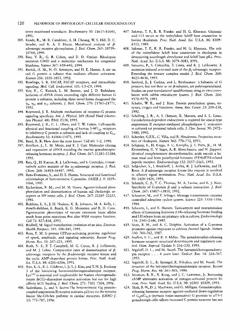
120 HANDBOOK OF PHYSIOLOGY-CELLULAR ENDOCRINOLOGY
toxin inactivated transducin. Biochemistry 30: 11637-1 1645, 1991.
389. Rands, R., M. R. Candelore, A. H. Cheung, W. S. Hill, D. C. Strader, and R. A. F. Dixon. Mutational analysis of p- adrenergic receptor glycosylation. 1. Biol. Chem. 265: 10759- 10764, 1990.
390. Rao, V. R., G. B. Cohen, and D. D. Oprian. Rhodopsin mutation G90D and a molecular mechanism for congenital blindness. Nature 367: 639-642, 1994.
391. Rarick, H. M., N. 0. Artemyev, and H. E. Hamm. A site on rod G protein a subunit that mediates effector activation. Science 256: 1031-1033, 1992.
392. Rawlings, S. R. PACAP, PACAP receptors, and intracellular signaling. Mol. Cell. Endocrinol. 101: C5-C9, 1994.
393. Ray, K., C. Kunsch, L. M. Bonner, and J. D. Robishaw. Isolation of cDNA clones enconding eight different human G protein y subunits, including three novel forms designated the y4, ylo and yl, subunits. 1. Biol. Chem. 270: 21765-21771, 1995.
394. Raymond, J. R. Multiple mechanisms of receptor-(; protein signaling specificity. Am. J. Physiol. 269 (Renal Fluid Electro- lyre Physiol. 40): F141-Fl58, 1995.
395. Raymond, J. R., C. L. Olsen, and T. W. Gettys. Cell-specific physical and functional coupling of human 5-HT,, receptors to inhibitory G protein a subunits and lack of coupling to Gsw. Biochemistry 32: 11064-11073, 1993.
396. Reed, R. R. How does the nose know? Cell 60: 1-2, 1990. 397. Reinhart, J., L. M. Mertz, and K. J. Catt. Molecular cloning
and expression of cDNA encoding the murine gonadotropin- releasing hormone receptor. J. Biol. Chem. 267: 21281-21284, 1992.
398. Ren, Q., H. Kurose, R. J. Lefkowitz, and S. Cotecchia. Consti- tutively active mutants of the qadrenergic receptor. 1. Biol. Chem. 268: 16483-16487, 1993.
399. Rens-Domiano, S., and H. E. Hamm. Structural and functional relationships of heterotrimeric G-proteins. FASEB J . 9: 1059- 1066, 1995.
400. Richardson, R. M., and M. M. Hosey. Agonist-induced phos- phorylation and desensitization of human m2 cholinergic re- ceptors in Sf9 insect cells. J . Biol. Chem. 267: 22249-22255, 1992.
401. Robbins, L. S., J. H. Nadeau, K. R. Johnson, M. A. Kelly, L. Roselli-Rehfuss, E. Baack, K. G. Mountjoy, and R. D. Cone. Pigmentation phenotypes of variant extension locus alleles result from point mutations that alter MSH receptor function. Cell 72: 827-834, 1993.
402. Rodbell, M. Signal transduction: evolution of an idea. Environ. Health Perspect. 103: 338-345, 1995.
403. Ross, E. M. G protein GTPase-activating proteins: regulation of speed, amplitude, and signaling selectivity. Recent Prog. Horm. Res. 50: 207-221, 1995.
404. Roth, N. S., P. T, Campbell, M. G. Caron, R. J. Lefkowitz, and M. J. Lohse. Comparative rates of desensitization of p- adrenergic receptors by the p-adrenergic receptor kinase and the cyclic AMP-dependent protein kinase. Proc. Natl. Acad. Sci. U.S.A. 88: 6201-6204, 1991.
405. Ryu, K.-S., R. L. Gilchrist, I. Ji, S.-J. Kim, andT. H. Ji. Exoloop 3 of the luteinizing hormone/choriogonadotropin receptor. LysSs3 is essential and irreplaceable for human choriogonado- tropin (hCG)-dependent receptor activation but not for high affinity hCG binding. 1. Biol. Chem. 271: 7301-7304, 1996.
406. Sadoshima, J., and S. 1zumo.The heterotrimeric Gq protein- coupled angiotensin I1 receptor activates p2lras via the tyrosine kinase Shc-Grb-Sos pathway in cardiac myocytes. EMBO I . 15: 775-787, 1996.
407. Sakmar, T. P., R. R. Franke, and H. G. Khorana. Glutamic acid-113 serves as the retinylidene Schiff base counterion in bovine rhodopsin. Proc. Natl. Acad. Sci. U.S.A. 86: 8309- 8313, 1989.
408. Sakmar, T. P., R. R. Franke, and H. G. Khorana. The role of the retinylidene Schiff base counterion in rhodopsin in determining wavelength absorbance and Schiff base pKa. Proc. Natl. Acad. Sci. U.S.A. 88: 3079-3083, 1991.
409. Samama, P., S. Cotecchia, T. Costa, and R. J. Lefkowitz. A mutation-induced activated state of the p,-adrenergic receptor. Extending the ternary complex model. I . Biol. Chem. 268: 4625-4636, 1993.
410. Sanford, J., J. Codina, and L. Birnbaumer. y-Subunits of G proteins, but not their a- or p-subunits, are polyisoprenylated. Studies on post-translational modifications using in vitro trans- lation with rabbit reticulocyte lysates. J . Biol. Chem. 266:
411. Schafer, W. R., and J. Rine. Protein prenylation: genes, en- zymes, targets and functions. Annu. Rev. Genet. 25: 209-238, 1992.
412. Schelling, J. R., A. S. Hanson, R. Marzen, and S. L. Linas. Cytoskeleton-dependent endocytosis is required for apical-type angiotensin I1 receptor-mediated phospholipase C activation in cultured rat proximal tubule cells. 1. Clin. Invest. 90: 2472- 2480, 1992.
413. Schertler, G.F.X., C. Villa, and R. Henderson. Projection struc- ture of rhodopsin. Nature 362: 770-772, 1993.
414. Schipani, E., H. Karga, A. C. Karaplis, J. T. Potts, Jr., H. M. Kronenberg, G. V. Segre, A. B. Abou-Samra, and H. Juppner. Identical complementary deoxyribonucleic acids encode a hu- man renal and bone parathyroid hormone (PTH)/PTH-related peptide receptor. Endocrinology 132: 2157-2165, 1993.
415. Schleicher, S., I. Boekhoff, J. Arriza, R. J. Lefkowitz, and H. Breer. A P-adrenergic receptor kinase-like enzyme is involved in olfatory signal termination. Proc. Natl. Acad. Scr. U.S.A.
416. Schmidt, C. J., T. C. Thomas, M. A, Levine, and E. J. Neer. Specificity of G-protein p and y subunit interaction. I . Biol. Chem. 267: 13807-13810, 1992.
417. Schramm, M., and Z. Selinger. Message transmission: receptor controlled adenylate cyclase system. Science 225: 1350-1356, 1984.
418. Schvarts, I., and E. Hazum. Tunicarnycin and neuraminidase effects of luteinizing hormone (LH)-releasing hormone binding and LH release from rat pituitary cels in culture. Endocrinology
419. Scott, R. H., and A. C. Dolphin. Activation of a G-protein promotes agonist responses to calcium channel ligands. Nature
420. Seaflon, S. C., and R. P. Millar. The gonadotrophin-releasing hormone receptor: structural determinants and regulatory con- trol. Hum. Reprod. Update 1: 216-230, 1995.
421. Segaloff, D. L., and M. Ascoli. The lutropidchoriogonadotro- pin receptor . . . 4 years later. Endocr. Rev. 14: 324-347, 1993.
422. Segaloff, D. L., R. Sprengel, K. Nikolics, and M. Ascoli. The structure of the lutropidchoriogonadotropin receptor. Recent Prog. Horm. Res. 46: 261-303, 1990.
423. Sevetson, B. R., X. Kong, and J. C. Lawrence, Jr. Increasing CAMP attenuates activation of mitogen-activated protein ki- nase. Proc. Natl. Acad. Sci. U.S.A. 90: 10305-10309, 1993.
424. Shah, B. H., D. J. MacEwan, and G. Milligan. Gonadotrophin- releasing hormone receptor agonist-mediated down-regulation of G,cu/Gl,a (pertusis toxin-insensitive) G proteins in aT3-1 gonadotroph cells reflects increased G protein turnover but not
9570-9579, 1991.
90: 1420-1424, 1993.
116: 2341-2346, 1985.
330: 760-762, 1987.
CHAPTER 6: G PROTEIN-COUPLED RECEPTORS 121
443. Stanislaus, D., J. A. Janovick, S. Brothers, and P. M. Conn. Regulation of Gq,llo by the gonadotropin-releasing hormone receptor. Mol. Endocrinol. 11: 738-746, 1997.
444. Sterne-Marr, R., and J. L. Benovic. Regulation of G protein- coupled receptors by receptor kinases and arrestins. Vitam.
445. Stiles, G. L., J. L. Benovic, M. G. Caron, and R. J. Lefkowitz. Mammalian beta-adrenergic receptors, distinct glycoprotein populations containing high mannose or complex type carbo- hydrate chains. J. Biol. Chem. 259: 8655-8663, 1984.
446. Stoffel, R. H., R. R. Randall, R. T. Premont, R. J. Lefkawitz, and J. Inglese. Palmitoylation of G protein-coupled receptor kinase, GRK6: lipid modification diversity in the GRK family. J. Biol. Chem. 269: 27791-27794,1994.
447. Stone, D. E., G. M. Cole, M. De Barros Lopez, M. Goebl, and S. I. Reed. N-Myristoylation is required for function of the pheromone-responsive G a protein of yeast: conditional activa- tion of the pheromone response by a temperature-sensitive N- myristoyl transferase. Genes Dev. 5: 1969-1981, 1991.
448. Strader, C. D., M. R. Candelore, W. S. Hill, I. S. Sigal, and R. A. Dixon. Identification of two serine residues involved in agonist activation of the P-adrenergic receptor. J. Biol. Chem.
449. Strader, C. D., R. A. Dixon, A. H. Cheung, M. R. Candelore, A. D. Blake, and I. S. Sigal. Mutations that uncouple the p- adrenergic receptor from Gs and increase agonist affinity. J. Biol. Chem. 262: 16439-16443, 1987.
450. Strader, C. D., T, M. Fong, M. P. Graziano, and M. R. Tota. The family of G-protein-coupled receptors. FASEB J. 9: 745- 754, 1995.
451. Strader, C. D., T. M. Fong, M. R. Tota, and D. Underwood. Structure and function of G protein-oupled receptors. Annu. Rev. Biochem. 63: 101-132, 1994.
452. Strader, C. D., T. Gaffney, E. E. Sugg, M. R. Candelore, R. Keys, A. A. Patchett, and R.A.F. Dixon. Allele-specific activa- tion of genetically engineered receptors. J. Biol. Chem. 266: 5- 8, 1991.
453. Strader, C. D., I. S. Sigal, M. R. Candelore, E. Rands, W. S. Hill, and R.A.F. Dixon. Conserved aspartate residues 79 and 113 of the P-adrenergic receptor have different roles in receptor function. J. Biol. Chem. 263: 10267-10271, 1988.
454. Strader, C. D., I. S. Sigal, and R.A.F. Dixon. Genetic ap- proaches to the determination of structure-function relation- ships of G protein-coupled receptors. Trends Pharmacol. Sci.
455. Strader, S. D., I. S. Sigal, and R. A. Dixon. Mapping the functional domains of the P-adrenergic receptor. Am. J. Resp. Cell Mol. Biol. 1: 81-86, 1989.
456. Strader, C. D., I. S. Sigal, R. B. Register, M. R. Candelore, E. Rands, and R. A. Dixon. Identification of residues required for ligand binding to the p-adrenergic receptor. Proc. Natl. Acad. Sci. U.S.A. 8 4 43844388, 1987.
457. Strathmann, M., and M. I. Simon. G protein diversity: a distinct class of Q subunits is present in vertebrates and inverte- brates. Proc. Natl. Acad. Sci. U.S.A. 87: 9113-9117, 1990.
458. Stryer, L. Visual excitation and recovery. J. Biol. Chem. 266:
459. Stryer, L., J. B. Hurley, and B. K. Fung. Transducin and the cyclic GMP phosphodiesterase of retinal rod outer segments. Methods Enzymol. 96: 617-627, 1983.
460. Su, Y.-F., T. K. Harden, and J. P. Perkins. Catecholamine- specific desensitization of adenylate cyclase: evidence for a multistep process. J. Biol. Chem. 255: 7410-7419, 1980.
461. Sullivan, K. A., R. T. Miller, S. B. Masters, B. Beiderman, W. Heideman, and H. R. Bourne. Identification of receptor contact
HOVTZ. 51: 193-234, 1995.
264: 13572-13578, 1989.
(SUPPI.): 26-30, 1989.
10711-10714, 1991.
alterations in mRNA levels. Proc. Natl. Acad. Sci. U.S.A. 92: 1886-1890, 1995.
425. Shapiro, R. A., and N. M. Nathanson. Deletion analysis of the mouse m l muscarinic acetylcholine receptor: effects on phosphoinositide metabolism and down-regulation. Biochemis-
426. Shear, M., P. A. Insel, K. L. Melmon, and P. Coffino. Agonist- specific refractoriness induced by isoproterenol: studies with mutant cells. J. Biol. Chem. 251: 7572-7576, 1976.
427. Shenker, A., L. Laue, S. Kosugl, J. Merendino, Jr., T. Minegishi, and G. B. Cutler. A constitutively activating mutation of the luteinizing hormone receptor in familial male precocious pu- berty. Nature 365: 652-654, 1993.
428. Shinozawa, T., S. Uchida, E. Martin, D. Cafiso, W. Hubbell, and M. Bitensky. Additional component required for activity and reconstitution of light activated vertebrate photoreceptor GTPase. Proc. Natl. Acad. Sci. U.S.A. 77: 1408-1411, 1980.
429. Simon, M. A., G. S. Dodson, and G. M. Rubin. An SH3-SH2- SH3 protein is required for p21RaS' activation and binds to sevenless and Sos proteins in vitro. Cell 73: 169-177, 1993.
430. Simon, M. I., P. Strathmann, and N. Gautam. Diversity of G proteins in signal transduction. Science 252: 802-808, 1991.
431. Simonds, W. F., J. E. Butrynski, N. Gautam, C. G. Unsion, and A.M. Spiegel. G-protein By dimers. Membrane targeting requires subunit co-expression and intact y CAAX domain. J. Biol. Chem. 266: 5363-5366, 1991.
432. Simonds, W. F., P. K. Goldsmith, J. Codina, C. G. Unson, and A. M. Spiegel. G,, mediates qadrenergic inhibition of adenylyl cyclase in platelet membranes: in situ identification with GQ C-terminal antibodies. Proc. Natl. Acad. Sci. U.S.A. 86: 7809- 7813, 1989.
433. Smrcka, A. V., J. R. Helper, K. 0. Brown, and P. C. Sternweiss. Regulation of polyphosphoinositide-specific phospholipase C activity by purifed Gq. Science 251: 804-807, 1991.
434. Smrcka, A. V., and P. C. Sternweis. Regulation of purified subtypes of phosphatidylinositol-specific phospholipase Cp by G protein a and Py subunits. J. Biol. Chem. 268: 9667-9674, 1993.
435. Sondek, J., A. Bohm, D. G. Lambright, H. E. Hamm, and P. B. Sigler. Crystal structure of a G, protein Py dimer at 2.1 A resolution. Nature 379: 369-374, 1996.
436. Sondek, J., D. G. Lambright, J. P. Noel, H. E. Hamm, and P. B. Sigler. GTPase mechanism of G proteins from the 1.7-A crystal structure of transducin a-GDP-AIF,- . Nature 372: 276-279, 1994.
437. Spalding, T., N. Birdsall, C. Curtis, and E. Hulme. Acetylcho- line mustard labels the binding site aspartate in muscarinic acetylcholine receptors. J. Biol. Chem. 269: 40924097, 1994.
438. Spengler, D., C. Waeber, C. Pantaloni, F. Holsboer, J. Bockaert, P. H. Seburg, and L. Journot. Differential signal transduction by five splice variants of the PACAP receptor. Nature 365:
439. Spiegel, A. M. Defects in G protein-coupled signal transduction in human disease. Annu. Rev. Physiol. 58: 143-170, 1995.
440. Spiegel, A. M., P. S. Backlund, J. E. Butyrinski, T. L. Z. Jones, and W. F. Simonds. The G protein connection: molecular basis of membrane association. Trends Biochem. Sci. 16: 338-341, 1991.
441. Spiegel, A. M., A. Shenker, and L. S. Weinstein. Receptor- effector coupling by G proteins: implications for normal and abnormal signal transduction. Endocr. Rev. 13: 536-565, 1992.
442. Spring, D. J., and E. J. Neer. A 14 amino acid region of the G protein y subunit is sufficient to confer the selectivity of a bindingtothep subunit. J. Biol. Chem.269:22882-22886,1994.
try 28: 8946-8950, 1989.
170-175, 1993.
122 HANDBOOK OF PHYSIOLOGY-CELLULAR ENDOCRINOLOGY
site involved in receptor-G protein coupling. Nature 330: 758- 760, 1987.
462. Sunahara, R. K., C. W. Dessauer, and A. G. Gilman. Complex- ity and diversity of mammalian adenylyl cyclases. Annu. Rev. Pharmacol. Toxicol. 36: 461480, 1996.
463. Suprenant, A., D. A. Horstman, H. Akbarali, and L. E. Lim- bird. A point mutation of the a2-adrenoceptor that blocks coupling to potassium but not calcium currents. Science 257:
464. Suzuki, H., G. N. Prado, N. Wilkinson, and J. Navarro. The N-terminus of interleukin-8 (IL-8) receptor confers high affinity binding to human IL-8. J . Biol. Chem. 269: 18263-18266, 1994.
465. Takeuchi, K., N. Takahashi, T. Abe, and K. Abe. Two isoforms of the rat kidney EP3 receptor derived by alternative RNA splicing: intrarenal expression co-localization. Biochem. Bio- phys. Res. Commun. 199: 834-840, 1994.
466. Tanabe, Y., M. Masu, T. Ishii, R. Shigemoto, and S. Nakanishi. A family of metabotropic glutamate receptors. Neuron 8: 169- 179, 1992.
467. Tang, W.-J., and A. G. Gilman. Type-specific regulation of adenylyl cyclase by G protein Py subunits. Science 254: 1500- 1504, 1991.
468. Tang, W.-J., and A. G. Gilman. Adenylyl cyclases. Cell 70:
469. Tapanainen, J. S., M. Bo, L. Dunkel, H. Billig, E. A. Perlas, I. Boime, and A.J.W. Hsueh. Deglycosylation of the human luteinizing hormone receptor does not affect ligand binding and signal transduction. Endocr. J . 1: 219-225, 1993.
470. Taussig, R., J. A. Iiiiguez-Lluhi, and A. G. Gilman. Inhibition of adenylyl cyclase by Gm,. Science 261: 218-221, 1993.
471. Taussig, R., L. M. Quarmby, and A. G. Gilman. Regulation of purified type-I and type-I1 adenylyl cyclases by G protein By subunits. J. Biol. Chem. 268: 9-12, 1993.
472. Taussig, R., W.-J. Tang, J. R. Hepler, and A. G. Gilman. Distinct patterns of bidirectional regulation of mammalian adenylyl cyclases. J . Biol. Chem. 269: 6093-6100, 1994.
473. Thomas, D., T. G. Rozell, X. Liu, and D. L. Segaloff. Muta- tional analyses of the extracellular domain of the full-length lutropinkhoriogonadotropin receptor suggest leucine-rich re- peats 1-6 are involved in hormone binding. Mol. Endocrinol.
474. Thomas, D. D., and L. Stryer. Transverse location of the retinal chromophore of rhodopsin in rod outer segment disc membranes. J . Mol. Biol. 154: 145-157, 1982.
475. Thomas, T. C., C. J. Schmidt, and E. J. Neer. G protein a, subunit: mutation of conserved cysteines identifies a subunit contact surface and alters GDP affinity. Proc. Natl. Acad. Sci.
476. Thompson, P., and J.B.C. Findlay. Phosphorylation of ovine rhodopsin. Biochem J . 220: 773-780, 1984.
477. Thorens, B. Expression cloning of the pancreatin p cell receptor for the gluco-incretin hormone glucagon-like peptide. Proc. Natl. Acad. Sci. U.S.A. 89: 8641-8645, 1992.
478. Tota, M. R., and C. D. Strader. Characterization of the binding domain of the P-adrenergic receptor with the fluorescent antag- onist carazolol. Evidence for a buried ligand binding site. J . Biol. Chem. 265: 16891-16897, 1990.
479. Touhara, K., B. E. Hawes, T. van Biesen, and R. J. Lefkowitz. G protein By subunits stimulate phosphorylation of Shc adapter protein. Proc. Natl. Acad. Sci. U.S.A. 92: 9284-9287, 1995.
480. Trumpp-Kallmeyer, S., B. Chini, B. Mouillac, C. Barberis, J. Hoflack, and M. Hibert. Towards understanding the role of the first extracellular loop for the binding of peptide hormones
977-980, 1992.
869-872, 1992.
10: 760-768, 1996.
U.S.A. 90: 10295-10299, 1993.
to G-protein coupled receptors. Pharm. Acta Helv. 70: 255- 262, 1995.
481. Tsai-Morris, C. H., E. Buczko, W. Wang, and M. L. Dufau. Intronic nature of the rat luteinizing hormone receptor gene defines a soluble receptor subspecies with hormone binding activity. J. Biol. Chem. 265: 19385-19388, 1990.
482. Tsai-Morris, C. H., E. Buczko, W. Wang, X. Z. Xie, and M. L. Dufau. Structural organization of the rat luteinizing hor- mone (LH) receptor gene. J. Biol. Chem. 266: 11355-11359, 1991.
483. Ullrich, A., and J. Schlessinger. Signal transduction by receptor with tyrosine kinase activity. Cell 61: 203-212, 1990.
484. Vaillancourt, R. R., A. M. Gardner, and G. L. Johnson. B-Raf- dependent regulation of the MEK-Umitogen-activated protein kinase pathway in PC12 cells and regulation by cyclic AMP. Mol. Cell. Biol. 14: 6522-6530, 1994.
485. van Biesen, T., B. E. Hawes, D. K. Luttrell, K. M. Krueger, K. Touhara, E. Porfiri, M. Sakaue, L. M. Luttrell, and R. J. Lef kowitz. Receptor-tyrosine-kinase- and Gpy-mediated MAP kinase activation by a common signalling pathway. Nature
486. van Biesen, T. M., L. M. Luttrell, B. E. Hawes, and R. J. Lef kowitz. Mitogenic signaling via G protein-coupled recep- tors. Endocr. Rev. 17: 698-714, 1996.
487. van Corven, E. J., P. L. Hordijk, R. H. Medema, J. L. Bos, and W. H. Moolenaar. Pertussis toxin-sensitive activation of p2lras by G protein-coupled receptor agonist in fibroblasts. Proc. Natl. Acad. Sci. U.S.A. 90: 1257-1261, 1993.
488. Van Dop, C., M. Tsubokawa, H. R. Bourne, and J. Ramachan- dran. Amino acid sequence of retinal transducin at the site ADP-ribosylated by cholera toxin. J . Biol. Chem. 259: 696- 699, 1984.
489. van Koppen, C. J., and N. M. Nathanson. The cysteine residue in the carboxy-terminal domain of the m2 muscarinic acetyl- choline receptor is not required for receptor-mediated inhibi- tion of adenylate cyclase. /. Neurochem. 57: 1873-1877, 1991.
490. Van Sande, J., E. Raspe, J. Perret, C. Lejeune, C. Maenhaut, G. Vassar, and J. E. Dumont. Thyrotropin activates both the CAMP and the PIP, cascades in C H O cells expressing the human cDNA of the TSH receptor. Mol. Cell. Endocrinol. 74:
491. Vara Prasad, M.V.V.S., J. M. Dermott, L. E. Heasley, G. L. Johnson, and N. Dhanasekaran. Activation of Jun kinase/ stress-activated protein kinase by GTPase-deficient mutants of Gal, and G G ~ , ~ . J . Biol. Chem. 270: 18655-18659, 1995.
492. Vassart, G., L. Desarnaud, L. Duprez, D. Eggerickx, 0. Labbk, F. Libert, C. Mollerau, J. Parma, R. Paschke, M. Tonacchera, P. Vanderhaeghen, J. Van Sande, J. Dumont, and M. Parmen- tier. The G protein-coupled receptor family and one of its members, the TSH receptor. Ann. N.Y. Acad. Sci. 766: 23-30, 1995.
493. Von Weizsacker, E., M. P. Strathmann, and M. I. Simon. Diversity among the p subunits of heterotrimeric GTP-binding proteins: characterization of a novel @-subunit. Biochem. Bio- phys. Res. Commun. 183: 350-356, 1992.
494. von Zastrow, M., and B. K. Kobilka. Ligand-regulated internal- ization and recycling of human &-adrenergic receptors be- tween the plasma membrane and endosomes containing trans- ferrin receptors. J . Biol. Chem. 267: 3530-3538, 1992.
495. von Zastrow, M., R. Link, D. Daunt, G. Barsh, and B. K. Kobilka. Subtype specific differences in the intracellular sorting of G protein-coupled receptors. J . Biol. Chem. 268: 763-766, 1993.
496. Voyno-Yasenetskaya, T., B. R. Conklin, R. L. Gilbert, R.
376: 781-784, 1995.
Rl-R6, 1990.
CHAPTER 6: G PROTEIN-COUPLED RECEPTORS 123
Cysteine-347 is the ADP ribose acceptor site. J. Biol. Chem.
513. Wheeler, G. L., and M. W. Bitensky. A light-activated GTPase in vertebrate photoreceptors: regulation of light-activated cyclic GMP phosphodiesterase. Proc. Natl. Acad. Sci. U.S.A. 74: 42384242, 1977.
514. Wheeler, M. B., M. Lu, J. S. Dillon, X.-H. Leng, C. Cheng, and A. E. Boyd 111. Functional expression of the rat glucagon- like peptide-I receptor: evidence for coupling to both adenylyl cyclase and phospholipase C. Endocrinology 133: 57-62, 1993.
515. Wickman, K., and D. E. Clapham. Ion channel regulation by G proteins. Physiol. Rev. 75: 865-885, 1995.
516. Wickman, K. D., J. Ifiiguez-Lluhi, P. Davenport, R. A. Taussig, G. B. Krapivinsky, M. E. Linder, A. Gilman, and D. E. Clap- ham. Recombinant GP-y activates the muscarinic-gated atrial potassium channel IKACh. Nature 368: 255-257, 1994.
517. Wieland, T., B. Nurnberg, I. Ulibarri, S. Kaldenberg-Stasch, G. Schultz, and K. H. Jakobs. Guanine nucleotide-specific phosphate transfer by guanine nucleotide-binding regulatory protein p-subunits: characterization of the phosphorylated amino acid. J. Biol. Chem. 268: 18111-18118, 1993.
518. Wilcox, C., J.-S. Hu, and E. N. Olson. Acylation of proteins with myristic acid occurs cotranslationally. Science 238: 1275- 1278, 1987.
519. Wilden, U., S. W. Hall, and H. Kuhn. Phosphodiesterase activa- tion by photoexcited rhodopsin is quenched when rhodopsin is phosphorylated and binds the intrinsic 48-kDA protein of rod outer segments. Proc. Natl. Acad. Sci. U.S.A. 83: 1174- 1178, 1986.
520. Wilden, U., and H. Kuhn. Light-dependent phosphorylation of rhodopsin: number of phosphorylation sites. Biochemistry 21:
521. Wilkie, T. M., P. A. Scherle, M. P. Strathmann, V. Z . Slepak, and M. I. Simon. Characterization of G-protein a subunits in the G, class: expression in murine tissues and in stromal and hematopoietic cell lines. Proc. Natl. Acad. Sci. U.S.A. 88:
522. Winitz, S., M. Russell, N, Qian, A. Gardner, L. Dwyer, and G. L. Johnson. Involvement of Ras and Raf in the Gi-coupled acetylcholine muscarinic m2 receptor activation of mitogen- activated protein (MAP) kinase kinase and MAP kinase. I. Biol. Chem. 268: 19196-19199, 1993.
523. Wise, A., M.-A.Watson-Koken, S. Rees, M. Lee, and G. Milli- gan. Interactions of the a,,-adrenoceptor with multiple G,- family G-proteins: studies with pertussis toxin-resistant G- protein mutants. Biochem. J. 321: 721-728, 1997.
524. Wong, S. K.-F., E. M. Parker, and E. M. Ross. Chimeric muscarinic cholinergic: P-adrenergic receptors that activate G, in response to muscarinic agonist. J. Biol. Chem. 265: 6219- 6224, 1990.
525. Wong, S. K.-F., C. Slaughter, A. E. Ruoho, and E. M. Ross. The catecholamine binding site of the P-adrenergic receptor is formed by juxtaposed membrane-spanning domains. I . Biol. Chem. 263: 7925-7928, 1988.
526. Wong, Y. H., B. R. Conklin, and H. R. Bourne. G,-mediated hormonal inhibition of cyclic AMP accumulation. Science 255:
527. Wong, Y. H., A. Federman, A. M. Pace, I. Zachary, T. Evans, J. Pouyssegur, and H. R. Bourne. Mutant a subunits of G,2 inhibit cyclic AMP accumulation. Nature 351: 63-65, 1991.
528. Wu, D., C. H. Lee, S. G. Ree, and M. I. Simon. Activation of phospholipase C by the a subunits of G, and GI, proteins in transfected Cos-7 cells. J. Biol. Chem. 267: 1811-1817, 1992.
260: 14428-14430, 1985.
3014-3022, 1982.
10049-10053, 1991.
339-341, 1992.
Hooley, H. R. Bourne, and D. L. Barber. Gal, stimulates Na- H exchange. J . Biol. Chem. 269: 472111724, 1994.
497. Vuong, T. M., and M. Chabre. Deactivation kinetics of the transduction cascade of vision. Proc. Natl. Acad. Sci. U.S.A.
498. Wadsworth, H. L., G . D. Chazenbalk, Y. Nagayama, D. Russo, and B. Rapoport. An insertion in the human thyrotropin recep- tor critical for high affinity hormone binding. Science 249:
499. Wall, M. A,, D. E. Coleman, E. Lee, J. A. Ifiiguez-Lluhi, B. A. Posner, A. G. Gilman, and S. R. Sprang. The structure of the G protein heterotrimer GlalPly2. Cell 83: 1047-1068, 1995.
500. Wang, C. D., M. A. Buck, and C. M. Fraser. Site-directed mutagenesis of a,,-adrenergic receptors: identification of amino acids involved in ligand binding and receptor activation by agonists. Mol. Pharmacol. 40: 168-179, 1991.
501. Wang, D. S., R. Shaw, J. C. Winkelmann, and G. Shaw. Binding of PH domains of P-adrenergic receptor kinase and P-spectrin to WD40/p-transducin repeat containing regions of the p- subunit of trimeric G-proteins. Biochem. Biophys. Res. Com- mun. 203: 29-35, 1994.
501a. Wang, Z . , R. W. Hipkin, and M. Ascoli. Progressive cyto- plasmic tail truncations of the lutropin-choriogonadotropin receptor prevent agonist- or phorbol ester-induced phosphory- lation, impair agonist- or phorbol ester-induced desensitization, and enhance agonist-induced receptor down-regulation. Mol. Endocrinol. 10: 748-759, 1996.
502. Wang, Z., X. Idu, and M. Ascoli. Phosphorylation of the lutropidchoriogonadotropin receptor facilitates uncoupling of the receptor from adenylyl cyclase and endocytosis of the bound hormone. Mol. Endocrinol. 11: 183-192, 1997.
503. Watson, A. J., A. Katz, and M. I. Simon. A fifth member of the mammalian G-protein P-subunit family. J . Biol. Chem.
504. Watson, N., M. E. Liknder, K. M. Druey, J. H. Kehrl, and K. J. Blumer. RGS family members: GTPase-activating proteins for heterotrimeric G-protein a-subunits. Nature 383: 172-177, 1996.
505. Wedegaertner, P. B., and H. R. Bourne. Activation and depalmi- toylation of G3a. Cell 77: 1063-1070, 1994.
506. Wedegaertner, P. B., H. R. Bourne, and M. von Zastrow. Activation-induced subcellular redistribution of Gsa. Mol. Biol. Cell 7: 1225-1233, 1996.
507. Wedegaertner, P. B., D. H. Chu, P. T. Wilson, M. J. Levis, and H. R. Bourne. Palmitoylation is required for signaling functions and membrane attachment of G,, and Gsa. J. Biol. Chem. 268:
508. Wess, J. Molecular basis of muscarinic acetylcholine receptor function. Trends Pharmacol. Sci. 14: 308-313, 1993.
509. Wess, J., T. I. Bonner, F. Dorje, and M. R. Brann. Delineation of muscarinic receptor domains conferring selectivity of cou- pling to guanine nucleotide-binding proteins and second mes- sengers. Mol. Pharmacol. 38: 517-523, 1990.
510. Wess, J., M. R. Brann, and T. I. Bonner. Delineation of musca- rinic receptor domains conferring selectivity of coupling to guanine nucleotide-binding proteins and second messengers.
511. Wess, J., D. Gdula, and M. R. Brand. Site-directed mutagenesis of the m3 muscarinic receptor: identification of a series of threonine and tyrosine residues involved in agonist but not antagonist binding. EMBO J . 10: 3729-3734, 1991.
512. West, R. E., J. Moss, M. Vaughan, T. Lui, and T. Y. Lin. Pertussis toxin-catalyzed ADP-ribosylation of transducin.
88: 9813-9817,1991.
1423-1425, 1990.
269: 22150-22156, 1994.
25001-25008, 1993.
FEBS Lett. 358: 133-136, 1989.
124 HANDBOOK OF PHYSIOLOGY-CELLULAR ENDOCRINOLOGY
529. Yang, K., and N. Gautman. Structural determinants for interac- tion with three different effectors on the G protein p subunit. J. Biol. Chem. 272: 2056-2059, 1997.
530. Yatani, A., J. Codina, Y. Imoto, J. P. Reeves, L. Birnbaumer, and A. M. Brown. A G-protein directly regulates mammalian cardiac calcium channels. Science 238: 1288-1292, 1987.
531. Yatani, A., R. Mattera, J. Codina, R. Graf, K. Okabe, E. Padrell, R. Iyenegar, A. M. Brown, and L. Birnbaumer. The G protein-gated atrial K+ channel is stimulated by three distinct Gla subunits. Nature 336: 680-682, 1988.
532. Yi, F., B. M. Denker, and E. J. Neer. Structural and functional studies of cross-linked G, protein subunits. I . Biol. Chem. 266:
533. Ykota, Y., C. Akazawa, H. Ohkubo, and S. Nakanishi. Delin- eation of structural domains involved in the subtype specificity of the tachykinin receptors through chimeric formation of substance €'/substance K receptors. EMBO J. 11: 3585-3591, 1992.
534. Yuzaki, M., and K. Mikoshiba. Pharmacological and immuno- cytochemical characterization of metabotropic glutamate re- ceptors in cultured Purkinje cells. J. Neurosci. 12: 42534263, 1992.
535. Zhang, R., C. H. Tsai-Morris, M. Kitamura, E. Buczko, and M. L. Dufau. Changes in binding activity of luteinizing hor- mone receptors by site directed mutagenesis of potential glyco-
2900-3906, 1991.
sylation sites. Biochem. Biophys. Res. Commun. 181: 804- 808, 1991.
536. Zhang, S., 0. A. Coso, C. Lee, J. S. Gutkind, and W. F. Simonds. Selective activation of effector pathways by brain- specific G protein &-. J. Biol. Chem. 271: 33575-33579, 1996.
537. Zheng, F., and J. P. Gallagher. (lS,3R)-l-Aminocyclopentane- 1,3-dicarboxylic acid-induced burst firing is mediated by native pertussis toxin-sensitive metabotropic receptor at rat dorsolat- era1 septa1 nucleus neurons. Neuroscience 68: 423434, 1995.
538. Zhou, W., C. Flanagan, J. A. Ballesteros, K. Konvicka, J. S. Davidson, H. Weinstein, R. P. Millar, and S. C. Seaflon. A reciprocal mutation supports helix 2 and helix 7 proximity in the gonadotropin-releasing hormone receptor. Mol. Pharma-
539. Zhou, W., V. Rodic, S. Kitanovic, C. A. Flanagan, L. Chi, H. Weinstein, S. Maayani, R. P. Millar, and S. C. Seaflon. A locus of the gonadotropin-releasing hormone receptor that differentiates agonist and antagonist binding sites. J. Biol. Chem. 270: 18853-18857, 1995.
540. Zhu, O., A. Wang, and M. Ascoli. The lutropidchoriogonado- tropin receptor is palmitoylated at intracellular cysteine resi- dues. Mol. Endocrinol. 9: 141-150, 1995.
541. Zhukovsky, E. A,, and D. D. Oprian. Effect of carboxylic acid side chains on the absorption maximum of visual pigments. Science 246: 928-930, 1989.
CO~. 45: 165-170, 1994.