complicated untreated apical periodontitis causing ... · endodontic treatment or retreatment was...
TRANSCRIPT
CASE REPORT
Complicated untreated apical periodontitis causing paraesthesia:A case reportDomenico Ricucci, MD, DDS
1,* ; Simona Loghin, DDS1; and Jos�e F. Siqueira Jr, DDS, MSc, PhD2
1 Private Practice, Cetraro, Italy
2 Department of Endodontics, Faculty of Dentistry, Est�acio de S�a University, Rio de Janeiro, RJ, Brazil
Keywords
apical periodontitis, calcium hydroxide,
endodontic retreatment, mandibular canal
paraesthesia, treatment decision-making.
Correspondence
Domenico Ricucci, Piazza Calvario, 7, 87022
Cetraro (CS), Italy. Email: [email protected]
doi: 10.1111/aej.12220
(Accepted for publication 10 July 2017.)
Abstract
The purpose of this article was to report a case of untreated apical periodontitis
resulting in severe late complications. A patient with an asymptomatic
crowned root canal-treated mandibular molar revealing a radiographic sub-
standard endodontic treatment and a slight periapical radiolucency was made
aware of the treatment options and opted for no treatment. The lesion slightly
increased in size after 6 years, but the tooth remained asymptomatic and
endodontic retreatment was again refused. After 4 more years, the patient pre-
sented with an abscess and severe pain, complicated by paraesthesia of the left
chin and lip. Radiographic examination revealed that the lesion had increased
considerably to involve the mandibular canal. The treatment protocol included
long-term intracanal medication with calcium hydroxide and follow-ups
revealed complete resolution of the periapical radiolucency and the paraesthe-
sia had completely subsided.
Introduction
Apical periodontitis is caused by bacterial infection of the
root canal system (1). Intracanal bacteria are usually
organised as biofilms adhering to the root canal walls (2).
In approximately 80% of teeth with primary or post-
treatment apical periodontitis, bacterial biofilms occur in
the apical third of the canal system (2). Consequently,
successful treatment of apical periodontitis is dependent
upon effective control of intraradicular biofilms. If bacte-
ria survive the effects of treatment, there is a significantly
increased risk for the lesion to persist and even progress
(3).
Apical periodontitis is commonly asymptomatic (4)
and in many cases it is diagnosed by chance (5), for
example, following a radiographic examination for
implants or restorative planning. As the tooth is asymp-
tomatic, it may be difficult to convince a patient
endodontic treatment or retreatment was indicated (6). If
left untreated, an apical periodontitis lesion may develop
into a chronic abscess with an associated intra- or extra-
oral draining sinus tract, large cyst, chronic focal scleros-
ing osteomyelitis or possible exacerbations such as an
acute abscess and cellulitis, depending on host immunity.
This case report illustrates the risks of leaving apical
periodontitis untreated and reinforces the need for
proper endodontic treatment to avoid possible serious
complications.
Case report
A 42-year-old woman presented for a routine dental
examination. She was found to be periodontally sound
with some interproximal carious lesions in the maxillary
posterior teeth. The mandibular left first molar was
crowned, and the patient stated that the tooth had been
root canal-treated approximately 10 years earlier. The
tooth was asymptomatic and responded normally to ver-
tical and lateral percussion and palpation.
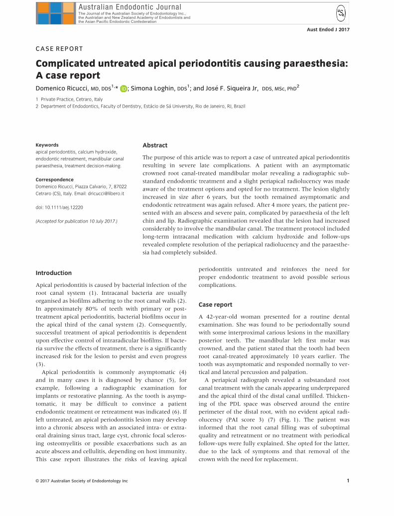
A periapical radiograph revealed a substandard root
canal treatment with the canals appearing underprepared
and the apical third of the distal canal unfilled. Thicken-
ing of the PDL space was observed around the entire
perimeter of the distal root, with no evident apical radi-
olucency (PAI score 3) (7) (Fig. 1). The patient was
informed that the root canal filling was of suboptimal
quality and retreatment or no treatment with periodical
follow-ups were fully explained. She opted for the latter,
due to the lack of symptoms and that removal of the
crown with the need for replacement.
© 2017 Australian Society of Endodontology Inc 1
Aust Endod J 2017
The patient returned 6 years later and requested the
possibility of replacement of her missing mandibular
right molar (46) with an implant. Tooth 36 was still
asymptomatic, and percussion and palpation gave
responses within normal limits. A periapical radiograph
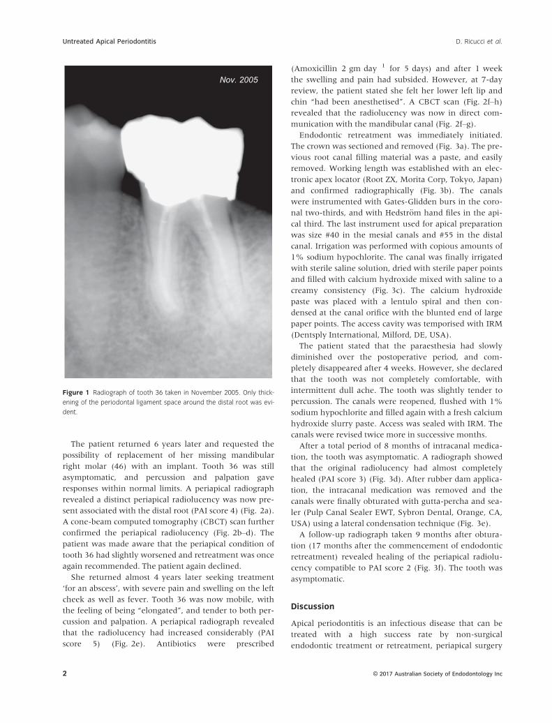
revealed a distinct periapical radiolucency was now pre-
sent associated with the distal root (PAI score 4) (Fig. 2a).
A cone-beam computed tomography (CBCT) scan further
confirmed the periapical radiolucency (Fig. 2b–d). The
patient was made aware that the periapical condition of
tooth 36 had slightly worsened and retreatment was once
again recommended. The patient again declined.
She returned almost 4 years later seeking treatment
‘for an abscess’, with severe pain and swelling on the left
cheek as well as fever. Tooth 36 was now mobile, with
the feeling of being “elongated”, and tender to both per-
cussion and palpation. A periapical radiograph revealed
that the radiolucency had increased considerably (PAI
score 5) (Fig. 2e). Antibiotics were prescribed
(Amoxicillin 2 gm day�1 for 5 days) and after 1 week
the swelling and pain had subsided. However, at 7-day
review, the patient stated she felt her lower left lip and
chin “had been anesthetised”. A CBCT scan (Fig. 2f–h)revealed that the radiolucency was now in direct com-
munication with the mandibular canal (Fig. 2f–g).Endodontic retreatment was immediately initiated.
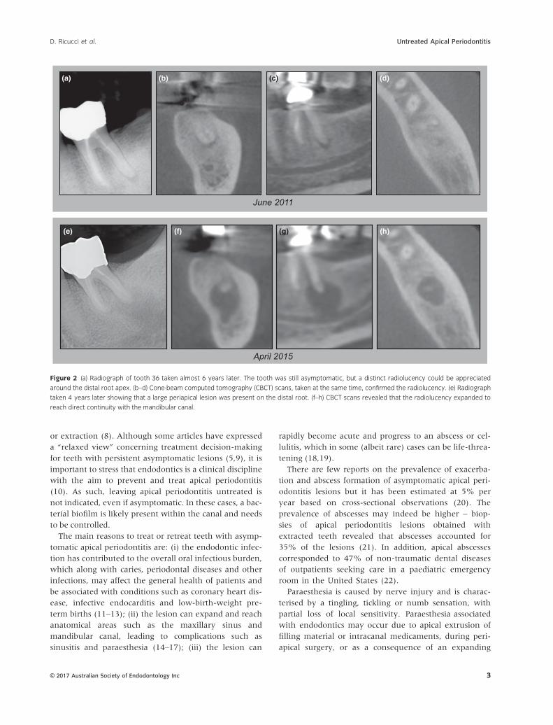
The crown was sectioned and removed (Fig. 3a). The pre-
vious root canal filling material was a paste, and easily
removed. Working length was established with an elec-
tronic apex locator (Root ZX, Morita Corp, Tokyo, Japan)
and confirmed radiographically (Fig. 3b). The canals
were instrumented with Gates-Glidden burs in the coro-
nal two-thirds, and with Hedstr€om hand files in the api-
cal third. The last instrument used for apical preparation
was size #40 in the mesial canals and #55 in the distal
canal. Irrigation was performed with copious amounts of
1% sodium hypochlorite. The canal was finally irrigated
with sterile saline solution, dried with sterile paper points
and filled with calcium hydroxide mixed with saline to a
creamy consistency (Fig. 3c). The calcium hydroxide
paste was placed with a lentulo spiral and then con-
densed at the canal orifice with the blunted end of large
paper points. The access cavity was temporised with IRM
(Dentsply International, Milford, DE, USA).
The patient stated that the paraesthesia had slowly
diminished over the postoperative period, and com-
pletely disappeared after 4 weeks. However, she declared
that the tooth was not completely comfortable, with
intermittent dull ache. The tooth was slightly tender to
percussion. The canals were reopened, flushed with 1%
sodium hypochlorite and filled again with a fresh calcium
hydroxide slurry paste. Access was sealed with IRM. The
canals were revised twice more in successive months.
After a total period of 8 months of intracanal medica-
tion, the tooth was asymptomatic. A radiograph showed
that the original radiolucency had almost completely
healed (PAI score 3) (Fig. 3d). After rubber dam applica-
tion, the intracanal medication was removed and the
canals were finally obturated with gutta-percha and sea-
ler (Pulp Canal Sealer EWT, Sybron Dental, Orange, CA,
USA) using a lateral condensation technique (Fig. 3e).
A follow-up radiograph taken 9 months after obtura-
tion (17 months after the commencement of endodontic
retreatment) revealed healing of the periapical radiolu-
cency compatible to PAI score 2 (Fig. 3f). The tooth was
asymptomatic.
Discussion
Apical periodontitis is an infectious disease that can be
treated with a high success rate by non-surgical
endodontic treatment or retreatment, periapical surgery
Nov. 2005
Figure 1 Radiograph of tooth 36 taken in November 2005. Only thick-
ening of the periodontal ligament space around the distal root was evi-
dent.
© 2017 Australian Society of Endodontology Inc2
Untreated Apical Periodontitis D. Ricucci et al.
or extraction (8). Although some articles have expressed
a “relaxed view” concerning treatment decision-making
for teeth with persistent asymptomatic lesions (5,9), it is
important to stress that endodontics is a clinical discipline
with the aim to prevent and treat apical periodontitis
(10). As such, leaving apical periodontitis untreated is
not indicated, even if asymptomatic. In these cases, a bac-
terial biofilm is likely present within the canal and needs
to be controlled.
The main reasons to treat or retreat teeth with asymp-
tomatic apical periodontitis are: (i) the endodontic infec-
tion has contributed to the overall oral infectious burden,
which along with caries, periodontal diseases and other
infections, may affect the general health of patients and
be associated with conditions such as coronary heart dis-
ease, infective endocarditis and low-birth-weight pre-
term births (11–13); (ii) the lesion can expand and reach
anatomical areas such as the maxillary sinus and
mandibular canal, leading to complications such as
sinusitis and paraesthesia (14–17); (iii) the lesion can
rapidly become acute and progress to an abscess or cel-
lulitis, which in some (albeit rare) cases can be life-threa-
tening (18,19).
There are few reports on the prevalence of exacerba-
tion and abscess formation of asymptomatic apical peri-
odontitis lesions but it has been estimated at 5% per
year based on cross-sectional observations (20). The
prevalence of abscesses may indeed be higher – biop-
sies of apical periodontitis lesions obtained with
extracted teeth revealed that abscesses accounted for
35% of the lesions (21). In addition, apical abscesses
corresponded to 47% of non-traumatic dental diseases
of outpatients seeking care in a paediatric emergency
room in the United States (22).
Paraesthesia is caused by nerve injury and is charac-
terised by a tingling, tickling or numb sensation, with
partial loss of local sensitivity. Paraesthesia associated
with endodontics may occur due to apical extrusion of
filling material or intracanal medicaments, during peri-
apical surgery, or as a consequence of an expanding
April 2015
(g) (h)(f)(e)
(a) (b) (c) (d)
June 2011
Figure 2 (a) Radiograph of tooth 36 taken almost 6 years later. The tooth was still asymptomatic, but a distinct radiolucency could be appreciated
around the distal root apex. (b–d) Cone-beam computed tomography (CBCT) scans, taken at the same time, confirmed the radiolucency. (e) Radiograph
taken 4 years later showing that a large periapical lesion was present on the distal root. (f–h) CBCT scans revealed that the radiolucency expanded to
reach direct continuity with the mandibular canal.
© 2017 Australian Society of Endodontology Inc 3
D. Ricucci et al. Untreated Apical Periodontitis
apical periodontitis lesion (16,23,24). This case of
paraesthesia of the lower left lip and chin was related
to the close proximity of the lesion to the mandibular
canal.
In the case reported, two reasons to not leave apical
periodontitis untreated were evident – the lesion devel-
oped into an acute abscess and expanded to affect the
mandibular canal, causing paraesthesia. These complica-
tions developed approximately 10 years after the patient
was initially seen. Patients should therefore be made
aware of the risks of leaving apical periodontitis
untreated.
Surgery was avoided in this case due to the tooth’s
location and that the lesion had reached and involved
the mandibular canal. Therefore, the canal was retreated
using an antimicrobial protocol with calcium hydroxide.
Paraesthesia fortunately resolved within 4 weeks. Ulti-
mately, the lesion completely healed 17 months after the
endodontic retreatment was initiated.
This case report has illustrated that, if left untreated,
apical periodontitis may exacerbate and lead to severe
complications such as paraesthesia. After appropriate
endodontic treatment, the patient’s symptoms and
paraesthesia had fortunately resolved.
Acknowledgements
The authors received no grants or other funding.
Disclosure
The authors have no interest to disclose.
Author contributions
All authors have contributed significantly. All authors
are in agreement with the manuscript.
References
1. Kakehashi S, Stanley HR, Fitzgerald RJ. The effects of sur-
gical exposures of dental pulps in germ-free and conven-
tional laboratory rats. Oral Surg Oral Med Oral Pathol
1965; 20: 340–9.
Dec 2015 (f)(e)
(b)(a) (c)
(d) Dec 2015 Sep 2016
Figure 3 (a) After removal of the crown, the tooth was isolated. (b) Working length was confirmed radiographically. (c) Intracanal medication with cal-
cium hydroxide. (d) Radiograph taken 8 months after beginning of retreatment. The radiolucent area had considerably reduced in size. (e) Postobtura-
tion radiograph. (f) Follow-up radiograph taken 9 months after root canal filling (17 months after beginning of retreatment). The periapical lesion
healed completely.
© 2017 Australian Society of Endodontology Inc4
Untreated Apical Periodontitis D. Ricucci et al.
2. Ricucci D, Siqueira JF Jr. Biofilms and apical periodontitis:
study of prevalence and association with clinical and
histopathologic findings. J Endod 2010; 36: 1277–88.
3. Fabricius L, Dahl�en G, Sundqvist G, Happonen RP, M€oller
AJ. Influence of residual bacteria on periapical tissue heal-
ing after chemomechanical treatment and root filling of
experimentally infected monkey teeth. Eur J Oral Sci
2006; 114: 278–85.
4. Tronstad L. Clinical endodontics. 3rd edn. Stuttgart:
Thieme; 2009.
5. Wesselink PR. The incidental discovery of apical periodon-
titis. Endod Topics 2014; 30: 23–8.
6. Friedman S, Mor C. The success of endodontic therapy–
healing and functionality. J Calif Dent Assoc 2004; 32:
493–503.
7. Ørstavik D, Kerekes K, Eriksen HM. The periapical index:
a scoring system for radiographic assessment of apical peri-
odontitis. Endod Dent Traumatol 1986; 2: 20–34.
8. Friedman S. Expected outcomes in the prevention and
treatment of apical periodontitis. In: Ørstavik D, Pitt Ford
T, eds. Essential endodontology. Oxford, UK: Blackwell
Munksgaard Ltd; 2008. pp. 408–69.
9. Bergenholtz G. Assessment of treatment failure in
endodontic therapy. J Oral Rehabil 2016; 43: 753–8.
10. Ørstavik D, Pitt Ford TR. Apical periodontitis: microbial
infection and host responses. In: Ørstavik D, Pitt Ford TR,
eds. Essential endodontology. 2nd edn. Oxford, UK:
Blackwell Munksgaard Ltd; 2008. pp. 1–9.
11. Siqueira JF Jr. Treatment of endodontic infections. Lon-
don: Quintessence Publishing; 2011.
12. Fouad AF. Endodontic infections and systemic disease. In:
Fouad AF, ed. Endodontic microbiology. Ames, Iowa:
Wiley-Blackwell; 2009. pp. 320–38.
13. Zhang J, Huang X, Lu B, Zhang C, Cai Z. Can apical peri-
odontitis affect serum levels of CRP, IL-2, and IL-6 as well
as induce pathological changes in remote organs? Clin
Oral Investig 2016; 20: 1617–24.
14. Pokorny A, Tataryn R. Clinical and radiologic findings in a
case series of maxillary sinusitis of dental origin. Int
Forum Allergy Rhinol 2013; 3: 973–9.
15. Patel NA, Ferguson BJ. Odontogenic sinusitis: an ancient
but under-appreciated cause of maxillary sinusitis. Curr
Opin Otolaryngol Head Neck Surg 2012; 20: 24–8.
16. Alves FR, Coutinho MS, Goncalves LS. Endodontic-
related facial paraesthesia: systematic review. J Can Dent
Assoc 2014;80:e13.
17. Mohammadi Z. Endodontics-related paraesthesia of the
mental and inferior alveolar nerves: an updated review.
J Can Dent Assoc 2010; 76: a117.
18. Li X, Tronstad L, Olsen I. Brain abscesses caused by oral
infection. Endod Dent Traumatol 1999; 15: 95–101.
19. Pappa H, Jones DC. Mediastinitis from odontogenic infec-
tion. A case report. Brit Dent J 2005; 198: 547–8.
20. Eriksen HM. Epidemiology of apical periodontitis. In:
Ørstavik D, Pitt Ford T, eds. Essential endodontology.
2nd edn. Oxford: Blackwell Science Ltd; 2008. pp.
262–74.
21. Nair PN, Pajarola G, Schroeder HE. Types and incidence of
human periapical lesions obtained with extracted teeth.
Oral Surg Oral Med Oral Pathol Oral Radiol Endod 1996;
81: 93–102.
22. Graham DB, Webb MD, Seale NS. Pediatric emergency
room visits for nontraumatic dental disease. Pediatr Dent
2000; 22: 134–40.
23. Ørstavik D, Brodin P, Aas E. Paraesthesia following
endodontic treatment: survey of the literature and report
of a case. Int Endod J 1983; 16: 167–72.
24. Tamse A, Kaffe I, Littner MM, Kozlovsky A. Paraesthesia
following overextension of AH-26: report of two cases and
review of the literature. J Endod 1982; 8: 88–90.
© 2017 Australian Society of Endodontology Inc 5
D. Ricucci et al. Untreated Apical Periodontitis