complementattackagainstaspergillusandcorresponding...
TRANSCRIPT
Hindawi Publishing CorporationInterdisciplinary Perspectives on Infectious DiseasesVolume 2012, Article ID 463794, 9 pagesdoi:10.1155/2012/463794
Review Article
Complement Attack against Aspergillus and CorrespondingEvasion Mechanisms
Cornelia Speth and Gunter Rambach
Division of Hygiene and Medical Microbiology, Innsbruck Medical University, Fritz-Pregl-Straße 3, 6020 Innsbruck, Austria
Correspondence should be addressed to Cornelia Speth, [email protected]
Received 27 February 2012; Revised 8 June 2012; Accepted 8 June 2012
Academic Editor: Emmanuel Roilides
Copyright © 2012 C. Speth and G. Rambach. This is an open access article distributed under the Creative Commons AttributionLicense, which permits unrestricted use, distribution, and reproduction in any medium, provided the original work is properlycited.
Invasive aspergillosis shows a high mortality rate particularly in immunocompromised patients. Perpetually increasing numbersof affected patients highlight the importance of a clearer understanding of interactions between innate immunity and fungi. Innateimmunity is considered to be the most significant host defence against invasive fungal infections. Complement represents a crucialpart of this first line defence and comprises direct effects against invading pathogens as well as bridging functions to other parts ofthe immune network. However, despite the potency of complement to attack foreign pathogens, the prevalence of invasive fungalinfections is increasing. Two possible reasons may explain that phenomenon: First, complement activation might be insufficient foran effective antifungal defence in risk patients (due to, e.g., low complement levels, poor recognition of fungal surface, or missinginterplay with other immune elements in immunocompromised patients). On the other hand, fungi may have developed evasionstrategies to avoid recognition and/or eradication by complement. In this review, we summarize the most important interactionsbetween Aspergillus and the complement system. We describe the various ways of complement activation by Aspergillus and theantifungal effects of the system, and also show proven and probable mechanisms of Aspergillus for complement evasion.
1. Aspergillus Evokes Invasive Infections inImmunocompromised Individuals
Aspergillus species are ascomycetes that are classified in theform subdivision Deuteromycotina, as many of them do notshow a sexual reproductive phase [1]. Generally, they arecommon ubiquitous saprophytes in soil and on dead organicsubstrates. Being classic opportunistic pathogens, invasiveinfections by Aspergillus species almost exclusively developin immunocompromised patients, while localized infectionsand allergic bronchopulmonary aspergillosis occur in indi-viduals without immunosuppression. Generally, the speciesAspergillus fumigatus represents the most common inducerof invasive and allergic manifestations, followed by A. terreus,A. flavus, and A. niger [1, 2].
Invasive aspergillosis (IA) considerably contributes tothe morbidity and mortality among immunocompromisedindividuals, including patients with haematological malig-nancies, recipients of haematological stem cell and solidorgan transplants, AIDS patients, and patients treated with
immunosuppressive regimens due to autoimmune diseases[3]. The most important single risk factor is prolonged andprofound neutropenia (<500 neutrophils/μL for more than10 days) [1, 4–6]. Over the last decades, invasive fungalinfections, particularly aspergillosis, have become morefrequent due to a higher number of immunocompromisedpatients (new chemotherapy regimens, increasing numberof solid organ transplant recipients, and immunosuppressiveregimens) and extended survival time in HIV patients(HAART therapy) [1, 7–9].
On the side of the pathogen, several characteristics andvarious putative virulence factors that may facilitate theinfection have been described for A. fumigatus. It differs fromnonpathogenic species by its growth at 37◦C; furthermore, itis rapidly growing and has very small conidiospores (3–5 μm)[1]. These include melanin and a hydrophobic protein-coatlayer on the surface of conidia that may help to protectthem against recognition, ingestion and/or elimination bycomplement and phagocytes [10–14]. Various proteases thatmay help to pass tissue barriers and to degrade proteins
2 Interdisciplinary Perspectives on Infectious Diseases
of the immune response are secreted by the fungus [15–17], and mycotoxins like gliotoxin might also contribute toundermine the host defence [18–20].
The most important path of Aspergillus infections isvia inhalation of the conidia into the respiratory tract. Asconidia of pathogenic Aspergillus species are very small,they can be inhaled deeply into the lung and even into thepulmonary alveoli [1]. In immunocompetent individuals,conidia are effectively phagocytosed and eliminated byalveolar macrophages and infiltrating neutrophils [12, 21,22], but in the case of immunologic deficits, they are ableto germinate and to penetrate the lung tissue, thus causingan invasive pulmonary aspergillosis.
Infections of the lung are the by far most frequenttype of IA. By penetration of blood vessels, Aspergillus candisseminate and invade other organs, including the heart,the liver, and the central nervous system (CNS). Cerebralaspergillosis occurs in 10%–20% of all cases of IA and thusis the most common extrapulmonary form [1]. Neuropatho-logic features include hemorrhagic infarcts and/or necrosis,vascular thrombosis, meningitis, granuloma, and formationof solitary as well as multiple abscesses [6, 23–25].
According to the Division of Bacterial and MycoticDiseases (DBMD), the incidence of aspergillosis is 1-2per 100,000 per year. Incidence rates of IA in high-riskpopulations depend on the respective group and rise up to24% in patients with prolonged and profound neutropenia[4]. Furthermore, IA is the most expensive opportunisticinfection in immunosuppressed patients, with annual treat-ing costs in Europe of approximately C 200 million. In-hospital stays complicated by IA cause additional costs of C75,000 per patient.
Despite antimycotic therapy and surgical interventions,the fatality of IA is high and depends on the degree ofimmunosuppression and on the affected organs. Withouttreatment, the mortality is nearly 100%, while under treat-ment the overall case-fatality rate is nearby 60% and rises tomore than 90% in cases of CNS aspergillosis [1, 6, 26].
2. Complement: An Innate and SophisticatedAntimicrobial Defence Mechanism
2.1. Three Activation Pathways Mediate Recognition of ForeignStructures. Complement consists of approximately 30 fluid-phase and membrane-bound proteins that cooperate toform the cascade. Regulatory factors control and modulateits activity, and cellular receptors mediate the interactionbetween complement factors and immune cells. Represent-ing a potent component of the innate host defence and aninterface to adaptive immunity, it displays a multitude ofphysiological activities and functions. The most outstandingroles are the direct and indirect defence against infections,the stimulation and regulation of B- and T-cell response,and the disposal of debris [27–31]. Hepatocytes are the mainproducers of complement factors; however, several other celltypes participate in the synthesis.
Activation of the complement system is triggered by amultiplicity of “danger signals,” such as pathogen-associated
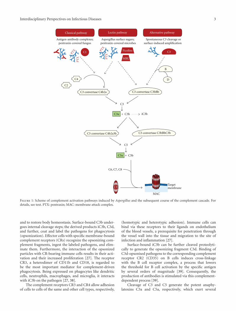
molecular patterns (PAMPs), antigen/antibody complexes,and the presence of transformed cells, apoptotic cells, orcell debris. Three different activation pathways start thecomplement cascade, all of them resulting in the cleavageof the central complement factor C3 by proteolytic enzymecomplexes (C3 convertases), and subsequently leading to thecommon terminal pathway (Figure 1) [2].
In the classical pathway, binding of complement factorC1q to immunoglobulin class G or M (IgG, IgM) ofantigen-antibody complexes represents the initial step [27].Alternatively, the globular heads of C1q can interact withmicrobial surfaces that had been covered by pentraxins,a class of soluble pattern recognition molecules [32]. Thethereby induced conformational changes of C1q subse-quently activate the associated proteases C1r and C1s, whichcleave the factors C4 and C2. The resulting fragments formthe C3 convertase C4b2a [27].
In the lectin pathway, foreign carbohydrate moleculeson the surface of pathogens are recognized by mannose-binding lectin (MBL) or the related ficolins [33]. MBL-associated serine proteases (MASPs) cleave C4 and C2, andthe fragments build up the C3 convertase C4b2a, which isidentically equal to the one of the classical pathway. Ficolin-2 can also interact with pentraxin-covered microbes, thusstarting the lectin pathway in an alternative manner [34].Interestingly, MBL was recently described to support C3cleavage by a C2 bypass mechanism [35], which results inactivation of the alternative pathway.
The alternative pathway is triggered via activating foreignsurfaces and creates an amplification loop by spontaneousreaction of C3 with H2O (C3(H2O)); alternatively, C3bgenerated by the other pathways represents the startingtrigger. Surface-bound C3b associates with factor B, which isthen cleaved by the plasma serine protease factor D. Thesesteps result in the formation of the C3 convertase C3bBb[27, 36].
2.2. All Activation Pathways End in a Common TerminalPathway. Proteolytic cleavage of C3 by one of the C3convertases is the common and central step of all threeactivation pathways. This split generates the fragmentsC3a and C3b, which are two important components thatmediate a multitude of complement functions (see below).The product C3b associates with the C3 convertases, thusforming the C5 convertases, which cleave factor C5 into C5aand C5b. This step initiates a chain of assembly processes ofthe proteins C6, C7, C8, and C9. The bound and polymerizedC9 units create the terminal complement complex (TCC)that can form a pore in the target lipid bilayer, calledmembrane attack complex (MAC). Targeted cells, bacteriaand viruses die or are inactivated by efficient disruption ofthe membrane integrity [31, 37].
2.3. Other Antimicrobial Functions of Complement ActivationProducts. Beneath the MAC formation and direct pathogendestruction, complement displays several additional antimi-crobial mechanisms aiming to neutralize invading microbes
Interdisciplinary Perspectives on Infectious Diseases 3
Classical pathway
Aspergillus surface sugars; pentraxin-covered microbes
Lectin pathway
Antigen-antibody complexes;pentraxin-covered fungus
Spontaneous C3 cleavage orsurface-induced amplification
iC3b
Alternative pathway
MAC
C3
B
C3 convertase C3bBb
C3bC3a +
C3
C5 convertase C3bBbC3b
C6, C7, C8
C1
MBL
Ficolins
C4
C2
C3 convertase C4b2a
C5 convertase C4b2a3b
C5bC5a +
C5
C9Targetmembrane
D
PTX PT
XP
TX
PT
X
Figure 1: Scheme of complement activation pathways induced by Aspergillus and the subsequent course of the complement cascade. Fordetails, see text. PTX: pentraxin; MAC: membrane attack complex.
and to restore body homeostasis. Surface-bound C3b under-goes internal cleavage steps; the derived products iC3b, C3d,and further, coat and label the pathogens for phagocytosis(opsonization). Effector cells with specific membrane-boundcomplement receptors (CRs) recognize the opsonizing com-plement fragments, ingest the labeled pathogens, and elim-inate them. Furthermore, the interaction of the opsonizedparticles with CR-bearing immune cells results in their acti-vation and their increased proliferation [27]. The receptorCR3, a heterodimer of CD11b and CD18, is regarded tobe the most important mediator for complement-drivenphagocytosis. Being expressed on phagocytes like dendriticcells, neutrophils, macrophages, and microglia, it interactswith iC3b on the pathogen [27, 38].
The complement receptors CR3 and CR4 allow adhesionof cells to cells of the same and other cell types, respectively,
(homotypic and heterotypic adhesion). Immune cells canbind via these receptors to their ligands on endotheliumof the blood vessels, a prerequisite for penetration throughthe vessel wall into the tissue and migration to the site ofinfection and inflammation [27].
Surface-bound iC3b can be further cleaved proteolyti-cally to generate the opsonizing fragment C3d. Binding ofC3d-opsonised pathogens to the corresponding complementreceptor CR2 (CD35) on B cells induces cross-linkagewith the B cell receptor complex, a process that lowersthe threshold for B cell activation by the specific antigenby several orders of magnitude [39]. Consequently, theproduction of antibodies is stimulated via this complement-dependent process [39].
Cleavage of C3 and C5 generate the potent anaphy-latoxins C3a and C5a, respectively, which exert several
4 Interdisciplinary Perspectives on Infectious Diseases
biological functions by binding to their correspondingcellular receptors C3aR, C5aR (CD88), and C5L2. Theyprovoke chemotactic attraction of immune cells to thesite of infection and an increase of vascular permeability[40, 41]. Furthermore, C3a and C5a trigger an efficientproinflammatory response by stimulating cytokine synthesisand secretion [40, 41]. Various cell types harboring thecorresponding anaphylatoxin receptors on their surface reacton ligand binding with cell activation, stimulation of cellspecific signaling pathways, or of oxidative burst [27, 42].
2.4. Regulator Molecules Strictly Control the Course of theComplement Cascade. The complement cascade needs atight control to prevent host damage by cell/tissue lysisand excessive inflammation. A variety of both soluble andmembrane-bound regulators can influence all steps of thecomplement cascade, with the C3/C5 convertases as maincontrol targets [27, 37, 43–45]. Under normal conditions,these regulators should protect all body cells against auto-attack by the complement system.
The serine protease factor I cleaves both C4b and C3band is thereby supported by various cofactor molecules.C4 binding protein (C4 bp) and factor H (FH) are fluid-phase proteins that enable the cleavage of C4b and C3b,respectively, Moreover, the membrane-anchored moleculescomplement receptor 1 (CR1, CD35) and membrane cofac-tor protein (MCP, CD46) support the degradation of bothC3b and C4b. In addition, FH and C4 bp accelerate thedecay of assembled C3 convertase; CR1 affects both C3 andC5 convertases. Decay accelerating factor (DAF, CD55) isanother notable membrane-bound regulator that efficientlyprevents the assembly and promotes the disintegration ofboth C3 and C5 convertases [27, 43–45].
In the terminal pathway, the membrane-anchored CD59(protectin) binds to C8 in the C5b-8 complex and thusinhibits further incorporation and polymerization of C9units to form the MAC [27, 43–45].
2.5. Complement in Infectious Diseases: Beneficial and Detri-mental Effects. The potency of complement represents avaluable tool to attack invading pathogens and to defend thehost against penetration and dissemination. One particularadvantage of complement lies in the fact that activationcan start within seconds after contact with the microbeand ends with a multifaceted spectrum of antimicrobialreactions. However, the fact that microbial infections occurin a considerable proportion, already implicates that thepathogens have developed appropriate counterstrategies toavoid elimination, thus starting a vicious circle of reactionand counterreaction.
Furthermore, the antimicrobial effector mechanismsof the complement system might also harbour harmfulconsequences for the affected host. As known from severalinfectious and noninfectious diseases, chronic or exceedingcomplement-mediated inflammation can also contribute totissue damage in the course of these diseases. Putative mech-anisms for such complement-induced tissue damage may
include a fulminant inflammatory reaction and opsonizationof surrounding “bystander” cells with subsequent lysis.
3. Complement Activation by Aspergillus
Aspergillus conidia and hyphae activate the complementsystem via all three pathways [46–48] (Figure 1).
Initiation of the complement cascade by resting conidiais mediated predominantly by the alternative pathway.However, when the conidia begin to swell and transform intohyphae, there is a progressive involvement of the classicalpathway [46]. These differences in the activation pathwaysare reflected by different kinetics; the slowest initiation is seenwith resting conidia [46].
Furthermore, MBL as a pattern recognition molecule ofthe lectin pathway is able to bind to carbohydrate structureson the surface of Aspergillus and promotes complement acti-vation via the lectin pathway, which results in the depositionof C4 [48]. As mentioned above, MBL can support C3cleavage by a C2 bypass mechanism after contact with A.fumigatus conidia, resulting in activation of the alternativepathway and avoiding formation of the classical pathwayC3 convertase [35]. This mechanism is not restricted to A.fumigatus, but can also take place in the presence of A. terreus,A. niger, and A. flavus.
MBL generally seems to be a molecule of high signif-icance for innate defence against a range of pathogens. Inseveral studies, it was shown to bind to various sugars on thesurfaces of viruses, bacteria, yeasts, fungi, and protozoa [48–51]. Further evidence for the crucial role of MBL arises fromfindings in patients with chronic necrotizing pulmonaryaspergillosis and mouse models of pulmonary aspergillosis[52, 53]; these facts suggest MBL as a promising molecule forprophylaxis and therapeutical treatment (see below).
A further mechanism for complement activation drivenby Aspergillus involves the interaction with the pattern recog-nition molecule pentraxin-3 (PTX-3). When A. fumigatusis opsonized with PTX-3, the complement cascade can beactivated either by interaction between PTX-3 and C1q viathe classical pathway [32], or by interaction between PTX-3and ficolin-2 via the lectin pathway [34].
After seroconversion, anti-Aspergillus antibodies in theserum can trigger the start of the classical complementpathway [46].
4. Complement Exerts Antimicrobial Functionsagainst Invasive Aspergillus Infections
The thesis that complement represents a central tool inantifungal host defence is supported by several findings.Complement deficiency is correlated with enhanced suscep-tibility to a disseminated infection by A. fumigatus [54].Furthermore, recognition by the complement system andactivation of the cascade seems to interfere with efficientdissemination in the host. This conclusion is stronglyindicated by the fact that the level of complement depositionon different Aspergillus species correlates inversely with theirpathogenicity: highly virulent species like A. fumigatus and
Interdisciplinary Perspectives on Infectious Diseases 5
A. flavus bind less C3 on their surface than nonpathogenicspecies like A. glaucus or A. nidulans [55].
The antimicrobial potency of the complement cascadeappears to be independent from direct killing via formationof a MAC; presumably, the thick fungal cell wall blockthe formation of a pore by the C9 polymers and thesubsequent lysis of the cells [56]. Other complement-derivedeffector molecules are more effective to cope with aspergillo-sis. Opsonization of the fungal surface with C3-derivedfragments are presumably the most relevant complement-associated weapon, stimulating efficient phagocytosis orrelease of damaging compounds, oxidative burst and killingby monocytes, bronchoalveolar macrophages, and polymor-phonuclear cells [46, 47, 57]. The capacity to opsonizepathogens and to exert antifungal effects strictly depends onthe available complement levels in the respective compart-ment of the body. The complement concentrations in thecentral nervous system (CNS) are low and thus only allow arather weak deposition [58]. Consequently, the complementamounts in the cerebrospinal fluid are unable to induce asignificant oxidative burst in immune cells and to resultin reduced fungal viability, thus making the CNS a highlyvulnerable organ. However, the brain cells react to the fungalpresence with an upregulation of complement synthesis toenable better opsonization and therefore a more efficientclearance of the fungus [58].
A second complement effector mechanism involvesmolecules of the terminal complement pathway. Micedeficient in complement factor C5 can exert the earlyopsonization processes with C3 fragments, but are unable tofulfil the complete cascade. When infected with A. fumigatus,these mice show decreased resistance and lower 50% lethalconidia dosage for a disseminated infection [59, 60]. Sincethis enhanced susceptibility is unlikely to be due to absentMAC formation in the fungal cell membrane, it mightbe supposed that the inability to form the anaphylatoxinC5a could be the relevant deficit in these mice [59, 60].C5a exerts a wide range of proinflammatory effects; bybinding to its receptor C5aR, C5a recruits inflammatorycells to the site of infection, enhances cellular adhesion,and stimulates oxidative metabolism. In addition, C5a cantrigger the release of lysosomal enzymes and of inflammatorymediators such as tumor necrosis factor-alpha (TNF-α)and interleukin-6 (IL-6) [61, 62]. Furthermore, a highersusceptibility to aspergillosis in C5-deficient mice might beattributed to missing TCC. Low doses of this soluble complexwere shown to bind to the membrane of a range of celltypes, thereby triggering various effects like activation, rescuefrom apoptosis, and secretion of prostaglandins, which areimportant regulators of the immune response [63–67].
5. Aspergillus Has Developeda Repertoire of Evasion Mechanisms toRepel Efficient Complement Attack
The potency of Aspergillus to cope with the complementsystem and to undermine its mechanisms for elimination
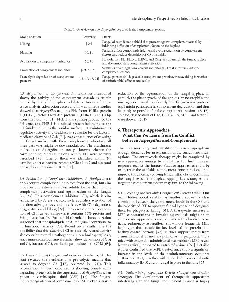
determines how successful the fungus can establish an infec-tion [68]. Aspergillus has developed a complex repertoire ofeffector mechanisms for this purpose (Table 1).
5.1. Hiding. Single or multiple abscess formation is a char-acteristic feature of aspergillosis, particularly in the centralnervous system (CNS). The fungal hyphae are found inbrain blood, vessels with invasion through vascular wallsinto adjacent parenchymal tissue. Swelling and inflammationdevelop in response to the infection. Fungal brain abscessesmay arise from these sites of localized parenchymal infection.In this case, white blood cells collect in the affected part ofthe brain, and fibrous tissue forms around this area, creatinga mass. CNS abscesses typically present with headache,focal neurological abnormalities, and/or seizure, which is theconsequence of local destruction or compression of adjacentbrain tissue [75]. Mature fungal abscesses exhibit a centralnecrotic area with fungal hyphae, surrounded by a capsuleof newly formed fibrous tissue. The formation of abscessesrepresents a host mechanism to inhibit further spreadingof invading pathogens. However, this “sealing off” not onlyinhibits fungal dissemination, but also forms some kindof protection shields against the complement attack [69].Immunohistochemical staining revealed that effect: whereasthe fibrous surrounding tissue was intensely stained forcomplement proteins, the central necrotic area containedonly minor complement concentrations. No deposition ofcomplement factors on the fungal surface in the abscess wasvisible, implying that the encapsulation protects the funguswithin the abscess from any efficient complement attack [69].
5.2. Masking. Putative complement recognition sites on theconidial surface of A. fumigatus are optimally masked tominimize the stimulus for complement activation [55].Experiments aiming to identify the relevant fungal structureindicated that melanin could play a substantial role formasking; for this purpose, knock out mutants lackingenzymes of the melanin biosynthesis pathway were used[10, 11, 76]. Disruption of the gene alb1, which encodesa polyketide synthase in the synthesis of melanin, resultsin increased opsonization of the conidia with C3 and in abetter ingestion by human neutrophils [11]. Deposition ofpigments on the conidial surface might mask the C3 bindingsites, and disruption of the alb1 gene might expose these sitesand thus allow enhanced C3 binding [11]. A mouse modelconfirms this function of alb1 in fungal pathogenesis, sincethe alb1-deficient mutant of A. fumigatus turned out to beless virulent than the wild-type fungus [11, 76]. Inactivationof the gene for the pigmentation protein arp1 similarlyincreased the deposition of C3 on conidia [10]. However,the detailed mechanism how melanin decreases complementdeposition remains unclear. This pigment seems to be acentral element of Aspergillus against the host defence, as itis also involved in scavenging reactive oxygen species (ROS)and inhibits the acidification of phagolysosomes of alveolarmacrophages, monocyte-derived macrophages, and humanneutrophil granulocytes after ingestion of conidia [12, 13,77].
6 Interdisciplinary Perspectives on Infectious Diseases
Table 1: Overview on how Aspergillus copes with the complement system.
Mode of action Reference Effects
Hiding [69]Fungal abscess forms a shield that protects against complement attack byinhibiting diffusion of complement factors to the hyphae
Masking [10, 11]Fungal surface compounds (pigments) avoid recognition by complementfactors and reduce deposition of C3 on conidia
Acquisition of complement inhibitors [70, 71]Host-derived FH, FHL-1, FHR-1, and C4bp are bound on the fungal surfaceand downmodulate complement activation
Production of complement inhibitors [69, 72, 73]Synthesis of a fungal complement inhibitor (CI) that interferes with thecomplement cascade
Proteolytic degradation of complementproteins
[15, 17, 47, 74]Fungal protease(s) degrade(s) complement proteins, thus avoiding formationof antimicrobial effector molecules
5.3. Acquisition of Complement Inhibitors. As mentionedabove, the activity of the complement cascade is strictlylimited by several fluid-phase inhibitors. Immunofluores-cence analysis, adsorption assays and flow cytometry studiesshowed that Aspergillus acquires FH, factor H-like protein1 (FHL-1), factor H-related protein 1 (FHR-1), and C4 bpfrom the host [70, 71]. FHL-1 is a splicing product of theFH gene, and FHR-1 is a related protein belonging to theFH family. Bound to the conidial surface, FH maintained itsregulatory activity and could act as a cofactor for the factor I-mediated cleavage of C3b [71]. As a consequence of coveringthe fungal surface with these complement inhibitors, allthree pathways might be downmodulated. The attachmentmolecules on Aspergillus are not yet known, whereas thecorresponding binding regions within FH were recentlydescribed [71]. One of them was identified within N-terminal short consensus repeats (SCRs) 1 to 7 and a secondone within C-terminal SCR 20 [71].
5.4. Production of Complement Inhibitors. A. fumigatus notonly acquires complement inhibitors from the host, but alsoproduces and releases its own soluble factor that inhibitscomplement activation and opsonization of the fungus[72, 73]. This complement inhibitor (CI), which is alsosynthesized by A. flavus, selectively abolishes activation ofthe alternative pathway and interferes with C3b-dependentphagocytosis and killing [72]. The exact chemical composi-tion of CI is as yet unknown; it contains 15% protein and5% polysaccharide. Further biochemical characterizationsuggested that phospholipids of A. fumigatus contribute toits functional activity [73]. Recent own results raise thepossibility that this described CI or a closely related activityalso contributes to the pathogenesis in cerebral aspergillosis,since immunohistochemical studies show deposition of C1qand C4, but not of C3, on the fungal hyphae in the CNS [69].
5.5. Degradation of Complement Proteins. Studies by Sturte-vant revealed the synthesis of a proteolytic enzyme thatis able to degrade C3 ([47], reviewed in: [74]). Thisis confirmed by own experiments showing complement-degrading proteolysis in the supernatant of Aspergillus whengrown in cerebrospinal fluid (CSF) [17]. The fungus-induced degradation of complement in CSF evoked a drastic
reduction of the opsonization of the fungal hyphae. Inparallel, the phagocytosis of the conidia by neutrophils andmicroglia decreased significantly. The fungal serine proteaseAlp1 might participate in complement degradation and thusbe partly responsible for the complement evasion [15, 17].To date, degradation of C1q, C3, C4, C5, MBL, and factor Dwere shown [15, 17].
6. Therapeutic Approaches:What Can We Learn from the Conflictbetween Aspergillus and Complement?
The high morbidity and lethality of invasive aspergillosisstrongly demands for an expansion of the current treatmentoptions. The antimycotic therapy might be completed bynew approaches aiming to strengthen the host immuneresponse against the fungus. Putative approaches could beto increase the available complement concentrations or toimprove the efficiency of complement attack by underminingthe fungal evasion strategies. Appropriate strategies thattarget the complement system may aim to the following .
6.1. Increasing the Available Complement Protein Levels. Ourown studies about cerebral aspergillosis showed a clearcorrelation between the complement levels in the CSF andthe capacity of CSF to opsonize fungal hyphae and designatethem for phagocytic killing [58]. A therapeutic increase ofMBL concentrations in invasive aspergillosis might be anappropriate approach, since patients with chronic necro-tizing pulmonary aspergillosis show more frequently MBLhaplotypes that encode for low levels of the protein thanhealthy control persons [52]. Further support comes froma murine model of invasive pulmonary aspergillosis: thosemice with externally administered recombinant MBL revealbetter survival, compared to untreated animals [53]. Detailedstudies confirmed that MBL-treated mice show a significantincrease in the levels of the proinflammatory cytokinesTNF-α and IL-1, together with a marked decrease of anti-inflammatory IL-10 and of fungal hyphae in the lung [53].
6.2. Undermining Aspergillus-Driven Complement EvasionStrategies. The development of therapeutic approachesinterfering with the fungal complement evasion is highly
Interdisciplinary Perspectives on Infectious Diseases 7
speculative. Blocking of the fungal surface pigments byspecific antibodies or peptides might be a hypotheticalapproach that could help to expose the C3 binding sitesand thus improve complement deposition and ingestion ofconidia by phagocytes. Another approach might target theacquisition of the negative complement regulators FH, FHL-1, and C4 bp to the fungal surface. For Candida albicans,some molecules that bind C4b and FH have recently beenidentified [78, 79], while the attachment sites on Aspergillusare still unknown but might include related molecules.Blocking antibodies, designed peptides or other inhibitorsagainst these complement regulator binding molecules mighthelp to make the fungus more vulnerable towards comple-ment attack. A similar approach might be developed forthe Aspergillus-derived complement inhibitor described byWashburn [72, 73].
Our own results open the possibility to neutralize thefungal protease(s) that is/are secreted by Aspergillus todegrade complement proteins [17]. Two different strategieswere tested by first in vitro experiments: the neutralizationof the protease by specific inhibitors or interference withthe production of this proteolytic enzyme. In our studies,we could prevent the complement degrading activity byserine protease inhibitors [17]. However, a therapeuticallyused protease inhibitor must be highly specific, since ageneral block of serine proteases might be fatal for the host.Alternatively, our experiments exhibited that the secretionof complement-degrading enzymes strictly depends on theavailability of nitrogen sources [17]. Thus, the supply ofamino acids in the infected host might downmodulate thesecretion of the relevant fungal protease that cleaves thecomplement factors of the host.
7. Summary and Conclusion
Despite new antifungal drugs and improved medical treat-ment, invasive aspergillosis remains a dangerous threatfor immunocompromised patients, as the innate immunedefence is the most crucial weapon against this infection.
The complement system is of particular importance, as itharbours multiple effects against infectious diseases, bridgesthe elements of the human defence network by a multitudeof factors, and helps to preserve the homeostasis of the body.The presence of fungal pathogens is detected by differentpattern recognition molecules; three pathways guarantee theactivation of the complement cascade by resting, swollen,and germinating conidia as well as by hyphae of Aspergillus.
Direct lysis of fungal cells by the membrane attachcomplex (MAC) appears to be of minor importance forthe antifungal defence. Presumably, attraction and activationof immune cells (monocytes, pulmonary macrophages,and polymorphonuclear neutrophils) are the most essentialmechanisms. Anaphylatoxins (C3a, C5a) chemotacticallyrecruit immune cells to the site of the infection and inducefurther inflammatory reactions. Opsonization of conidiaand hyphae with complement fragments like C3b and iC3bmediate phagocytosis, oxidative burst, and release of dam-aging compounds by binding to corresponding receptors onimmune cells.
However, highly virulent Aspergillus species have evolvedmechanisms to evade the attack by complement. They hidefrom recognition, acquire complement regulatory moleculesfrom the host, and secrete proteases to degrade complementfactors.
The multifaceted interactions between complement andAspergillus represent promising approaches for future ther-apeutic strategies that may help to improve the outcome ofinvasive aspergillosis.
References
[1] D. W. Denning, “Invasive aspergillosis,” Clinical InfectiousDiseases, vol. 26, no. 4, pp. 781–803, 1998.
[2] C. Speth, G. Rambach, R. Wurzner, and C. Lass-Florl,“Complement and fungal pathogens: an update,” Mycoses, vol.51, no. 6, pp. 477–496, 2008.
[3] M. Ellis, “Invasive fungal infections: evolving challenges fordiagnosis and therapeutics,” Molecular Immunology, vol. 38,no. 12-13, pp. 947–957, 2002.
[4] M. J. G. T. Ruping, J. J. Vehreschild, and O. A. Cornely,“Patients at high risk of invasive fungal infections: when andhow to treat,” Drugs, vol. 68, no. 14, pp. 1941–1962, 2008.
[5] V. Balloy, M. Huerre, J. P. Latge, and M. Chignard, “Differencesin patterns of infection and inflammation for corticosteroidtreatment and chemotherapy in experimental invasive pul-monary aspergillosis,” Infection and Immunity, vol. 73, no. 1,pp. 494–503, 2005.
[6] M. Ruhnke, G. Kofla, K. Otto, and S. Schwartz, “CNSaspergillosis: recognition, diagnosis and management,” CNSDrugs, vol. 21, no. 8, pp. 659–676, 2007.
[7] D. W. Denning, “Invasive aspergillosis in AIDS-an overview,”Journal of Medical Mycology, vol. 2, supplement 1, pp. 34–41,1998.
[8] A. N. Malani and C. A. Kauffman, “Changing epidemiologyof rare mould infections: implications for therapy,” Drugs, vol.67, no. 13, pp. 1803–1812, 2007.
[9] M. A. Pfaller, P. G. Pappas, and J. R. Wingard, “Invasive fungalpathogens: current epidemiological trends,” Clinical InfectiousDiseases, vol. 43, supplement 1, pp. S3–S14, 2006.
[10] H. F. Tsai, R. G. Washburn, Y. C. Chang, and K. J. Kwon-Chung, “Aspergillus fumigatus arp1 modulates conidial pig-mentation and complement deposition,” Molecular Microbiol-ogy, vol. 26, no. 1, pp. 175–183, 1997.
[11] H. F. Tsai, Y. C. Chang, R. G. Washburn, M. H. Wheeler, andK. J. Kwon-Chung, “The developmentally regulated alb1 geneof Aspergillus fumigatus: its role in modulation of conidialmorphology and virulence,” Journal of Bacteriology, vol. 180,no. 12, pp. 3031–3038, 1998.
[12] A. A. Brakhage, S. Bruns, A. Thywissen, P. F. Zipfel, and J.Behnsen, “Interaction of phagocytes with filamentous fungi,”Current Opinion in Microbiology, vol. 13, no. 4, pp. 409–415,2010.
[13] A. Thywissen, T. Heinekamp, H. M. Dahse et al., “Conidialdihydroxynaphthalene melanin of the human pathogenic fun-gus Aspergillus fumigatus interferes with the host endocytosispathway,” Frontiers in Microbiology, vol. 2, p. 96, 2011.
[14] B. Jahn, F. Boukhallouk, J. Lotz, K. Langfelder, G. Wanner,and A. A. Brakhage, “Interaction of human phagocytes withpigmentless Aspergillus conidia,” Infection and Immunity, vol.68, no. 6, pp. 3736–3739, 2000.
8 Interdisciplinary Perspectives on Infectious Diseases
[15] J. Behnsen, F. Lessing, S. Schindler et al., “Secreted Aspergillusfumigatus protease Alp1 degrades human complement pro-teins C3, C4, and C5,” Infection and Immunity, vol. 78, no. 8,pp. 3585–3594, 2010.
[16] M. Monod, S. Capoccia, B. Lechenne, C. Zaugg, M. Holdom,and O. Jousson, “Secreted proteases from pathogenic fungi,”International Journal of Medical Microbiology, vol. 292, no. 5-6, pp. 405–419, 2002.
[17] G. Rambach, D. Dum, I. Mohsenipour et al., “Secretion of afungal protease represents a complement evasion mechanismin cerebral aspergillosis,” Molecular Immunology, vol. 47, no.7-8, pp. 1438–1449, 2010.
[18] S. Spikes, R. Xu, C. K. Nguyen et al., “Gliotoxin production inAspergillus fumigatus contributes to host-specific differencesin virulence,” Journal of Infectious Diseases, vol. 197, no. 3, pp.479–486, 2008.
[19] P. Sutton, N. R. Newcombe, P. Waring, and A. Mullbacher,“In vivo immunosuppressive activity of gliotoxin, a metaboliteproduced by human pathogenic fungi,” Infection and Immu-nity, vol. 62, no. 4, pp. 1192–1198, 1994.
[20] C. Speth, C. Kupfahl, K. Pfaller et al., “Gliotoxin as putativevirulence factor and immunotherapeutic target in a cell cul-ture model of cerebral aspergillosis,” Molecular Immunology,vol. 48, no. 15-16, pp. 2122–2129, 2011.
[21] J. Sturtevant and J. P. Latge, “Participation of complementin the phagocytosis of the conidia of Aspergillus fumigatusby human polymorphonuclear cells,” Journal of InfectiousDiseases, vol. 166, no. 3, pp. 580–586, 1992.
[22] M. Hasenberg, J. Behnsen, S. Krappmann, A. Brakhage,and M. Gunzer, “Phagocyte responses towards Aspergillusfumigatus,” International Journal of Medical Microbiology, vol.301, no. 5, pp. 436–444, 2011.
[23] W. W. Hope, T. J. Walsh, and D. W. Denning, “The invasiveand saprophytic syndromes due to Aspergillus spp,” MedicalMycology, vol. 43, pp. S207–S238, 2005.
[24] B. K. Kleinschmidt-DeMasters, “Central nervous systemaspergillosis: a 20-year retrospective series,” Human Pathology,vol. 33, no. 1, pp. 116–124, 2002.
[25] T. J. Walsh, D. B. Hier, and L. R. Caplan, “Fungal infections ofthe central nervous system: comparative analysis of risk factorsand clinical signs in 57 patients,” Neurology, vol. 35, no. 11, pp.1654–1657, 1985.
[26] S. J. Lin, J. Schranz, and S. M. Teutsch, “Aspergillosis case-fatality rate: systematic review of the literature,” ClinicalInfectious Diseases, vol. 32, no. 3, pp. 358–366, 2001.
[27] C. Speth, W. Prodinger, R. Wurzner, H. Stoiber, and M. P.Dierich, “Complement,” in Fundamental Immunology, W. E.Paul, Ed., pp. 1047–1078, Lippincott Williams & Wilkins,Philadelphia, Pa, USA, 2008.
[28] M. G. Strainic, J. Liu, D. Huang et al., “Locally produced com-plement fragments C5a and C3a provide both costimulatoryand survival signals to naive CD4+ T cells,” Immunity, vol. 28,no. 3, pp. 425–435, 2008.
[29] J. Lu, X. Wu, and B. K. Teh, “The regulatory roles of C1q,”Immunobiology, vol. 212, no. 4-5, pp. 245–252, 2007.
[30] F. R. Toapanta and T. M. Ross, “Complement-mediatedactivation of the adaptive immune responses: role of C3din linking the innate and adaptive immunity,” ImmunologicResearch, vol. 36, no. 1–3, pp. 197–210, 2006.
[31] P. Gasque, “Complement: a unique innate immune sensor fordanger signals,” Molecular Immunology, vol. 41, no. 11, pp.1089–1098, 2004.
[32] F. Moalli, A. Doni, L. Deban et al., “Role of complement andFcγ receptors in the protective activity of the long pentraxin
PTX3 against Aspergillus fumigatus,” Blood, vol. 116, no. 24,pp. 5170–5180, 2010.
[33] R. C. Duncan, L. C. Wijeyewickrema, and R. N. Pike, “Theinitiating proteases of the complement system: controlling thecleavage,” Biochimie, vol. 90, no. 2, pp. 387–395, 2008.
[34] Y. J. Ma, A. Doni, T. Hummelshøj et al., “Synergy betweenficolin-2 and pentraxin 3 boosts innate immune recognitionand complement deposition,” Journal of Biological Chemistry,vol. 284, no. 41, pp. 28263–28275, 2009.
[35] C. Dumestre-Perard, B. Lamy, D. Aldebert et al., “Aspergillusconidia activate the complement by the mannan-bindinglectin C2 bypass mechanism,” Journal of Immunology, vol. 181,no. 10, pp. 7100–7105, 2008.
[36] S. D. Fleming and G. C. Tsokos, “Complement, naturalantibodies, autoantibodies and tissue injury,” AutoimmunityReviews, vol. 5, no. 2, pp. 89–92, 2006.
[37] J. Van Beek, K. Elward, and P. Gasque, “Activation ofcomplement in the central nervous system: roles in neu-rodegeneration and neuroprotection,” Annals of the New YorkAcademy of Sciences, vol. 992, pp. 56–71, 2003.
[38] G. Raivich, M. Bohatschek, C. U. A. Kloss, A. Werner, L. L.Jones, and G. W. Kreutzberg, “Neuroglial activation repertoirein the injured brain: graded response, molecular mechanismsand cues to physiological function,” Brain Research Reviews,vol. 30, no. 1, pp. 77–105, 1999.
[39] Z. Chen, S. B. Koralov, and G. Kelsoe, “Regulation of humoralimmune responses by CD21/CD35,” Immunological Reviews,vol. 176, pp. 194–204, 2000.
[40] J. Kohl, “Anaphylatoxins and infectious and non-infectiousinflammatory diseases,” Molecular Immunology, vol. 38, no. 2-3, pp. 175–187, 2001.
[41] M. M. Markiewski and J. D. Lambris, “The role of complementin inflammatory diseases from behind the scenes into thespotlight,” American Journal of Pathology, vol. 171, no. 3, pp.715–727, 2007.
[42] J. A. Ember and T. E. Hugli, “Complement factors and theirreceptors,” Immunopharmacology, vol. 38, no. 1-2, pp. 3–15,1997.
[43] D. D. Kim and W. C. Song, “Membrane complement regula-tory proteins,” Clinical Immunology, vol. 118, no. 2-3, pp. 127–136, 2006.
[44] J. M. Thurman and B. Renner, “Dynamic control of thecomplement system by modulated expression of regulatoryproteins,” Laboratory Investigation, vol. 91, no. 1, pp. 4–11,2011.
[45] M. V. Carroll and R. B. Sim, “Complement in health anddisease,” Advanced Drug Delivery Reviews, vol. 63, no. 12, pp.965–975, 2011.
[46] T. R. Kozel, M. A. Wilson, T. P. Farrell, and S. M. Levitz,“Activation of C3 and binding to Aspergillus fumigatus conidiaand hyphae,” Infection and Immunity, vol. 57, no. 11, pp. 3412–3417, 1989.
[47] J. E. Sturtevant and J. P. Latge, “Interactions between conidiaof Aspergillus fumigatus and human complement componentC3,” Infection and Immunity, vol. 60, no. 5, pp. 1913–1918,1992.
[48] O. Neth, D. L. Jack, A. W. Dodds, H. Holzel, N. J. Klein, andM. W. Turner, “Mannose-binding lectin binds to a range ofclinically relevant microorganisms and promotes complementdeposition,” Infection and Immunity, vol. 68, no. 2, pp. 688–693, 2000.
[49] W. K. Eddie Ip, K. Takahashi, R. Alan Ezekowitz, and L.M. Stuart, “Mannose-binding lectin and innate immunity,”Immunological Reviews, vol. 230, no. 1, pp. 9–21, 2009.
Interdisciplinary Perspectives on Infectious Diseases 9
[50] E. C. Van Asbeck, A. I. M. Hoepelman, J. Scharringa, B. L.Herpers, and J. Verhoef, “Mannose binding lectin plays acrucial role in innate immunity against yeast by enhancedcomplement activation and enhanced uptake of polymor-phonuclear cells,” BMC Microbiology, vol. 8, article 229, 2008.
[51] M. W. Turner, “The role of mannose-binding lectin in healthand disease,” Molecular Immunology, vol. 40, no. 7, pp. 423–429, 2003.
[52] D. J. Crosdale, K. V. Poulton, W. E. Ollier, W. Thomson, and D.W. Denning, “Mannose-binding lectin gene polymorphismsas a susceptibility factor for chronic necrotizing pulmonaryaspergillosis,” Journal of Infectious Diseases, vol. 184, no. 5, pp.653–656, 2001.
[53] S. Kaur, V. K. Gupta, S. Thiel, P. U. Sarma, and T. Madan, “Pro-tective role of mannan-binding lectin in a murine model ofinvasive pulmonary aspergillosis,” Clinical and ExperimentalImmunology, vol. 148, no. 2, pp. 382–389, 2007.
[54] R. F. Hector, E. Yee, and M. S. Collins, “Use of DBA/2N micein models of systemic candidiasis and pulmonary and systemicaspergillosis,” Infection and Immunity, vol. 58, no. 5, pp. 1476–1478, 1990.
[55] S. Henwick, S. V. Hetherington, and C. C. Patrick, “Com-plement binding to Aspergillus conidia correlates withpathogenicity,” Journal of Laboratory and Clinical Medicine,vol. 122, no. 1, pp. 27–35, 1993.
[56] T. R. Kozel, “Activation of the complement system bypathogenic fungi,” Clinical Microbiology Reviews, vol. 9, no. 1,pp. 34–46, 1996.
[57] J. Sturtevant and J. P. Latge, “Participation of complementin the phagocytosis of the conidia of Aspergillus fumigatusby human polymorphonuclear cells,” Journal of InfectiousDiseases, vol. 166, no. 3, pp. 580–586, 1992.
[58] G. Rambach, M. Hagleitner, I. Mohsenipour et al., “Antifungalactivity of the local complement system in cerebral aspergillo-sis,” Microbes and Infection, vol. 7, no. 13, pp. 1285–1295, 2005.
[59] A. R. Waldorf and R. D. Diamond, “Neutrophil chemotacticresponses induced by fresh and swollen Rhizopus oryzaespores and Aspergillus fumigatus conidia,” Infection andImmunity, vol. 48, no. 2, pp. 458–463, 1985.
[60] E. V. Svirshchevskaya, M. A. Shevchenko, D. Huet et al.,“Susceptibility of mice to invasive aspergillosis correlates withdelayed cell influx into the lungs,” International Journal ofImmunogenetics, vol. 36, no. 5, pp. 289–299, 2009.
[61] N. P. Gerard and C. Gerard, “The chemotactic receptor forhuman C5a anaphylatoxin,” Nature, vol. 349, no. 6310, pp.614–617, 1991.
[62] L. Kacani, Z. Banki, J. Zwirner et al., “C5a and C5adesArg
enhance the susceptibility of monocyte-derived macrophagesto HIV infection,” Journal of Immunology, vol. 166, no. 5, pp.3410–3415, 2001.
[63] R. H. Daniels, W. A. J. Houston, M. M. Petersen, J. D.Williams, B. D. Williams, and B. P. Morgan, “Stimulation ofhuman rheumatoid synovial cells by non-lethal complementmembrane attack,” Immunology, vol. 69, no. 2, pp. 237–242,1990.
[64] M. Schonermark, R. Deppisch, G. Riedasch, K. Rother, andG. M. Hansch, “Induction of mediator release from humanglomerular mesangial cells by the terminal complement com-ponents c5b-9,” International Archives of Allergy and AppliedImmunology, vol. 96, no. 4, pp. 331–337, 1991.
[65] P. Kalinski, “Regulation of immune responses by prostaglan-din E2,” The Journal of Immunology, vol. 188, no. 1, pp. 21–28,2012.
[66] S. M. Dashiell, H. Rus, and C. L. Koski, “Terminal complementcomplexes concomitantly stimulate proliferation and rescue ofSchwann cells from apoptosis,” Glia, vol. 30, no. 2, pp. 187–198, 2000.
[67] L. Soane, H. Rus, F. Niculescu, and M. L. Shin, “Inhibitionof oligodendrocyte apoptosis by sublytic C5b-9 is associatedwith enhanced synthesis of Bcl-2 and mediated by inhibitionof caspase-3 activation,” Journal of Immunology, vol. 163, no.11, pp. 6132–6138, 1999.
[68] R. Wurzner, “Complement and infectious diseases,” Contribu-tions to Microbiology, vol. 10, pp. 1–17, 2003.
[69] G. Rambach, H. Maier, G. Vago et al., “Complement inductionand complement evasion in patients with cerebral aspergillo-sis,” Microbes and Infection, vol. 10, no. 14-15, pp. 1567–1576,2008.
[70] G. Vogl, I. Lesiak, D. B. Jensen et al., “Immune evasion byacquisition of complement inhibitors: the mould Aspergillusbinds both factor H and C4b binding protein,” MolecularImmunology, vol. 45, no. 5, pp. 1485–1493, 2008.
[71] J. Behnsen, A. Hartmann, J. Schmaler, A. Gehrke, A. A.Brakhage, and P. F. Zipfel, “The opportunistic humanpathogenic fungus Aspergillus fumigatus evades the hostcomplement system,” Infection and Immunity, vol. 76, no. 2,pp. 820–827, 2008.
[72] R. G. Washburn, C. H. Hammer, and J. E. Bennett, “Inhibitionof complement by culture supernatants of Aspergillus fumiga-tus,” Journal of Infectious Diseases, vol. 154, no. 6, pp. 944–951,1986.
[73] R. G. Washburn, D. J. DeHart, D. E. Agwu, B. J. Bryant-Varela, and N. C. Julian, “Aspergillus fumigatus complementinhibitor: production, characterization, and purification byhydrophobic interaction and thin-layer chromatography,”Infection and Immunity, vol. 58, no. 11, pp. 3508–3515, 1990.
[74] J. F. C. Tomee and H. F. Kauffman, “Putative virulence factorsof Aspergillus fumigatus,” Clinical and Experimental Allergy,vol. 30, no. 4, pp. 476–484, 2000.
[75] E. P. Scully, L. R. Baden, and J. T. Katz, “Fungal braininfections,” Current Opinion in Neurology, vol. 21, no. 3, pp.347–352, 2008.
[76] K. Langfelder, B. Jahn, H. Gehringer, A. Schmidt, G. Wanner,and A. A. Brakhage, “Identification of a polyketide synthasegene (pksP) of Aspergillus fumigatus involved in conidialpigment biosynthesis and virulence,” Medical Microbiologyand Immunology, vol. 187, no. 2, pp. 79–89, 1998.
[77] K. Langfelder, M. Streibel, B. Jahn, G. Haase, and A. A.Brakhage, “Biosynthesis of fungal melanins and their impor-tance for human pathogenic fungi,” Fungal Genetics andBiology, vol. 38, no. 2, pp. 143–158, 2003.
[78] I. Lesiak-Markowicz, G. Vogl, T. Schwarzmuller et al., “Can-dida albicans Hgt1p, a multifunctional evasion molecule:complement inhibitor, CR3 analogue, and human immun-odeficiency virus-binding molecule,” Journal of InfectiousDiseases, vol. 204, no. 5, pp. 802–809, 2011.
[79] S. Luo, A. M. Blom, S. Rupp et al., “The pH-regulated antigen1 of Candida albicans binds the human complement inhibitorC4b-binding protein and mediates fungal complement eva-sion,” Journal of Biological Chemistry, vol. 286, no. 10, pp.8021–8029, 2011.

Submit your manuscripts athttp://www.hindawi.com
Stem CellsInternational
Hindawi Publishing Corporationhttp://www.hindawi.com Volume 2014
Hindawi Publishing Corporationhttp://www.hindawi.com Volume 2014
MEDIATORSINFLAMMATION
of
Hindawi Publishing Corporationhttp://www.hindawi.com Volume 2014
Behavioural Neurology
EndocrinologyInternational Journal of
Hindawi Publishing Corporationhttp://www.hindawi.com Volume 2014
Hindawi Publishing Corporationhttp://www.hindawi.com Volume 2014
Disease Markers
Hindawi Publishing Corporationhttp://www.hindawi.com Volume 2014
BioMed Research International
OncologyJournal of
Hindawi Publishing Corporationhttp://www.hindawi.com Volume 2014
Hindawi Publishing Corporationhttp://www.hindawi.com Volume 2014
Oxidative Medicine and Cellular Longevity
Hindawi Publishing Corporationhttp://www.hindawi.com Volume 2014
PPAR Research
The Scientific World JournalHindawi Publishing Corporation http://www.hindawi.com Volume 2014
Immunology ResearchHindawi Publishing Corporationhttp://www.hindawi.com Volume 2014
Journal of
ObesityJournal of
Hindawi Publishing Corporationhttp://www.hindawi.com Volume 2014
Hindawi Publishing Corporationhttp://www.hindawi.com Volume 2014
Computational and Mathematical Methods in Medicine
OphthalmologyJournal of
Hindawi Publishing Corporationhttp://www.hindawi.com Volume 2014
Diabetes ResearchJournal of
Hindawi Publishing Corporationhttp://www.hindawi.com Volume 2014
Hindawi Publishing Corporationhttp://www.hindawi.com Volume 2014
Research and TreatmentAIDS
Hindawi Publishing Corporationhttp://www.hindawi.com Volume 2014
Gastroenterology Research and Practice
Hindawi Publishing Corporationhttp://www.hindawi.com Volume 2014
Parkinson’s Disease
Evidence-Based Complementary and Alternative Medicine
Volume 2014Hindawi Publishing Corporationhttp://www.hindawi.com