comparison of maturation, fertilization, development, and gene expression of mouse oocytes grown in...
TRANSCRIPT
Journal of Assisted Reproduction and Genetics, Vol. 21, No. 7, July 2004 ( C© 2004)
Animal Experimentation
Comparison of Maturation, Fertilization, Development,and Gene Expression of Mouse Oocytes GrownIn Vitro and In Vivo
Dong-Hoon Kim,1,6 Duck-Sung Ko,2 Hoi-Chang Lee,2 Ho-Joon Lee,3 Won-Il Park,4 S. Samuel Kim,4
Jin-Ki Park,1 Byoung-Chul Yang,1 Soo-Bong Park,1 Won-Kyong Chang,1 and Hoon-Taek Lee5
Submitted March 31, 2003; accepted May 4, 2004
Purpose : To investigate the difference of in vitro and in vivo grown oocytes, we comparedmaturation, fertilization, development, and maternal gene expression from both in vitro andin vivo grown mouse oocytes.Methods : The preantral follicles isolated from 12-day-old mice were cultured on Transwell-COL membrane inserts. After culture, maturation, fertilization, and developmental rates wereassessed. RT-PCR (reverse transcription—polymerase chain reaction) was performed to ex-amine the expression of β-actin, GDF-9, and IGF-II in matured oocytes.Results : No difference in the nuclear maturation was detected between in vitro and in vivogrown oocytes, but the mean oocyte diameter of the in vitro group was smaller than that ofthe in vivo group. The fertilization rate was significantly lower in the in vitro group than inthe in vivo group (p < 0.05). The capacities of in vitro grown oocyte to cleave and developto blastocysts were significantly lower than those of the in vivo grown oocytes (p < 0.001).Moreover, blastocyst of in vitro group had fewer total cells than those of in vivo group (p <
0.05). In regards to the expression of genes in mature oocytes, growth differentiation factor-9(GDF-9) expression was similar between the two groups, but β-actin was significantly reducedin the in vitro group compared to the in vivo group. Particularly, the expression of insulin-likegrowth factor II (IGF-II) was not found in the in vitro grown oocytes.Conclusions : These results showed that in vitro grown oocytes did not have the same de-velopmental capacity as in vivo grown oocytes. We assume that the aberrant expression ofmaternal-derived genes in the in vitro grown oocytes may cause the poor embryo viability.
KEY WORDS: Gene expression; in vitro; maturation; mouse; preantral follicle.
INTRODUCTION
There are thousands of oocytes present in the mam-malian ovaries. However, most of them undergo de-generation during the growth and maturation, onlya very few oocytes become available for ovula-
1 Animal Biotechnology Division, National Livestock ResearchInstitute, Suwon, Korea.
2 Medical Science Institute, Eulji General Hospital. Eulji Univer-sity School of Medicine, Seoul, Korea.
3 Department of Physiology, Eulji University School of Medicine,Seoul, Korea.
tion (1). The mammalian oocytes isolated fromantral follicles can be matured, fertilized and de-veloped into live young in mice (2), rats (3), sheep(4), cattle (5), pigs (6), and humans (7). How-ever, the mammalian ovaries have only limited num-ber of antral follicles available for the in vitromaturation.
4Department of Obstretics and Gynacology, Eulji UniversitySchool of Medicine, Seoul, Korea.
5Department of Animal Science, Konkuk University, Seoul, Korea.6To whom correspondence should be addressed; e-mail:[email protected].
233 1058-0468/04/0700-0233/0 C© 2004 Springer Science+Business Media, Inc.
234 Kim, Ko, Lee, Lee, Park, Kim, Park, Yang, Park, Chang, and Lee
The ovarian preantral follicles are an enormous andpotentially valuable source of oocytes that could beused for clinical, agricultural, and zoological purposes.However, developing an optimal in vitro culture tech-nique to bring the immature oocytes in preantral folli-cles to mature oocytes is an important prerequisite. Todate, many researchers have published their efforts todevelop culture techniques and their insights into themetabolism of follicles, production of steroids duringthe growth, and effects of gonadotrophins and growthfactors on the follicular development in mice (8–10),cattle (11), pigs (12), and even humans (13).
Murine species have been widely used to developthe culture system for preantral follicles and to studyfollicular growth, since they are small, readily avail-able, and grow to Graafian follicle within a shorttime-span. Particularly, the studies for the cultureof mouse preantral follicles have demonstrated thatnot only the growth and maturation of the oocytesbut also fertilization and embryos development werepossible (10,14,15). Furthermore, live young wereobtained by transferring embryos derived from invitro grown oocytes (14,15). Earlier studies show thatmouse oocytes grown, matured, and fertilized in vitrohave limited developmental capacity. However, infor-mation on the cause of lower developmental capacityof in vitro grown oocytes is lacking.
Thus, examination of the difference of mouseoocytes grown in vitro and in vivo to verify the causeof lower developmental capacity is necessary. The aimof this study was to examine maturation, fertilization,and developmental rates and expression of maternalgenes in the mouse oocytes derived from the in vitrogrown preantral follicles, and to compare these find-ings with those obtained from oocytes grown and ma-tured in vivo.
MATERIALS AND METHODS
Animals
The mice used in this study (ICR) were housedin a temperature- and light-controlled room, undera photoperiod of 12-h light:12-h dark. All experimen-tal protocols and animal handling procedures werereviewed and approved by the Animal Ethics Com-mittee of Eulji General Hospital and Eulji UniversitySchool of Medicine.
Isolation of Preantral Follicles
The ovaries were aseptically removed from 12-day-old ICR female mice (n = 30). They were immersed
into Leibovitz L-15 medium containing 1 mg/mL col-lagenase (Type 1A; C-2674, Sigma, St. Louis, MO)and 0.2 mg/mL DNase I (DN-25, Sigma, St. Louis,MO) for 20 min at 37◦C and repeatedly drawn in andout of the pipette until the ovaries were dissociatedinto individual follicles. A total of 1588 preantral fol-licles to be cultured were selected by the followingcriteria: i) intact round follicular structure with twoto three layers of granulosa cells; ii) the oocyte hadto be visible, round and centrally located within thefollicle.
In Vitro Growth and Maturationof Preantral Follicles
The culture medium was αMEM medium supple-mented with 5% fetal bovine serum, 100 mIU/mLFSH (Metrodin-HP, Sereno, Aubonne, Switzerland),and 10 mIU/mL LH (L-5259, Sigma, St. Louis, MO).The follicles were cultured on Transwell-COL mem-brane inserts (3.0-µm pore size, 24.5-mm diameter;Costar, Cambridge, MA) in six-well cluster dishesto prevent the loss of structural integrity betweenthe oocyte and granulosa cells. The follicles werecultured for 10 days at 37◦C and 5% CO2 in air.Half of the medium was changed every 2 days. Af-ter 10 days of growth in vitro, follicles were al-lowed to mature for 16–18 h in medium supplementedwith 1.5 IU/mL hCG (Profasi, Sereno, Aubonne,Switzerland).
As a control, in vivo grown oocytes were collectedfrom the ovaries of 22-day-old ICR mice and maturedin the same medium in which in vitro grown oocyteswere matured.
A part of matured follicles were selected randomlyand adherent cumulus cells were removed. The mat-uration status and the diameter of the oocytes, ex-cluding the zona pellucida, were examined with aninverted microscope and the ocular micrometer.
In Vitro Fertilization
In vitro matured oocyte-granulosa cell complexeswere placed into 50-µL drops of Whittingham’s T6
medium supplemented with 30 mg/mL bovine serumalbumin (A-3311, Sigma, St. Louis, MO) under min-eral oil.
As a control, in vivo matured oocytes were obtainedfrom 23-day-old ICR mice. The mice were superovu-lated with 5 IU pregnant mare serum gonadotrophin(PMSG) followed 48 h later by 5 IU of human chori-onic gonadotrophin (HCG). Fifteen to sixteen hours
Journal of Assisted Reproduction and Genetics, Vol. 21, No. 7, July 2004
Maturation, Fertilization, Development, and Gene Expression of Mouse Oocytes 235
after the HCG injection, oocytes were recovered fromthe oviducts then placed into the drops of fertilizationmedium.
The sperms collected from the cauda epididymis ofmature ICR male mice were incubated for 2 h in fer-tilization medium before insemination. The oocyteswere inseminated with spermatozoa with concentra-tion of 1–2 × 10⁶ per mL. Four to six hours after theinsemination, oocytes were washed and cultured forin vitro development.
In Vitro Development
The fertilized oocytes were cultured in 20-µL dropsof KSOM medium supplemented with 10% (v/v)serum substitute supplement (SSS, Irvine, Santa Ana,CA), 1% (v/v) essential amino acids (M-7145, Sigma,St. Louis, MO), and 0.5% (v/v) nonessential aminoacids (B-6766, Sigma, St. Louis, MO) under mineraloil. The oocytes were examined the following day, andthe number of 2-cell embryos was noted. The 2-cellembryos were further cultured for 4 days until theblastocyst stage.
Flourescence Staining
The chromatin configuration of GV (germinal vesi-cle) oocytes (10 days after culture), the detailed sta-tus of fertilized oocytes (6 h after insemination) andthe cell number of blastocysts (5 days after insemi-nation) were evaluated by fluoresence staining. Theoocytes and blastocysts were fixed in 2% formalde-hyde in PBS for 10 min at room temperature. Theoocytes and blastocysts were then placed on slideswith a drop of mounting medium consisting of the3:1 ratio of glycerol versus PBS containing 2.5 mg/mLsodium azide and 2.5 µg/mL Hoechst 33342 (B-2261,Sigma, St. Louis, MO). A cover slip was placed ontop of the oocytes and blastocysts, and the edge wassealed with fingernail polish. The stained oocytesand blastocysts were examined under a fluorescencemicroscope.
Table I. Primers Used for RT-PCR and Product Size
Gene Primer sequence Product (bp)
β-actin 5′-GTGGGCCGCTCTAGGCACCAA-3′ 5395′-CTCTTTGATGTCACGCACGATTTC-3′
GDF-9 5′-GGTTCTATCTGATAGGCGAGG-3′ 4465′-GGGGCTGAAGGAGGGAGG-3′
IGF-II 5′-GGCCCCGGAGAGACTCTGTGC-3′ 2555′-GCCCACGGGGTATCTGGGGAA-3′
Assessment of Zona Pellucida Hardening
In vitro and in vivo grown and matured oocyteswere treated with 1% hyalruonidase (H-3506, Sigma,St. Louis, MO) and denuded of adherent granulosacells by drawing them in and out a pasteur pipetteand collected in PBS. The time required for the diges-tion of the zona pellucida by 1 mg/mL α-chymotrypsin(C-4129, Sigma, St. Louis, MO) was used as an indi-cator of zona hardening as described previously (16).The zona pellucida digestion time is defined as thetime required for 50% of the oocytes to become com-pletely zona-pellucida-free as observed under the in-verted microscope.
Gene Expression
Mature oocytes were washed in three changes ofPBS containing 0.1% PVP and then transferred ina minimal volume of PBS into 300 µl of TRIzolTM
Reagent (Gibco BRL, USA.). Total RNA was ex-tracted according to the manufacturer’s instructions.Reverse transcription (RT) was performed with to-tal RNA isolated from 30 oocytes by using 1st
strand cDNA Synthesis Kit (Boehringer Mannheim,Germany). Polymerase chain reaction (PCR) wasconducted on three oocyte equivalents for β-actin,growth differentiation factor-9 (GDF-9), and Insulin-like growth factor-II (IGF-II). The sequences of theprimer for PCR (listed in Table I) were designedaccording to published cDNA sequences. The PCRproducts were resolved on 2% agarose gels con-taining 0.5 µg/mL ethidium bromide and visualizedwith UV transilluminator. The amounts of specificmRNAs were analyzed using an image-analyzingsystem with a computer software (Vilber Lourmat,France).
Embryo Transfer
Morphologically normal 2-cell embryos that wasderived from in vitro growth, maturation, and
Journal of Assisted Reproduction and Genetics, Vol. 21, No. 7, July 2004
236 Kim, Ko, Lee, Lee, Park, Kim, Park, Yang, Park, Chang, and Lee
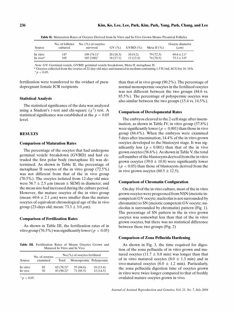
Table II. Maturation Rates of Oocytes Derived from In Vitro and In Vivo Grown Mouse Preantral Follicles
No. of follicles No. (%) of oocytes Oocyte diameterSource cultured survived GV (%) GVBD (%) Meta II (%) (µm)
In vitro 147 109 (74.1)∗ 20 (18.3) 10 (9.2) 79 (72.5) 69.6 ± 2.1∗In vivoa 105 105 (100)∗ 18 (17.1) 13 (12.4) 74 (70.5) 73.3 ± 3.0∗
Note. GV: Germinal vesicle, GVBD: germinal vesicle breakdown, Meta II: metaphase II.a Oocytes collected from the ovaries of 22-day-old mice and matured in medium containing 1.5 IU/mL hCG for 16–18 h.∗ p < 0.05.
fertilization were transferred to the oviduct of pseu-dopregnant female ICR recipients.
Statistical Analysis
The statistical significance of the data was analyzedusing a Student’s t-test and chi-square (χ2) test. Astatistical significance was established at the p < 0.05level.
RESULTS
Comparison of Maturation Rates
The percentage of the oocytes that had undergonegerminal vesicle breakdown (GVBD) and had ex-truded the first polar body (metaphase II) was de-termined. As shown in Table II, the percentage ofmetaphase II oocytes of the in vitro group (72.5%)was not different from that of the in vivo group(70.5%). The oocytes isolated from 12-day-old micewere 56.7 ± 2.5 µm (mean ± SEM) in diameter, andthe mean size had increased during the culture period.However, the mature oocytes of the in vitro group(mean: 69.6 ± 2.1 µm) were smaller than the matureoocytes of equivalent chronological age of the in vivogroup (23-days old; mean: 73.3 ± 3.0 µm).
Comparison of Fertilization Rates
As shown in Table III, the fertilization rates of invitro group (76.5%) was significantly lower (p < 0.05)
Table III. Fertilization Rates of Mouse Oocytes Grown andMatured In Vitro and In Vivo
No.(%) of oocytes fertilizedNo. of oocytesSource examined Total Monospermic Polyspermic
In vitro 85 65 (76.5)∗ 55 (84.6) 10 (15.4)In vivo 92 83 (90.2)∗ 71 (85.5) 12 (14.5)
∗ p < 0.05.
than that of in vivo group (90.2%). The percentage ofnormal monospermic oocytes in the fertilized oocyteswas not different between the two groups (84.6 vs.85.5%). The percentage of polyspermic oocytes wasalso similar between the two groups (15.4 vs. 14.5%).
Comparison of Developmental Rates
The embryos cleaved to the 2-cell stage after insem-ination, as shown in Table IV, in vitro group (57.8%)were significantly lower (p < 0.001) than those in vivogroup (84.4%). When the embryos were examined5 days after insemination, 14.4% of the in vitro grownoocytes developed to the blastocyst stage. It was sig-nificantly less (p < 0.001) than that of the in vivogrown oocytes (56.6%). As shown in Table V, the totalcell number of the blastocysts derived from the in vitrogrown oocytes (39.0 ± 10.8) were significantly lower(p < 0.05) than those of blastocysts derived from thein vivo grown oocytes (60.5 ± 12.5).
Comparison of Chromatin Configuration
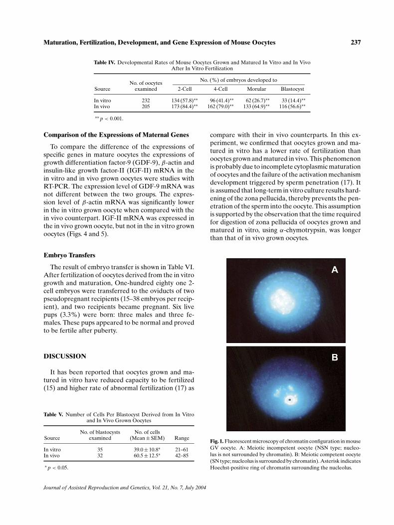
On day 10 of the in vitro culture, most of the in vitrogrown oocytes were progressed from NSN (meiotic in-competent GV oocyte; nucleolus is not surrounded bychromatin) to SN (meiotic competent GV oocyte; nu-cleolus is surrounded by chromatin) pattern (Fig. 1).The percentage of SN pattern in the in vivo grownoocytes was somewhat less than that of the in vitrogrown oocytes, but there was no statistical differencebetween these two groups (Fig. 2)
Comparison of Zona Pellucida Hardening
As shown in Fig. 3, the time required for diges-tion of the zona pellucida of in vitro grown and ma-tured oocytes (11.7 ± 0.8 min) was longer than thatof in vitro matured oocytes (8.0 ± 1.3 min) and invivo-matured oocytes (6.0 ± 1.2 min). Particularly,the zona pellucida digestion time of oocytes grownin vitro were twice longer compared to that of freshlyovulated mature oocytes grown in vivo.
Journal of Assisted Reproduction and Genetics, Vol. 21, No. 7, July 2004
Maturation, Fertilization, Development, and Gene Expression of Mouse Oocytes 237
Table IV. Developmental Rates of Mouse Oocytes Grown and Matured In Vitro and In VivoAfter In Vitro Fertilization
No. (%) of embryos developed toNo. of oocytesSource examined 2-Cell 4-Cell Morular Blastocyst
In vitro 232 134 (57.8)∗∗ 96 (41.4)∗∗ 62 (26.7)∗∗ 33 (14.4)∗∗In vivo 205 173 (84.4)∗∗ 162 (79.0)∗∗ 133 (64.9)∗∗ 116 (56.6)∗∗
∗∗ p < 0.001.
Comparison of the Expressions of Maternal Genes
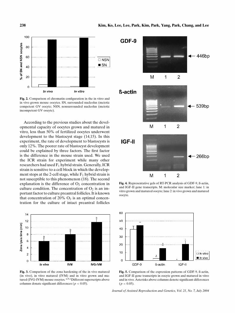
To compare the difference of the expressions ofspecific genes in mature oocytes the expressions ofgrowth differentiation factor-9 (GDF-9), β-actin andinsulin-like growth factor-II (IGF-II) mRNA in thein vitro and in vivo grown oocytes were studies withRT-PCR. The expression level of GDF-9 mRNA wasnot different between the two groups. The expres-sion level of β-actin mRNA was significantly lowerin the in vitro grown oocyte when compared with thein vivo counterpart. IGF-II mRNA was expressed inthe in vivo grown oocyte, but not in the in vitro grownoocytes (Figs. 4 and 5).
Embryo Transfers
The result of embryo transfer is shown in Table VI.After fertilization of oocytes derived from the in vitrogrowth and maturation, One-hundred eighty one 2-cell embryos were transferred to the oviducts of twopseudopregnant recipients (15–38 embryos per recip-ient), and two recipients became pregnant. Six livepups (3.3%) were born: three males and three fe-males. These pups appeared to be normal and provedto be fertile after puberty.
DISCUSSION
It has been reported that oocytes grown and ma-tured in vitro have reduced capacity to be fertilized(15) and higher rate of abnormal fertilization (17) as
Table V. Number of Cells Per Blastocyst Derived from In Vitroand In Vivo Grown Oocytes
No. of blastocysts No. of cellsSource examined (Mean ± SEM) Range
In vitro 35 39.0 ± 10.8∗ 21–61In vivo 32 60.5 ± 12.5∗ 42–85
∗ p < 0.05.
compare with their in vivo counterparts. In this ex-periment, we confirmed that oocytes grown and ma-tured in vitro has a lower rate of fertilization thanoocytes grown and matured in vivo. This phenomenonis probably due to incomplete cytoplasmic maturationof oocytes and the failure of the activation mechanismdevelopment triggered by sperm penetration (17). Itis assumed that long-term in vitro culture results hard-ening of the zona pellucida, thereby prevents the pen-etration of the sperm into the oocyte. This assumptionis supported by the observation that the time requiredfor digestion of zona pellucida of oocytes grown andmatured in vitro, using α-chymotrypsin, was longerthan that of in vivo grown oocytes.
Fig. 1. Fluorescent microscopy of chromatin configuration in mouseGV oocyte. A: Meiotic incompetent oocyte (NSN type; nucleo-lus is not surrounded by chromatin). B: Meiotic competent oocyte(SN type; nucleolus is surrounded by chromatin). Asterisk indicatesHoechst-positive ring of chromatin surrounding the nucleolus.
Journal of Assisted Reproduction and Genetics, Vol. 21, No. 7, July 2004
238 Kim, Ko, Lee, Lee, Park, Kim, Park, Yang, Park, Chang, and Lee
Fig. 2. Comparison of chromatin configuration in the in vitro andin vivo grown mouse oocytes. SN, surrounded nucleolus (meioticcompetent GV oocyte; NSN, nonsurrounded nucleolus (meioticincompetent GV oocyte).
According to the previous studies about the devel-opmental capacity of oocytes grown and matured invitro, less than 50% of fertilized oocytes underwentdevelopment to the blastocyst stage (14,15). In thisexperiment, the rate of development to blastocysts isonly 12%. The poorer rate of blastocyst developmentcould be explained by three factors. The first factoris the difference in the mouse strain used. We usedthe ICR strain for experiment while many otherresearchers had used F1 hybrid strain. Generally, ICRstrain is sensitive to a cell block in which the develop-ment stops at the 2-cell stage, while F1 hybrid strain isnot susceptible to this phenomenon (18). The secondexplanation is the difference of O2 concentration inculture condition. The concentration of O2 is an im-portant factor to culture preantral follicles. It is knownthat concentration of 20% O2 is an optimal concen-tration for the culture of intact preantral follicles
Fig. 3. Comparison of the zona hardening of the in vivo matured(in vivo), in vitro matured (IVM) and in vitro grown and ma-tured (IVG-IVM) mouse oocytes. a,b,cDifferent superscripts abovecolumns donate significant differences (p < 0.05).
Fig. 4. Representative gels of RT-PCR analysis of GDF-9, ß-actin,and IGF-II gene transcripts. M: molecular size marker; lane 1: invitro grown and matured oocyte; lane 2: in vivo grown and maturedoocyte.
Fig. 5. Comparison of the expression patterns of GDF-9, ß-actin,and IGF-II gene transcripts in oocyte grown and matured in vitroand in vivo. Asterisks above columns denote significant differences(p < 0.05).
Journal of Assisted Reproduction and Genetics, Vol. 21, No. 7, July 2004
Maturation, Fertilization, Development, and Gene Expression of Mouse Oocytes 239
Table VI. Result of Transfer of Embryos Derived from the In VitroGrown, Matured, and Fertilized Oocytes
No. of No. of embryos No. of No.(%) ofRecipient transferred Pregnant offsprings
8 181 2 6 (3.3) M: 3, F: 3
Note. M: Male, F: female.
isolated by manual dissection (19). On the contrary,for preantral follicles isolated by enzyme treatment,5% O2 is believed as an optimal concentration be-cause partial damage of theca cells and basal laminacause more oxidative stress. Eppig and Wigglesworth(20) reported that 20% O2 concentration resultedlower survival and these oocytes could not developto blastocysts after fertilization. Finally, we did notadd EGF in maturation medium. It is known thatEGF promotes nuclear and cytoplasmic maturationof oocytes. Previous studies for the preantral follicleculture showed impressive beneficial effects ofEGF, combined with FSH or LH, on maturation,fertilization, and development to blastocysts (9,15).
The diameter of oocytes is an important index forthe cytoplasmic maturation and developmental com-petency of oocytes. In our experiment, none of thein vitro grown oocytes grew as large as the oocytesisolated from the antral follicles of 23-day-old mice,which were at the same chronological age as the invitro grown oocytes isolated 12 days of age and cul-tured for 11 days. Thus, in vitro grown oocytes, underthese conditions, were not equivalent in size to the invivo grown oocytes. This result is very similar to otherstudies (14,17).
Initially, all oocytes in preantral follicles are in theNSN state, but while growing they either continueto develop to the NSN configuration or are shiftedinto the SN configuration (21). The transition of GVoocytes from the NSN to SN configuration is impor-tant to acquire the meiotic competency to resume thefirst meiosis (22). Hartshorne et al. (23) reported thatin cultured mouse ovarian follicles, the chromatin or-ganization in the oocyte normally changed from theNSN to SN configuration. This study demonstratedthat the transition rate of oocyte chromatin organi-zation from NSN to SN in the in vitro grown oocytewas similar to that of the in vivo grown oocytes at thesame chronological age (23-day old). Thus, this studyconfirmed that in vitro grown oocytes may normallyacquire the meiotic competence to resume the firstmeiotic division during the culture period.
During the oocyte growth, gene expression is highlyactive in order to accumulate the maternal productsthat are required, after fertilization, to sustain the first
step of the embryonic development (24). Several ex-periments have shown that maternal factors are ableto modify developmental ability (25) and gene expres-sion in the zygote (26).
β-actin is a housekeeping gene, which is a keycomponent of the cytoskeleton. In this study, theexpression of β-actin mRNA in the in vitro grownoocyte group was significantly lower compared tothe in vivo group. GDF-9 is an oocyte-specific factorand a member of the transforming growth factor-β(TGFβ) superfamily that is expressed at the highlevel in mammalian oocytes beginning from thetype 3a primary follicles (one-layer growing primaryfollicles) but not in oocytes of quiescent primordialfollicles (27). The GDF-9 knockout mice showedan arrest of follicle development beyond the type3b stage (one-layer primary follicle stage) (28). Inaddition, this mice showed the defect in oocytemeiotic competence, including abnormal GVBD,and spontaneous parthenogenetic activation of theoocytes (29). Moreover, it reported that GDF-9 is animportant factor for the growth and differentiationof cultured early ovarian follicles (30). In our study,there was no difference in the expression of GDF-9mRNA between in vitro and in vivo grown oocyte.
IGF-II has been known to stimulate the growth andmetabolism of early embryos (31). It is also an impor-tant survival factor that protect a range of cell typesfrom apoptosis (32). The report of Stojanov et al. (33)showed that IGF-II was present in the oocyte andearly zygote as maternal transcripts, and was rapidlylost after fertilization. This transcript was not detectedby the early 2-cell stage, but detected again in thelate 2-cell stage. Our study demonstrated that IGF-IImRNA was not present in the in vitro grown oocyte,whereas there was IGF-II mRNA expression in thein vivo grown oocyte. The specific gene expressionpatterns in this study suggest that the aberrations inthe expression of specific genes in the in vitro grownoocyte may cause the poor embryo viability.
There were several reports of production of liveyoung after transfer of embryos derived from the invitro grown, matured, and fertilized oocytes. How-ever, only about 5% of the transferred embryos devel-oped to the live young (14,15). We also demonstratedthe production of live young from oocytes grown invitro from preantral follicles which were fertilized andtransferred at the 2-cell embryo stage. The rate of liveyoung produced in our laboratory was 3.3%. The lowbirth rate may be a consequence of the compromiseddevelopmental capacity of the in vitro grown oocytes.
Of note, the total cell number is regarded as animportant index for developmental capacity of the
Journal of Assisted Reproduction and Genetics, Vol. 21, No. 7, July 2004
240 Kim, Ko, Lee, Lee, Park, Kim, Park, Yang, Park, Chang, and Lee
blastocyst. We could also speculate that insufficientcytoplasmic maturation and aberrant gene expres-sions of the in vitro grown oocyte caused the smallercell number of the blastocyst. Further studies areneeded to improve the culture system for preantralfollicles and to verify the cause of the lower develop-mental capacity of in vitro grown oocytes.
REFERENCES
1. Gosden RG, Telfer E: Numbers of follicles and oocytes inmammalian ovaries and their allometric relationships. J Zool1987;211:169–175
2. Schroeder AC, Eppig JJ: The developmental capacity of mouseoocytes that matured spontaneously in vitro is normal. Dev Biol1984;102:493–497
3. Vanderhyden BC, Armstrong DT: Role of cumulus cells andserum in the in vitro maturation, fertilization and subsequentdevelopment of rat oocytes. Biol Reprod 1989;40:720–728
4. Staigmiller RB, Moor RM: Effect of follicle cells on the matura-tion and developmental competence of ovine oocytes maturedoutside the follicle. Gamete Res 1984;9:221–229
5. Goto K, Kajihara Y, Kosaka S, Koba M, Nakanishi Y, OgawaK: Pregnancies after co-culture of cumulus cells with bovineembryos derived from in-vitro fertilization of in-vivo maturedfollicular oocytes. J Reprod Fertil 1988;83:753–758
6. Mattioli M, Bacci ML, Galeati G, Seren E: Developmentalcompetence of pig oocytes matured and fertilized in vitro. The-riogenology 1989;31:1201–1207
7. Cha KY, Choi DH, Koo JJ, Han JJ, Ko JJ, Yoon TK: Pregnancyafter in vitro fertilization of human follicular oocytes collectedfrom nonstimulated cycles, their culture in vitro and their trans-fer in a donor oocyte program. Fertil Steril 1991;55:109–113
8. Nayudu PL, Osborn SM: Factors influencing the rate of pre-antral and antral growth of mouse ovarian follicles in vitro. JReprod Fertil 1992;95:349–362.
9. Boland NI, Gosden RG: Effects of epidermal growth factoron the growth and differentiation of cultured mouse ovarianfollicles. J Reprod Fertil 1994;101:369–374
10. Cortvrindt R, Smitz J, Van Steirteghem AC: In-vitro mat-uration, fertilization and embryo development of immatureoocytes from early preantral follicles from prepuberal mice ina simplified culture system. Hum Reprod 1996;11:2656–2666
11. Figueiredo JR, Hulshof SCJ, Van den Hurk R, Bevers MM,Nusgens B, Beckers JF: Development of a combined new me-chanical and enzymatic method for the isolation of intact pre-antral follicles from fetal, calf and adult bovine ovaries. Theri-ogenology 1993;40:789–799
12. Hirao Y, Nagai T, Kubo M, Miyano T, Miyake M, Kato S: Invitro growth and maturation of pig oocytes. J Reprod Fertil1994;100:333–339
13. Roy SK, Treacy BJ: Isolation and long-term culture of humanpreantral follicles. Fertil Steril 1993;59:783–790
14. Eppig JJ, Schroeder AC: Capacity of mouse oocytes from pre-antral follicles to undergo embryogenesis and development tolive young after growth, maturation and fertilization in vitro.Biol Reprod 1989;41:268–276
15. Spears N, Boland NI, Murray AA, Gosden RG: Mouse oocytesderived from in vitro grown primary ovarian follicles are fertile.Hum Reprod 1994;9:527–532
16. Schroeder AC, Schultz RM, Kopf GS, Taylor FR, Backer RB,Eppig JJ: Fetuin inhibits zona pellucida hardening and conver-sion of ZP2 to ZP2f during spontaneous mouse oocyte matura-tion in vitro in the absence of serum. Biol Reprod;1990;43:891–897
17. Hirao Y, Miyano T, Kato S: Fertilization of in vitro grownmouse oocytes. Theriogenology 1990;34:1071–1077
18. Whitten WK, Biggers JD: Complete development in vitro ofthe preimplantation stage of the mouse in a simple, chemicallydefined medium. J Reprod Fertil 1968;17:399–401
19. Smitz J, Cortvrintz A, Van Steirteghem A: Normal oxygenatmosphere is essential for the solitary long-term culture ofearly preantral mouse follicles. Mol Reprod Dev 1996;45:466–475
20. Eppig JJ, Wigglesworth K: Factors affecting the developmentalcompetence of mouse oocytes grown in vitro oxygen concen-tration. Mol Reprod Dev 1995;42:447–456
21. Zuccotti M, Piccinelli A, Giorgi Rossi P, Garagna S, Redi CA:Chromatin organization during mouse oocyte growth. Mol Re-prod Dev 1995;41:479–485
22. Wickramasinghe D, Ebert KM, Albertini DF: Meiotic com-petence acquisition is associated with the appearance of M-phase characteristics in growing mouse oocytes. Dev Biol1991;143:162–172
23. Hartshorne GM, Sargent IL, Barlow DH: Meiotic progressionof mouse oocytes throughout follicle growth and ovulation invitro. Hum Reprod 1994;9:352–359
24. Telford NA, Watson AJ, Schultz GA: Transition from mater-nal to embryonic control in early mammalian development: Acomparison of several species. Mol Reprod Dev 1990;26:90–100
25. Latham KE, Solter D: Effect of egg composition on the devel-opmental capacity of androgenetic mouse embryos. Develop-ment 1991;113:561–568
26. Chastant S, Christians E, Campion E, Renard JP: Quantitativecontrol of gene expression by nucleocytoplasmic interactionsin early mouse embryos: Consequence for reprogramming bynuclear transfer. Mol Reprod Dev 1996;44:423–432
27. McGrath SA, Esquela SF, Lee S-J: Oocyte-specific expressionof growth differentiation factor-9. Mol Endocrinol 1995;9:131–136
28. Dong J, Albertini DF, Nishimori K, Rajendra Kumar T, Lu N,Matzuk MM: Growth differentiation factor-9 is required duringearly ovarian folliculogenesis. Nature 1996;383:531–535
29. Carabatsos MJ, Elvin J, Matzuk MM, Albertini DF: Charac-terization of oocyte and follicle development in growth dif-ferentiation factor-9-deficient mice. Dev Biol 1998;204:373–384
30. Hayashi M, McGee EA, Min G, Klein D, Rose UM, Van DuinM, Hsueh AJ: Recombinant growth differentiation factor-9(GDF-9) enhances growth and differentiation of cultured earlyovarian follicles. Endocrinology 1999;140:1236–1244
31. Harvey MB, Kaye PL: IGF-2 stimulates growth andmetabolism of early mouse embryos. Mech Dev 1992;38:169–173
32. Ueda K, Ganem D: Apoptosis is induced by N-myc expres-sion in hepatocytes, a frequent event in hepadnavirus oncoge-nesis and is blocked by insulin-like growth factor II. J Virol1996;70:1375–1383
33. Stojanov T, Alechna S, O’Neill C: In-vitro fertilization and cul-ture of mouse embryos in vitro significantly retards the on-set of insulin-like growth factor-II expression from the zygoticgenome. Mol Hum Reprod 1999;5:116–124
Journal of Assisted Reproduction and Genetics, Vol. 21, No. 7, July 2004