comparison of corticotropin-releasing factor...
TRANSCRIPT
1
Comparison of Corticotropin-Releasing Factor,
Dexamethasone and Temozolomide: Treatment Efficacy and
Toxicity in U87 and C6 Intracranial Gliomas. Authors: Maxim A. Moroz1, Ruimin Huang1, Tatiana Kochetkov1, Weiji Shi4, Howard Thaler4, Elisa de Stanchina3, Idoia Gamez5, Robert P. Ryan5 and Ronald G. Blasberg1, 2, 3
Running title: Comparison of hCRF and DEX efficacy in glioma models.
Key words: glioma, corticotropin-releasing factor, dexamethasone.
Authors Affiliations: Departments of 1Neurology and 2Radiology, 3Sloan Kettering Institute Molecular Pharmacology and Chemistry Program, 4Department of Epidemiology and Biostatistics. Memorial Sloan-Kettering Cancer Center, New York, New York. 5Celtic Pharmaceutical Development Services America, Inc., New York, New York.
Research funding: Celtic Pharmaceutical Development Services America, Inc., New York, NY and
Corresponding author: Ronald G. Blasberg, M.D. Departments of Neurology and Radiology, MH (Box 52) Molecular Pharmacology & Chemistry Program, SKI Memorial Sloan Kettering Cancer Center Zuckerman Research Center, Z-2060 1275 York Avenue New York, N.Y. 10065 Telephone: (646) 888-2211 e-mail: [email protected] FAX: (646) 422-0408
on July 7, 2018. © 2011 American Association for Cancer Research.clincancerres.aacrjournals.org Downloaded from
Author manuscripts have been peer reviewed and accepted for publication but have not yet been edited. Author Manuscript Published OnlineFirst on March 8, 2011; DOI: 10.1158/1078-0432.CCR-10-3203

2
Abstract.
Treatment of cerebral tumors and peritumoral brain edema remains a clinical
challenge and is associated with high morbidity and mortality. Dexamethasone
(DEX) is an effective drug to treat brain edema, but is associated with well-
described side effects. Corticorelin acetate (Xerecept) or human corticotrophin
releasing factor (hCRF) is a comparatively new drug and was evaluated in two
orthotopic glioma models (U87 and C6), by a direct comparison with
dexamethasone and temozolomide.
In vitro mono- and combination-treatments showed a variable response in 6
different glioma cell lines. In vivo studies showed a dose-dependent effect of
hCRF (0.03 and 0.1 mg/kg/q12h) on survival of U87 intracranial xenograft-
bearing animals [median survival: control 41 days (95% CI 25-61 d); “low-hCRF”
74.5 d (95% CI 41-88 d); “high-hCRF” >130 d (95% CI not reached)].
Dexamethasone treatment had no effect on survival, but significant toxicity was
observed. A survival benefit was observed with TMZ and TMZ + hCRF - treated
animals, but with significant TMZ toxicity. C6-bearing animals showed no survival
benefit, but similar treatment toxicities. The difference in hCRF-treatment
response between U87- and C6-intracranial gliomas can be explained by a
difference in receptor expression. RT-PCR identified CRF2r mRNA in U87-
xenografts; no CRF-receptors were identified in C6-xenografts.
HCRF was more effective than either dexamethasone or temozolomide in the
treatment of U87 xenografts, with long-term survivors and only mild toxicity.
HCRF therapeutic efficacy appears to be dependent on tumor hCRF-receptor
expression. These results support further clinical assessment hCRF therapeutic
efficacy and levels of CRFr expression in different human gliomas.
on July 7, 2018. © 2011 American Association for Cancer Research.clincancerres.aacrjournals.org Downloaded from
Author manuscripts have been peer reviewed and accepted for publication but have not yet been edited. Author Manuscript Published OnlineFirst on March 8, 2011; DOI: 10.1158/1078-0432.CCR-10-3203

3
Translational Relevance
Tumor-associated edema is a major negative factor for patients with brain
tumors, and is responsible for high morbidity and mortality in this patient group.
Dexamethasone has been the most effective and commonly used drug to treat
brain edema for decades, but is associated with well-known side effects.
Recently, bevacizumab has shown remarkable transient responses in patients
with high-grade gliomas, both on MRI and in clinical performance that is at least
partially related to its anti-edematous effect. In this study we evaluated the
efficacy and toxicity of human corticotropin-releasing factor (hCRF) in the
treatment of two orthotopic intercranial brain tumors (C6, rat; and U87, human) in
a SCID-mouse model. Comparisons were also made with dexamethasone and
temozolomide treatment regimens.
Three important observations were made: 1) hCRF was more effective
than either dexamethasone or temozolomide in the treatment of U87 intracranial
xenografts; 2) hCRF treatment was associated with significantly less toxicity in
comparison to either dexamethasone or temozolomide treatment; 3) hCRF
efficacy was dependent on hCRF receptor expression in the tumor (e.g., C6
gliomas showed no response to hCRF and had no measurable levels of CRF
mRNA on RT-PCR). These results support the development of hCRF-releasing
formulations to optimize the therapeutic antitumoral effect in human subjects with
brain tumors, since several clinical trials have shown that hCRF is safe and
enables reductions of steroid-dosing.
on July 7, 2018. © 2011 American Association for Cancer Research.clincancerres.aacrjournals.org Downloaded from
Author manuscripts have been peer reviewed and accepted for publication but have not yet been edited. Author Manuscript Published OnlineFirst on March 8, 2011; DOI: 10.1158/1078-0432.CCR-10-3203
4
Introduction.
Peritumoral brain edema is a significant cause of morbidity and mortality and
there have been relatively few advances in brain edema treatment since the
introduction of dexamethasone in the 1970s. Relatively new treatments which
have demonstrated a significant impact on tumor-associated brain edema include
human corticotropin-releasing factor (hCRF) (1-4) and anti-angiogenic therapy (5,
6), although the latter (bevacizumab) has recently been associated with
potentiation of tumor cell invasion and rapid progression following the cessation
of therapy (7-10). This study focuses on hCRF monotherapy in two xenograft
mouse models, and provides a direct comparison between hCRF,
dexamethasone and temozolomide monotherapy.
Human corticotropin-releasing factor (hCRF) is a 41 residue neuropeptide,
initially isolated from sheep hypothalamic extracts in 1981 by Vale et al (11). It is
produced in the hypothalamus and is an important component in regulating the
hypothalamic-pituitary-adrenal (HPA) axis. CRF is the predominant regulator of
adrenocorticotropic-hormone (ACTH) formation and release by the pituitary (12).
In addition to its primary location in the hypothalamic paraventricular nucleus, it
has also been identified in cerebral cortical interneurons, the limbic system, brain
stem and spinal cord (13).
It has been demonstrated that hCRF also possesses anti-edema properties.
As an anti-edematous agent hCRF prevents vascular leakage induced by
inflammatory mediators that selectively act on postcapillary venules and veins in
skin (14), from alveolar capillaries (15-18), and from muscle capillaries (19). The
anti-edematous effects of hCRF on systemic vessels appears to be mediated in
part through activation of CRF2 receptors, whereas in the brain and in brain
tumors, this effect is mediated through both CRF1 and CRF2 receptors (20).
These observations suggest that hCRF acts throughout the microcirculation to
preserve endothelial cell integrity. Several possible mechanisms of hCRF action
have been reviewed in the literature (21), but the exact mechanisms are not yet
fully established.
on July 7, 2018. © 2011 American Association for Cancer Research.clincancerres.aacrjournals.org Downloaded from
Author manuscripts have been peer reviewed and accepted for publication but have not yet been edited. Author Manuscript Published OnlineFirst on March 8, 2011; DOI: 10.1158/1078-0432.CCR-10-3203

5
HCRF has been proposed as a new treatment for peritumoral brain edema.
Strong pre-clinical data supported hCRF as an effective anti-brain edema agent,
which has substantially less toxicity than dexamethasone (22). The first study of
exogenous CRF administration and toxicity assessment in patients was reported
by Chrousos at al (23). More recently, Xerecept (Corticorelin acetate or hCRF)
has been shown to be a well-tolerated drug based on data from ongoing clinical
trials involving nearly 200 patients. The side effects of clinical treatment with
hCRF in i.v. single doses (1-5 µg/kg) and continuous i.v. infusions of up to 2,000
µg / 24 hours / patient were not associated with any significant side effects. Only
at much higher i.v. single doses (up to 30 µg/kg) did significant symptoms
develop, including hypotension, tachycardia, arrhythmias and mental 'absences'.
Ongoing clinical trials involving nearly 200 patients that have received hCRF
indicate that subcutaneous (sc) administration of the drug, often for extended
periods, is well tolerated (24-26). The evolving clinical efficacy and safety data
supports the use of hCRF as a dexamethasone-sparing treatment (if not an
alternative to dexamethasone) for the management of symptomatic peri-tumoral
brain edema (4). In contrast to the well known systemic side effects associated
with chronic Dexamethasone administration, which can be even more debilitating
than the primary disease process, hCRF has demonstrated much less toxicity
(27, 28), with the primary effect being transient local reactions at or near injection
site.
The evolving clinical efficacy and safety data indicate that Xerecept might
provide a dexamethasone-sparing treatment (if not an alternative to
dexamethasone) for the management of symptomatic peri-tumoral brain edema
(24, 25). In addition, our previous preclinical studies indicated that hCRF may
have a direct anti-tumor effect (1, 29), as well as a well-documented anti-
edematous effect (1-4). In this study, we assess the efficacy and toxicity of hCRF
(Xerecept, Celtic Pharma, New York) (30) treatment, both in vitro and in two
orthotopic glioma animal models. First, we assessed hCRF and dexamethasone
(DEX) for additive effects in cell culture viability assays involving BCNU and
temozolomide treatment of six different glioma cell lines. Then we focused on a
on July 7, 2018. © 2011 American Association for Cancer Research.clincancerres.aacrjournals.org Downloaded from
Author manuscripts have been peer reviewed and accepted for publication but have not yet been edited. Author Manuscript Published OnlineFirst on March 8, 2011; DOI: 10.1158/1078-0432.CCR-10-3203

6
comparison of hCRF and dexamethasone monotherapy and a comparison
between temozolomide (TMZ) monotherapy and TMZ plus hCRF combined
therapy.
on July 7, 2018. © 2011 American Association for Cancer Research.clincancerres.aacrjournals.org Downloaded from
Author manuscripts have been peer reviewed and accepted for publication but have not yet been edited. Author Manuscript Published OnlineFirst on March 8, 2011; DOI: 10.1158/1078-0432.CCR-10-3203

7
Materials and Methods.
Cell lines, transduction and FACS sorting. RG2 and C6 rat glioma, and U87
and LN229 human glioma cells were obtained from the American Type Culture
Collection (ATCC). The cell lines were maintained in 75 cm2 flasks with MEM
(RG2 and U87) or DMEM (C6 and LN229) media plus fetal bovine serum (10%),
penicillin (100 U / ml) and streptomycin (100 µg/ml). All cells were maintained in
a humidified atmosphere (5% CO2/95% air) at 37°C.
To monitor intracranial glioma growth by bioluminescence imaging, we
transduced U87 and C6 cells with a retroviral reporter vector containing a Firefly
luciferase-IRES-Green Fluorescent protein (GFP) cassette previously developed
in our laboratory (31). C6 and U87 cell lines were stably transduced as previously
described (32). After transduction, the cells were expanded in culture for several
days and prepared for subsequent FACS analysis and cell sorting as previously
described (33).
Therapeutic drugs. Human corticotropin-realizing factor (hCRF) (Corticorelin
acetate, Xerecept®) was generously provided by Celtic Pharmaceutical
Development Services America, Inc., New York, NY. Dexamethasone (Dex) and
BCNU were obtained from the MSKCC pharmacy. Temozolomide (TMZ) was
generously provided by the Developmental Therapeutics Program, National
Cancer Institute (Rockville, Maryland).
In vitro cytotoxicity assays. Cytotoxicity assays were performed using wild-
type and FLuc-transduced cell lines, as previously described (34). Briefly, cells
(5000 per well) were seeded into 96-well microplates and incubated for 4 , 24
and 72 hours incubation in control (non-treated) condition and with different
doses of BiCNU and TMZ, both alone and in combination with low and high
doses of DEX (0.01 µM and 1µM) and hCRF (0.01 nM and 1 nM), treated
simultaneously. The in vitro exposure times for hCRF were chosen based on
on July 7, 2018. © 2011 American Association for Cancer Research.clincancerres.aacrjournals.org Downloaded from
Author manuscripts have been peer reviewed and accepted for publication but have not yet been edited. Author Manuscript Published OnlineFirst on March 8, 2011; DOI: 10.1158/1078-0432.CCR-10-3203

8
unpublished data from Celtic Pharma. The exposure time for BCNU was chosen
based on previously published work (35, 36). Total number of experiments was
at least 6 for each cell line. WST-1 reagent (Roche, Mannheim, Germany), was
used to measure cell viability after drug incubation. Measurement of the
absorbance of the samples against a background control as blank was done
using Packard ELISA reader (Packard Bioscience Company, IL). EC50 was
assessed using Excel and Sigma Plot programs.
Semi-quantitative reverse transcription-PCR
Expression level of CRF receptors (CRF1 and CRF2) was determined by
semi-quantitative reverse transcription-PCR. Briefly, total RNA from cultured cells
and tumor xenografts were isolated using TRIzol reagent (Invitrogen, Carlsbad,
CA) and treated with RNase-free DNase I (AmBion, Austin, TX) according to the
manufacturer’s instructions. The first strand of cDNA was synthesized by
GoScript Reverse Transcriptase (Promega, Madison, WI). PCR was performed
using the following primer sets, which are located in the conserved domains of
CRF1, CRF2 and β-actin gene from human, mouse and rat species:
CRF1: (F) 5´- cctggtggcctttgtcctc-3´; (R) 5´- tggggccctggtagatgta -3´;
CRF2: (F) 5´- cctactgcaacacgacctt -3´; (R) 5´- tagcagccttccacaaaca -3´;
β-actin: (F) 5'- ggctggccgggacctgac -3'; (R) 5'- tactcctgcttgctgat -3'.
In vivo studies
Male rnu/rnu mice were obtained from NCI (Bethesda, MD) and all studies
were performed under a Memorial Sloan Kettering Cancer Center IACUC-
approved protocol. Xenografts were established in 3 month-old mice by
intracranial injection of 5 x 10⁴ of either U87FLuc or C6FLuc reporter cells, using
a stereotactic device as previously described (29).
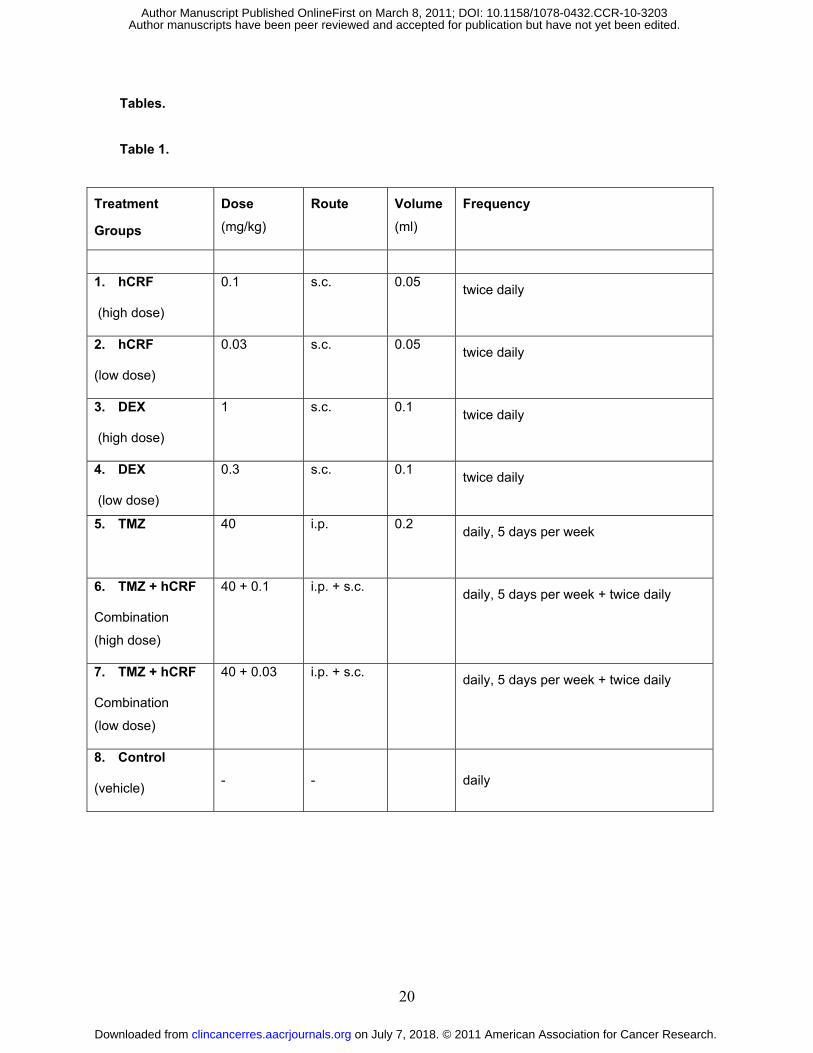
Eight treatment groups of 10 athymic rnu/rnu mice per group, each bearing a
U87 FLuc or a C6 FLuc xenograft, were developed as outlined in Table 1.
Treatment with hCRF, dexamethasone, TMZ or a combination of TMZ plus hCRF
was initiated three weeks after tumor cell implantation and lasted for 7 weeks.
on July 7, 2018. © 2011 American Association for Cancer Research.clincancerres.aacrjournals.org Downloaded from
Author manuscripts have been peer reviewed and accepted for publication but have not yet been edited. Author Manuscript Published OnlineFirst on March 8, 2011; DOI: 10.1158/1078-0432.CCR-10-3203

9
For animal groups 1 and 2, the stock clinical grade solution of hCRF
(Xerecept®) (1 mg/ml) was diluted with 0.9% sodium chloride solution to the
required “low” and “high” concentrations prior to each injection (Table 1). For
animal groups 3 and 4, clinical grade, i.v. injectable Dexamethasone (DEX) (1
mg/ml) was diluted with 0.9% sodium chloride solution to the required
concentrations). Treatment with either hCRF or DEX was administered by
subcutaneous (s.c.) injections every 12 hours for 7 weeks, starting 2nd week after
implantation. For animal group 5, animals were treated with a single daily dose
of Temozolomide (TMZ). TMZ solution was freshly prepared daily and
administered by intraperitoneal (i.p.) injection 5 of 7 days each week, for 7
weeks. TMZ powder was dissolved in pure DMSO and further diluted 1:5 with
0.9% sodium chloride solution to the desired concentration. For animal groups 6
and 7, the effects of combined TMZ and “low” or “high” hCRF were studied
(Table 1). The preparation and administration of the drugs were the same as
described above, respectively. For animal group 8 (control), animals were
treated with a single daily dose of drug vehicle. All surviving animals were
euthanized on day 150.
In vivo bioluminescence imaging was performed 7 days after implantation
of the xenografts and repeated weekly for the duration of the experiment, using
an IVIS-200 Imaging System (Caliper, CA). Imaging was performed 10 minutes
after i.p. injection of D-luciferin (2 mg per animal; Xenogen), with mice lying in the
prone position. Five mice were imaged at the same time with a field of view of 25
cm. An imaging time of 3 minutes with medium binning and an f-stop of 1 were
used initially; this was sequentially reduced as the xenografts grew and
saturation levels of BLI signal intensity were approached. Measurements of
signal intensity were obtained from region of interest analysis using Living Image
software (Xenogen). The images displayed in each data set were normalized to
the appropriate color intensity scale. BLI intensity was expressed as total
photons per region of interest per 1 second of imaging.
on July 7, 2018. © 2011 American Association for Cancer Research.clincancerres.aacrjournals.org Downloaded from
Author manuscripts have been peer reviewed and accepted for publication but have not yet been edited. Author Manuscript Published OnlineFirst on March 8, 2011; DOI: 10.1158/1078-0432.CCR-10-3203

10
Statistical analyses. Results are reported as Mean ± SD. We applied
analysis of variance (ANOVA) to the EC50 results for each drug (BCNU and TMZ)
and each cell line (C6, LN229, RG2 and U87) separately, and included date of
experiment as an effect in the model for quality control. We compared each of 4
treatments low- and high-dose dexamethasone and low- and high-dose hCRF vs
BCNU or TMZ alone and adjusted for multiple comparisons within each
experiment (combination of drug and cell line) using Dunnett’s method. We
considered p<0.05 after adjustment for multiple comparisons to be statistically
significant.
In order to account for possible differences in global levels or calibration of
BLI measurement between animals, we used the change from the average of
week 1 and week 2 to the average of week 5 and week 6 as a summary value.
Survival was measured from time of administration of treatment until death,
sacrifice, or end of study. Animals that were moribund at time of sacrifice were
considered the same as animals that died, and others were considered as
censored observations in survival analysis. Using parallel factorial designs, we
applied analysis of variance to change in BLI measurements and the Cox
proportional hazard model to survival data. We also included each animal’s
change in BLI as a potential predictor of survival in the Cox model. Analysis
showed that there was no significantly different effect on survival between high
versus low dose of hCRF, DEX, or TMZ + hCRF. We combined high and low
dose of each treatment in further analyses.
on July 7, 2018. © 2011 American Association for Cancer Research.clincancerres.aacrjournals.org Downloaded from
Author manuscripts have been peer reviewed and accepted for publication but have not yet been edited. Author Manuscript Published OnlineFirst on March 8, 2011; DOI: 10.1158/1078-0432.CCR-10-3203

11
Results.
Effects of hCRF and dexamethasone on BCNU and TMZ cytotoxicity in cell
culture. Cytotoxicity studies were performed in 6 glioma cell lines (U87, U87FLuc,
C6, C6FLuc, Ln229, RG2) with increasing doses of hCRF (0.0001 to 1000 nM)
and dexamethasone (0.001 to 10 µM). Minimal loss of cell viability (less than
20%) was observed following a 3 day exposure to 1 nM hCRF and 1 µM
dexamethasone, and less than 10% loss of cell viability following a 3 day
exposure to 0.01 nM hCRF and 0.01 µM dexamethasone. On the basis of these
results, the above concentrations of “high” and “low” hCRF and dexamethasone,
respectively, were selected for the in vitro drug-combination cytotoxicity studies.
BCNU and TMZ, both alkylating drugs extensively used to treat high grade
gliomas, were assessed as monotherapy and in drug-combination cytotoxicity
studies with either hCRF or dexamethasone. Combining BCNU with either hCRF
or dexamethasone had a variable effect; whereas, there were no significant
differences in cytotoxicity between TMZ treatment alone and combined treatment
with hCRF or dexamethasone (data not shown). In a second experimental series,
the EC50 of BCNU (Table 2A) and TMZ (Table 2B) was assessed in 4 different
cell lines. Although the EC50 values varied over a ~3-fold range for each of the
two drugs, the rank-order of sensitivity was quite different for the four cell lines. A
comparison of the results, accounting for multiple comparisons, demonstrated
only a few statistically significant effects. In the TMZ experiments, the addition of
hCRF significantly lowered the EC50 values in RG2 cells and a nonsignificant
trend toward a lower EC50 was observed in U87 cells. There was no significant
effect of dexamethasone on TMZ EC50 values with any of the cell lines.
The in vitro studies showed little evidence of a consistent additive effect when
hCRF or dexamethasone was combined with BCNU or TMZ treatment. Despite
these inconsistent results, we decided to study and compare the effect of hCRF,
dexamethasone and TMZ monotherapy in two orthotopic glioma xenografts in
immunodeficient rnu/rnu mice, and we also performed one combined therapy
study involving TMZ and hCRF. We chose a human (U87) and a rodent (C6)
on July 7, 2018. © 2011 American Association for Cancer Research.clincancerres.aacrjournals.org Downloaded from
Author manuscripts have been peer reviewed and accepted for publication but have not yet been edited. Author Manuscript Published OnlineFirst on March 8, 2011; DOI: 10.1158/1078-0432.CCR-10-3203
12
glioma cell line for comparison. Both cell lines were transduced with a reporter
gene that constitutively expressed firefly luciferase (U87FLuc and C6FLuc).
Importantly, there were no significant differences in cytotoxicity observed
between U87 and U87FLuc cells and between C6 and C6FLuc cells (Table 2).
The intracranial xenografts generated from these reporter-transduced cell lines
allowed us to monitor tumor growth non-invasively using bioluminescence
imaging (BLI).
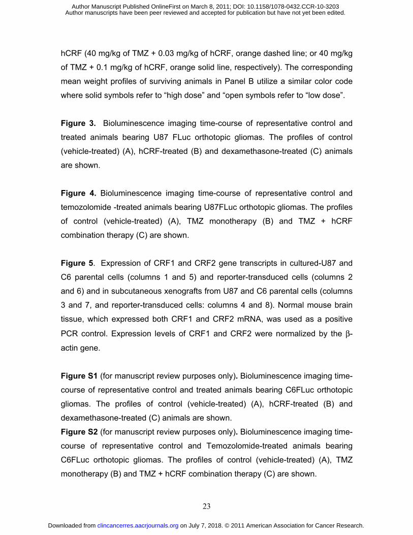
hCRF treatment of intracranial gliomas. A dose dependant effect of hCRF on
the survival of animals bearing U87 FLuc intracranial xenografts was observed
(Figure 1A). Both “low” (0.03 mg/kg/q12h) and “high” doses (0.1 mg/kg/q12h) of
hCRF increased survival compared to controls, median survival from 41 days
(95% CI 25-61 d) to 74.5 days (95% CI 41-88 d) (p<0.05) and >130 days (95%
CI not reached) (p<0.05), respectively. In contrast, hCRF had no effect on the
survival of animals bearing intracranial C6 FLuc xenografts (Figure 2A).
U87 FLuc intracranial xenografts treated with hCRF usually showed a BLI
signal intensity pattern that paralleled survival (Figure 3). For example, a rapid
decrease in BLI signal intensity that remained low for the duration of the
experiment was associated with a long survival, whereas, an increasing signal
was usually associated with a shorter survival (Figure 3B). HCRF treatment had
no effect on the pattern of increasing BLI signal intensity of C6FLuc gliomas,
consistent with the absence of any survival benefit in these animals (Figure 2).
Further analysis of treatment effects on overall survival (OS), adjusting for
experiment and change in BLI effects, showed that hCRF (low or high doses)
significantly prolonged OS (p<0.0001).
Very little or no toxicity was observed in animals treated with hCRF alone.
The only minor complication was a random non-specific skin infection that
probably reflects the systemic effects of corticosteroids on the immune system. In
addition, hCRF-treated animals bearing U87FLuc gliomas continued to gain
weight, similar to control animals (Figure 1B). No difference in animal weight
was noted between control and “low” or “high” hCRF-treated animals.
on July 7, 2018. © 2011 American Association for Cancer Research.clincancerres.aacrjournals.org Downloaded from
Author manuscripts have been peer reviewed and accepted for publication but have not yet been edited. Author Manuscript Published OnlineFirst on March 8, 2011; DOI: 10.1158/1078-0432.CCR-10-3203

13
Dexamethasone treatment of intracranial gliomas. Dexamethasone at both
low (0.3 mg/kg) and high doses (1 mg/kg) was not effective in prolonging animal
survival compared to control non-treated animals bearing either U87FLuc
(Figure 1A) or C6FLuc (Figure 2A) gliomas. Similarly, the patterns of increasing
bioluminescence intensity were indistinguishable from that of non-treated control
animals (Figure 3C). Further statistical analysis showed that animals treated with
hCRF had significantly longer survival than DEX-treated animals (p=0.02),
whereas no differences between “low” and “high” doses of Dex were noted.
Treatment-related toxicity was greater in the dexamethasone-treated animals
and included significant weight loss (Figure 1B and Figure 2B), more sever skin
infections and some necrosis at the injection sites.
TMZ treatment of intracranial gliomas. A significant effect of TMZ alone
(p=0.008) or in combination with different doses of hCRF (p=0.003) on the
survival of animals bearing U87FLuc xenografts was observed (Figure 1C), but
not on the survival of animals bearing C6FLuc xenografts (Figure 2C). The
temporal profiles of the bioluminescence images were similar to the survival data
in both U87FLuc (Figures 1B and 4B, respectively) and C6FLuc (data not
shown) xenograft-bearing animals. Many of the animals treated with TMZ
showed a decrease and stabilization of the BLI signal at a low or moderate level
for most of the treatment and post-treatment period, followed by a rapid increase
in last two weeks of the animal’s life. However, TMZ monotherapy and TMZ
combined with hCRF was accompanied with high toxicity. There was a
substantial loss of weight (up to 40%), which tended to recover during the post-
treatment period if the animal survived. This weight loss is reflected in the
photographic images of the animals at week 9, just after completing the course of
TMZ therapy (Figure 4B and 4C). A very high incidence (27/30) of skin
neoplasms was observed in animals treated with TMZ and there were rare
occurrences (2/30) of internal abdominal tumors. Spontaneous tumorigenesis
was not observed in any of the other treatment groups.
CRF1 and CRF2 receptor expression. U87-FLuc and C6-FLuc cell lines and
xenografts were examined for CRF1 and CRF2 mRNA levels by RT-PCR
on July 7, 2018. © 2011 American Association for Cancer Research.clincancerres.aacrjournals.org Downloaded from
Author manuscripts have been peer reviewed and accepted for publication but have not yet been edited. Author Manuscript Published OnlineFirst on March 8, 2011; DOI: 10.1158/1078-0432.CCR-10-3203

14
(Figure 5). U87 cultured-cells and xenografts express measurable levels of
CRF2, but not CRF1 mRNA transcript. In contrast, neither CRF1 nor CRF2
transcripts could be detected in the C6 cultured-cells and xenografts.
on July 7, 2018. © 2011 American Association for Cancer Research.clincancerres.aacrjournals.org Downloaded from
Author manuscripts have been peer reviewed and accepted for publication but have not yet been edited. Author Manuscript Published OnlineFirst on March 8, 2011; DOI: 10.1158/1078-0432.CCR-10-3203

15
Discussion.
We previously reported a dose-dependent decrease in vasogenic peritumoral
brain edema following treatment of immunocompetent Fischer 344 rats, bearing
RG2 intracranial gliomas, with human corticotropin-releasing factor (Xerecept,
Corticorelin acetate) (29). We also reported a similar result in a single-dose
hCRF study involving Sprague-Dawley rats bearing W256 intracranial gliomas (1,
17). Both studies demonstrated a significant anti-edematous effect in tumor and
peritumoral brain tissue by proton density-weighted and T1-weighted contrast-
enhanced MRI, and by ex vivo measures of tissue water content. The initial study
also reported a significant survival advantage for hCRF-treated animals
compared to dexamethasone-treated as well as to control animals (29).
In this study, an immunocompromised animal model harboring either an
intracranial human (U87) or a rat (C6) glioma xenograft was studied, and
therapeutic efficacy was evaluated in terms of animal survival. We focused on a
comparison of hCRF and dexamethasone (Dex) monotherapy; although a
comparison between temozolomide (TMZ) monotherapy and TMZ plus hCRF
combined therapy was also performed. Interestingly, a highly significant, dose-
dependent effect of hCRF monotherapy on survival was observed in animals
bearing U87 gliomas, but not C6 gliomas. These results were highly consistent
with tumor growth, regression and regrowth patterns visualized by sequential-
weekly bioluminescence reporter gene imaging of the xenografts prior-to, during
and following hCRF treatment. Interestingly, dexamethasone treatment had little
or no effect on either the survival or bioluminescence imaging profiles of animals
bearing either U87 or C6 gliomas, and this may reflect in part the toxic side-
effects of twice-daily s.c. injections of dexamethasone. TMZ monotherapy and
TMZ plus hCRF combined therapy of U87 gliomas yielded similar survival
profiles that were significantly longer than control animals. Again, no survival
advantage was observed in C6 gliomas.
To further explore the dichotomy in hCRF treatment response between U87
and C6 gliomas, we evaluated the levels of CRF1 and CRF2 receptor mRNA in
on July 7, 2018. © 2011 American Association for Cancer Research.clincancerres.aacrjournals.org Downloaded from
Author manuscripts have been peer reviewed and accepted for publication but have not yet been edited. Author Manuscript Published OnlineFirst on March 8, 2011; DOI: 10.1158/1078-0432.CCR-10-3203

16
the two cell lines and xenografts. This investigation was based on our previous
study which showed that significant levels of CRF1 mRNA (0.25±0.01 pg/µg total
RNA) were detectable in W256 cells, and presumably reflects CRF1 receptor
expression on tumor and/or endothelial cell membranes (22). Blockade of CRF
receptors with alpha-helical CRF-(9-41) analog abolished the growth inhibitory
and differentiation inducing effects of hCRF. Together, these findings suggested
that W256 cells express functional rat CRF receptors in vitro and that these
receptors are likely to mediate the effects observed following exposure to hCRF.
Our current results suggest that C6 cells and xenografts express neither CRF1
nor CRF2 receptors, whereas U87 cells and xenografts express CRF2, but not
CRF1 receptors. This difference between the two cell lines and xenografts could
contribute to the difference in hCRF treatment response that was observed in this
study. In addition, these results suggest that CRF1 and/or CRF2 receptor
expression is required for the anti-tumor and anti-edematous effects of hCRF.
It was previously shown that CRFR1 is expressed in tumor cells and that
urocortin and corticotropin-releasing factor (UCN/CRF), both members of the
CRF family, reduce tumor cell growth via CRFR1 (37, 38). Multiple malignancies
have been reported to have high leves of CRFR1 and CRFR2 expression, and to
be sensitive to the suppressive effects of CRF and its agonists. Graziani et al.
reported that UCN/CRF inhibited the growth of adenocarcinoma Ishikawa cells in
a concentration-dependent manner, and that this effect was mediated by CRFR1
(38). It was reported that UCN inhibited the proliferation of melanoma cells in
vitro and in vivo, also through CRFR1 (39) and in human mammary cancer cells,
CRF acted on CRFR1 to inhibit the proliferative effects of estrogens on MCF7
cells in both a paracrine and autocrine manner (40). Activation of CRFR2 was
observed to suppress angiogenesis and rearrange the vasculature. It was also
reported that CRFR2 agonists inhibited hepatocellular carcinoma tumor
angiogenesis in vitro and reduced tumor microvessel density in vivo. Our results
are consistent with these observations.
on July 7, 2018. © 2011 American Association for Cancer Research.clincancerres.aacrjournals.org Downloaded from
Author manuscripts have been peer reviewed and accepted for publication but have not yet been edited. Author Manuscript Published OnlineFirst on March 8, 2011; DOI: 10.1158/1078-0432.CCR-10-3203

17
Reports from other animal and human studies confirm that the anti-
edematous effects of hCRF- and dexamethasone-treated animals are similar.
Patients treated with hCRF and decreasing doses of dexamethasone
demonstrate clinical improvement in neurologic symptoms and less steroid-
associated myopathy and insulin dependence (41). The substitution of a less
toxic, but equally effective drug for dexamethasone to treat tumor-associated
cerebral edema has been a long-sought challenge for clinicians. Our pre-clinical
studies add to the body of data that indicates that hCRF has the potential to meet
this challenge. We observed higher therapeutic efficacy with hCRF than with
either dexamethasone or temozolomide monotherapy in U87 glioma xenografts.
Also, no toxic effects or signs of discomfort were observed in mice who have
received hCRF treatment in low or high dose for 2-6 months. In comparison,
animals who have received dexamethasone or temozolomide therapy exhibited
significant toxicity, manifested by a decrease in weight, increased irritability, local
skin necrosis and even spontaneous tumorigenesis.
The current clinical development program for hCRF has focused on the
treatment of peritumoral brain edema in patients with metastatic and primary
brain tumors requiring dexamethasone to control symptoms. Three major clinical
trials (0303, 0501 and 0302) have established two important points: 1) hCRF is
safe in man, and 2) hCRF enables significant reductions or elimination of steroid-
dosing in patients with cerebral tumors, with no apparent impairment of
neurocognitive status (26). Blinded independent review of magnetic resonance
imaging (MRI) scans from patients receiving hCRF demonstrated that most of
these patients experienced prolonged periods of stable disease, and a minority
have achieved a measurable level of tumor regression (24). In a phase 1 clinical
trial, 10 of the 15 patients who received hCRF had improvement in neurological
symptoms or physical findings, with little or no toxicity (4). It is notable that there
was a measurable decrease in steroid related side-effects, especially myopathy
and the appearance of Cushingoid features. One patient, who had lymphoma-
related pruritus and a long-standing rash resistant to steroids, noted
improvement while receiving hCRF (25). In this preclinical study, we also noted
on July 7, 2018. © 2011 American Association for Cancer Research.clincancerres.aacrjournals.org Downloaded from
Author manuscripts have been peer reviewed and accepted for publication but have not yet been edited. Author Manuscript Published OnlineFirst on March 8, 2011; DOI: 10.1158/1078-0432.CCR-10-3203

18
differences in toxicity and efficacy of hCRF, dexamethasone and temozolomide
treatment of the glioma-bearing animals.
Overall we have shown therapeutic efficacy and low toxicity of hCRF in the
treatment of a human-derived glioma in an orthotopic nude mouse model. Our
results are consistent with the requirement of CRF-receptor expression in the
tumor cells for hCRF therapeutic efficacy. Notably, hCRF treatment was less
toxic in comparison to that with Dexamethasone or with Temozolomide. These
results support the development of novel hCRF-releasing formulations or
platforms for extended constant administration and studies to optimize the
therapeutic antitumoral effect. Further clinical studies will need to evaluate
whether long-term hCRF treatment (or the rapid cessation of treatment) is
associated with increased glioma cell invasion and rapid tumor progression, as
has been described for bevacizumab.
on July 7, 2018. © 2011 American Association for Cancer Research.clincancerres.aacrjournals.org Downloaded from
Author manuscripts have been peer reviewed and accepted for publication but have not yet been edited. Author Manuscript Published OnlineFirst on March 8, 2011; DOI: 10.1158/1078-0432.CCR-10-3203

19
Acknowledgements.
This work was supported by Celtic Pharma award # SK#12715.
The MSKCC Small Animal Imaging Core Facility was supported by NIH Small-
Animal Imaging Research Program grant R24 CA83084 and NIH Center grant P30
CA08748); and we thank Dr. Pat Zanzonico and Ms. Valerie Longo for imaging support;
and we also thank Dr. Ekaterina Moroz for technical support.
on July 7, 2018. © 2011 American Association for Cancer Research.clincancerres.aacrjournals.org Downloaded from
Author manuscripts have been peer reviewed and accepted for publication but have not yet been edited. Author Manuscript Published OnlineFirst on March 8, 2011; DOI: 10.1158/1078-0432.CCR-10-3203
20
Tables.
Table 1.
Treatment
Groups
Dose
(mg/kg)
Route
Volume
(ml)
Frequency
1. hCRF
(high dose)
0.1 s.c. 0.05 twice daily
2. hCRF
(low dose)
0.03 s.c. 0.05 twice daily
3. DEX
(high dose)
1 s.c. 0.1 twice daily
4. DEX
(low dose)
0.3 s.c. 0.1 twice daily
5. TMZ
40 i.p. 0.2 daily, 5 days per week
6. TMZ + hCRF
Combination
(high dose)
40 + 0.1 i.p. + s.c. daily, 5 days per week + twice daily
7. TMZ + hCRF
Combination
(low dose)
40 + 0.03 i.p. + s.c. daily, 5 days per week + twice daily
8. Control
(vehicle)
-
-
daily
on July 7, 2018. © 2011 American Association for Cancer Research.clincancerres.aacrjournals.org Downloaded from
Author manuscripts have been peer reviewed and accepted for publication but have not yet been edited. Author Manuscript Published OnlineFirst on March 8, 2011; DOI: 10.1158/1078-0432.CCR-10-3203

21
Table 2
A. hCRF and dexamethasone effects on BCNU EC50
In vitro cell viability studies: EC50 (µM)
Cell Line BCNU alone BCNU +
low hCRF (0.01nM)
BCNU + high hCRF
(1.0nM)
BCNU + low DEX (0.01mM)
BCNU + high DEX (1.0mM)
U87 226 ± 56 (9) 264 ± 91 (9) 193 ± 54 (9) 311 ± 65 (9)* 260 ± 59 (9)
C6 414 ± 43 (8) 419 ± 33 (8) 388 ± 59 (8) 346 ± 94 (8) 304 ± 102 (9)*
Ln229 234 ± 43 (7) 261 ± 58 (7) 219 ± 36 (7) 239 ± 47 (7) 201 ± 91 (7)
RG2 140 ± 22 (6) 259 ± 96 (6) 173 ± 60 (6) 329 ± 165 (6)* 147 ± 72 (6)
B. hCRF and dexamethasone effects on TMZ EC50
In vitro cell viability studies: EC50 (mM)
Cell Line TMZ alone TMZ +
low hCRF (0.01nM)
TMZ + high hCRF
(1.0nM)
TMZ + low DEX (0.01mM)
TMZ + high DEX (1.0mM)
U87 4.8 ± 3.5 (5) 3.1 ± 1.8 (5) 3.8 ± 2.8 (5) 4.5 ± 3.3 (5) 3.9 ± 3.2 (5)
C6 2.9 ± 1.1 (5) 3.6 ± 1.7 (5) 2.9 ± 1.0 (5) 4.5 ± 3.0 (5) 3.8 ± 3.5 (5)
Ln229 3.1 ± 0.6 (6) 3.2 ± 0.5 (6) 3.3 ± 0.6 (6) 3.4 ± 1.0 (6) 3.5 ± 1.0 (6)
RG2 9.8 ± 2.8 (5) 6.9 ± 1.4 (5)* 6.2 ± 0.3 (5)* 7.8 ± 2.1 (5) 7.9 ± 1.6 (5)
* Assays were performed after 24 hour exposure to the drugs. Significant difference compared to BCNU or TMZ alone; p<0.05 after adjustment for multiple comparisons using Dunnett’s method.
on July 7, 2018. © 2011 American Association for Cancer Research.clincancerres.aacrjournals.org Downloaded from
Author manuscripts have been peer reviewed and accepted for publication but have not yet been edited. Author Manuscript Published OnlineFirst on March 8, 2011; DOI: 10.1158/1078-0432.CCR-10-3203

22
Figure Legends.
Figure 1. Time profiles of survival (A, C) and body weight (B, D) of animals
bearing orthotopic U87-FLuc intracranial xenografts. The treatment period and
end of the experiment (euthanasia of all surviving animals) is indicated by the
bracket and arrow, respectively. Control animals received vehicle injections
(black dashed line in Panels A and C). Treated animals in Panel A received
either “low dose” hCRF (0.03 mg/kg twice daily; red dotted line), “high dose”
hCRF (0.1 mg/kg twice daily; red solid line), “low dose” dexamethasone (0.3
mg/kg twice daily; blue dotted line), or “high dose” dexamethasone (1 mg/kg
twice daily; blue solid line). The corresponding mean weight profiles of surviving
animals in Panel B utilize a similar color code where solid symbols refer to “high
dose” and “open symbols refer to “low dose”. Temozolomide (TMZ) treated
animals are shown in Panel C. Animals received monotherapy (40 mg/kg daily, 5
of 7 days; green solid line), or combination therapy with “low” and “high” doses of
hCRF (40 mg/kg of TMZ + 0.03 mg/kg of hCRF, orange dashed line; or 40 mg/kg
of TMZ + 0.1 mg/kg of hCRF, orange solid line, respectively). The corresponding
mean weight profiles of surviving animals in Panel B utilize a similar color code
where solid symbols refer to “high dose” and “open symbols refer to “low dose”.
Figure 2. Time profiles of survival (A, C) and body weight (B, D) of animals
bearing orthotopic C6-FLuc intracranial xenografts. Control animals received
vehicle injections (black dashed line in Panels A and C); treated animals in Panel
A received either “low dose” hCRF (0.03 mg/kg twice daily; red dotted line), “high
dose” hCRF (0.1 mg/kg twice daily; red solid line), “low dose” dexamethasone
(0.3 mg/kg twice daily; blue dotted line), or “high dose” dexamethasone (1 mg/kg
twice daily; blue solid line). The corresponding mean weight profiles of surviving
animals in Panel B utilize a similar color code where solid symbols refer to “high
dose” and “open symbols refer to “low dose”. Temozolomide (TMZ) treated
animals are shown in Panel C. Animals received monotherapy (40 mg/kg daily, 5
of 7 days; green solid line), or combination therapy with “low” and “high” doses of
on July 7, 2018. © 2011 American Association for Cancer Research.clincancerres.aacrjournals.org Downloaded from
Author manuscripts have been peer reviewed and accepted for publication but have not yet been edited. Author Manuscript Published OnlineFirst on March 8, 2011; DOI: 10.1158/1078-0432.CCR-10-3203
23
hCRF (40 mg/kg of TMZ + 0.03 mg/kg of hCRF, orange dashed line; or 40 mg/kg
of TMZ + 0.1 mg/kg of hCRF, orange solid line, respectively). The corresponding
mean weight profiles of surviving animals in Panel B utilize a similar color code
where solid symbols refer to “high dose” and “open symbols refer to “low dose”.
Figure 3. Bioluminescence imaging time-course of representative control and
treated animals bearing U87 FLuc orthotopic gliomas. The profiles of control
(vehicle-treated) (A), hCRF-treated (B) and dexamethasone-treated (C) animals
are shown.
Figure 4. Bioluminescence imaging time-course of representative control and
temozolomide -treated animals bearing U87FLuc orthotopic gliomas. The profiles
of control (vehicle-treated) (A), TMZ monotherapy (B) and TMZ + hCRF
combination therapy (C) are shown.
Figure 5. Expression of CRF1 and CRF2 gene transcripts in cultured-U87 and
C6 parental cells (columns 1 and 5) and reporter-transduced cells (columns 2
and 6) and in subcutaneous xenografts from U87 and C6 parental cells (columns
3 and 7, and reporter-transduced cells: columns 4 and 8). Normal mouse brain
tissue, which expressed both CRF1 and CRF2 mRNA, was used as a positive
PCR control. Expression levels of CRF1 and CRF2 were normalized by the β-
actin gene.
Figure S1 (for manuscript review purposes only). Bioluminescence imaging time-
course of representative control and treated animals bearing C6FLuc orthotopic
gliomas. The profiles of control (vehicle-treated) (A), hCRF-treated (B) and
dexamethasone-treated (C) animals are shown.
Figure S2 (for manuscript review purposes only). Bioluminescence imaging time-
course of representative control and Temozolomide-treated animals bearing
C6FLuc orthotopic gliomas. The profiles of control (vehicle-treated) (A), TMZ
monotherapy (B) and TMZ + hCRF combination therapy (C) are shown.
on July 7, 2018. © 2011 American Association for Cancer Research.clincancerres.aacrjournals.org Downloaded from
Author manuscripts have been peer reviewed and accepted for publication but have not yet been edited. Author Manuscript Published OnlineFirst on March 8, 2011; DOI: 10.1158/1078-0432.CCR-10-3203

24
References.
1. Tjuvajev J, Uehara H, Desai R, Beattie B, Matei C, Zhou Y, et al. Corticotropin-releasing factor decreases vasogenic brain edema. Cancer Res. 1996 Mar 15;56(6):1352-60. 2. Villalona-Calero MA, Eckardt J, Burris H, Kraynak M, Fields-Jones S, Bazan C, et al. A phase I trial of human corticotropin-releasing factor (hCRF) in patients with peritumoral brain edema. Ann Oncol. 1998 Jan;9(1):71-7. 3. Bale TL, Giordano FJ, Vale WW. A new role for corticotropin-releasing factor receptor-2: suppression of vascularization. Trends Cardiovasc Med. 2003 Feb;13(2):68-71. 4. Moliterno JA, Henry E, Pannullo SC. Corticorelin acetate injections for the treatment of peritumoral brain edema. Expert Opin Investig Drugs. 2009 Sep;18(9):1413-9. 5. Norden AD, Drappatz J, Wen PY. Antiangiogenic therapies for high-grade glioma. Nat Rev Neurol. 2009 Nov;5(11):610-20. 6. Argirios Moustakas TNK. New treatment options in the managment of glioblastoma multiforme: a focus on bevacizumab. OncoTargets and Therapy. 2010(3):27-38. 7. Pavlidis ET, Ballas KD, Symeonidis NG, Psarras K, Koliakos G, Kouzi-Koliakos K, et al. The effect of bevacizumab on colon anastomotic healing in rats. Int J Colorectal Dis. 2010 Dec;25(12):1465-73. 8. Lucio-Eterovic AK, Piao Y, de Groot JF. Mediators of glioblastoma resistance and invasion during antivascular endothelial growth factor therapy. Clin Cancer Res. 2009 Jul 15;15(14):4589-99. 9. Iwamoto FM, Abrey LE, Beal K, Gutin PH, Rosenblum MK, Reuter VE, et al. Patterns of relapse and prognosis after bevacizumab failure in recurrent glioblastoma. Neurology. 2009 Oct 13;73(15):1200-6. 10. Verhoeff JJ, van Tellingen O, Claes A, Stalpers LJ, van Linde ME, Richel DJ, et al. Concerns about anti-angiogenic treatment in patients with glioblastoma multiforme. BMC Cancer. 2009;9:444. 11. Vale W, Spiess J, Rivier C, Rivier J. Characterization of a 41-residue ovine hypothalamic peptide that stimulates secretion of corticotropin and beta-endorphin. Science. 1981 Sep 18;213(4514):1394-7. 12. Brown JK HA. Toxic encephalopathy and acute brain-swelling in children. Dev Med Child Neurology. 1975;17(5):659-79. 13. Taylor AL, Fishman LM. Corticotropin-releasing hormone. N Engl J Med. 1988 Jul 28;319(4):213-22. 14. Kiang JG, Poree L, Wei ET. Anti-inflammatory activity of corticotropin releasing factor: II. Mechanisms of action. Proc West Pharmacol Soc. 1987;30:63-5. 15. Serda SM, Wei ET. Epinephrine-induced pulmonary oedema in rats is inhibited by corticotropin-releasing factor. Pharmacol Res. 1992 Jul-Aug;26(1):85-91. 16. Wei ET, Kiang JG, Tian JQ. Anti-inflammatory activity of corticotropin releasing factor: I. Efficacy studies. Proc West Pharmacol Soc. 1987;30:59-62.
on July 7, 2018. © 2011 American Association for Cancer Research.clincancerres.aacrjournals.org Downloaded from
Author manuscripts have been peer reviewed and accepted for publication but have not yet been edited. Author Manuscript Published OnlineFirst on March 8, 2011; DOI: 10.1158/1078-0432.CCR-10-3203

25
17. Wei ET, Thomas HA. Anti-inflammatory peptide agonists. Annu Rev Pharmacol Toxicol. 1993;33:91-108. 18. Thomas HA, Ling N, Wei ET. CRF and related peptides as anti-inflammatory agonists. Ann N Y Acad Sci. 1993 Oct 29;697:219-28. 19. Wei ET, Gao GC. Corticotropin-releasing factor: an inhibitor of vascular leakage in rat skeletal muscle and brain cortex after injury. Regul Pept. 1991 Apr 25;33(2):93-104. 20. Reubi JC, Waser B, Vale W, Rivier J. Expression of CRF1 and CRF2 receptors in human cancers. J Clin Endocrinol Metab. 2003 Jul;88(7):3312-20. 21. Wei ET, Gao GC, Thomas HA. Peripheral anti-inflammatory actions of corticotropin-releasing factor. Ciba Found Symp. 1993;172:258-68; discussion 68-76. 22. Tjuvajev J, Kolesnikov Y, Joshi R, Sherinski J, Koutcher L, Zhou Y, et al. Anti-neoplastic properties of human corticotropin releasing factor: involvement of the nitric oxide pathway. In Vivo. 1998 Jan-Feb;12(1):1-10. 23. Chrousos GP, Schulte HM, Oldfield EH, Gold PW, Cutler GB, Jr., Loriaux DL. The corticotropin-releasing factor stimulation test. An aid in the evaluation of patients with Cushing's syndrome. N Engl J Med. 1984 Mar 8;310(10):622-6. 24. Recht LD ML PS, Hormigo A, Hines V, Milsted E, O'Connor PC, Ryan RP, Wong ET. A placebo-controlled stud investigating the dexamethasone-sparing effects of corticorelin acetate in patience with primary or metastatic brain tumors abd peritumoral edema. Journal of Clinical Oncology. 2009;27:Abstract 2078. 25. Shapiro WR ML CL, Wheeler H, Hines V, Milsted E, O'Connor OC, Ryan RP, Recht L,. A randomized, double-blind study comparing corticorelin acetate with dexamethasone in patients with primary malignant glioma who require increased dexamethasone to control symptoms of peritumoral brain edema. Journal of Clinical Oncology. 2009;27:Abstract 2080. 26. Mechtler L WE, Hormigo A, Pannullo S, Hines V, Milsted R, O'Connor PO, Ryan RP, Recht L A long-term open-label extension study examining the steroid-sparing effects of corticorelin acetate in patients with cerebral tumors. Journal of Clinical Oncology. 2009 27. 27. Leff RS, Thompson JM, Daly MB, Johnson DB, Harden EA, Mercier RJ, et al. Acute neurologic dysfunction after high-dose etoposide therapy for malignant glioma. Cancer. 1988 Jul 1;62(1):32-5. 28. Treadwell BL, Sever ED, Savage O, Copeman WS. Side-Effects of Long-Term Treatment with Corticosteroids and Corticotrophin. Lancet. 1964 May 23;1(7343):1121-3. 29. Tjuvajev J, Gansbacher B, Desai R, Beattie B, Kaplitt M, Matei C, et al. RG-2 glioma growth attenuation and severe brain edema caused by local production of interleukin-2 and interferon-gamma. Cancer Res. 1995 May 1;55(9):1902-10. 30. Corticorelin: ACTH RF, corticoliberin, corticotrophin-releasing hormone, corticotropin-releasing factor, human corticotropin-releasing hormone, ovine corticotrophine-releasing factor, Xerecept. . Drugs R D. 2004;5(4):218-9. 31. Moroz E, Carlin S, Dyomina K, Burke S, Thaler HT, Blasberg R, et al. Real-time imaging of HIF-1alpha stabilization and degradation. PLoS One. 2009;4(4):e5077.
on July 7, 2018. © 2011 American Association for Cancer Research.clincancerres.aacrjournals.org Downloaded from
Author manuscripts have been peer reviewed and accepted for publication but have not yet been edited. Author Manuscript Published OnlineFirst on March 8, 2011; DOI: 10.1158/1078-0432.CCR-10-3203

26
32. Ponomarev V, Doubrovin M, Serganova I, Vider J, Shavrin A, Beresten T, et al. A novel triple-modality reporter gene for whole-body fluorescent, bioluminescent, and nuclear noninvasive imaging. Eur J Nucl Med Mol Imaging. 2004 May;31(5):740-51. 33. Serganova I, Doubrovin M, Vider J, Ponomarev V, Soghomonyan S, Beresten T, et al. Molecular imaging of temporal dynamics and spatial heterogeneity of hypoxia-inducible factor-1 signal transduction activity in tumors in living mice. Cancer Res. 2004 Sep 1;64(17):6101-8. 34. Ngamwongsatit P, Banada PP, Panbangred W, Bhunia AK. WST-1-based cell cytotoxicity assay as a substitute for MTT-based assay for rapid detection of toxigenic Bacillus species using CHO cell line. J Microbiol Methods. 2008 Jun;73(3):211-5. 35. Ciusani E, Balzarotti M, Calatozzolo C, de Grazia U, Boiardi A, Salmaggi A, et al. Valproic acid increases the in vitro effects of nitrosureas on human glioma cell lines. Oncol Res. 2007;16(10):453-63. 36. Tannock I, Guttman P. Misonidazole increases the toxicity of BCNU for hypoxic cells. Int J Radiat Oncol Biol Phys. 1982 Mar-Apr;8(3-4):663-6. 37. Schoeffter P, Feuerbach D, Bobirnac I, Gazi L, Longato R. Functional, endogenously expressed corticotropin-releasing factor receptor type 1 (CRF1) and CRF1 receptor mRNA expression in human neuroblastoma SH-SY5Y cells. Fundam Clin Pharmacol. 1999;13(4):484-9. 38. Graziani G, Tentori L, Portarena I, Barbarino M, Tringali G, Pozzoli G, et al. CRH inhibits cell growth of human endometrial adenocarcinoma cells via CRH-receptor 1-mediated activation of cAMP-PKA pathway. Endocrinology. 2002 Mar;143(3):807-13. 39. Carlson KW, Nawy SS, Wei ET, Sadee W, Filov VA, Rezsova VV, et al. Inhibition of mouse melanoma cell proliferation by corticotropin-releasing hormone and its analogs. Anticancer Res. 2001 Mar-Apr;21(2A):1173-9. 40. Graziani G, Tentori L, Muzi A, Vergati M, Tringali G, Pozzoli G, et al. Evidence that corticotropin-releasing hormone inhibits cell growth of human breast cancer cells via the activation of CRH-R1 receptor subtype. Mol Cell Endocrinol. 2007 Jan 29;264(1-2):44-9. 41. CelticPharma. Celtic Pharma Announces Results of a Phase III Program Evaluating XERECEPT® in Patients with Primary and Metastatic Brain Tumors. Celtic Pharma press release. 2009.
on July 7, 2018. © 2011 American Association for Cancer Research.clincancerres.aacrjournals.org Downloaded from
Author manuscripts have been peer reviewed and accepted for publication but have not yet been edited. Author Manuscript Published OnlineFirst on March 8, 2011; DOI: 10.1158/1078-0432.CCR-10-3203

Published OnlineFirst March 8, 2011.Clin Cancer Res Maxim A Moroz, Ruimin Huang, Tatiana Kochetkov, et al. C6 Intracranial Gliomasand Temozolomide: Treatment Efficacy and Toxicity in U87 and Comparison of Corticotropin-Releasing Factor, Dexamethasone
Updated version
10.1158/1078-0432.CCR-10-3203doi:
Access the most recent version of this article at:
Material
Supplementary
http://clincancerres.aacrjournals.org/content/suppl/2011/05/18/1078-0432.CCR-10-3203.DC1
Access the most recent supplemental material at:
Manuscript
Authoredited. Author manuscripts have been peer reviewed and accepted for publication but have not yet been
E-mail alerts related to this article or journal.Sign up to receive free email-alerts
Subscriptions
Reprints and
To order reprints of this article or to subscribe to the journal, contact the AACR Publications
Permissions
Rightslink site. Click on "Request Permissions" which will take you to the Copyright Clearance Center's (CCC)
.http://clincancerres.aacrjournals.org/content/early/2011/03/05/1078-0432.CCR-10-3203To request permission to re-use all or part of this article, use this link
on July 7, 2018. © 2011 American Association for Cancer Research.clincancerres.aacrjournals.org Downloaded from
Author manuscripts have been peer reviewed and accepted for publication but have not yet been edited. Author Manuscript Published OnlineFirst on March 8, 2011; DOI: 10.1158/1078-0432.CCR-10-3203