comparison of cells free in coelomic and water-vascular system of sea cucumber, apostichopus...
TRANSCRIPT

lable at ScienceDirect
Fish & Shellfish Immunology 35 (2013) 1654e1657
Contents lists avai
Fish & Shellfish Immunology
journal homepage: www.elsevier .com/locate/ fs i
Short communication
Comparison of cells free in coelomic and water-vascular system of seacucumber, Apostichopus japonicus
Qiang Li a, Rui-rong Qi a, Yi-nan Wang a, Shi-gen Ye a, Guo Qiao b, Hua Li a,*aKey Laboratory of Mariculture & Stock Enhancement in North China’s Sea, Ministry of Agriculture, Dalian Ocean University, Dalian 116023, ChinabDepartment of Aquaculture, National Fisheries Research & Development Institute, Incheon 400-420, Republic of Korea
a r t i c l e i n f o
Article history:Received 9 September 2012Received in revised form11 July 2013Accepted 11 July 2013Available online 2 August 2013
Keywords:Apostichopus japonicusCoelomocytesWater-vascular systemMonoclonal antibody
* Corresponding author. Tel.: þ86 411 84763680; faE-mail address: [email protected] (H. Li).
1050-4648/$ e see front matter � 2013 Elsevier Ltd.http://dx.doi.org/10.1016/j.fsi.2013.07.020
a b s t r a c t
The sea cucumber, Apostichopus japonicus possesses a variety of cells populating in both the coelomic(cells in the coelomic are called coelomocytes) and water-vascular system. In this study, we comparedcells in these two systems of A. japonicus on total cell number, cell types and surface antigens throughmonoclonal antibodies against coelomocytes. The results demonstrated that the cell types observed incoelomic also could be found in water-vascular system, but the total cell number and percentages of eachtype were different. The total number of coelomocytes was 2e3 times of that in water-vascular system.Lymphoid cells were numerically dominant in coelomic system, while spherulocytes with pseudopods inwater-vascular system. Results of indirect immunofluorescence assay technique showed that both coe-lomocytes and cells in water-vascular system could be recognized by the corresponding MAbs, and thedistribution of its positive signals was not different. In conclusion, cell types and surface antigens incoelomic and water-vascular system were same, but the total cell number and percentages of each typewere different. And further researches are needed on whether there are differences in functions of thedifferent composition.
� 2013 Elsevier Ltd. All rights reserved.
1. Introduction
One of the most distinctive characteristics of echinoderms istheir water-vascular system-a unique arrangement of fluid-filledcoelomic passages and associated parts. There are three mainparts of the system, including the ring canal, stone canal and pe-ripheral system [1,2]. The central parts of the system maintain asupply of coelomic fluid to each radial canal as required. Some ofthe accessory structures branched from the ring canal, such as themuscular and bag-like polian vesicle which are present in somemembers of all groups except crinoids [3], appear to hold water-vascular fluid in reverse and under slight pressure until it isrequired by the animal. The function of the system is to generate,distribute, and control the hydrostatic pressure necessary for theoperation of the tube-feet, but secondarily it may also serve otherfunctions, for instance, assistance in gaseous exchange to the innerparts of the body and in removal of waste [2,4]. However, water-vascular system is much more complex in both structure andfunction than general realization, and it presents many attributes
x: þ86 411 84762871.
All rights reserved.
that are puzzled and not yet studied. Many free cells float in thewater-vascular system and are considered to be the same with thecoelomocytes in coelom of Apostichopus japonicus [2].
Sea cucumbers, especially A. japonicus, are economically impor-tant breeding species in East Asia. Most of studies on sea cucumbershave focused on culture and management, and some studies oncytology and histology. A. japonicus has a coelom filled withcoelomicfluid andmany cells suspended in thisfluid. Those cells arecalled coelomocytes. Coelomocytes are believed to participate in gasexchange, nutrient storage, excretion of waste products, productionof some components of the connective tissue, and immune defense[4]. There is a complicated water-vascular system in A. japonicuswhich is fluid-filledwith a dense population of cells suspended in it,and this system may be expected to assume considerable impor-tance in the life of A. japonicus. We want to find out the origin ofcoelomocytes at first, we studied cells and tissues of A. japonicus.When we studied cells in the coelomic and water-vascular system,we found out that both the coelomic and water-vascular system arefluid-filled with dense cells, and cells in these two systems areconsidered to be same without enough and detailed studies.Therefore, comparative studies of coelomocytes and cells in thewater-vascular system are necessary and important for under-standing the origin and functions of coelomocytes, particularly their
Q. Li et al. / Fish & Shellfish Immunology 35 (2013) 1654e1657 1655
role in immune responses. The present study shows a first approachtowards the comparisons/relationship between coelomocytes andcells in water-vascular system of A. japonicus.
2. Materials and methods
2.1. Animals
Healthy A. japonicus juveniles (15 animals, body weight 20e35 g) and adults (15 animals, body weight 85e120 g) were obtainedfrom a local aquatic farm (Dalian, China) and kept in the aquaria(the size of aquaria is 60 � 25 � 40 cm3,5 animals each aquarium)with aerated seawater (aerated seawater is filtering seawatercoming from Dalian Heishijiao waters) in the laboratory.
2.2. Preparation of monoclonal antibodies
The monoclonal antibodies (MAbs) against A. japonicus coelo-mocytes were generated following the method of Li et al. [5].Briefly, spleen cells from the immunized mouse were fused withmyeloma cells, positive hybridomas were cloned by limiting dilu-tion, monoclonal antibodies were secreted by the positive hybrid-omas obtained from repeatedly cloning.
2.3. Preparation of cell samples
Coelomic fluid of A. japonicus was drawn from the right lateralside of the body and immediately diluted (1:1) in antiaggregantmodified Alsever’s solution (MAS) [5]. Then, fluid in water-vascularsystem was drawn from the polian vesicle (accessory structure ofwater-vascular system) as soon as you can with a 1 ml medicalsyringe penetrating into the polian vesicle, after the residual fluid inthe coelomic were suctioned clean with filter papers immediately.
2.4. Cell count
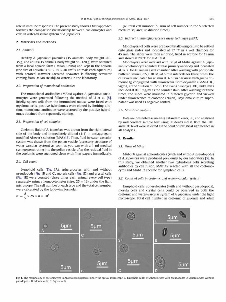
Lymphoid cells (Fig. 1A), spherulocytes with and withoutpseudopods (Fig. 1B and C), morula cells (Fig. 1D) and crystal cells(Fig. 1E) were counted (three times each animal every cell type)separately using a hemocytometer (size: 25 � 16) under the lightmicroscope. The cell number of each type and the total cell numberwere calculated by the following formula:
N ¼ A5� 25� B� 104
Fig. 1. The morphology of coelomocytes in Apostichopus japonicas under the optical microspseudopods; D: Morula cells; E: Crystal cells.
(N: total cell number; A: sum of cell number in the 5 selectedmedium squares; B: dilution times).
2.5. Indirect immunofluorescence assay technique (IIFAT)
Monolayers of cells were prepared by allowing cells to be settledonto glass slides and incubated at 37 �C in a wet chamber for45 min. The slides were then air dried, fixed in acetone for 15 minand stored at﹣20 �C for IIFAT test.
Monolayers were overlaid with 50 ml of MAbs against A. japo-nicus coelomocytes diluted 1:10 as primary antibody and incubatedat 37 �C for 45min in awet chamber. After washing with phosphatebuffered saline (PBS, 0.01 M) at 5 min intervals for three times, thecells were incubated for 45 min at 37 �C in darkness with goat-anti-mouse Ig conjugated with fluorescein isothiocyanate (GAM-FITC,Sigma) at the dilution of 1:256. The Evans blue dye (EBD, Fluka) wasincluded at 0.01 mg/ml as the counter stain. After washing for threetimes, the slides were mounted in buffered glycerin and viewedunder fluorescence microscope (Nikon). Myeloma culture super-natant was used as negative control.
2.6. Statistical analysis
Data are presented as means (�standard error, SE) and analyzedby independent sample test using Student’s t-test. Both the 0.01and 0.05 level were selected as the point of statistical significance inall analyses.
3. Results
3.1. Panel of MAbs
MAb3F6 against spherulocytes (with and without pseudopods)of A. japonicus were produced previously by our laboratory [5]. Inthis study, we obtained another two hybridoma cells secretingantibodies by cell fusion, MAb1C2 reacted with all the coelomo-cytes and MAb1E2 specific for lymphoid cells.
3.2. Count of cells in coelomic and water-vascular system
Lymphoid cells, spherulocytes (with and without pseudopods),morula cells and crystal cells could be observed in both thecoelomic and water-vascular system of A. japonicus under the lightmicroscope. Total cell number in coelomic of juvenile and adult
cope. A: Lmyphoid cells; B: Spherulocytes with pseudopods; C: Spherulocytes without

Fig. 2. Total cell number in coelomic and water-vascular system of juvenile and adult A. japonicus. 1e15: Cell number of juvenile A. japonicus; 16e30: Cell number of adultA. japonicus. COE: Coelomocytes; CIW: Cells in water-vascular system.
Q. Li et al. / Fish & Shellfish Immunology 35 (2013) 1654e16571656
A. japonicuswas (3.13 � 1.03) � 107 cells/ml and (2.14 � 1.05) � 107
cells/ml, while (1.23 � 0.37) � 107 cells/ml and(1.13 � 0.34) � 107cells/ml in water-vascular system, respectively(Fig. 2). From the data, total cell number in the coelomic showedsignificant statistical differences compared to total cell number inthe water-vascular system (P < 0.01), showing that total cellnumber in the two parts are significantly difference.
The percentages of five types of the cells (lymphoid cells,spherulocytes with pseudopods, spherulocytes without pseudo-pods, morula cells and crystal cells) in these two systems of bothjuvenile and adult A. japonicus would be listed as follows.
(Fig. 3): 1), juvenile: (73.12 � 6.77) %, (41.30 � 7.85) %(coelomicand water-vascular system); (14.13 � 7.42) %, (45.19 � 6.80) %;(8.70 � 7.30) %, (7.68 � 5.09) %; (2.82 � 1.37) %, (4.10 � 3.02) % and(1.23 � 0.58) %, (1.73 � 1.54) %; 2), adult: (62.82 � 7.95) %,(37.67� 11.71) %; (20.64� 6.61) %, (46.95�18.84) %; (8.77� 5.29) %,(9.83� 14.79) %; (6.42� 2.75) %, (4.23� 2.72) % and (1.35� 0.49) %,(1.32� 0.99) %; respectively. Percentages of different type cells, boththe percentages of lymphoid cells and spherulocytes with pseudo-pods showed significant statistical differences (P < 0.01), percent-ages of spherulocytes without pseudopods, morula cells and crystalcells are no statistical difference (P > 0.05).
3.3. Results of IIFAT
Spherulocytes with and without pseudopods, those two celltypes had an average size of 8e10 mm. Spherulocytes withoutpseudopods were smooth, spherulocytes with pseudopods hadmanyfliopodias,whichmade those cells look like a sunflowerunderthe microscope. MAb3F6 reacted with spherulocytes in coelomic orwater-vascular system (Fig. 4A and B), the specific green fluores-cence signals were detected in the membrane and cytoplasm of thecells. Although the distribution of positive signals in the cells wasnot different, the percentage of positive cells in water-vascularsystem (Fig. 4B) was higher than that in coelomic system (Fig. 4A).
0
1020
30
4050
60
7080
90
A B C
Perc
enta
ges(
%)
Fig. 3. Percentages of each type of the cells in coelomic and water-vascular system of juveniA. japonicus; B: Percentages of each type of the cells in water-vascular system of juvenile A.Percentages of each type of the cells in water-vascular system of adult A. japonicus; Data a
MAb1C2 reacted with all the cells in coelomic (Fig. 4C) or water-vascular system (Fig. 4D), and there was no significant difference.
Lymphoid cells had a large nucleus surrounded by a very thinlayer of cytoplasmwithout visible organelles or inclusions. The sizewas about 5e6 mm and 1e3 filopodias were often visible. MAb1E2reactedwith lymphocytes in coelomic orwater-vascular systemwithgreen fluorescence signals at the membrane and cytoplasm of thecells (Fig. 4E and F), and there was no significant difference in thedistributionof positive signals in the lymphocytes. However, positivecells inwater-vascular systemwere slightly smaller (Fig. 4F) than thatin coelomic system (Fig. 4E), and percentage of the cells was lower.
4. Discussion
Until now, there haven’t been uniform criteria on classificationof coelomocytes. According to a generally accepted classification,there are five types of coelomocytes in echinoderms: phagocytes(phagocytic amoebocytes), morula (or spherule) cells, vibratilecells, progenitor (lymphocyte-like) cells, and hemocytes [6e8].Hetzel [9] reported six types of holothurian coelomocytes. Eli-seikina and Magarlamov [10] described nine types of coelomo-cytes in A. japonicus using light microscopy, transmission electronmicroscopy (TEM) and histochemistry. Xing et al. [11] distin-guished six coelomocytes types in A. japonicus based on cellmorphological and ultrastructural features. Li et al. [12] identifiedcoelomocytes of A. japonicus into lymphoid cells, spherulocytes,amoebocytes, hyaline cells, fusiform cells and crystal cells basedon microstructural and ultrastructural characteristics. In thisstudy, we classified coelomocytes into lymphoid cells, spher-ulocytes with pseudopods, spherulocytes without pseudopods,morula cells and crystal cells.
We compared lymphoid cells, spherulocytes (with and withoutpseudopods), morula cells and crystal cells in coelomic and water-vascular system (polian vesicle) from various fields, including totalcell number, percentages of each cell type and the distribution of
D
Lymphocytes
Spherulocytes with pseudopods
Spherulocytes without pseudopods
Morula cells
Crystal cells
le and adult A. japonicus. A: Percentages of each type of the cells in coelomic of juvenilejaponicus; C: Percentages of each type of the cells in coelomic of adult A. japonicus; D:re presented as means � SE.

Fig. 4. Detection of MAbs reacting with coelomocytes and cells in water-vascular system of A. Japonicus. AB:MAb3F6; CD:MAb1C2; EF:MAb1E2; A, C, E: Coelomocytes were stainedby MAb3F6,1C2 and 1E2; G:Negative control of coelomocytes in coelomic; B, D, F: Cells in water-vascular system were stained by MAb3F6, 1C2 and 1E2; H:Negative control of cellsin water-vascular system; Short and long arrows indicated spherulocytes and lymphoid cells, respectively.
Q. Li et al. / Fish & Shellfish Immunology 35 (2013) 1654e1657 1657
the cell antigens. We found that all the cell types mentioned abovecould be observed in both coelomic and water-vascular system, andthere was not significant difference among cell antigens distribu-tion. However, there was great difference in the total cell numberand percentages of each cell type. The total cell number of coelo-mocytes was 2e3 times of that in water-vascular system, whetherit’s in the juvenile or the adult A. japonicus. The lymphoid cells arenumerically dominant in coelomic, and the spherulocytes withpseudopods are the secondly most abundant, which is consistentwith Eliseikina and Magarlamov’s [10]. Whereas, the spherulocyteswith pseudopods are predominant and the lymphoid cells take thesecond place in the water-vascular system.
All parts of water-vascular system of echinoderms are fluid-filled, and the fluid is virtually sea-water with a dense populationof cells suspended in it [2], which was also proved in our presentstudy. Cells in water-vascular system are also the coelomocytes.However, there was significant difference in the total cell number,whichmay be relatedwith the functions of cells. Further research isrequested on whether there are different functions.
As we know, fluid in water-vascular system sometimes migratesthrough its walls to other body cavities and also out to the sur-rounding sea water [2], which suggests that the water-vascularsystem is interconnected with the environment in vitro. Some re-searchers found out that the spherulocytes with pseudopodia mayhave phagocytic ability [11,13], which helps us to explain the reasonwhy the spherulocytes are more in the water-vascular system. Thephagocytosis spherulocytes may be the first defensive line whilethe body is invaded by pathogens [14]. Although from the point ofour data, cell number of coelomocytes in coelomic is more than thatin the water-vascular system, so polian vesicle may be not a se-lective recruitment of coelomocytes, we suggest polian vesicle maybe the origin tissue of coelomocytes when polian vesicle work as a“pump” [15] pumping cells to the coelomic. In Fig. 3,lymphoid cellsinwater-vascular systemmay be the early stage of cell type, so cellsin water-vascular system reacting with MAb1E2 is slightly smallerthan coelomocytes reacting the MAb. It proves that polian vesiclemay be the origin of coelomocytes from one side, if it is really anearly stage lymphoid cell type.
In conclusion, cells in coelomic and water-vascular system ofA. japonicus were not thoroughly same, although there were
common cell types and antigens distribution. They had differentcomposition which will determine its functions for further studies.
Acknowledgments
This study was supported by National Natural Science Founda-tion of China (30800853), National Key Projects, National Science &Technology Pillar Program during the twelfth Five-Year-Plan period(2011BAD13B03), and Educational Commission Project of Liaoning(LJQ2011075).
References
[1] Liao YL. China fauna Echinodermata Holothuroidea. Beijing: Science Press;1997. p. 2e38 [in Chinese].
[2] Nichols D. The water-vascular system in living and fossil Echinoderms. Pale-ontology 1972;15:519e38.
[3] Haugh BN. Water vascular system of the crinoidea camerata. Journal ofPaleontology 1973;47:77e90.
[4] Baccetti B, Rosati F. The fine structure of polian vesicles of Holothurians. Celland Tissue Research 1968;90:148e60.
[5] Li Q, Li Y, Li H, Wang YN, Xu DH. Production, characterization and applicationof monoclonal antibody to spherulocytes: a subpopulation of coelomocytes ofApostichopus japonicus. Fish & Shellfish Immunology 2010;29:832e8.
[6] Chia F, Xing J. Echinoderm coelomocytes. Zoological Studies 1996;35:231e54.[7] Smiley S. Holothuroidea. In: Harrison FW, Chia FS, editors. Microscopic
anatomy of invertebrates, vol. 14. New York: Wiley-Liss; 1994. p. 401e72.[8] Smith VJ. The echinoderms. In: Invertebrate blood cells. London: Academic
Press; 1981. p. 513e62.[9] Hetzel HR. Studies on holothurian coelomocytes.Ⅰ.A survey of coelomocyte
types. Biological Bulletin 1963;125:289e301.[10] Eliseikina MG, Magarlamov TY. Coelomocyte morphology in the holothurians
Apostichopus japonicus (Aspidochirota: Stichopodidae) and Cucumariajaponica (Dendrochirotida: Cucumariidae). Russian Journal of Marine Biology2002;28:197e202.
[11] Xing K, Yang HS, Chen MY. Morphological and ultrastructural characterizationof the coelomocytes in Apostichopus japonicus. Aquatic Biology 2008;2:85e92.
[12] Li H, Chen J, Lu J, Li Q, Park SI. Type and quantity of blood cells and coelo-mocytes in Apostichopus japonicus. Acta Hydrobiologica Sinica 2009;2:207e13[In Chinese].
[13] Xing J, Leung MF, Chia FS. Quantitative analysis of phagocytosis by amebo-cytes of a sea cucumber, Holothuria leucospilota. Invertebrate Biology1998;117(1):67e74.
[14] Smith AC. A proposed phylogenetic relationship between sea cucumber polianvesicles and the vertebrate lymphoreticular system. Journal of InvertebratePathology 1978;31(3):353e7.
[15] Barnes Robert D. Invertebrate zoology. Philadelphia, PA: Holt-SaundersInternational; 1982, ISBN 0-03-056747-5; 1982. p. 991e2.