comparative transcriptome analysis of salivary glands of ... · comparative transcriptome analysis...
TRANSCRIPT
Comparative Transcriptome Analysis of Salivary Glandsof Two Populations of Rice Brown Planthopper,Nilaparvata lugens, That Differ in VirulenceRui Ji1., Haixin Yu1., Qiang Fu2., Hongdan Chen1, Wenfeng Ye1, Shaohui Li3, Yonggen Lou1*
1 State Key Laboratory of Rice Biology, Institute of Insect Science, Zhejiang University, Hangzhou, China, 2 Research and Development Center of Rice Production
Technology, China National Rice Research Institute, Hangzhou, China, 3 State Key Laboratory of Rice Biology, Institute of Biotechnology, Zhejiang University, Hangzhou,
China
Abstract
Background: The brown planthopper (BPH), Nilaparvata lugens (Stal), a destructive rice pest in Asia, can quickly overcomerice resistance by evolving new virulent populations. Herbivore saliva plays an important role in plant–herbivoreinteractions, including in plant defense and herbivore virulence. However, thus far little is known about BPH saliva at themolecular level, especially its role in virulence and BPH–rice interaction.
Methodology/Principal Findings: Using cDNA amplification in combination with Illumina short-read sequencingtechnology, we sequenced the salivary-gland transcriptomes of two BPH populations with different virulence; thepopulations were derived from rice variety TN1 (TN1 population) and Mudgo (M population). In total, 37,666 and 38,451unigenes were generated from the salivary glands of these populations, respectively. When combined, a total of 43,312unigenes were obtained, about 18 times more than the number of expressed sequence tags previously identified fromthese glands. Gene ontology annotations and KEGG orthology classifications indicated that genes related to metabolism,binding and transport were significantly active in the salivary glands. A total of 352 genes were predicted to encodesecretory proteins, and some might play important roles in BPH feeding and BPH–rice interactions. Comparative analysis ofthe transcriptomes of the two populations revealed that the genes related to ‘metabolism,’ ‘digestion and absorption,’ and‘salivary secretion’ might be associated with virulence. Moreover, 67 genes encoding putative secreted proteins weredifferentially expressed between the two populations, suggesting these genes may contribute to the change in virulence.
Conclusions/Significance: This study was the first to compare the salivary-gland transcriptomes of two BPH populationshaving different virulence traits and to find genes that may be related to this difference. Our data provide a rich molecularresource for future functional studies on salivary glands and will be useful for elucidating the molecular mechanismsunderlying BPH feeding and virulence differences.
Citation: Ji R, Yu H, Fu Q, Chen H, Ye W, et al. (2013) Comparative Transcriptome Analysis of Salivary Glands of Two Populations of Rice Brown Planthopper,Nilaparvata lugens, That Differ in Virulence. PLoS ONE 8(11): e79612. doi:10.1371/journal.pone.0079612
Editor: Daniel Doucet, Natural Resources Canada, Canada
Received May 13, 2013; Accepted September 23, 2013; Published November 14, 2013
Copyright: � 2013 Ji et al. This is an open-access article distributed under the terms of the Creative Commons Attribution License, which permits unrestricteduse, distribution, and reproduction in any medium, provided the original author and source are credited.
Funding: The study was jointly sponsored by the National Basic Research Program of China (2010CB126200; http://www.most.gov.cn/index.htm), the InnovationResearch Team Program of the National Natural Science Foundation of China (31021003; http://www.nsfc.gov.cn/Portal0/default152.htm), and the ChinaAgriculture Research System (CARS-01-25; http://www.moa.gov.cn/). The funders had no role in study design, data collection and analysis, decision to publish, orpreparation of the manuscript.
Competing Interests: The authors have declared that no competing interests exist.
* E-mail: [email protected]
. These authors contributed equally to this work.
Introduction
Insect herbivore saliva contains digestive enzymes, such as
alkaline phosphatase, esterase, amylase and b-glucosidase, as well
as other components, such as elicitors that induce plant defense,
effectors that inhibit plant defense, and proteins related to
pathogen transmission [1–3]. Some studies have also found a
relationship between saliva components and herbivore virulence
[4]. Therefore, herbivore saliva, the first substance to come into
chemical contact with the plant, plays important roles in both food
ingestion and interactions between herbivores and their host plants
[1–5]. Characterizing herbivore saliva will provide new insights
into plant–herbivore interactions, including induced plant defense
and herbivore virulence.
To characterize herbivore saliva, the transcriptome and/or
proteome of the salivary glands and/or saliva of several herbivore
species – mostly hemipterans such as rice brown planthopper
(BPH; Nilaparvata lugens (Stal)); Hemiptera: Delphacidae) [6,7], pea
aphid (Acyrthosiphon pisum; Hemiptera: Aphididae) [8,9,10], green
peach aphid (Myzus persicae; Hemiptera: Aphididae) [11,12],
whitefly (Bemisia tabaci (Gennadius); Hemiptera: Aleyrodidae)
[13], and potato leafhopper (Empoasca fabae (Harris); Hemiptera:
Cicadellidae) [14] – have been analyzed. These studies found
several hundred proteins in the saliva [4,6–14]. However, whether
differences in salivary components exist between herbivore
PLOS ONE | www.plosone.org 1 November 2013 | Volume 8 | Issue 11 | e79612
populations with different virulence traits and what functions these
components have remain largely unanswered questions.
BPH, one of the most destructive insect pests of the rice plant
(Oryza sativa L.) in Asia, causes substantial losses of rice yield every
year by sucking phloem sap and transmitting plant viruses, such as
the rice ragged stunt virus and the rice grassy stunt virus [15]. The
cultivation of resistant rice varieties is an important control
measure for the BPH. However, the BPH rapidly overcomes rice
resistance by evolving new virulent populations [16]. BPH
virulence strains generally correspond to particular resistance
genes in rice. For example, rice varieties TN1 (a susceptible
variety) and Mudgo (carrying the resistance gene bph1) host
virulent BPH strains 1 and 2, respectively [16]. Although some
minor differences in morphology and in the composition of
bacterial symbionts among virulence genotypes have been
reported, the mechanisms underlying changes in BPH virulence
are not clear [16–19].
When sucking sap, BPHs and other phloem feeders secrete two
primary kinds of saliva: coagulable and watery saliva. Coagulable
saliva forms salivary sheaths, which help to stabilize the insects’
stylets and suppress plant defense responses to components of the
watery saliva [1,4]. Watery saliva, which contains a mixture of
amino acids, proteins, and digestive enzymes, assists the movement
of the stylets inside the salivary sheath, the digestion of plant
material, and the suppression of plant defense responses [1–
4,9,12]. Given the important roles of herbivore saliva in plant-
herbivore interactions, BPH saliva is likely to play a central role in
the interaction between BPHs and rice, including in the evolution
of BPH virulence.
Salivary gland morphology and several salivary proteins of
BPHs have been reported [20–24]. Moreover, Noda et al. (2008)
identified 2383 expressed sequence tags (ESTs) in the salivary
glands of the BPH by Sanger’s method [6]. However, these data
are insufficient to elucidate the nature of BPH saliva. Moreover,
whether BPH populations with different virulence have different
saliva components remains unclear. To explore these issues, we
compared the salivary-gland transcriptomes of two BPH popula-
tions with different virulence that were maintained on one of two
rice varieties: TN1 (TN1 population) or Mudgo (M population).
Using next-generation high-throughput sequencing technology, a
total of 37,666 and 38,451 unigenes were identified in the
respective populations, and 3,757 unigenes were differentially
expressed between the two populations. Moreover, among the
differentially expressed genes, 67 unigenes encoded putative
secretory proteins. These findings provide an exciting opportunity
to elucidate the components of BPH saliva and suggest a possible
correlation between BPH saliva and virulence.
Results and Discussion
Illumina Sequencing and Read AssemblyThe cDNA libraries of the salivary glands of BPH TN1 and M
populations were sequenced using an Illumina platform, resulting
in 38,487,746 and 40,350,780 reads, respectively. After cleaning
and quality checks, short sequences were assembled into 97,103
and 101,885 contigs, respectively. Using paired end-joining and
gap-filling methods, these contigs were further assembled into
73,510 and 76,195 scaffolds, which were then clustered into
37,666 and 38,451 unigenes, respectively (Table 1). Finally,
sequence data from the two libraries were combined to obtain
43,312 unigenes with a mean length of 746 bp. The length
distributions of the unigenes were similar between the two
populations, suggesting there was no bias in the cDNA library
construction (Figure S1). However, some unigenes were found in
only one population (data not shown). We believe these differences
may have been caused by differential long-term ecological
adaptations to the different rice hosts. The total number of
unigenes obtained was much higher than the number of BPH
salivary gland ESTs identified by Noda et al. [6], indicating the
depth of coverage possible using HiSeq technology.
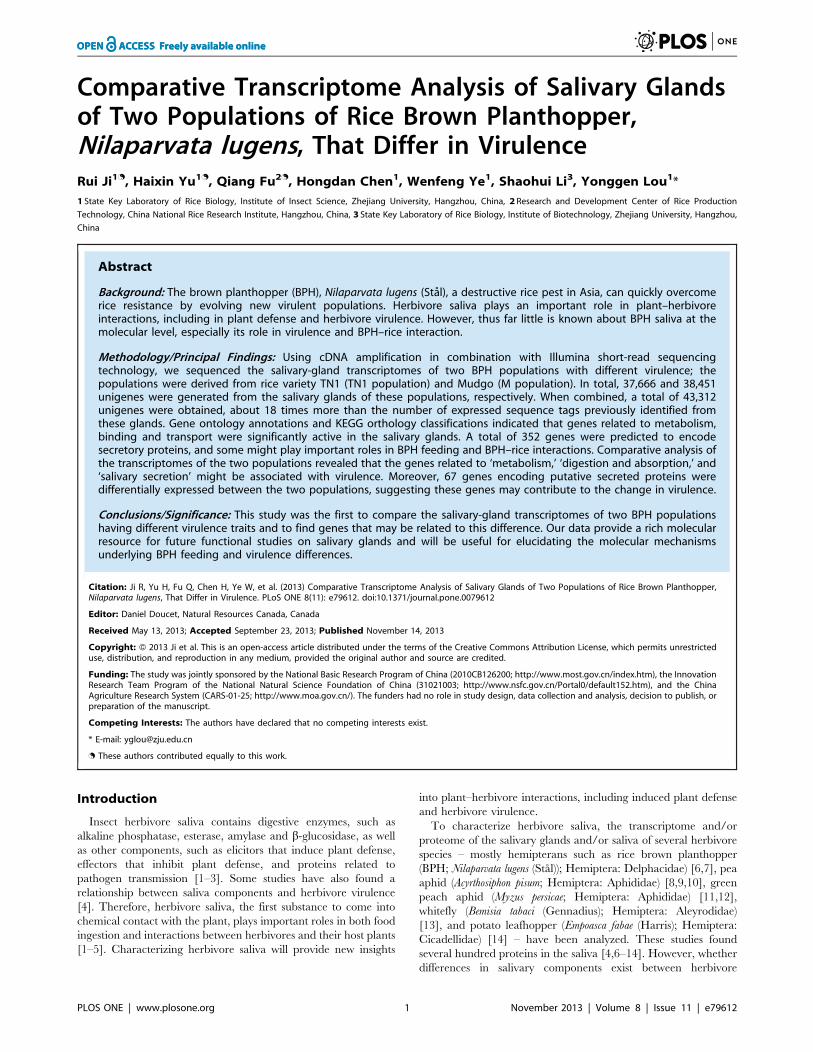
Annotation of Salivary-gland TranscriptsFor functional annotation, the 43,312 unigenes were searched
using BLASTx against the non-redundant (nr) NCBI protein
database. A total of 19,771 unigenes (45.65% of all distinct
sequences) yielded significant BLAST hits with a cut-off E-value of
1.0E–5 (Table S1). The species distributions of the best matches for
each sequence are shown in Figure 1. The highest percentages of
unique sequences matched genes of the red flour beetle (Tribolium
castaneum; 14.34%), A. pisum (11.48%), and a parasitoid wasp
(Nasonia vitripennis; 7.90%). A similar result was also reported by
Xue et al. [25], Peng et al. [26] and Bao et al. [27]. Further
research should explore why the highest percentage of unique
sequences matches genes of T. castaneum, a coleoptera beetle,
rather than A. pisum, a hemiptera aphid which is much more
closely related to BPH.
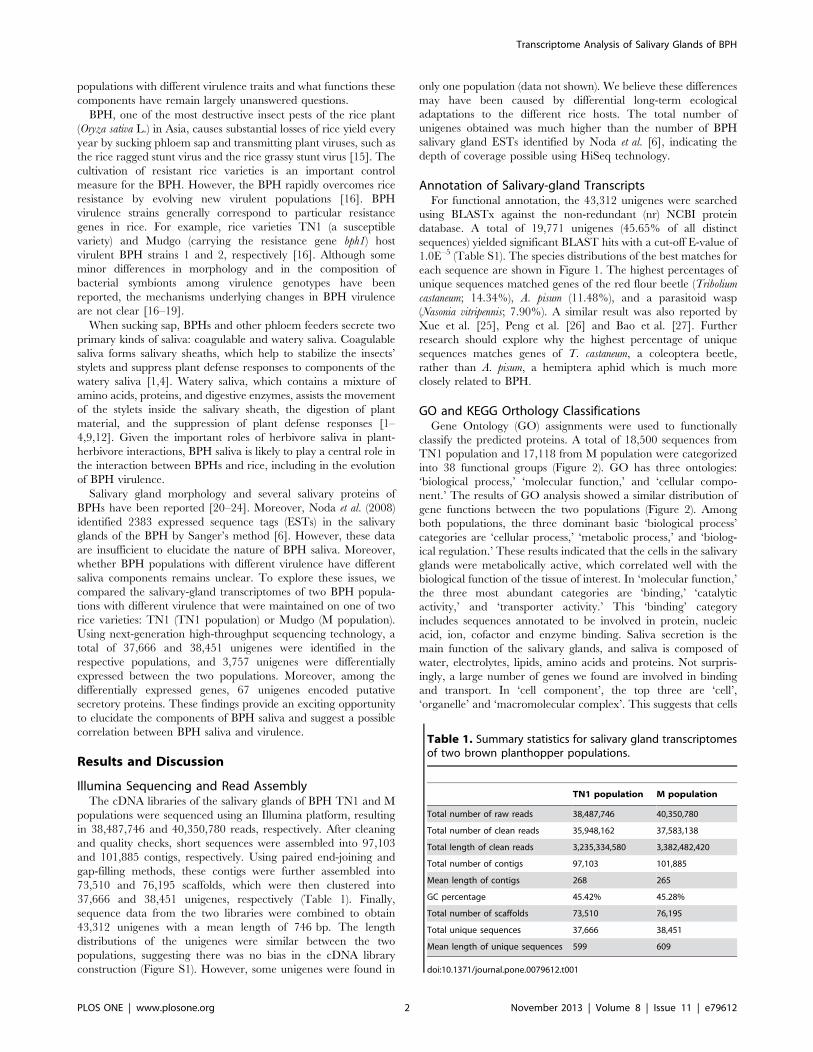
GO and KEGG Orthology ClassificationsGene Ontology (GO) assignments were used to functionally
classify the predicted proteins. A total of 18,500 sequences from
TN1 population and 17,118 from M population were categorized
into 38 functional groups (Figure 2). GO has three ontologies:
‘biological process,’ ‘molecular function,’ and ‘cellular compo-
nent.’ The results of GO analysis showed a similar distribution of
gene functions between the two populations (Figure 2). Among
both populations, the three dominant basic ‘biological process’
categories are ‘cellular process,’ ‘metabolic process,’ and ‘biolog-
ical regulation.’ These results indicated that the cells in the salivary
glands were metabolically active, which correlated well with the
biological function of the tissue of interest. In ‘molecular function,’
the three most abundant categories are ‘binding,’ ‘catalytic
activity,’ and ‘transporter activity.’ This ‘binding’ category
includes sequences annotated to be involved in protein, nucleic
acid, ion, cofactor and enzyme binding. Saliva secretion is the
main function of the salivary glands, and saliva is composed of
water, electrolytes, lipids, amino acids and proteins. Not surpris-
ingly, a large number of genes we found are involved in binding
and transport. In ‘cell component’, the top three are ‘cell’,
‘organelle’ and ‘macromolecular complex’. This suggests that cells
Table 1. Summary statistics for salivary gland transcriptomesof two brown planthopper populations.
TN1 population M population
Total number of raw reads 38,487,746 40,350,780
Total number of clean reads 35,948,162 37,583,138
Total length of clean reads 3,235,334,580 3,382,482,420
Total number of contigs 97,103 101,885
Mean length of contigs 268 265
GC percentage 45.42% 45.28%
Total number of scaffolds 73,510 76,195
Total unique sequences 37,666 38,451
Mean length of unique sequences 599 609
doi:10.1371/journal.pone.0079612.t001
Transcriptome Analysis of Salivary Glands of BPH
PLOS ONE | www.plosone.org 2 November 2013 | Volume 8 | Issue 11 | e79612
and organelles are the prominent parts of the salivary glands, and
macromolecular complex secretion is also critical in the salivary
glands.
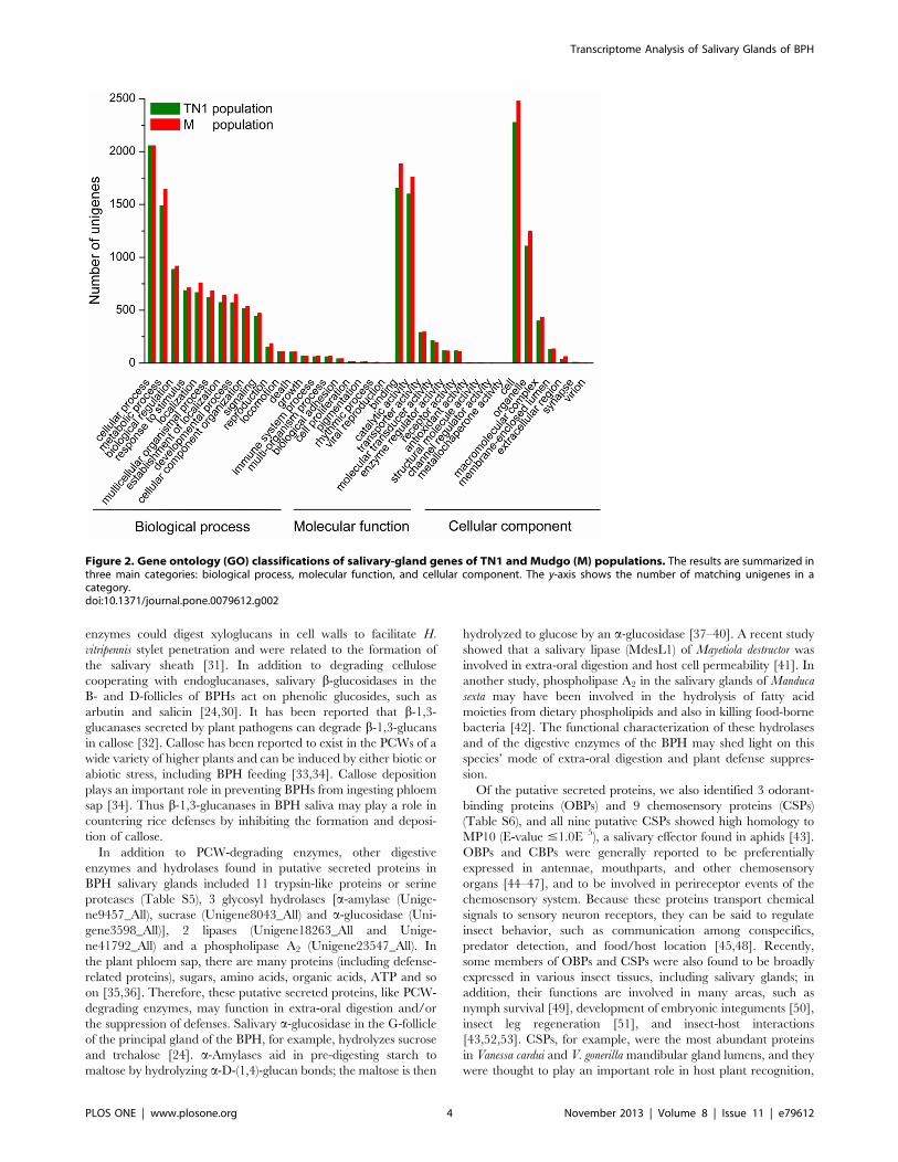
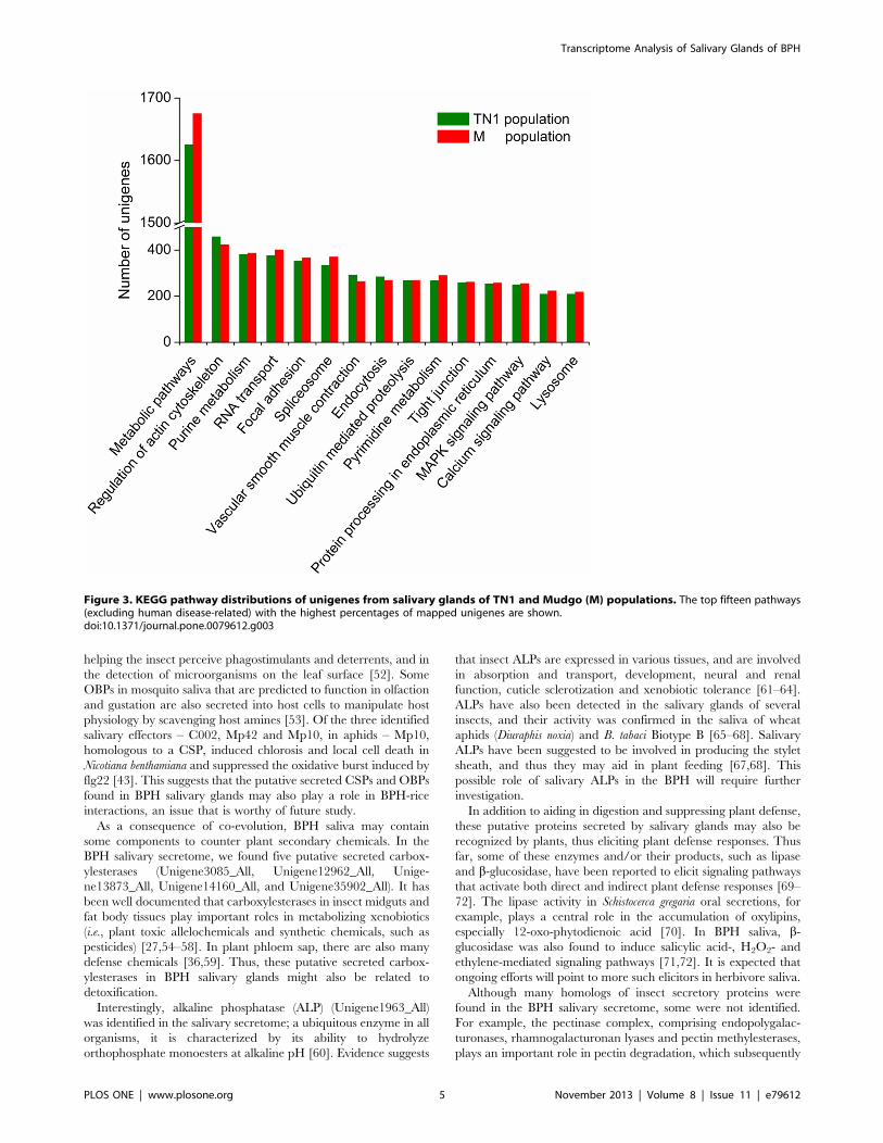
To investigate which biological pathways are active in the
salivary glands, all of the sequences were assigned to the reference
canonical pathways in the Kyoto Encyclopedia of Genes and
Genomes (KEGG). A similar distribution of biological pathways
for TN1 and M populations was also found by KEGG mapping,
and a total of 11449 unigenes from TN1 population and 11502
unigenes from M population were mapped separately to 239 and
240 pathways in total (Tables S2 and S3). The salivary glands are
specific organs for salivary macromolecule production and,
consequently, have a high level of metabolic activity. This
characteristic was implied in a previous study by observing the
dense cytoplasm and organized whorls of rough endoplasmic
reticulum in the salivary-gland cells [28]. Consequently, among
these pathways, ‘‘metabolism’’ is important in the salivary glands
(1625 unigenes from TN1 population and 1675 unigenes from M
population, Figure 3, in pathways associated with human diseases
were excluded). Since there are abundant proteins in the saliva of
BPHs, as a major original location of these proteins, the salivary
glands should be active in protein synthesis and catabolism.
Indeed, the transcriptome contained many sequences involved in
the ‘‘protein processing in endoplasmic reticulum’’ pathway (254
genes in TN1 population and 257 genes in M population), which is
related to the formation and transport of nascent secretory
proteins, and the ‘lysosome’ pathway (209 genes in TN1
population and 218 genes in M population), a major pathway
related to protein degradation. The GO annotations and KEGG
orthology (KO) classifications indicated that the salivary glands
might be active in metabolism, binding and transport, and that a
high percentage of genes might be involved in secretory protein
processing and secretion.
Unigenes Encoding Putative Secreted ProteinsWe characterized the salivary secretome of the BPH by
combining the transcripts of the salivary glands of the two
populations. Secreted proteins are probably delivered into saliva
through the eukaryotic endoplasmic reticulum–Golgi pathway
[29]. Proteins secreted through this pathway have an N-terminal
signal peptide. Therefore, all amino acid sequences were analyzed
for the presence of signal peptides and potential cleavage sites. A
total of 464 unique genes encoded a secretory signal peptide. Of
these, 112 were predicted to have at least one transmembrane
domain besides the signal peptide, indicating that their proteins
were likely embedded in cell membranes of the salivary glands.
Thus, 352 potential secretory proteins were retained (Table S4),
which is comparable to the numbers of putative secreted proteins
reported in A. pisum (324) and B. tabaci (295) [10,13]. Interestingly,
the possible functions of some putative secreted proteins were
closely related to the known roles of insect saliva, such as digestion
and suppressing or eliciting plant defenses.
Among the putative secreted proteins in the BPH, a set of
digestive enzymes and hydrolases, including plant cell wall (PCW)-
degrading enzymes were found. These putative PCW-degrading
enzymes included one b-1,4-endoglucanase (Unigene1860_All),
one b-glucosidase (Unigene26172_All), and two b-1,3-glucanases
(Unigene10762_All and Unigene23029_All). PCW, a thick, rigid
polysaccharide structure comprising an extensive network of
celluloses, hemicelluloses and pectins, forms a formidable barrier
to herbivores. To overcome this barrier, herbivores have evolved
to secrete salivary PCW-degrading enzymes, such as cellulases
(consisting of endoglucanases and b-glucosidases) and pectinases
[1,29,30]. For example, an extract of Homalodisca vitripennis salivary
gland contained a variety of enzymes capable of hydrolyzing
glycosidic linkages in the polysaccharides of PCWs, including b-
1,4-endoglucanase, xyloglucanase and b-1,4-endoxylanase; these
Figure 1. Species distribution of the BLASTX results of brown planthopper salivary-gland genes. This figure shows the speciesdistribution of unigene BLASTX results against the NCBI nr protein database with a cutoff E-value #1.0E–5 and the proportions of each species,represented by different colors. Species with proportions greater than 1% are shown.doi:10.1371/journal.pone.0079612.g001
Transcriptome Analysis of Salivary Glands of BPH
PLOS ONE | www.plosone.org 3 November 2013 | Volume 8 | Issue 11 | e79612
enzymes could digest xyloglucans in cell walls to facilitate H.
vitripennis stylet penetration and were related to the formation of
the salivary sheath [31]. In addition to degrading cellulose
cooperating with endoglucanases, salivary b-glucosidases in the
B- and D-follicles of BPHs act on phenolic glucosides, such as
arbutin and salicin [24,30]. It has been reported that b-1,3-
glucanases secreted by plant pathogens can degrade b-1,3-glucans
in callose [32]. Callose has been reported to exist in the PCWs of a
wide variety of higher plants and can be induced by either biotic or
abiotic stress, including BPH feeding [33,34]. Callose deposition
plays an important role in preventing BPHs from ingesting phloem
sap [34]. Thus b-1,3-glucanases in BPH saliva may play a role in
countering rice defenses by inhibiting the formation and deposi-
tion of callose.
In addition to PCW-degrading enzymes, other digestive
enzymes and hydrolases found in putative secreted proteins in
BPH salivary glands included 11 trypsin-like proteins or serine
proteases (Table S5), 3 glycosyl hydrolases [a-amylase (Unige-
ne9457_All), sucrase (Unigene8043_All) and a-glucosidase (Uni-
gene3598_All)], 2 lipases (Unigene18263_All and Unige-
ne41792_All) and a phospholipase A2 (Unigene23547_All). In
the plant phloem sap, there are many proteins (including defense-
related proteins), sugars, amino acids, organic acids, ATP and so
on [35,36]. Therefore, these putative secreted proteins, like PCW-
degrading enzymes, may function in extra-oral digestion and/or
the suppression of defenses. Salivary a-glucosidase in the G-follicle
of the principal gland of the BPH, for example, hydrolyzes sucrose
and trehalose [24]. a-Amylases aid in pre-digesting starch to
maltose by hydrolyzing a-D-(1,4)-glucan bonds; the maltose is then
hydrolyzed to glucose by an a-glucosidase [37–40]. A recent study
showed that a salivary lipase (MdesL1) of Mayetiola destructor was
involved in extra-oral digestion and host cell permeability [41]. In
another study, phospholipase A2 in the salivary glands of Manduca
sexta may have been involved in the hydrolysis of fatty acid
moieties from dietary phospholipids and also in killing food-borne
bacteria [42]. The functional characterization of these hydrolases
and of the digestive enzymes of the BPH may shed light on this
species’ mode of extra-oral digestion and plant defense suppres-
sion.
Of the putative secreted proteins, we also identified 3 odorant-
binding proteins (OBPs) and 9 chemosensory proteins (CSPs)
(Table S6), and all nine putative CSPs showed high homology to
MP10 (E-value #1.0E–5), a salivary effector found in aphids [43].
OBPs and CBPs were generally reported to be preferentially
expressed in antennae, mouthparts, and other chemosensory
organs [44–47], and to be involved in perireceptor events of the
chemosensory system. Because these proteins transport chemical
signals to sensory neuron receptors, they can be said to regulate
insect behavior, such as communication among conspecifics,
predator detection, and food/host location [45,48]. Recently,
some members of OBPs and CSPs were also found to be broadly
expressed in various insect tissues, including salivary glands; in
addition, their functions are involved in many areas, such as
nymph survival [49], development of embryonic integuments [50],
insect leg regeneration [51], and insect-host interactions
[43,52,53]. CSPs, for example, were the most abundant proteins
in Vanessa cardui and V. gonerilla mandibular gland lumens, and they
were thought to play an important role in host plant recognition,
Figure 2. Gene ontology (GO) classifications of salivary-gland genes of TN1 and Mudgo (M) populations. The results are summarized inthree main categories: biological process, molecular function, and cellular component. The y-axis shows the number of matching unigenes in acategory.doi:10.1371/journal.pone.0079612.g002
Transcriptome Analysis of Salivary Glands of BPH
PLOS ONE | www.plosone.org 4 November 2013 | Volume 8 | Issue 11 | e79612
helping the insect perceive phagostimulants and deterrents, and in
the detection of microorganisms on the leaf surface [52]. Some
OBPs in mosquito saliva that are predicted to function in olfaction
and gustation are also secreted into host cells to manipulate host
physiology by scavenging host amines [53]. Of the three identified
salivary effectors – C002, Mp42 and Mp10, in aphids – Mp10,
homologous to a CSP, induced chlorosis and local cell death in
Nicotiana benthamiana and suppressed the oxidative burst induced by
flg22 [43]. This suggests that the putative secreted CSPs and OBPs
found in BPH salivary glands may also play a role in BPH-rice
interactions, an issue that is worthy of future study.
As a consequence of co-evolution, BPH saliva may contain
some components to counter plant secondary chemicals. In the
BPH salivary secretome, we found five putative secreted carbox-
ylesterases (Unigene3085_All, Unigene12962_All, Unige-
ne13873_All, Unigene14160_All, and Unigene35902_All). It has
been well documented that carboxylesterases in insect midguts and
fat body tissues play important roles in metabolizing xenobiotics
(i.e., plant toxic allelochemicals and synthetic chemicals, such as
pesticides) [27,54–58]. In plant phloem sap, there are also many
defense chemicals [36,59]. Thus, these putative secreted carbox-
ylesterases in BPH salivary glands might also be related to
detoxification.
Interestingly, alkaline phosphatase (ALP) (Unigene1963_All)
was identified in the salivary secretome; a ubiquitous enzyme in all
organisms, it is characterized by its ability to hydrolyze
orthophosphate monoesters at alkaline pH [60]. Evidence suggests
that insect ALPs are expressed in various tissues, and are involved
in absorption and transport, development, neural and renal
function, cuticle sclerotization and xenobiotic tolerance [61–64].
ALPs have also been detected in the salivary glands of several
insects, and their activity was confirmed in the saliva of wheat
aphids (Diuraphis noxia) and B. tabaci Biotype B [65–68]. Salivary
ALPs have been suggested to be involved in producing the stylet
sheath, and thus they may aid in plant feeding [67,68]. This
possible role of salivary ALPs in the BPH will require further
investigation.
In addition to aiding in digestion and suppressing plant defense,
these putative proteins secreted by salivary glands may also be
recognized by plants, thus eliciting plant defense responses. Thus
far, some of these enzymes and/or their products, such as lipase
and b-glucosidase, have been reported to elicit signaling pathways
that activate both direct and indirect plant defense responses [69–
72]. The lipase activity in Schistocerca gregaria oral secretions, for
example, plays a central role in the accumulation of oxylipins,
especially 12-oxo-phytodienoic acid [70]. In BPH saliva, b-
glucosidase was also found to induce salicylic acid-, H2O2- and
ethylene-mediated signaling pathways [71,72]. It is expected that
ongoing efforts will point to more such elicitors in herbivore saliva.
Although many homologs of insect secretory proteins were
found in the BPH salivary secretome, some were not identified.
For example, the pectinase complex, comprising endopolygalac-
turonases, rhamnogalacturonan lyases and pectin methylesterases,
plays an important role in pectin degradation, which subsequently
Figure 3. KEGG pathway distributions of unigenes from salivary glands of TN1 and Mudgo (M) populations. The top fifteen pathways(excluding human disease-related) with the highest percentages of mapped unigenes are shown.doi:10.1371/journal.pone.0079612.g003
Transcriptome Analysis of Salivary Glands of BPH
PLOS ONE | www.plosone.org 5 November 2013 | Volume 8 | Issue 11 | e79612
facilitates the decomposition of (hemi)cellulose and makes the
PCWs more susceptible to further breakdown by other enzymes
[29,69,73]. Pectinases occur mainly in Hemiptera and Coleoptera
[69]. However, in the BPH salivary secretome, pectinases were not
identified. This suggests that BPH may use unique feeding
strategies. Alternatively, these enzymes may have been filtered
out during the prediction of secretory proteins because they have
incomplete 59 sequences. In our experiments, for instance, the
unigene Unigene1860_All was annotated to encode a cellulase.
However, because it lacks the 59 end, this unigene was filtered out.
Therefore, to completely explore the protein composition of BPH
saliva, other research approaches, such as the proteome analysis of
BPH saliva, should also be used.
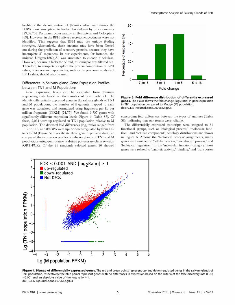
Differences in Salivary-gland Gene Expression Profilesbetween TN1 and M Populations
Gene expression levels can be estimated from Illumina
sequencing data based on the number of raw reads [74]. To
identify differentially expressed genes in the salivary glands of TN1
and M populations, the number of fragments mapped to each
gene was calculated and normalized using fragments per kb per
million fragments (FPKM) [74,75]. We found 3,757 genes with
significantly different expression levels (Figure 4, Table S7). Of
these, 2,084 were up-regulated in TN1 population relative to M
population. The detected fold differences (log2 ratio) ranged from
217 to +16, and 89.88% were up- or down-regulated by from 1.0-
to 5.0-fold (Figure 5). To validate these gene expression data, we
compared the expression profiles of salivary glands of TN1 and M
populations using quantitative real-time polymerase chain reaction
(QRT-PCR). Of the 21 randomly selected genes, 20 showed
concordant fold differences between the types of analyses (Table
S8), indicating that our results were reliable.
The differentially expressed transcripts were assigned to 31
functional groups, such as ‘biological process,’ ‘molecular func-
tion,’ and ‘cellular component’; ontology distributions are shown
in Figure 6. Among the ‘biological process’ assignments, many
genes were assigned to ‘cellular process,’ ‘metabolism process,’ and
‘biological regulation.’ In the ‘molecular function’ category, most
genes were related to ‘catalytic activity,’ ‘binding,’ and ‘transporter
Figure 4. Bitmap of differentially expressed genes. The red and green points represent up- and down-regulated genes in the salivary glands ofTN1 population, respectively; the blue points represent genes with no differences in expression based on the criteria of the false discovery rate (FDR)#0.001 and an absolute value of the log2 ratio $1.doi:10.1371/journal.pone.0079612.g004
Figure 5. Fold difference distribution of differently expressedgenes. The x-axis shows the fold change (log2 ratio) in gene expressionin TN1 population compared to Mudgo (M) population.doi:10.1371/journal.pone.0079612.g005
Transcriptome Analysis of Salivary Glands of BPH
PLOS ONE | www.plosone.org 6 November 2013 | Volume 8 | Issue 11 | e79612
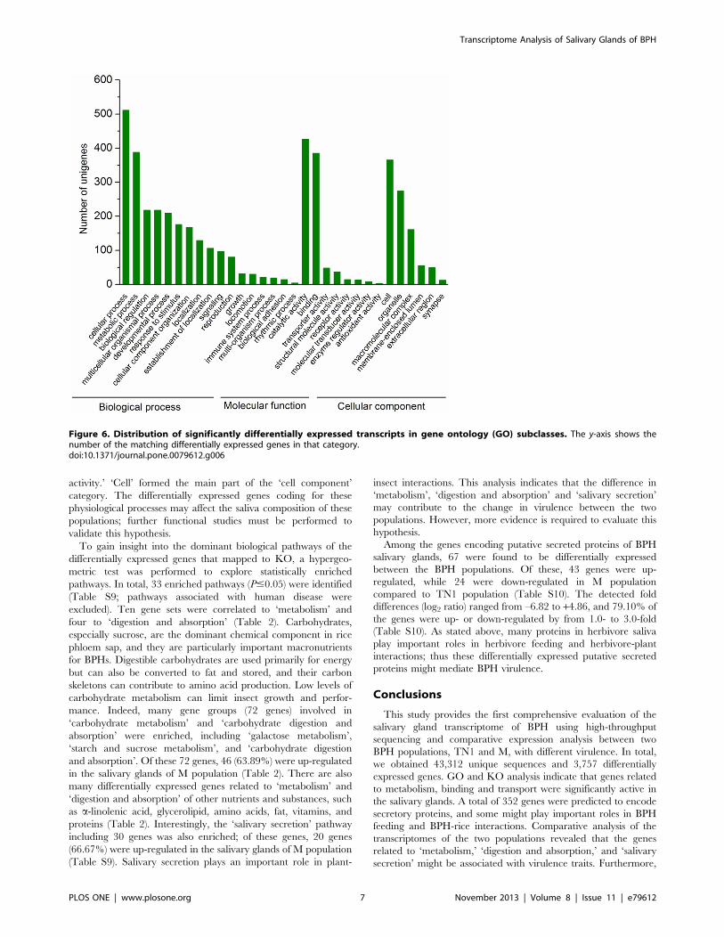
activity.’ ‘Cell’ formed the main part of the ‘cell component’
category. The differentially expressed genes coding for these
physiological processes may affect the saliva composition of these
populations; further functional studies must be performed to
validate this hypothesis.
To gain insight into the dominant biological pathways of the
differentially expressed genes that mapped to KO, a hypergeo-
metric test was performed to explore statistically enriched
pathways. In total, 33 enriched pathways (P#0.05) were identified
(Table S9; pathways associated with human disease were
excluded). Ten gene sets were correlated to ‘metabolism’ and
four to ‘digestion and absorption’ (Table 2). Carbohydrates,
especially sucrose, are the dominant chemical component in rice
phloem sap, and they are particularly important macronutrients
for BPHs. Digestible carbohydrates are used primarily for energy
but can also be converted to fat and stored, and their carbon
skeletons can contribute to amino acid production. Low levels of
carbohydrate metabolism can limit insect growth and perfor-
mance. Indeed, many gene groups (72 genes) involved in
‘carbohydrate metabolism’ and ‘carbohydrate digestion and
absorption’ were enriched, including ‘galactose metabolism’,
‘starch and sucrose metabolism’, and ‘carbohydrate digestion
and absorption’. Of these 72 genes, 46 (63.89%) were up-regulated
in the salivary glands of M population (Table 2). There are also
many differentially expressed genes related to ‘metabolism’ and
‘digestion and absorption’ of other nutrients and substances, such
as a-linolenic acid, glycerolipid, amino acids, fat, vitamins, and
proteins (Table 2). Interestingly, the ‘salivary secretion’ pathway
including 30 genes was also enriched; of these genes, 20 genes
(66.67%) were up-regulated in the salivary glands of M population
(Table S9). Salivary secretion plays an important role in plant-
insect interactions. This analysis indicates that the difference in
‘metabolism’, ‘digestion and absorption’ and ‘salivary secretion’
may contribute to the change in virulence between the two
populations. However, more evidence is required to evaluate this
hypothesis.
Among the genes encoding putative secreted proteins of BPH
salivary glands, 67 were found to be differentially expressed
between the BPH populations. Of these, 43 genes were up-
regulated, while 24 were down-regulated in M population
compared to TN1 population (Table S10). The detected fold
differences (log2 ratio) ranged from –6.82 to +4.86, and 79.10% of
the genes were up- or down-regulated by from 1.0- to 3.0-fold
(Table S10). As stated above, many proteins in herbivore saliva
play important roles in herbivore feeding and herbivore-plant
interactions; thus these differentially expressed putative secreted
proteins might mediate BPH virulence.
Conclusions
This study provides the first comprehensive evaluation of the
salivary gland transcriptome of BPH using high-throughput
sequencing and comparative expression analysis between two
BPH populations, TN1 and M, with different virulence. In total,
we obtained 43,312 unique sequences and 3,757 differentially
expressed genes. GO and KO analysis indicate that genes related
to metabolism, binding and transport were significantly active in
the salivary glands. A total of 352 genes were predicted to encode
secretory proteins, and some might play important roles in BPH
feeding and BPH-rice interactions. Comparative analysis of the
transcriptomes of the two populations revealed that the genes
related to ‘metabolism,’ ‘digestion and absorption,’ and ‘salivary
secretion’ might be associated with virulence traits. Furthermore,
Figure 6. Distribution of significantly differentially expressed transcripts in gene ontology (GO) subclasses. The y-axis shows thenumber of the matching differentially expressed genes in that category.doi:10.1371/journal.pone.0079612.g006
Transcriptome Analysis of Salivary Glands of BPH
PLOS ONE | www.plosone.org 7 November 2013 | Volume 8 | Issue 11 | e79612
some putative secretory proteins that were differentially expressed
in the two populations were identified. These findings provide a
valuable resource for future investigations of the composition of
BPH saliva and the role of saliva in changes in BPH virulence and
interactions between BPH and rice.
Materials and Methods
BPH Cultures, Salivary-gland Collection, and RNAIsolation
Two different virulent populations of BPH, TN1 and M, were
maintained on rice varieties TN1 and Mudgo, respectively, for
more than 170 generations at 2761uC and 70610% relative
humidity under a 14/10 h light/dark photoperiod. The original
insects were provided by the Chinese National Rice Research
Institute (Hangzhou, China). BPH adult females, within 2 days
after eclosion, were placed in a Petri dish on ice, and their salivary
glands were dissected in phosphate buffer saline (pH 7.2) using
forceps. For RNA extraction, the salivary glands of about 150
females were pooled for each population and directly dipped in
RNA Lysis Buffer (Promega, Fitchburg, WI, USA). Total RNA
was isolated using the SV Total RNA Isolation System (Promega)
according to the manufacturer’s protocol. The concentration and
quality of total RNA were determined by a NanoDrop spectro-
photometer (Thermo Fisher, Waltham, MA, USA).
Library Construction of the Salivary Glands and IlluminaSequencing
The salivary-gland cDNA library was prepared using a
SMARTerTM PCR cDNA Synthesis Kit (Clontech, Mountain
View, CA, USA) and an Advantage 2 PCR Kit (Clontech)
following the kit’s instructions. After end repair and adaptor
ligation, the products were amplified by PCR and purified using
QIAquick PCR Purification Kit (Qiagen, Hilden, Germany). This
cDNA library was then sequenced on a Hiseq2000 Illumina
sequencing platform in BGI-Shenzhen (Shenzhen, China). Raw
reads were generated using a Solexa GA Pipeline 1.6 (Illumina).
After the removal of empty and low-quality reads with undeter-
mined nucleotides (‘N’), processed reads were assembled using
SOAPdenovo software and clustered with TGI Clustering tools
[76,77]. All raw transcriptome data was deposited in the NCBI
short read archive (SRA) with accession number SRX276865
(TN1 population) and SRX276866 (M population). The assem-
bled sequences whose orientations were predicted via blast or
ESTScan (recorded in 59 to 39) have been deposited in the NCBI
transcriptome shotgun assembly (TSA) database with accession
number 214044 TSA. The generated unigenes were screened with
BLAST (http://blast.ncbi.nlm.nih.gov) and annotated against the
nr and SwissProt databases with an E-value cut-off of 1.0E–5. GO
and KO annotations of the unigenes were determined using
BLAST2GO (http://www.blast2go.org/) and Inter-ProScan soft-
ware [13,25].
Secretory Protein PredictionTo translate the unigene nucleotide sequences into amino acid
sequences, we first searched all the combined transcriptome
unigenes against protein databases using BLASTx (E-value
#1.0E–5, switch -F F) in the following order – nr, Swiss-Prot,
KEGG, and clusters of orthologous groups (COG) – until the
sequences had significant hits. The BLAST results were used to
extract coding sequences (CDSs). The CDSs of unigenes with no
significant BLAST hits were predicted by ESTScan [13]. We then
used the SignalP 4.0 Server (http://www.cbs.dtu.dk/services/
SignalP/) to predict the presence of signal peptides and cleavage
sites in the amino acid sequences. To predict transmembrane
domains, we submitted each amino acid sequence with a signal
peptide to the TMHMM Server v. 2.0 (http://www.cbs.dtu.dk/
services/TMHMM/) [13,43]. Putative proteins with a signal
peptide and 0–1 transmembrane domain (the signal peptide can
Table 2. Differentially-expressed genes related to ‘metabolism’ and ‘digestion and absorption’ pathways.
KEGG Pathway P-value Total1 Up-regulated2 Down-regulated3
etabolism
Alpha-linolenic acid metabolism 6.76E-05 14 8 6
Glycerolipid metabolism 4.89E-04 26 12 14
Amino sugar and nucleotide sugar metabolism 1.98E-03 24 21 3
Starch and sucrose metabolism 3.01E-03 30 17 13
Pyrimidine metabolism 3.15E-03 52 15 37
Retinol metabolism 4.24E-03 15 5 10
Galactose metabolism 4.56E-03 22 14 8
Glycerophospholipid metabolism 5.26E-03 27 18 9
Drug metabolism - cytochrome P450 1.76E-02 12 4 8
Linoleic acid metabolism 4.41E-02 5 4 1
Digestion and absorption
Fat digestion and absorption 8.13E-07 29 13 16
Vitamin digestion and absorption 1.29E-05 25 7 18
Protein digestion and absorption 8.98E-03 39 15 24
Carbohydrate digestion and absorption 1.14E-02 20 15 5
1Number of differentially expressed genes in salivary glands belonging to each KEGG pathway.2Number of genes up-regulated in brown planthopper M population relative to TN1 population in each KEGG pathway.3Number of genes down-regulated in brown planthopper M population relative to TN1 population in each KEGG pathway.doi:10.1371/journal.pone.0079612.t002
Transcriptome Analysis of Salivary Glands of BPH
PLOS ONE | www.plosone.org 8 November 2013 | Volume 8 | Issue 11 | e79612
be a transmembrane domain) were considered to be potential
secreted proteins [13].
Analysis of Differential Gene ExpressionSalivary gland genes that were differentially expressed between
TN1 and M populations were identified using a table of counts
constructed with FPKM values, which adjusted the number of
fragments by the total number of fragments mapped and the
length of the gene [74,75]. The false discovery rate (FDR) was
used to determine threshold P-values in multiple test and analysis.
An FDR,0.001 and an absolute value of the log2 ratio .1
provided significance thresholds for gene expression differences.
QRT-PCR AnalysisTo confirm the results of FPKM analysis, the expression levels
of 21 randomly selected salivary-gland genes were measured in
TN1 and M populations by QRT-PCR. Total RNA from each
sample (salivary glands of about 120 females per sample) was
extracted using the SV Total RNA Isolation System. For each
total RNA sample, 0.75 mg of RNA was reverse-transcribed using
the PrimeScript RT–PCR Kit (TaKaRa, Otsu, Japan). QRT-
PCR was carried out in a CFX96TM Real-Time System (Bio-
Rad, Hercules, CA, USA) using iQTM SYBR Green (Bio-Rad)
under the following conditions: 95uC for 3 min, then 40 cycles of
95uC for 5 s, 60uC for 15 s, and melting curve generation from 65
to 95uC. Primers used for QRT–PCR are listed in Table S11.
Each gene was analyzed in three biological replications, after
which the average threshold cycle (Ct) per sample was calculated.
The endogenous actin gene was used for normalization. A no-
template control sample (using nuclease-free water) was run to
detect contamination and to determine the degree of dimer
formation (data not shown). Finally, relative expression levels were
calculated as 2–DDCt.
Identification of Statistically Enriched KEGG PathwaysThe differentially expressed genes were used for KO enrich-
ment analysis using the hypergeometric test to measure signifi-
cantly enriched terms. The formula was:
P~1{Xm{1
i~0
Mi
� �N{Mn{i
� �
Nn
� �
In this equation, N indicates the number of genes with KO
annotations and n the number of differentially expressed genes in
N. The variables M and m represent the numbers of genes and of
differentially expressed genes, respectively, in each KO term. The
threshold to determine significant enrichment of the gene sets was
corrected to P-value #0.05.
Supporting Information
Figure S1 Length distribution of unigenes in salivary-gland transcriptomes of brown planthopper popula-tions. The x-axis shows the calculated lengths of unigenes in the
salivary-gland library and the y-axis shows the number of
unigenes. (A) TN1 population. (B) M population.
(TIF)
Table S1 Top BLASTX hits from NCBI non-redundant(nr) database. All unigenes were screened using BLASTX
against the NCBI nr database with a cut-off E-value #1.0E–5.
(XLSX)
Table S2 KEGG orthology (KO) annotation of unigenesfrom the salivary-gland transcriptome of TN1 popula-tion of the brown planthopper (BPH).
(XLSX)
Table S3 KEGG orthology (KO) annotation of unigenesfrom the salivary-gland transcriptome of Mudgo (M)population of the brown planthopper (BPH).
(XLSX)
Table S4 Genes encoding putative secreted proteins.Based on the coding sequences of translated unigenes, the
presence of a signal peptide was predicted using SignalP v. 4.0,
and cleavage position was calculated, resulting in 464 proteins
with putative secretion signals. Transmembrane domains in these
proteins were predicted with the TMHMM Server v. 2.0. After
removing sequences for proteins that were likely embedded in cell
membranes, 352 predicted secretory proteins were retained. They
were annotated by searching against the nr, Swiss-Prot, and COG
databases.
(XLSX)
Table S5 Genes encoding putative secreted trypsin-likeproteins and serine proteases.
(XLSX)
Table S6 Genes encoding putative secreted odorant-binding and chemosensory proteins.
(XLSX)
Table S7 Differentially expressed genes indicated bythe FPKM statistical analysis. FPKM, FDR-value, fold
change (log2 ratio) of gene expression, and best hits against nr
(E-value #1.0E–5) for all gene pairs are listed in this table.
(XLSX)
Table S8 Gene expression data verified by quantitativereal-time polymerase chain reaction (QRT-PCR). Twen-
ty-one unigenes were selected for expression-level validation using
QRT-PCR.
(XLSX)
Table S9 Statistically significantly enriched KEGGpathways. Human disease-associated pathways were excluded
from this analysis.
(XLSX)
Table S10 Differentially expressed genes encodingputative secreted proteins indicated by FPKM values.
(XLSX)
Table S11 Primers used in quantitative real-timepolymerase chain reaction (QRT-PCR) for validation ofdifferentially expressed genes.
(XLSX)
Acknowledgments
We thank Jian Xue and Yunlin Su (Institute of Insect Sciences, Zhejiang
University, China) for advice on data analysis and Emily Wheeler for
editorial assistance.
Author Contributions
Conceived and designed the experiments: YL RJ. Performed the
experiments: RJ HY QF HC. Analyzed the data: RJ HY QF HC WY
SL YL. Contributed reagents/materials/analysis tools: QF YL. Wrote the
paper: RJ HY SL YL.
Transcriptome Analysis of Salivary Glands of BPH
PLOS ONE | www.plosone.org 9 November 2013 | Volume 8 | Issue 11 | e79612
References
1. Miles PW (1999) Aphid saliva. Biol Rev 74: 41–85.
2. DE Vos M, Jander G (2009) Myzus persicae (green peach aphid) salivary
components induce defence responses in Arabidopsis thaliana. Plant Cell Environ
32: 1548–1560.
3. Hogenhout SA, Bos JIB (2011) Effector proteins that modulate plant-insect
interactions. Curr Opin Plant Biol 14: 422–428.
4. Nicholson SJ, Hartson SD, Puterka GJ (2012) Proteomic analysis of secreted
saliva from Russian Wheat Aphid (Diuraphis noxia Kurd.) biotypes that differ in
virulence to wheat. J Proteomics 75: 2252–2268.
5. Will T, Tjallingii WF, Thonnessen A, van Bel AJE (2007) Molecular sabotage of
plant defense by aphid saliva. Proc Natl Acad Sci USA 104: 10536–10541.
6. Noda H, Kawai S, Koizumi Y, Matsui K, Zhang Q, et al. (2008) Annotated
ESTs from various tissues of the brown planthopper Nilaparvata lugens: a genomic
resource for studying agricultural pests. BMC Genomics 9: 117.
7. Konishi H, Noda H, Tamura Y, Hattori M (2009) Proteomic analysis of the
salivary glands of the rice brown planthopper, Nilaparvata lugens (Stal)
(Homoptera: Delphacidae). Appl Entomol Zool 44: 525–534.
8. Mutti NS (2006) Molecular studies of the salivary glands of the pea aphid,
Acyrthosiphon pisum. Ph.D. thesis, Kansas State University.
9. Carolan JC, Fitzroy CIJ, Ashton PD, Douglas AE, Wilkinson TL (2009) The
secreted salivary proteome of the pea aphid Acyrthosiphon pisum characterised by
mass spectrometry. Proteomics 9: 2457–2467.
10. Carolan JC, Caragea D, Reardon KT, Mutti NS, Dittmer N, et al. (2011)
Predicted effector molecules in the salivary secretome of the pea aphid
(Acyrthosiphon pisum): A dual transcriptomic/proteomic approach. J Proteome
Res 10: 1505–1518.
11. Ramsey J, Wilson A, De Vos M, Sun Q, Tamborindeguy C, et al. (2007)
Genomic resources for Myzus persicae: EST sequencing, SNP identification, and
microarray design. BMC Genomics 8: 423.
12. Harmel N, Letocart E, Cherqui A, Giordanengo P, Mazzucchelli G, et al. (2008)
Identification of aphid salivary proteins: a proteomic investigation of Myzus
persicae. Insect Mol Biol 17: 165–174.
13. Su YL, Li JM, Li M, Luan JB, Ye XD, et al. (2012) Transcriptomic analysis of
the salivary glands of an invasive whitefly. PLoS ONE 7: e39303.
14. DeLay B, Mamidala P, Wijeratne A, Wijeratne S, Mittapalli O, et al. (2012)
Transcriptome analysis of the salivary glands of potato leafhopper, Empoasca
fabae. J Insect Physiol 58: 1626–1634.
15. Hibino H (1996) Biology and epidemiology of rice viruses. Annu Rev
Phytopathol 34: 249–274.
16. Claridge MF, Hollander JD (1980) The ‘‘biotypes’’ of the rice brown
planthopper, Nilaparvata lugens. Entomol Exp Appl 27: 23–30.
17. Claridge MF, Hollander JD, Haslam D (1984) The significance of morphometric
and fecundity differences between the ‘‘biotypes’’ of the brown planthopper,
Nilaparvata lugens. Entomol Exp Appl 36: 107–114.
18. Hollander JD, Pathak PK (1981) The genetics of the ‘‘biotypes’’ of the rice
brown planthopper, Nilaparvata lugens. Ent Exp Appl 29: 76–86.
19. Tang M, Lav L, Jing SL, Zhu LL, He GC (2010) Bacterial symbionts of the
brown planthopper. Appl Environ Microbiol 76: 1740–1745.
20. Sogawa K (1965) Studies on the salivary glands of rice plant leafhoppers. I.
Morphology and histology. Jpn J Appl Entomol Zool 19: 275–290.
21. Sogawa K (1967a) Chemical nature of the sheath materials secreted by
leafhoppers (Homoptera). Appl Entomol Zool 2: 13–21.
22. Sogawa K (1967b) Studies on the salivary glands of rice plant leafhoppers. II.
Origins of the structural precursors of the sheath material. Appl Entomol Zool 2:
195–202.
23. Sogawa K (1968a) Studies of the salivary glands of rice leafhoppers. III. Salivary
phenolase. Appl Entomol Zool 3: 13–25.
24. Sogawa K (1968b) Studies on the salivary glands of rice plant leafhoppers. IV.
Carbohydrate activities. Appl Entomol Zool 3: 67–73.
25. Xue J, Bao YY, Li BL, Cheng YB, Peng ZY, et al. (2010) Transcriptome analysis
of the brown planthopper Nilaparvata lugens. PLoS One 5: e14233.
26. Peng X, Zha W, He R, Lu T, Zhu L, et al. (2011). Pyrosequencing the midgut
transcriptome of the brown planthopper, Nilaparvata lugens. Insect Mol Biol 20:
745–762.
27. Bao YY, Wang Y, Wu WJ, Zhao D, Xue J, et al. (2012) De novo intestine-specific
transcriptome of the brown planthopper Nilaparvata lugens revealed potential
functions in digestion, detoxification and immune response. Genomics 99: 256–
264.
28. Ghanim M, Rosell RC, Campbell LR, Czosnek H, Brown JK, et al. (2001)
Digestive, salivary, and reproductive organs of Bemisia tabaci (Gennadius)
(Hemiptera: Aleyrodidae) B type. J Morphol 248: 22–40.
29. Cherqui A, Tjallingii WF (2000) Salivary proteins of aphids, a pilot study on
identification, separation and immunolocalisation. J Insect Physiol 46: 1177–
1186.
30. Watanabe H, Tokuda G (2010) Cellulolytic systems in insects. Annu Rev
Entomol 55: 609–632.
31. Backus EA, Andrews KB, Shugart HJ, Greve CL, Labavitch JM, et al. (2012)
Salivary enzymes are injected into xylem by the glassy-winged sharpshooter, a
vector of Xylella fastidiosa. J Insect Physiol 58: 949–959.
32. Tenberge KB, Brockmann B, Tudzynski P (1999) Immunogold localization of
an extracellular b-1,3-glucanase of the ergot fungus Claviceps purpurea duringinfection of rye. Mycol Res 103: 1103–1118.
33. Jacobs AK, Lipka V, Burton RA, Panstruga R, Strizhov N, et al. (2003) An
Arabidopsis callose synthase, GSL5, is required for wound and papillary calloseformation. Plant Cell 15: 2503–2513.
34. Hao P, Liu CX, Wang YY, Chen RZ, Tang M, et al. (2008) Herbivore-induced
callose deposition on the sieve plates of rice: an important mechanism for host
resistance. Plant Physiol 146: 1810–1820.35. Hayashi H, Fukuda A, Suzui N, Fujimaki S (2000) Proteins in the sieve element-
companion cell complexes: their detection, localization and possible functions.
Funct Plant Biol 27: 489–496.36. Kehr J (2006) Phloem sap proteins: their identities and potential roles in the
interaction between plants and phloem-feeding insects. J Exp Bot 57: 767–774.
37. Grossman G, James A (1993) The salivary glands of the vector mosquito, Aedes
aegypti, express a novel member of the amylase gene family. Insect Mol Biol 11:223–232.
38. Feng GH, Richardson M, Chen MS, Kramer KJ, Morgan TD, et al. (1996)
Alpha-amylase inhibitors from wheat: amino acid sequences and patterns ofinhibition of insect and human alpha-amylases. Insect Biochem Mol Biol 26:
419–426.
39. Ohashi K, Natori S, Kubo T (1999) Expression of amylase and glucose oxidasein the hypopharyngeal gland with an age-dependent role change of the worker
honeybee (Apis mellifera L.). Eur J Biochem 265: 127–133.
40. Ngernyuang N, Kobayashi I, Promboon A, Ratanapo S, Tamura T, et al. (2011)Cloning and expression analysis of the Bombyx mori a-amylase gene (Amy) from
the indigenous Thai silkworm strain, Nanglai. J Insect Sci 11: 38.
41. Shukle RH, Mittapalli O, Morton PK, Chen MS (2009) Characterization andexpression analysis of a gene encoding a secreted lipase-like protein expressed in
the salivary glands of the larval Hessian fly, Mayetiola destructor (Say). J Insect
Physiol 55: 104–111.42. Tunaz H, Stanley DW (2004) Phospholipase A2 in salivary glands isolated from
tobacco hornworms, Manduca sexta. Comp Biochem Phys B 139: 27–33.
43. Bos JIB, Prince D, Pitino M, Maffei ME, Win J, et al. (2010) A functional
genomics approach identifies candidate effectors from the aphid species Myzus
persicae (green peach aphid). PLoS Genet 6: e1001216.
44. Jacobs SP, Liggins AP, Zhou JJ, Pickett JA, Jin X, et al. (2005) OS-D-like genes
and their expression in aphids (Hemiptera: Aphididae). Insect Mol Biol 14: 423–432.
45. Xu YL, He P, Zhang L, Fang SQ, Dong SL, et al. (2009) Large-scale
identification of odorant-binding proteins and chemosensory proteins fromexpressed sequence tags in insects. BMC Genomics 10: 632.
46. Pelletier J, Leal WS (2011) Characterization of olfactory genes in the antennae of
the southern house mosquito, Culex quinquefasciatus. J Insect Physiol 57: 915–929.
47. Iovinella I, Dani FR, Niccolini A, Sagona S, Michelucci E, et al. (2011)Differential expression of odorant-binding proteins in the mandibular glands of
the honey bee according to caste and age. J Proteome Res 10: 3439–3449.
48. Whiteman N, Pierce N (2008) Delicious poison: genetics of Drosophila host plantpreference. Trends Ecol Evol 23: 473–478.
49. He P, Zhang J, Liu NY, Zhang YN, Yang K, et al. (2011) Distinct expression
profiles and different functions of odorant binding proteins in Nilaparvata lugens
Stal. PLoS ONE 6: e28921.
50. Maleszka J, Foret S, Saint R, Maleszka R (2007) RNAi-induced phenotypes
suggest a novel role for a chemosensory protein CSP5 in the development ofembryonic integument in the honeybee (Apis mellifera). Dev Genes Evol 217: 189–
196.
51. Nomura A, Kawasaki K, Kubo T, Natori S (1992) Purification and localizationof p10, a novel protein that increases in nymph regenerating legs of Periplaneta
americana (American cockroach). Int J Dev Biol 36: 391–398.
52. Celorio-Mancera MP, Sundmalm SM, Vogel H, Rutishauser D, Ytterberg AJ, et
al. (2012) Chemosensory proteins, major salivary factors in caterpillarmandibular glands. Insect Biochem Mol Biol 42: 796–805.
53. Calvo E, Mans BJ, Andersen JF, Ribeiro JM (2005) Function and evolution of a
mosquito salivary protein family. J Biol Chem 281: 1935–1942.54. Despres L, David JP, Gallet C (2007) The evolutionary ecology of insect
resistance to plant chemicals. Trends Ecol Evol 22: 298–307.
55. Ross MK, Streit TM, Herring KL, Xie S (2012) Carboxylesterases: dual roles in
lipid and pesticide metabolism. J Pestic Sci 35: 257–264.56. Pauchet Y, Wilkinson P, van Munster M, Augustin S, Pauron D, et al. (2009)
Pyrosequencing of the midgut transcriptome of the poplar leaf beetle Chrysomela
tremulae reveals new gene families in Coleoptera. Insect Biochem Mol Biol 39:403–413.
57. Karatolos N, Pauchet Y, Wilkinson P, Chauhan R, Denholm I, et al. (2011)
Pyrosequencing the transcriptome of the greenhouse whitefly, Trialeurodes
vaporariorum reveals multiple transcripts encoding insecticide targets and
detoxifying enzymes. BMC Genomics 12: 56.
58. Ramsey JS, Rider DS, Walsh TK, De Vos M, Gordon KHJ, et al. (2010)Comparative analysis of detoxification enzymes in Acyrthosiphon pisum and Myzus
persicae. Insect Mol Biol 19: 155–164.
59. Givovich A, Sandstrom J, Niemeyer HM, Pettersson J (1994) Presence of ahydroxamic acid glucoside in wheat phloem sap, and its consequences for
Transcriptome Analysis of Salivary Glands of BPH
PLOS ONE | www.plosone.org 10 November 2013 | Volume 8 | Issue 11 | e79612
performance of Rhopalosiphum padi (L) (Homoptera, Aphididae). J Chem Ecol 20:
1923–1930.60. Eguchi M (1995) Alkaline phosphatase isozymes in insects and comparison with
mammalian enzyme. Comp Biochem Phys B 111: 151–162.
61. Yan Y, Peng L, Liu WX, Wan FH, Harris MK (2011) Host plant effects onalkaline phosphatase activity in the whiteflies, Bemisia tabaci Biotype B and
Trialeurodes vaporariorum. J Insect Sci 11: 9.62. Nathan SS (2006) Effects of Melia azedarach on nutritional physiology and enzyme
activities of the rice leaffolder Cnaphalocrocis medinalis (Guenee) (Lepidoptera:
Pyralidae). Pestic Biochem Phys 84: 98–108.63. Nathan SS, Choi MY, Paik CH, Seo HY (2007) Food consumption, utilization,
and detoxification enzyme activity of the rice leaffolder larvae after treatmentwith Dysoxylum triterpenes. Pestic Biochem Phys 88: 260–267.
64. Wang ZX, Liu HH, Yang BJ, Liu ZW (2011) Characterization of soluble andmembrane-bound alkaline phosphatase in Nilaparvata lugens and their potential
relation to development and insecticide resistance. Arch Insect Biochem 78: 30–
45.65. Srivastava JP, Saxena SC (1967) On the alkaline and acid phosphatase in the
alimentary tract of Periplaneta americana L. (Blattaria: Blattidae) Appl EntomolZool 2: 85–92.
66. Kumar D, Ray A, Ramanurt PS (1980) Studies on the salivary gland of Lygaeus
sp. (Lygaeidae: Heteroptera) - histological, histochemical, autoradiographic andelectron microscopic investigations. Z Mikrosk Anat Forsch 4: 669–695.
67. Copper WR, Dillwith JW, Puterka GJ (2010) Salivary proteins of Russian WheatAphid (Hemiptera: Aphididae). Environ Entomol 39: 223–231.
68. Funk CJ (2001) Alkaline phosphatase activity in whitefly salivary glands andsaliva. Arch Insect Biochem 46: 165–174.
69. Calderon-Cortes N, Quesada M, Watanabe H, Cano-Camacho H, Oyama K
(2012) Endogenous plant cell wall digestion: a key mechanism in insect
evolution. Annu Rev Ecol Evol S 43: 45–71.
70. Schafer M, Fischer C, Meldau S, Seebald E, Oelmuller R, et al. (2011) Lipase
activity in insect oral secretions mediates defense responses in Arabidopsis. Plant
Physiol 156: 1520–1534.
71. Mattiacci L, Dicke M, Posthumus MA (1995) b-glucosidase, an elicitor of
herbivore-induced plant odor that attracts host-searching parasitic wasps. Proc
Natl Acad Sci USA 92: 2036–2040.
72. Wang X (2006) Influence of infestation by herbivores with different feeding
habits or treatment by b-glucosidase on levels of main defense-related signals in
rice plants. MSc Thesis, Zhejiang University.
73. Zhang MJ, Su RX, Qi W, He ZM (2002) Enhanced enzymatic hydrolysis of
lignocellulose by optimizing enzyme complexes. Appl Biochem Biotechnol 160:
1407–1414.
74. Mortazavi A, Williams BA, McCue K, Schaeffer L, Wold B (2008) Mapping and
quantifying mammalian transcriptomes by RNA-Seq. Nat Methods 5: 621–628.
75. Audic S, Claverie JM (1997) The significance of digital gene expression profiles.
Genome Res 7: 986–995.
76. Li RQ, Zhu HM, Ruan J, Qian WB, Fang XD, et al. (2010) De novo assembly
of human genomes with massively parallel short read sequencing. Genome Res
20: 265–272.
77. Pertea G, Huang X, Liang F, Antonescu V, Sultana R, et al. (2003) TIGR Gene
Indices clustering tools (TGICL): a software system for fast clustering of large
EST datasets. Bioinformatics 19: 651–652.
Transcriptome Analysis of Salivary Glands of BPH
PLOS ONE | www.plosone.org 11 November 2013 | Volume 8 | Issue 11 | e79612