collective vibrations of an alpha-helix. a molecular dynamics study
TRANSCRIPT
Collective vibrations of an a-helixA molecular dynamics study
Jurgen Pleiss and Fritz JahnigMax-Planck-lnstitut fur Biologie, D7400 Tubingen, Federal Republic of Germany
ABSTRACT The internal dynamics of a 20-residue polyalanine helix was investigated by molecular dynamics simulations. Specialattention was paid to the collective vibrations of the helix backbone. The stretch and bend vibrations could be assignedunambiguously to oscillations with periods of 1.4 and 4.3 ps, respectively. The influence of the environment on the dynamics of thecollective vibrations was studied by coupling the helix to a heat bath and by adding water molecules. In the presence of water, thestretch vibration becomes more strongly damped, but still exists as a vibration, while the bend vibration becomes overdamped anddegenerates into a relaxation process. The results are compared with available experimental data.
INTRODUCTION
It is by now an accepted fact that proteins are not rigidstructures but dynamic entities able to undergo struc-tural changes (for a recent review see reference 1).These changes range from atomic fluctuations in thefemtosecond range up to conformational changes in therange of seconds. Conformational changes are involvedin enzymatic activity and, therefore, are agreed to befunctionally relevant. Fluctuations in the femtosecondand picosecond range, however, have long been ques-tioned as being relevant for protein activity. It is only inthe last few years that data have been presented indicat-ing that actually the fast fluctuations are the origin of theslow conformational changes (2, 3). A hierarchical orderhas been postulated for the relation between the dif-ferent kinds of motions (2). Fast thermal fluctuationsgive rise to slower ones, which cause still slower ones,until finally a conformational change occurs. Hence, fastinternal fluctuations appear to be relevant to proteinactivity.
Several different experimental techniques have beenapplied to study fast internal fluctuations of proteins,among them Raman (4, 5) and infrared (6) spectros-copy, microwave absorption (7), inelastic neutron scatter-ing (8), fluorescence anisotropy decay (3, 9), Mo6bauerspectroscopy (10), and nuclear magnetic resonance (11).Theoretical techniques which have contributed to in-crease the understanding of the internal dynamics ofproteins involve normal mode analysis (12, 13) andmolecular dynamics simulations (14).For the present studies we used the molecular dynam-
ics (MD) technique. With this technique a time range upto some hundreds of picoseconds is accessible withpresent-day computers. Even in this limited time range,however, it is not yet clear whether the temporal
behavior of proteins is described correctly by MD (15,16). The localized oscillations of bond lengths andangles which occur in the femtosecond range are treatedcorrectly, but this is not astonishing because spectro-scopic data have been used to determine the parametersof the bonded interactions (17). Slower delocalized or
collective oscillations such as stretch or bend vibrationsof helices which occur in the picosecond range havebeen observed in MD simulations on proteins (18, 19)and peptides (20, 21), but were not tested againstexperimental data. Still slower modes in the range ofhundreds of picoseconds such as the diffusive reorienta-tion of side chain rings turned out as too fast in MDsimulations compared to experimental data (22).Two reasons for this failure are conceivable. First, the
description of the nonbonded interactions within theproteins may be inadequate and, second, the interac-tions of the proteins with the solvent may be treatedincompletely. Of the internal nonbonded interactions,especially the electrostatic interaction is still underdebate (23, 24). Concerning interactions with the sol-vent, water has recently been shown to have a consider-able effect on the internal dynamics of a protein (25).To investigate these two possible error sources in
more detail, we performed MD simulations on a 20-residue polyalanine helix. This peptide is small enoughto permit simulations over an extended time range andlarge enough to adopt an a-helical structure (26).Spectroscopic data on the internal vibrations are alsoavailable (4-6). These vibrations range from fast local-ized bond vibrations to slower delocalized collectivevibrations such as stretch and bend vibrations of thehelix. The MD result obtained is in good agreement withthe experimental data, especially for the stretch vibra-
Biophys. J. c Biophysical SocietyVolume 59 April 1991 795-804
rl%! -L-. ,-. --!- - L- -.-- , ---0 -.349.59 1/0 79 1. $2.00006-3495/91/04/795/1 0 $2.00 795

tion. This lends support to the description of theinteractions used in the MD simulations, including thenonbonded interactions.The influence of the solvent on the internal dynamics
of the helix was studied by introducing different kinds ofcoupling of the helix to the environment. In the simplestcase, any coupling was omitted with the consequencethat the temperature did not remain constant. In thesecond case, the helix was coupled to a heat bath inorder to keep the temperature constant (27). Finally, inthe third case, the helix was embedded into 570 watermolecules. In each case, the internal dynamics of thehelix was analyzed with special care devoted to thestretch and bend vibrations. A strong influence of theenvironment on the damping of these vibrations was
observed, while the frequencies of the vibrations were
hardly affected. Most pronounced was the effect on thebend vibration of the helix which in the presence ofwater became overdamped.
Helix length and bend angleIf the length of a helix is calculated as the distance between two atomsat both ends of the helix, all the local fluctuations of the two atoms are
reflected by the length. To eliminate this effect, the helix length was
calculated as the distance between two groups of atoms at the NH2-and COOH-terminus. We determined the centers of mass of thebackbone atoms of Ala2-Ala5 and Alal6-Alal9, and used theirdistance 1 as a measure for the helix length. It is smaller than theend-to-end distance, therefore, only relative changes were compared,(1 - (l))/(l), with (1) denoting the temporal average.
In a similar way, the bend angle of the helix was calculated as theangle between the axes n-, and n2 of the NH2- and COOH-terminalsegments. The axes ni and "2 were determined by adding the unitvectors between the atom pairs N3-C,6, C83-C6, C3-N7, N4-C 7,C04-C7, C4-N8, and the atom pairs C11-N15, N12-C.15, C.12-C15,C12-N16, N13-C.16, Ca13-C16, respectively.
Correlation functionsTo analyze the dynamic behavior of the helix appropriate correlationfunctions were calculated. They are defined as temporal averages (30),
C%(t) = (AX(t') AX(t' + t))
METHODS
MD simulationsFor initial conformations a regular helix was generated with thebackbone dihedrals 4), * set equal to -57°, -47°, and all peptide bondsin trans. When water molecules were included explicitly, the helix wasplaced in the center of a box of size 2 x 2 x 4.7 nm filled with 570 watermolecules, and periodic boundary conditions were applied.
Initial conformations were energy-minimized by the method ofsteepest descent. For this and the ensuingMD simulation the programGromos (W. F. van Gunsteren and H. J. C. Berendsen, BIOMOSB. V., University of Groningen, Groningen, Netherlands) was used.The MD simulations were performed on a WVAX II and a CRAY-XMP.
All simulations were started with a heating period of 2 ps in whichthe temperature of the system was increased to 300 K under strongcoupling to a heat bath. After this period, the system was inequilibrium as judged by the potential energy. The bond lengths wereconstrained by the SHAKE algorithm (28). Then the potential energyis a sum of terms involving bond angles, dihedrals, improper dihedrals,and terms describing the nonbonded electrostatic and Lennard-Jonesinteractions (29). The electrostatic interaction is calculated by takinginto account only neutral charge groups and a dielectric constant ofone. For the simulation with an explicit hydrogen bond interaction, anadditional 10-12 potential was introduced (29). For all nonbondedinteractions a cutoff of 1 nm was used with the neighbor list updatedevery 10 time steps. The time step was chosen as 2 fs. Coordinates werestored routinely at every 10 time steps, in some simulations at eachtime step.To couple the helix to an external heat bath without explicitly
simulating solvent molecules, an algorithm of Berendsen et al. (27) wasadopted. After each integration step, the temperature of the helix iscalculated from the velocities of the atoms. If it deviates from 300 K,the velocities are rescaled with a relaxation time T. Formally, therelaxation time is inversely proportional to the viscosity of thesurrounding solvent, hence, a small relaxation time corresponds to a
highly viscous solvent. Three values for r were used: T = 9,000 ps for nocoupling, = 100 fs for weak coupling, and r = 10 fs for strongcoupling.
1 rT= limr fAx(t') Ax(t' + t) dt', (1)
with Ax(t) = x(t) - (x) denoting the displacement of the quantity x(t)from its temporal average (t). At time zero, C,(0) = ((Ax)2), themean-square displacement, and at long times CQ(oo) = 0.
Especially when the quantityx oscillates, it is advantageous to studythe Fourier transform of its correlation function
T
QW~~~~~Ct)= JiM Cx,(t)e'o dt. (2)
Because in the numerical calculation the time interval T for theintegration is finite, the Fourier transform C,Qw) is defined only atfrequencies w = 2rmr/T, with n integer. Often instead of the angularfrequency w the frequency v = w/27r is used or the correspondingwavenumber v = v/c, with c denoting the velocity of light.The quantityx may be the position of an atom, a dihedral angle, or
the length or bend angle of the helix. Ifx is a vector like the position xof an atom, the scalar product is taken to calculate CQ(t). Sometimestemporal derivatives ordinary v = dx/dt of these quantities are used,e.g., the velocity of an atom with v = dx/dt. The information containedin C, is the same as in C.. This is especially obvious from their Fouriertransforms, because
(3)However, the high-frequency motions are weighted more strongly inC,(X). Hence, for their analysis it is preferable to use CQ(w), whereasfor the low-frequency motions C,(w) is preferable.
Harmonic oscillator modelA harmonic oscillator in contact with a heat bath may be described bythe Langevin equation (30, 31)
d2xmd + mp t +gTadV=A(t).
Here, m is the mass of the oscillating particle, the damping
796 Bipyia ora
2e.(O)).CXW) = w
(4)
796 Biophysical Journal Volume 59 April 1991

coefficient (sometimes called friction coefficient), V = 1/2 mcix42 theharmonic potential, andA (t) the stochastic force giving rise to thermalfluctuations. In the case w0 > ,B/2, the oscillator is periodic, in the case'o < 13/2 it is overdamped. The displacement and velocity autocorrela-tion functions for such an oscillator have been calculated (31). In thelimit w0 > ,B/2, they are given by
C.(t) = (2) exp (- Pt/2) cos (wot)
C,(t) = (v2) exp (- 3t/2) cos (w0t),
and in the limit wo0 < 1/2 by
CQ(t) = ( 2) exp (-_ot/3)
C.(t) = (v2) exp (-_t).
t>
(5)L
(6) -4.-)
(7)
(8)
The Fourier transforms of the autocorrelation functions are, forboth cases w0 > ,B/2 and c0 < 1/2,
Il I I I
0.5
n% f
-0.51 1 ' 1
I~~ ~i-LI 1_|l
0 2 4 6 8 10TIME t (ps)
WAVENUMBER v (cm-')kT 1
=
m ( 2 _( 2)2 + P2(2 (9)
and C,(X) = w2C,(w).
Elastic rod modelThe properties of an isotropic elastic rod are determined by itselasticity modulus Y (32). For a rod with free ends, the frequencies ofthe stretch and bend vibrations result as
v(s) 2-L (Y/P)1,
and
v(b) = 1.12 R (Y/p)"2
(10)
(11) FREQUENCY v (ps-1)
with p denoting the density of the rod, R the radius, and L the length.For a helix of 20 alanine residues, one may assume L = 3 nm, R = 0.45nm, and p = 1.24 g/cm3. The ratio v(s)/vQ) is independent of Y, and withthese numbers for a helix becomes v(s)/v(b) = 3.0.
FIGURE 1 Velocity autocorrelation function CQ(t) for Ca11 of an(Ala)20-helix obtained from a 160 ps trajectory with an output at every2 fs (A), and its Fourier transform Ce(v) derived from the time range0-10 ps (B). The inset inA shows the first 500 fs on an extended timescale. The helix was weakly coupled to a heat bath at 300 K.
RESULTS AND DISCUSSION
Atomic fluctuationsThe fluctuations of individual atoms were investigatedby calculating their velocity autocorrelation functionCv(). Fig. 1 A shows this function for the C. atom ofAlall, a residue in the middle of the (Ala),-helix. Thehelix was weakly coupled to a heat bath at 300 K to keepthe temperature constant. For other backbone atoms,similar results were obtained. After a rapid initial decaywhich occurs within 10-20 fs, CQt) exhibits dampedoscillations with a period of 60 fs. In addition, there is
a weak indication of an oscillation with a period of 1Ps.
In the Fourier transform Ce(v) shown in Fig. 1 B, the
60 fs oscillation appears as a peak at v = 17 ps-',corresponding to a wavenumber v = 560 cm-'. Inaddition, there are other peaks in the range of 8-22 ps-'(260-730 cm-') and 0-3 ps-' (0-100 cm-'). If the bondlengths are not kept constant by the SHAKE algorithm,essentially the same spectrum in the range up to 25 ps-tis obtained, whereas at higher frequencies new peaksappear due to bond length vibrations. These results arein fair agreement with experimental data on polyalaninefrom Raman spectroscopy (4) and with the results of anormal coordinate analysis (13).The fast initial decay of C,(t) reflects the relaxation of
the atomic velocities to a Maxwell distribution and hasalready been observed in other cases (16, 25). Oscilla-
Plis an .a i Coleciv Virtin of an.a-heli.797
A1.a
T, 0.5C-
,>.4 c. 5
0 100 200 300 400TIME t (fs)
niul.t n1
C:
Pleiss and Jahnig Collective Vibrations of an a-helix 797
-0.5
I.L -k.ihL- AiLib- -d AdL
tions with a period of 60 fs were also found in theautocorrelation function of the bond and dihedral an-
gles of the helix backbone (data not shown) and,therefore, are interpreted as orginating from oscillationsof bond and dihedral angles.To analyze the slow oscillations with a period of - 1
ps, it is advantageous to employ the displacementautocorrelation function CQt). For the Ca atom of Alall,this function is shown in Fig. 2A. It was normalized byits value at time zero, C5(0) = ((Ai)2) = 0.3 A2. This valuefor the mean-square displacement is typical for Ca atomsof peptides or proteins (19, 20). Included in Fig. 2A isthe displacement autocorrelation function for the C.atom of Alal. Both functions exhibit oscillations with a
period of S ps. In the Fourier transforms CQ(v) shownin Fig. 2 B, these oscillations give rise to a peak at 0.2
ps-', corresponding to a wavenumber of 7 cm-'. Theseslow oscillations may tentatively be assigned to collectivevibrations of the helix. It is not possible at this stage,however, to distinguish between the two collective vibra-tions of a helix, the stretch and bend vibrations. Thisrequires a more detailed analysis as presented in thefollowing paragraph.
1.01
o 0.5t..
Nt) 0
5 10TIME t (ps)
15 21
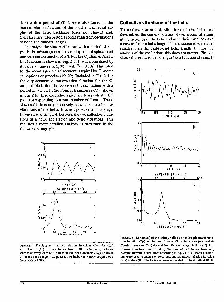
Collective vibrations of the helixTo analyze the stretch vibrations of the helix, we
determined the centers of mass of two groups of atomsat the two ends of the helix and used their distance I as a
measure for the helix length. This distance is somewhatsmaller than the end-to-end helix length, but for theanalysis of the oscillations this does not matter. Fig. 3Ashows this reduced helix length I as a function of time. It
2.2 lA
E
=2.1
TIME t (ps)
5 1.0 1.5FREQUENCY v (ps-')
FIGURE 3 Length l(t) of the (Ala)20-helix (A), the length autocorrela-tion function C,Qt) as obtained from a 400 ps trajectory (B), and itsFourier transform e,(v) derived from the time range 0-10 ps (C). TheFourier transform was fitted by the sum of two terms describingdamped harmonic oscillators according to Eq. 9 (... ). The fit parame-ters were used to calculate the corresponding autocorrelation function(...) in time (B). The helix was weakly coupled to a heat bath at 300 K.
Biophysical Journal Volume 59 April 1991
A
FIGURE 2 Displacement autocorrelation functions CQt) for CQ11( ) and CQ1 (- * *) as obtained from a 400 ps trajectory with anoutput at every 20 fs (A), and their Fourier transforms CI(v) derivedfrom the time range 0-20 ps (B). The helix was weakly coupled to aheat bath at 300 K.
U-h
Biophysical Journal Volume 59 April 1991798

performs oscillations with a period of 1.4 ps and a
root-mean-square (rms) amplitude ((Al)2)1'2 = 0.26 Acorresponding to 1.3% of the average length (1). Thelength autocorrelation function C,(t) is shown in Fig. 3 Band exhibits oscillations with the same period. Theamplitude of the oscillations decreases rapidly duringthe first 5 ps, but thereafter the decrease is much slower.The oscillations in C,(t) at long times arise from
oscillations of the helix length over several periodswithout appreciable damping (Fig. 3 A). Outside suchregions, the oscillations of the length are more stochas-tic, i.e., more strongly damped, giving rise to the oscilla-tions in C,(t) at short times with their rapidly decreasingamplitude. Both types of correlations may be describedby damped harmonic oscillators. To analyze the short-time correlations, we performed a Fourier transforma-tion of C,(t) within the time range 0-10 ps and obtained a
curve C,(v) with one peak at a finite frequency andanother one at zero frequency (Fig. 3 C). The peak atfinite frequency describes damped oscillations, the stretchvibrations of the helix, and the peak at zero frequency anoverdamped relaxation of the helix length, which maytentatively be assigned to the effect of helix bending on
the helix length. The spectrum was fitted by the sum oftwo displacement autocorrelation functions for dampedharmonic oscillators, Eq. 9. For the peak at finitefrequency we obtained the eigen frequency v0 = 0.73ps' and the damping coefficient A = 0.80 ps', and forthe peak at zero frequency the relaxation rate 2/I =
0.36 ps '. The autocorrelation function in time corre-
sponding to this superposition of two damped harmonicoscillators is included in Fig. 3 B. The short-time behav-ior of C,(t) is well described by the harmonic oscillatormodel, but the sustained oscillations in C,(t) at longertimes, say after 5 ps, are not. They might be described bya third harmonic oscillator with the same frequency v0 =
0.73 ps-1 but a much smaller damping coefficient P. Thetwo oscillators might then be interpreted as two differentsubstates of the helix (2). In both substates the helixwould undergo stretch vibrations, but with differentdamping coefficients.To study the influence of the hydrogen bonds on the
stretch vibration, a simulation was performed withhydrogen bonds explicitly taken into account by an
additional term in the interaction energy. The eigenfrequency v0 of the stretch vibration increased by 40%(Table 1) and its amplitude decreased by 20%.
Spectroscopic data for the vibrations of long polyala-nine helices have been analyzed previously within theframework of a normal coordinate analysis, and thewavenumbers for the stretch vibration of polyalaninehelices of arbitrary length have been deduced (33). Foran (Ala)20-helix two different sets of experimental data
TABLE 1 Frequencies v, of the stretch and bend vibrations ofan (Ala)20-helix
Method v(S) v(b)
ps psSCalculated without explicit Hbonds 0.73 0.23
Calculated with explicit Hbonds 1.06 0.33
0.69Experimental (reference 33) 1.26
yield 23 and 42 cm-' corresponding to frequencies of0.69 and 1.26 ps-' (Table 1). These numbers are compat-ible with our MD result, their spread is even larger thanthe difference between the two values obtained with andwithout an explicit potential for hydrogen bonds. For therms amplitude of the stretch vibration, the analysis ofthe spectroscopic data yielded 1.4% (33), in goodagreement with our result.To analyze the bend vibrations of the helix, the
directions of two segments at the two ends of the helixwere determined and the angle between them used as
the bend angle 0 of the helix. Inspection of helices with a
finite 0 on a graphics system indicated that the bendextends more or less homogeneously along the helix. Fig.4 shows the autocorrelation function CQt) of the bendangle exhibiting damped oscillations. A fit of the Fouriertransform C9(v) (data not shown) by the expression for a
damped harmonic oscillator yielded v0 = 0.23 ps-' andP = 0.11 ps-'. Thus, the bend vibration is a factor ofthree slower than the stretch vibration (Table 1) and itsdamping coefficient is a factor of seven smaller.One might have attempted to analyze the stretch
vibrations of the helix by using the distance between two
TIME t (ps)
FIGURE 4 Autocorrelation function C,(t) of the bend angle 0(t) of the(Ala)20-helix as obtained from a 400 ps trajectory. The curve was fittedas described in the legend to Fig. 3 (.. ). The helix was weakly coupledto a heat bath at 300 K.
Plis an .ani Coleciv Virtin ot an ..heli 799.Pleiss and Jahnig Collective Vibrations of an a-helix 799

single atoms at the two ends of the helix as a measure forthe helix length (18, 21). The corresponding autocorrela-tion function is shown in Fig. 5. It exhibits oscillationswith a period of 5 ps, superimposed on oscillationswith a period of 1 ps. Hence, both the stretch and thebend vibration are reflected in the distance between twosingle atoms. However, it is not possible from such ananalysis to individually assign the stretch and the bendvibration to the two oscillations at 1.4 and 4.3 ps.The ratio of the frequencies for the stretch and bend
vibrations results as 4.3/1.4 = 3.1. This is the valueexpected if the helix behaves as an isotropic elastic rod(see Methods). Using Eqs. 14 and 15 and the numericalvalues for the stretch and bend vibrations, the elasticitymodulus of the polyalanine helix is obtained as Y(s)2.1- 1011 dyn/cm2 and Y(") = 2.3- 1011 dyn/cm2, respec-tively. These values are compatible with the value(1.7-4.8) - 1011 dyn/cm2 calculated previously for a (Gly)1O-helix by a normal coordinate analysis (13). Lacking otherdata, our result may be compared with experimentalvalues for the elasticity modulus of collagen helicesdetermined as (1.5-2)* 1011 dyn/cm2 by Brillouin scatter-ing (34) and as (3-5)* 1011 dyn/cm2 by measurement ofthe persistence length (35). Although the collagen heli-ces differ from our regular a-helix, the values for theelasticity modulus lie in the same range.
Different environmentsThe MD simulations discussed above were performedon an (Ala)20-helix under weak coupling to a heat bath at300 K. To study the influence of the environment on thecollective vibrations of the helix, three further kinds ofcoupling to the environment were investigated: (a) nocoupling, (b) strong coupling to a heat bath at 300 K, and(c) coupling to 570 surrounding water molecules kept at300 K.To characterize thermodynamically the systems stud-
ied under the different conditions, the mean potential
1.0
° 0.5
L0.0
-0.50 5 10
TIME t (ps)15 20
and kinetic energies of the helix and their rms fluctua-tions were calculated and are presented in Table 2 forweak and strong coupling to a heat bath and for couplingto water. For weak coupling, the potential energyfluctuates with an amplitude of +22 kJ/mol around amean value of zero. The kinetic energy fluctuates withan amplitude of +18 kJ/mol around a mean value of
300 kJ/mol. This mean value is expected for a canoni-
cal system of 123 atoms with fixed bond length at 300 K.Hence, one might conclude that the helix behaves as acanonical ensemble. This would imply that the potentialand kinetic energies are uncorrelated and the fluctua-tions of the total energy are given by the sum ofthe fluctuations of the potential and kinetic energy,(AE2)cnnc = (AE2o ) + (AE 2 ). Insertion of numbersfor (E~pot) and (kEin) yields (tEocanonical-= +28 kJ/mol.This is about twice the actual value of (AE 2 t) I2 obtainedby first calculating the total energy as Etot = Epot + Ekinand then its fluctuations. For a microcanonical ensemble,on the other hand, one would expect (AE2ot)microcanonicai = 0,
i.e., the potential and kinetic energy would be perfectlyanticorrelated. Hence, the helix coupled weakly to aheat bath behaves as a system somewhere between acanonical and a microcanonical ensemble, and the sameis true for strong coupling to a heat bath and forcoupling to water. This deficiency is partially a conse-quence of the algorithm used to describe the coupling toa heat bath. However, two additional sources of errorhave been proposed and would explain the deviationfrom a canonical ensemble even in the case of couplingto water: the inaccuracy of the integration algorithm andthe introduction of a cutoff radius (27). In the case of nocoupling of the helix to the environment, the potentialand kinetic energies and with them the total energyincreased continuously, as observed previously (27).This drift of the energy was the reason to introduce thecoupling to a heat bath.The autocorrelation functions C,(t) of the helix length
for the different cases of coupling the helix to theenvironment are shown in Fig. 6. Without any coupling,the stretch vibrations are well pronounced, the initialdecrease extends up to 10 ps and thereafter long-timecorrelations exist as in the case of weak coupling to aheat bath. The Fourier transform C,(v) was calculatedusing a time window of 10 ps and fitted by the sum of twoterms describing damped harmonic oscillators, Eq. 9.The result for the oscillating term is presented in Table3. The eigen frequency vo of the oscillator is the same asfor the case of weak coupling to a heat bath, but thedamping coefficient is smaller by a factor of 1.4. In thecase of strong coupling to a heat bath, the amplitude ofthe oscillations in C,t) decreases more rapidly than forweak coupling, and the amplitude of the superimposedrelaxation is increased. The long-time correlations are
non BJ
FIGURE 5 Autocorrelation function Cd(t) of the helix length deter-mined as the distance C.3-C.18, obtained from a 400 ps trajectory. Thehelix was weakly coupled to a heat bath at 300 K.
-U.J
800 Biophysical Journal Volume 59 April 1991

TABLE 2 Mean values and rms deviations of the potential, kinetic, and total energies of an (Ala)2,-helix for three diftrent couplings of thehelix to the environment
Coupling (Epo) (Ebf) (E,E1) 2 (AE2i (ot)1
kJ/mol kl/mol kf/mol kfImol kf/mol kd/molWeak coupling to heat bath -9 297 288 22 18 12Strong coupling to heat bath 16 298 314 37 13 36Coupling to water 14 304 318 26 20 24
more complex. Therefore, a time window of 5 ps was
used to calculate the Fourier transform C1(v). A fit bythe sum of two terms describing damped harmonicoscillators yielded for the oscillating term a slightlylower eigen frequency and a damping coefficient threetimes higher than for weak coupling to a heat bath(Table 3). If the helix is surrounded by water molecules,the stretch vibration is also strongly damped as ex-pressed by the rapid decay of the autocorrelation func-tion C#(). The long-time correlations are more regularthan for strong coupling to a heat bath and resemble
TIME t (ps)
those for weak coupling. A fit of the Fourier transformC,(v) calculated for a time window of 5 ps yieldedroughly the same eigen frequency as for weak couplingand a damping coefficient 1 about twice as large.The autocorrelation functions CQt) of the helix bend
angle for the different cases of coupling the helix to theenvironment are shown in Fig. 7. Without any coupling,the bend oscillations are well pronounced and weaklydamped. For the case of strong coupling to a heat bath,the autocorrelation function rapidly decays to zero andprovides only a very weak indication of a S ps oscillation.Obviously, the bend vibration has become overdamped.The same is true for the case of coupling to water. Thedecay of the autocorrelation function is less rapid, butthe 5 ps oscillations are even less pronounced. Thedamping coefficient may be estimated by fitting theautocorrelation function by the expression for a har-monic oscillator in the overdamped case, Eq. 7. Usingthe time range 0-10 ps for the fit, one obtains for therelaxation time wI/3 = 0.2 ps'. Under the assumptionthat the angular frequency wo = 1.5 ps' of the bendvibration is not altered by the presence of water, thedamping coefficient results as ,B = 11 ps'. Hence, thecondition for overdamping, o0 < P/2, is fulfilled. Sur-
TABLE 3. Results of fits of the Fourier transform C,(v) of thedisplacement autocorrelation function for the helix length bythe sum of two terms describing damped harmonicoscillators
Coupling vo
PS -1 Ps-No coupling* 0.70 0.56 0.014Weak coupling to heat
bath* 0.73 0.80 0.013Strong coupling to heat
batht 0.69 2.4 0.022Coupling to watert 0.83 2.0 0.012
The values for the eigen frequencies v0 and the damping coefficients 1of the stretch vibration are presented for four different cases ofcoupling of the helix to the environment. The relative rms fluctuations((Ai)2)112/(i) of the length are included. *The time window for theFourier transformation was 10 ps. tThe time window for the Fouriertransformation was 5 ps.
Plis an J. ni Colctv Virtin of an ..hel..801
FIGURE 6 Autocorrelation functions CQt) of the helix length fordifferent cases of coupling the helix to the environment: no coupling(A), strong coupling to a heat bath at 300 K (B), and surrounded by570 water molecules at 300 K (C). The curves were obtained fromtrajectories extended over 40 ps (A), 400 ps (B), and 60 ps (C).
Pleiss and Jahnig Collective Vibrations of an a-helix 801

° 0.5
O-,,0.0
TIME t (ps)
FIGURE 7 Autocorrelation functions C,(t) of the bend angle of thehelix for different cases of coupling the helix to the environment, asspecified in the legend to Fig. 6.
rounding water molecules increase the damping coeffi-cient for the bend vibration by a factor of 100 relative tothe value for weak coupling to a heat bath.
CONCLUSION
The internal dynamics of a 20-residue polyalanine helixwas analyzed in terms of autocorrelation functions forthe displacement and the velocity of individual atoms,derived from the trajectories of MD simulations. Theautocorrelations exhibit oscillations in the range of 50 fsto 5 ps. The fast oscillations correspond to localizedvibrations of the bond and dihedral angles, vibrations ofthe bond lengths do not occur in our MD simulationsbecause the bond lengths were kept constant. Thespectrum of the fast oscillations is consistent withexperimental data from Raman and infrared spectros-copy. This is not astonishing because as mentioned inthe Introduction the parameters of the bonded interac-tions were chosen to fit these data. Of more interest arethe slow oscillations which correspond to delocalizedcollective vibrations of the helix. They are strongly
influenced by the nonbonded interactions whose descrip-tion lacks a direct experimental justification. To assign
the slow oscillations unambiguously to stretch and bendvibrations of the helix, the relative motions of two groups
of atoms at the two ends of the helix were analyzed. Inthis way the 1.4 ps oscillation could be assigned to thestretch vibration and the 4.3 ps oscillation to the bendvibration. The frequency and amplitude of the stretchvibration as well as the deduced elastic modulus of thehelix are compatible with experimental data from Ra-man and infrared spectroscopy, Brillouin scattering, andpersistence length measurements. This indicates thatthe description of the nonbonded interactions is notunrealistic. The description of, for example, the hydro-gen bond interaction might even be improved, if moreaccurate experimental data on the stretch vibration andthe elastic modulus become available.The influence of the environment on the internal
dynamics of the helix was studied by coupling the helixto a heat bath or to water. Because the delocalizedcollective motions are supposed to be more sensitive tothe environment than the localized motions, we concen-
trated our analysis on the collective motions. Thefrequencies of the stretch and bend vibrations of thehelix turned out to be essentially unaffected by theenvironment, whereas their dampingwas affected consid-erably. This implies that the frequencies are determinedessentially by the interactions among the helix atoms as
expected, whereas the coupling of the helix atoms to theenvironment determines the damping (25). Water in-creases the damping of the stretch vibration by a factorof two compared to the case of weak coupling to a heatbath, but the vibration remains underdamped. By con-
trast, the damping of the bend vibration increases by a
factor of 100, it becomes overdamped, i.e., its dynamicbehavior changes from oscillation to relaxation. That thebend vibration is more strongly damped by water thanthe stretch vibration is conceivable. The displacement ofthe helix ends is much larger for the bend than for thestretch vibration, so that more water molecules have tomove away for an oscillation to occur. This overdampingof the bend vibration by water is probably the reason
that it is not detected in Raman and infrared measure-
ments. It might be observable as a relaxation process influorescence anisotropy decay measurements with pico-second resolution or in microwave absorption measure-
ments. Such experiments would be useful to test thedescription of damping in the picosecond range by MDsimulations. An attempt in that direction has recentlybeen made by measuring the overall rotational diffusionof tryptophan in water and comparing it with the resultobtained from an MD simulation (36). Such studies
802 Biophysical Journal Volume 59 April1991802 Biophysical Journal Volume 59 April 1991

might be extended to the internal dynamics of peptidesand proteins.Although we could show that the stretch and bend
vibration of helices are rather well described in terms ofdamped harmonic oscillators, such a model does notaccount for all aspects of these collective motions.Inspection of the fluctuations of the helix length indi-cates that the polyalanine helix flips randomly betweenat least two states, one with weakly damped and one withmore strongly damped stretch vibrations. Such behavioris reminiscent of chaotic systems with strange attractors(37). More work is required to enlighten this aspect ofthe dynamic behavior of peptides and proteins.
We thank Olle Edholm for introducing us to Gromos, and for manystimulating discussions. We also thank W. F. van Gunsteren and H. J.C. Berendsen for providing the Gromos program. The cooperativity ofR. D. Kloth from this institute, U. Wandel from the Max-Planck-Institut fur Biologische Kybemetik, and the staff of the Rechenzen-trum Garching is gratefully acknowledged.
Received for publication 14 June 1990 and in final form 21November 1990.
REFERENCES
1. Frauenfelder, H., F. Parak, and R. D. Young. 1988. Conforma-tional substates in proteins. Annu. Rev. Biophys. Biophys. Chem.17:451-479.
2. Ansari, A., J. Berendzen, S. F. Bowne, H. Frauenfelder, I. E. T.lben, T. B. Sauke, E. Shyamsunder, and R. D. Young. 1985.Protein states and proteinquakes. Proc. Natl. Acad. Sci. USA.82:5000-5004.
3. Dornmair, K., and F. Jahnig. 1989. Internal dynamics of lactosepermease. Proc. Natl. Acad. Sci. USA. 86:9827-9831.
4. Tipping, M., K. Viras, and T. A. King. 1984. Low-frequencydynamics of solid poly(L-alanine) from Raman spectroscopy.Biopolymers. 23:2891-2899.
5. Fanconi, B., E. W. Small, and W. L. Peticolas. 1971. Phonondispersion curves and normal coordinate analysis of a-poly-L-alanine. Biopolymers. 10:1277-1298.
6. Itoh, K., and T. Shimanouchi. 1970. Vibrational frequencies andmodes of a-helix. Biopolymers. 9:383-399.
7. Genzel, L., F. Kremer, A. Poglitsch, and G. Bechtold. 1983.Relaxation processes on a picosecond time scale in hemoglobinand poly(L-alanine) observed by millimeter-wave spectroscopy.Biopolymers. 22:1715-1729.
8. Cusack, S., J. Smith, J. Finney, B. Tidor, and M. Karplus. 1988.Inelastic neutron scattering analysis of picosecond internalprotein dynamics. J. Mol. BioL 202:903-908.
9. Beechem, J. M., and L. Brand. 1985. Time-resolved fluorescenceof proteins.Annu. Rev. Biochem. 54:43-71.
10. Parak, F., and L. Reinisch. 1986. Mossbauer effect in the study ofstructure dynamics. Methods Enzymol. 131:568-607.
11. Wuthrich, K 1976. Nuclear magnetic resonance in biologicalresearch. Peptides and proteins. North-Holland Publishing Co.,Amsterdam.
12. Go, N., T. Noguti, and T. Nishikawa. 1983. Dynamics of a smallglobular protein in terms of low-frequency vibrational modes.Proc. Natl. Acad. Sci. USA. 80:3696-3700.
13. Levy, R. M., and M. Karplus. 1979. Vibrational approach to thedynamics of an a-helix. Biopolymers. 18:2465-2495.
14. Karplus, M., and J. A. McCammon. 1983. Dynamics of proteins:elements and function.Annu. Rev. Biochem. 53:263-300.
15. Parak, F., and E. W. Knapp. 1984. A consistent picture of proteindynamics. Proc. Natl. Acad. Sci. USA. 81:7088-7092.
16. Nadler, W., A. T. Brunger, K. Schulten, and M. Karplus. 1987.Molecular and stochastic dynamics of proteins. Proc. Natl. Acad.Sci. USA. 84:7933-7937.
17. Levitt, M. 1982. Protein conformation, dynamics, and folding bycomputer simulation.Annu. Rev. Biophys. Bioeng. 11:251-271.
18. Aqvist, J., W. F. van Gunsteren, M. Leijonmarck, and 0. Tapia.1985. A molecular dynamics study of the C-terminal fragment ofthe L7/L12 ribosomal protein. J. Mol. Biol. 183:461-477.
19. Swaminathan, S., T. Ichiye, W. F. van Gunsteren, and M. Karplus.1982. Time dependence of atomic fluctuations in proteins:analysis of local and collective motions in bovine pancreatictrypsin inhibitor. Biochemisby. 21:5230-5241.
20. Levy, R. M., D. Perahia, and M. Karplus. 1982. Moleculardynamics of an a-helical polypeptide: temperature dependenceand deviation from harmonic behavior. Proc. Natl. Acad. Sci.USA. 79:1346-1350.
21. Edholm, O., and F. Jahnig. 1988. The structure of a membrane-spanning polypeptide studied by molecular dynamics. Biophys.Chem. 30:279-292.
22. Ghosh, I., and J. A. McCammon. 1987. Sidechain rotationalisomerization in proteins. Biophys. J. 51:637-641.
23. Gilson, M. K., and B. H. Honig. 1986. The dielectric constant of afolded protein. Biopolymers. 25:2097-2119.
24. Russell, S. T., and A. Warshel. 1985. Calculations of electrostaticenergies in proteins. J. Mol. Biol. 185:389-404.
25. Brooks, C. L., and M. Karplus. 1989. Solvent effects on proteinmotion and protein effects on solvent motion. J. Mol. Biol.208:159-181.
26. Marqusee, S., V. H. Robbins, and R. L. Baldwin. 1989. Unusuallystable helix formation in short alanine-based peptides. Proc.Natl. Acad. Sci. USA. 86:5286-5290.
27. Berendsen, H. J. C., J. P. M. Postma, W. F. van Gunsteren, A.DiNola, and J. R. Haak. 1984. Molecular dynamics with cou-pling to an external bath. J. Chem. Phys. 81:3684-3690.
28. Ryckaert, J. P., G. Ciccotti, and H. J. C. Berendsen. 1977.Numerical integration of the cartesian equations of motion of asystem with constaints: molecular dynamics of n-alkanes. J.Comp. Phys. 23:327-341.
29. van Gunsteren, W. F., and M. Karplus. 1982. Effect of constraintson the dynamics of macromolecules. Macromolecules. 15:1528-1544.
30. Springer, T. 1972. Quasielastic neutron scattering for the investiga-tion of diffusive motions in solids and liquids. Springer-Verlag,Berlin.
31. Chandrasekhar, S. 1943. Stochastic problems in physics andastronomy. Rev. Mod. Phys. 15:1-89.
Pleiss and Jahnig Collective Vibrations of an a-helix 803

32. Landau, L. D., and E. M. Lifshitz. 1970. Course of theoreticalphysics. Vol. 7. Pergamon Press Inc., New York.
33. Peticolas, W. L. 1979. Mean-square amplitudes of the longitudinalvibrations of helical polymers. Biopolymners. 18:747-755.
34. Harley, R., D. James, A. Miller, and J. W. White. 1977. Phononsand the elastic moduli of collagen and muscle. Nature (Lond.).267:285-287.
35. Hofmann, H., T. Voss, and K Kuhn. 1984. Localization of flexible
sites in thread-like molecules from electron micrographs. J. MoLBiol. 172:325-343.
36. Chen, L. X.-Q., R. A. Engh, and G. R. Fleming. 1988. Reorienta-tion of tryptophan and simple peptides: onset of internalflexibility and comparison with molecular dynamics simulation.J. Phys. Chem. 92:4811-4816.
37. Lorenz, E. N. 1963. Deterministic nonperiodic flow. J. Atmos. Sci.20:130-141.
804 Biophysical Journal Volume 59 April 1991