cognitive neuroscience: colourful language

1
COGNITIVE NEUROSCIENCE Colourful language Synaesthesia — a phenomenon that occurs when one sense is stimulated by another — has intrigued scientists for more than a century. Recently, imaging techniques have advanced studies of this condition, allowing scientists to finally show that synaes- thesia is the result of cross-activation between different brain areas — possibly caused by a failure in spine pruning — and that it can aid per- ception. Reporting in Neuron , Boynton, Ramachandran and co- workers provide further support for the cross-activation theory, and show a link between two particular brain areas in one type of synaesthesia. The authors studied a relatively common form of synaesthesia — grapheme–colour synaesthesia — in which individuals see colours when viewing particular letters and num- bers. They first used number and letter patterns to show that synaes- thetes really do see colours when viewing certain graphemes, and that this helps them to identify shapes within the patterns more easily than control subjects. However, the use of synaesthetic colours was found to be less effective than real colours in discriminating such shapes. Boynton et al. then went on to use a combination of behavioural and imaging techniques to investi- gate the idea that grapheme–colour synaesthesia might be caused by cross-activation between areas of the fusiform gyrus that are involved in grapheme processing and areas that are involved in colour processing. During a grapheme-viewing task, functional MRI scans showed acti- vation of grapheme areas in all par- ticipants. In grapheme–colour synaesthetes the colour-selective area V4 was also activated, whereas for control subjects significantly less activation was seen in this region. The lack of variation seen in the grapheme areas of synaes- thetes and control subjects indi- cates that the observed differences between the two groups were not related to the attentional states of the participants. Boynton and colleagues also present evidence that synaesthetes and control subjects showed no dif- ferences in their brain activation in response to colours, which indicates that synaesthesia does not arise from any serious differences in brain anat- omy or function. Importantly, although the authors propose that the strength of the synaesthetic colours experienced depends on the degree of activation of area V4, they do not believe that this entirely accounts for these differences. Synaesthetes, they suggest, might be a highly heterogeneous group, so it would be a mistake to simply pool data from a group of synaesthetes and compare them with data from non- synaesthetes. Further research with many more subjects will be required to establish whether this is the case. Sarah Archibald References and links ORIGINAL RESEARCH PAPER Hubbard, E. M., Cyrus Arman, A., Ramachandran, V. S. & Boynton, G. M. Individual differences among grapheme–color synesthetes: brain–behavior correlations. Neuron 45, 975–985 (2005) FURTHER READING Rich, A. N. & Mattingley, J. B. Anomalous perception in synaesthesia: a cognitive neuroscience perspective. Nature Rev. Neurosci. 3, 43–52 (2002) 344 | MAY 2005 | VOLUME 6 www.nature.com/reviews/neuro HIGHLIGHT ADVISORS NANCY ANDREASEN UNIVERSITY OF IOWA, IA, USA ALLAN BASBAUM UNIVERSITY OF CALIFORNIA SAN FRANCISCO, CA, USA RANDY BUCKNER WASHINGTON UNIVERSITY, MO, USA DAVID CLAPHAM HARVARD MEDICAL SCHOOL, MA, USA PIETRO DE CAMILLI YALE UNIVERSITY SCHOOL OF MEDICINE, CT, USA BARRY EVERITT UNIVERSITY OF CAMBRIDGE, UK GORDON FISHELL SKIRBALL INSTITUTE, NY, USA MARY KENNEDY CALIFORNIA INSTITUTE OF TECHNOLOGY, CA, USA LYNN NADEL UNIVERSITY OF ARIZONA, AZ, USA DENNIS O’LEARY THE SALK INSTITUTE, CA, USA TERRY SEJNOWSKI THE SALK INSTITUTE, CA, USA WOLF SINGER MAX-PLANCK-INSTITUT FÜR HIRNFORSCHUNG, GERMANY CLAUDIO STERN UNIVERSITY COLLEGE LONDON, UK PATRICK TAM CHILDREN'S MEDICAL RESEARCH INSTITUTE, SYDNEY, AUSTRALIA RICHARD W. TSIEN STANFORD UNIVERSITY SCHOOL OF MEDICINE, CA, USA RAFAEL YUSTE COLUM UNIVERSITY, NY, USA RESEARCH HIGHLIGHTS © 2005 Nature Publishing Group

Upload: sarah

Post on 21-Jul-2016

216 views

Category:

Documents


2 download

TRANSCRIPT

URLsC O G N I T I V E N E U R O S C I E N C E

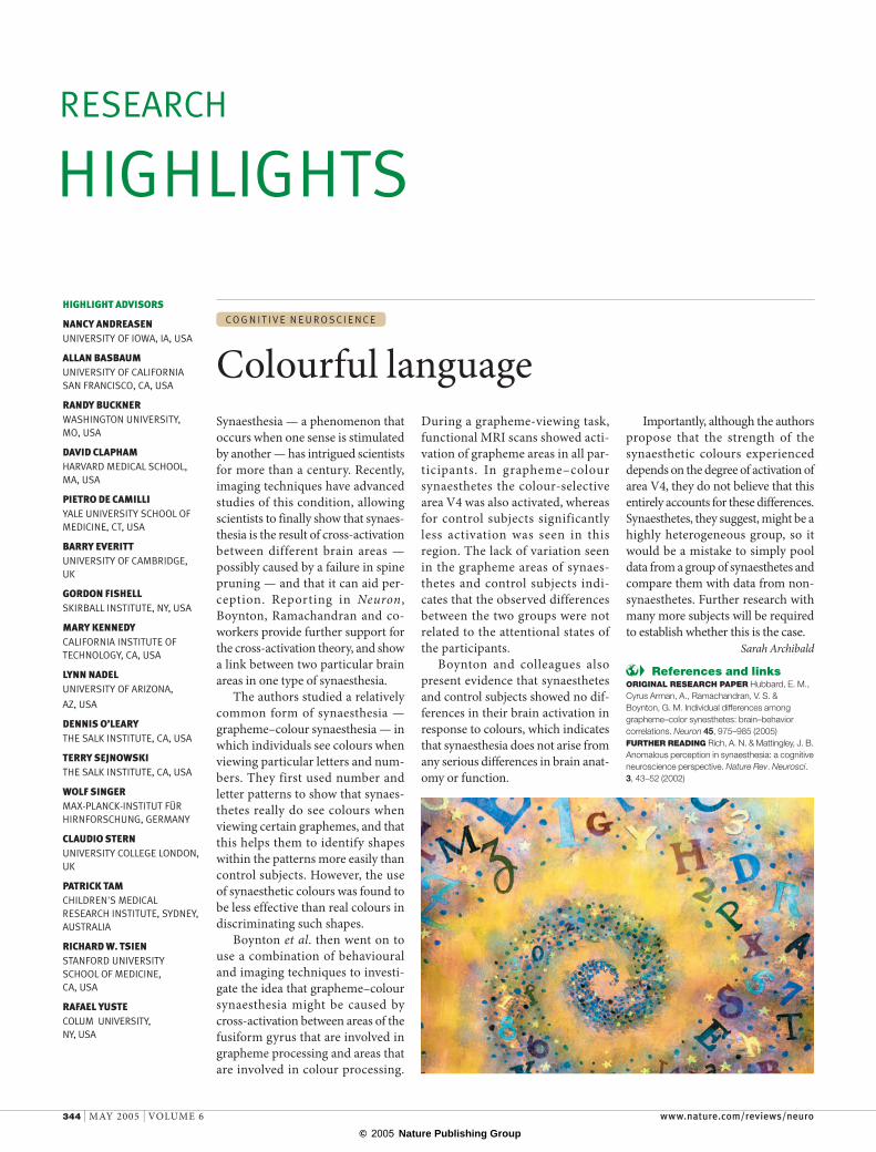
Colourful languageSynaesthesia — a phenomenon that occurs when one sense is stimulated by another — has intrigued scientists for more than a century. Recently, imaging techniques have advanced studies of this condition, allowing scientists to finally show that synaes-thesia is the result of cross-activation between different brain areas — possibly caused by a failure in spine pruning — and that it can aid per-ception. Reporting in Neuron, Boynton, Ramachandran and co-workers provide further support for the cross-activation theory, and show a link between two particular brain areas in one type of synaesthesia.

The authors studied a relatively common form of synaesthesia — grapheme–colour synaesthesia — in which individuals see colours when viewing particular letters and num-bers. They first used number and letter patterns to show that synaes-thetes really do see colours when viewing certain graphemes, and that this helps them to identify shapes within the patterns more easily than control subjects. However, the use of synaesthetic colours was found to be less effective than real colours in discriminating such shapes.

Boynton et al. then went on to use a combination of behavioural and imaging techniques to investi-gate the idea that grapheme–colour synaesthesia might be caused by cross-activation between areas of the fusiform gyrus that are involved in grapheme processing and areas that are involved in colour processing.

During a grapheme-viewing task, functional MRI scans showed acti-vation of grapheme areas in all par-ticipants. In grapheme–colour synaesthetes the colour-selective area V4 was also activated, whereas for control subjects significantly less activation was seen in this region. The lack of variation seen in the grapheme areas of synaes-thetes and control subjects indi-cates that the observed differences between the two groups were not related to the attentional states of the participants.

Boynton and colleagues also present evidence that synaesthetes and control subjects showed no dif-ferences in their brain activation in response to colours, which indicates that synaesthesia does not arise from any serious differences in brain anat-omy or function.

Importantly, although the authors propose that the strength of the synaesthetic colours experienced depends on the degree of activation of area V4, they do not believe that this entirely accounts for these differences. Synaesthetes, they suggest, might be a highly heterogeneous group, so it would be a mistake to simply pool data from a group of synaesthetes and compare them with data from non-synaesthetes. Further research with many more subjects will be required to establish whether this is the case.

Sarah Archibald

References and linksORIGINAL RESEARCH PAPER Hubbard, E. M., Cyrus Arman, A., Ramachandran, V. S. & Boynton, G. M. Individual differences among grapheme–color synesthetes: brain–behavior correlations. Neuron 45, 975–985 (2005)FURTHER READING Rich, A. N. & Mattingley, J. B. Anomalous perception in synaesthesia: a cognitive neuroscience perspective. Nature Rev. Neurosci. 3, 43–52 (2002)

344 | MAY 2005 | VOLUME 6 www.nature.com/reviews/neuro

HIGHLIGHT ADVISORS

NANCY ANDREASENUNIVERSITY OF IOWA, IA, USA

ALLAN BASBAUMUNIVERSITY OF CALIFORNIA SAN FRANCISCO, CA, USA

RANDY BUCKNERWASHINGTON UNIVERSITY, MO, USA

DAVID CLAPHAMHARVARD MEDICAL SCHOOL, MA, USA

PIETRO DE CAMILLIYALE UNIVERSITY SCHOOL OF MEDICINE, CT, USA

BARRY EVERITTUNIVERSITY OF CAMBRIDGE, UK

GORDON FISHELLSKIRBALL INSTITUTE, NY, USA

MARY KENNEDYCALIFORNIA INSTITUTE OF TECHNOLOGY, CA, USA

LYNN NADELUNIVERSITY OF ARIZONA,

AZ, USA

DENNIS O’LEARYTHE SALK INSTITUTE, CA, USA

TERRY SEJNOWSKITHE SALK INSTITUTE, CA, USA

WOLF SINGERMAX-PLANCK-INSTITUT FÜR HIRNFORSCHUNG, GERMANY

CLAUDIO STERN UNIVERSITY COLLEGE LONDON, UK

PATRICK TAMCHILDREN'S MEDICAL RESEARCH INSTITUTE, SYDNEY, AUSTRALIA

RICHARD W. TSIENSTANFORD UNIVERSITY SCHOOL OF MEDICINE, CA, USA

RAFAEL YUSTECOLUM UNIVERSITY, NY, USA

RESEARCH HIGHLIGHTS

© 2005 Nature Publishing Group