coarse-grained modeling of vesicle responses to...
TRANSCRIPT
Nanoscale
PAPER
Cite this: Nanoscale, 2015, 7, 13458
Received 13th March 2015,Accepted 18th June 2015
DOI: 10.1039/c5nr01652e
www.rsc.org/nanoscale
Coarse-grained modeling of vesicle responses toactive rotational nanoparticles
Liuyang Zhang and Xianqiao Wang*
In recent years, magnetically-driven-rotating superparamagnetic nanoparticles have been emerging as a
valuable component in designing targeted drug delivery carriers and cellular killers via membranes’
physical rupture. The lack of an in-depth understanding of how to control the interaction of rotational
nanoparticles (RNPs) with vesicles has hindered progress in the development of their relevant biomedical
applications. Here we perform dissipative particle dynamics simulations to analyze the rotation frequen-
cies, size, and coating patterns of the RNPs as they interact with the vesicle so as to provide novel designs
of drug delivery applications. Results have revealed that the RNPs are capable of triggering local disturb-
ance around the vesicle and therefore promoting the vesicle translocation toward the RNPs. By investi-
gating the translocation time and driving forces required for RNPs to enter inside the vesicle at various
rotation frequencies as well as the interaction energy between coated RNPs and the vesicle, we have
tuned the coating pattern of the ligands on the surface of RNPs to open a specified channel in the vesicle
for promoting drug delivery. Our findings can provide useful guidelines for the molecular design of
patterned RNPs for controllable bio/inorganic interfaces and help establish qualitative rules for the
organization and optimization of ligands on the surface of the desired drug delivery carriers.
Introduction
Recent years have witnessed the explosive growth of interest innanoparticles (NPs) with a wealth of biomedical applicationssince they are widely used as carriers to translocate drug mole-cules and useful materials into cell interiors.1 A number ofsimulations suggested that the membrane translocation of aniso-tropic NPs is often accompanied by spontaneous and con-tinuous rotation of the NPs. Taking the spontaneous rotationfor example, the ligand coated NPs with anisotropic patternsrotated to a preferred orientation while penetrating throughthe membrane.2 Similar behavior was observed for the trans-location of graphene sheets across the membrane.3 In theendocytosis process of anisotropic NPs, the anisotropic NPsgenerally undergo a transient rotation during the wrappingprocess to minimize the free energy.4,5 Different from thelimited effect of spontaneous rotation of anisotropic NPs, thepromoting effect of continuous NP rotation is ascribed to theenhanced membrane monolayer protrusion as well as it exertsa shearing force to rupture the membrane.6 However, the fun-damental mechanism of how a vesicle responds to the activeRNP remains poorly understood.
From the experimental viewpoints, the active rotation ofNPs can be controlled by the underlying dynamic magneticfield. For example, the rotational movement of superparamag-netic iron oxide nanoparticles induced by a dynamic magneticfield can cause membrane permeabilization and lead to apop-tosis.7 NPs are enclosed inside intracellular endosome vesicles,which can be deformed to probe the intracellular membrane,or turn into an intracellular heater when subjected to a mag-netic field.8,9 Another promising application of RNPs is drugdelivery. RNPs are widely used as carriers to translocate drugmolecules and other useful materials into cell interiors.10–16
The targeted RNPs have been described to control the deliveryand release of anticancer drugs with advanced controllabil-ity.17,18 RNPs in an appropriate magnetic field can give rise toa local effect to encourage drug release, for example by way ofthe hyperthermia effect.19 However the efficiency of drugloading and unloading remains challenging for RNPs. By un-raveling the rudimentary behavior of a single cell interactingwith a single RNP, it will offer insight into the development ofdrug delivery research. In this paper, we propose a simplemagnetic field geometry to create a RNP channel on a singlevesicle with a hollow hybrid RNP-filled channel. There hasbeen much interest in developing synthetic analogues of bio-logical membrane channels with high efficiency and remark-able selectivity for transporting ions and molecules.20,21 Byusing dissipative dynamics simulation, we investigate theinteraction between RNPs and a vesicle at different rotational
College of Engineering and NanoSEC, University of Georgia, Athens, GA 30602, USA.
E-mail: [email protected]; Tel: +706-5426251
13458 | Nanoscale, 2015, 7, 13458–13467 This journal is © The Royal Society of Chemistry 2015
Publ
ishe
d on
19
June
201
5. D
ownl
oade
d by
Uni
vers
ity o
f G
eorg
ia o
n 06
/08/
2015
20:
24:2
8.
View Article OnlineView Journal | View Issue
frequencies as well as the effect of RNP size and coating pat-terns. We also demonstrate a possible pathway of formation ofa RNP-based channel for drug delivery which is executed via adiffusion process through the channel. The main advantagesof RNP channels are that they (i) can be visualized, (ii) guidethe drug through the channel by diffusion, and (iii) do notneed a trigger for drug release. Analysis of the mechanismunderlying the interaction between RNPs and a single vesicleprovides insight into the molecular dynamics of cellular fea-tures. Our simulation results have established RNPs as a prom-ising prototype of synthetic membrane channel with inherentrobustness geared towards biological challenges and excep-tional biocompatibility for drug delivery.
Computational model and methodology
Simulations are based on dissipative particle dynamics (DPD),a mesoscopic coarse-grained simulation method for softmatter and biomembrane systems.22,23 In a typical DPDmodel, a small group of atoms is treated as a single beadlocated at the center of mass of the group. Beads i and j inter-act with each other via a pairwise force consisting of a conser-vative force FCij (representing the excluded volume effect), adissipative force FDij (representing viscous drag betweenmoving beads), and a random force FRij (representing stochasticimpulse). Both FDij and FRij act together as a thermostat for thebeads. Similar to molecular dynamics simulation, time evol-ution is also governed by the Newton’s equation of motion.The total force on bead i can be expressed as
Fi ¼Xi=j
ðFCij þ FD
ij þ FRij Þ
¼Xi=j
aijωðrijÞr̂ij � γω2ðrijÞðr̂ij � vijÞr̂ij þ σωðrijÞζijΔt�1=2 r̂ij� �
ð1Þwhere aij is the maximum repulsive force, rij is the distance, r̂ijis the unit vector, and vij is the relative velocity between beads iand j; ζij denotes a random number with zero mean and unitvariance, and
ωðrijÞ ¼ 1� rijrc; rij , rc
0; rij > rc
(ð2Þ
is a normalized distribution function, with rc being the cutoffradius; γ and σ are parameters related to each other as σ2 =2γkBT, and kBT being the unit of energy. The standard values σ= 3.0 and γ = 4.5 are used in our study.2 The mass, length andtime scales are all normalized in the DPD simulations, withthe unit of length taken to be the cutoff radius rc, the unit ofmass to be that of the solvent beads, and the unit of energy tobe kBT. All other quantities are expressed in terms of thesebasic units. The reduced DPD unit can be converted to the SIunit by examining the membrane thickness and the lipiddiffusion coefficient. The simulated value of bilayer thicknessis 5rc and the effective time scale of the simulation can be
determined from the simulated lateral diffusion constants ofthe lipid bilayer.24 The DPC bilayer thickness is about 4 nmwith a diffusion coefficient around 5 μm2 s−1.25 By comparisonwith typical experimental values, it can be shown that oneDPD length unit corresponds to approximately 0.8 nm inphysical units and the time unit to τ = 24.32 ps. The force unitis kBT/rc = 5.175 pN. The time step is taken as Δt = 0.01τ. Allsimulations are performed using LAMMPS.26
The dimensions of the simulation box are 42rc × 42rc × 50rc.The system contains a total of 271 215 particles, 241 214 waterbeads, and 29 445 lipid beads with a particle density of approxi-mately 3.27 The solvent beads are not shown for clarity in thefollowing figures. The simulation system is comprised of avesicle consisting of 2265 lipid molecules and a rigid cylindricalRNP (556 hydrophobic beads) coated with a selected pattern ofligand and solvent particles. The lipid molecule is representedby the coarse-grained model proposed by Groot and Rabone22
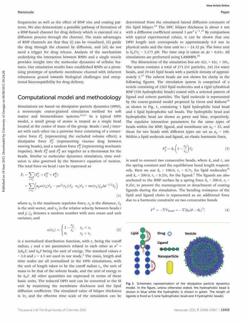
as shown in Fig. 1, containing 1 lipid hydrophilic head beadand 4 lipid hydrophobic tail beads. The hydrophilic bead andhydrophobic bead are shown as green and blue, respectively.The repulsive interaction parameters for the same types ofbeads within the RNP, ligand, and membrane are aij = 25, andthose for two beads with different types are set as aij = 100.Within a lipid molecule and ligand, an elastic harmonic force,
FSij ¼ ks 1� rij
rs
� �r̂ij ð3Þ
is used to connect two consecutive beads, where ks and rs arethe spring constant and the equilibrium bond length respecti-vely. Here we use ks = 100.0, rs = 0.7rc for lipid molecules28
and ks = 200.0, rs = 0.25rc for the ligand.2 The ligands are alsoanchored to the RNP surface by a spring force ks = 200.0, rs =0.25rc to prevent the rearrangement or detachment of coatingligands during the simulation. The bending resistance of thelipid and ligand chain is represented as an additional forcedue to a harmonic constraint on two consecutive bonds
F θ ¼ �rVbend ¼ �r½kθðθ � θ0Þ2� ð4Þ
Fig. 1 Schematic representation of the dissipative particle dynamicsmodel. In the figure, unless otherwise stated, the hydrophobic bead isshown in blue while the hydrophilic is shown in green. The length ofligands is fixed as 5 (one hydrophobic bead and 4 hydrophilic beads).
Nanoscale Paper
This journal is © The Royal Society of Chemistry 2015 Nanoscale, 2015, 7, 13458–13467 | 13459
Publ
ishe
d on
19
June
201
5. D
ownl
oade
d by
Uni
vers
ity o
f G
eorg
ia o
n 06
/08/
2015
20:
24:2
8.
View Article Online
where kθ, θ and θ0 are the bending constant, inclination angleand equilibrium angle, respectively. For three consecutive lipidtail beads or three consecutive lipid head beads in a lipidmolecule, we take k1 = 6 and θ = 180°; for the last head-beadand the top two tail-beads k2 = 3 and θ = 120°; for the bottomtwo consecutive head beads and the first bead in each tail k3 =4.5 and θ = 120°.2 For three consecutive ligand beads, we takekθ = 5 and θ = 180°.
Results and discussionEffect of rotational frequencies
It has been shown that bacteria cells can move forward byrotating the long thin helical filaments counterclockwise.29,30
Artificial bacterial flagella, those consisting of helical tailsresembling natural flagella, show a linear relationship betweenthe frequencies of their rotation and the translational vel-ocity.31 Different from a filament-based motor, the motor-likeRNP can tremendously affect its local biological environment,thereby exerting undeniable influence on its penetration capa-bility into the biological tissues. To demonstrate the effect ofRNP on the surrounding environment, we place the RNP onthe planar membrane and characterize the membrane disturb-ance by calculating the local membrane curvature based onthe method developed by Yue et al.32 First of all, the RNP isput 5rc away from the 40rc × 40rc membrane surface. The RNProtates along the vertical axis. The membrane is divided into2rc × 2rc vertical square prism units, and lipids within eachunit determine the center of mass of the unit. The geo-metric surface representing the membrane via least-squaresfitting is described in the form of quantic polynomial,zðx; yÞ ¼ P
iþj�5aijxiyj, where aij are fitted parameters. And the
curvature kx and ky at point (x, y) can be evaluated via
kx ¼ d2zdx2
= 1þ dzdx
� �2� �3=2; ky ¼ d2z
dx2= 1þ dz
dy
� �2� �3=2ð5Þ
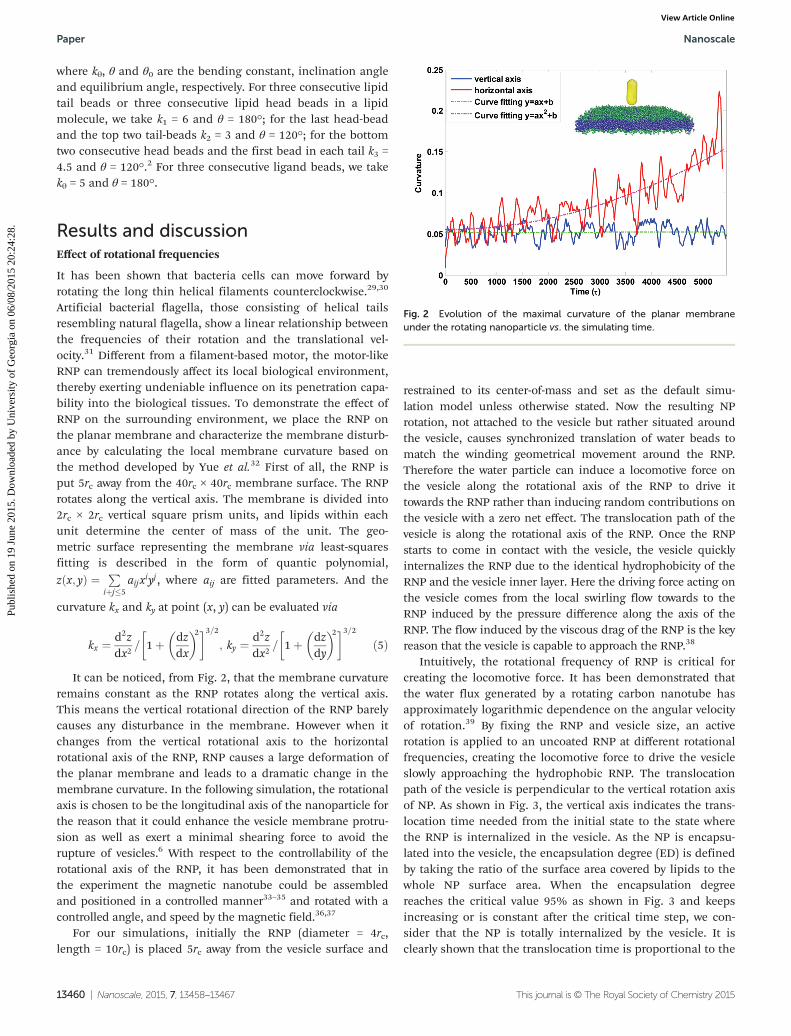
It can be noticed, from Fig. 2, that the membrane curvatureremains constant as the RNP rotates along the vertical axis.This means the vertical rotational direction of the RNP barelycauses any disturbance in the membrane. However when itchanges from the vertical rotational axis to the horizontalrotational axis of the RNP, RNP causes a large deformation ofthe planar membrane and leads to a dramatic change in themembrane curvature. In the following simulation, the rotationalaxis is chosen to be the longitudinal axis of the nanoparticle forthe reason that it could enhance the vesicle membrane protru-sion as well as exert a minimal shearing force to avoid therupture of vesicles.6 With respect to the controllability of therotational axis of the RNP, it has been demonstrated that inthe experiment the magnetic nanotube could be assembledand positioned in a controlled manner33–35 and rotated with acontrolled angle, and speed by the magnetic field.36,37
For our simulations, initially the RNP (diameter = 4rc,length = 10rc) is placed 5rc away from the vesicle surface and
restrained to its center-of-mass and set as the default simu-lation model unless otherwise stated. Now the resulting NProtation, not attached to the vesicle but rather situated aroundthe vesicle, causes synchronized translation of water beads tomatch the winding geometrical movement around the RNP.Therefore the water particle can induce a locomotive force onthe vesicle along the rotational axis of the RNP to drive ittowards the RNP rather than inducing random contributions onthe vesicle with a zero net effect. The translocation path of thevesicle is along the rotational axis of the RNP. Once the RNPstarts to come in contact with the vesicle, the vesicle quicklyinternalizes the RNP due to the identical hydrophobicity of theRNP and the vesicle inner layer. Here the driving force acting onthe vesicle comes from the local swirling flow towards to theRNP induced by the pressure difference along the axis of theRNP. The flow induced by the viscous drag of the RNP is the keyreason that the vesicle is capable to approach the RNP.38
Intuitively, the rotational frequency of RNP is critical forcreating the locomotive force. It has been demonstrated thatthe water flux generated by a rotating carbon nanotube hasapproximately logarithmic dependence on the angular velocityof rotation.39 By fixing the RNP and vesicle size, an activerotation is applied to an uncoated RNP at different rotationalfrequencies, creating the locomotive force to drive the vesicleslowly approaching the hydrophobic RNP. The translocationpath of the vesicle is perpendicular to the vertical rotation axisof NP. As shown in Fig. 3, the vertical axis indicates the trans-location time needed from the initial state to the state wherethe RNP is internalized in the vesicle. As the NP is encapsu-lated into the vesicle, the encapsulation degree (ED) is definedby taking the ratio of the surface area covered by lipids to thewhole NP surface area. When the encapsulation degreereaches the critical value 95% as shown in Fig. 3 and keepsincreasing or is constant after the critical time step, we con-sider that the NP is totally internalized by the vesicle. It isclearly shown that the translocation time is proportional to the
Fig. 2 Evolution of the maximal curvature of the planar membraneunder the rotating nanoparticle vs. the simulating time.
Paper Nanoscale
13460 | Nanoscale, 2015, 7, 13458–13467 This journal is © The Royal Society of Chemistry 2015
Publ
ishe
d on
19
June
201
5. D
ownl
oade
d by
Uni
vers
ity o
f G
eorg
ia o
n 06
/08/
2015
20:
24:2
8.
View Article Online
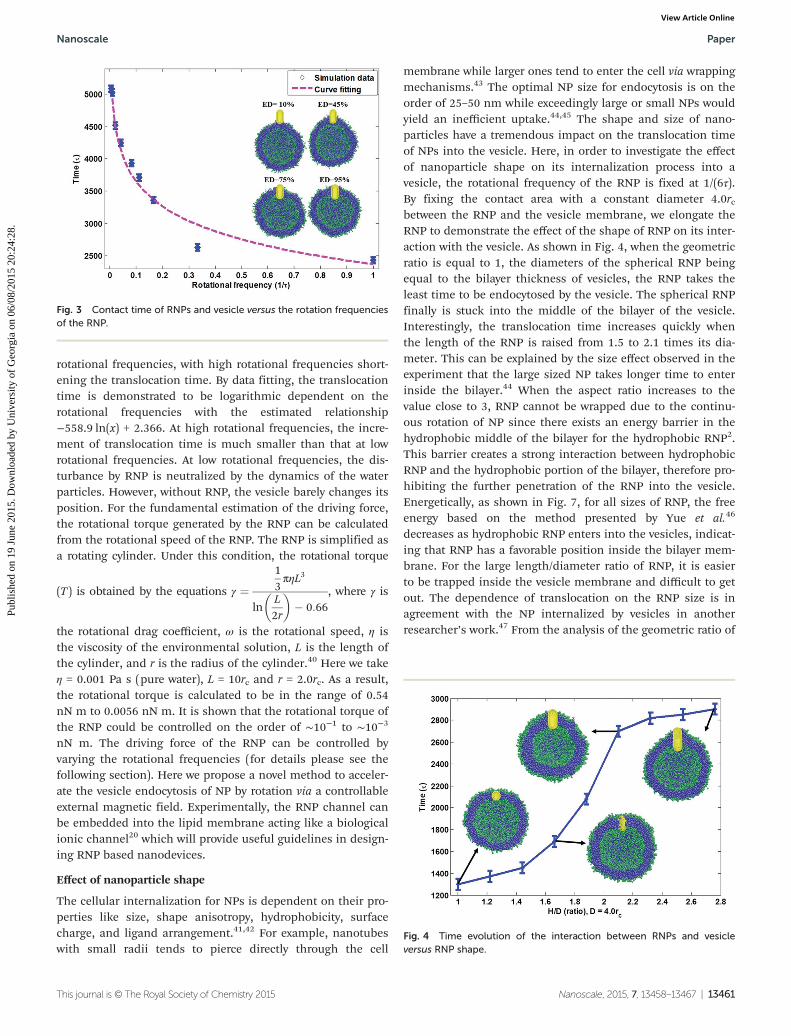
rotational frequencies, with high rotational frequencies short-ening the translocation time. By data fitting, the translocationtime is demonstrated to be logarithmic dependent on therotational frequencies with the estimated relationship−558.9 ln(x) + 2.366. At high rotational frequencies, the incre-ment of translocation time is much smaller than that at lowrotational frequencies. At low rotational frequencies, the dis-turbance by RNP is neutralized by the dynamics of the waterparticles. However, without RNP, the vesicle barely changes itsposition. For the fundamental estimation of the driving force,the rotational torque generated by the RNP can be calculatedfrom the rotational speed of the RNP. The RNP is simplified asa rotating cylinder. Under this condition, the rotational torque
(T ) is obtained by the equations γ ¼13πηL3
lnL2r
� �� 0:66
, where γ is
the rotational drag coefficient, ω is the rotational speed, η isthe viscosity of the environmental solution, L is the length ofthe cylinder, and r is the radius of the cylinder.40 Here we takeη = 0.001 Pa s (pure water), L = 10rc and r = 2.0rc. As a result,the rotational torque is calculated to be in the range of 0.54nN m to 0.0056 nN m. It is shown that the rotational torque ofthe RNP could be controlled on the order of ∼10−1 to ∼10−3
nN m. The driving force of the RNP can be controlled byvarying the rotational frequencies (for details please see thefollowing section). Here we propose a novel method to acceler-ate the vesicle endocytosis of NP by rotation via a controllableexternal magnetic field. Experimentally, the RNP channel canbe embedded into the lipid membrane acting like a biologicalionic channel20 which will provide useful guidelines in design-ing RNP based nanodevices.
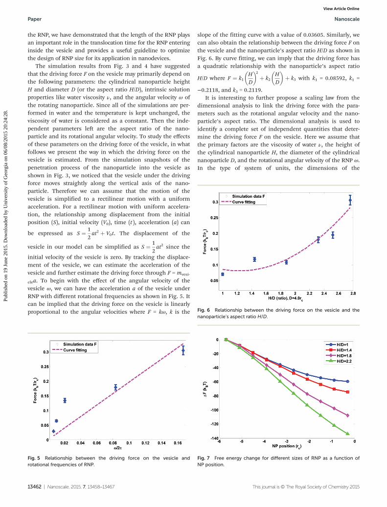
Effect of nanoparticle shape
The cellular internalization for NPs is dependent on their pro-perties like size, shape anisotropy, hydrophobicity, surfacecharge, and ligand arrangement.41,42 For example, nanotubeswith small radii tends to pierce directly through the cell
membrane while larger ones tend to enter the cell via wrappingmechanisms.43 The optimal NP size for endocytosis is on theorder of 25–50 nm while exceedingly large or small NPs wouldyield an inefficient uptake.44,45 The shape and size of nano-particles have a tremendous impact on the translocation timeof NPs into the vesicle. Here, in order to investigate the effectof nanoparticle shape on its internalization process into avesicle, the rotational frequency of the RNP is fixed at 1/(6τ).By fixing the contact area with a constant diameter 4.0rcbetween the RNP and the vesicle membrane, we elongate theRNP to demonstrate the effect of the shape of RNP on its inter-action with the vesicle. As shown in Fig. 4, when the geometricratio is equal to 1, the diameters of the spherical RNP beingequal to the bilayer thickness of vesicles, the RNP takes theleast time to be endocytosed by the vesicle. The spherical RNPfinally is stuck into the middle of the bilayer of the vesicle.Interestingly, the translocation time increases quickly whenthe length of the RNP is raised from 1.5 to 2.1 times its dia-meter. This can be explained by the size effect observed in theexperiment that the large sized NP takes longer time to enterinside the bilayer.44 When the aspect ratio increases to thevalue close to 3, RNP cannot be wrapped due to the continu-ous rotation of NP since there exists an energy barrier in thehydrophobic middle of the bilayer for the hydrophobic RNP2.This barrier creates a strong interaction between hydrophobicRNP and the hydrophobic portion of the bilayer, therefore pro-hibiting the further penetration of the RNP into the vesicle.Energetically, as shown in Fig. 7, for all sizes of RNP, the freeenergy based on the method presented by Yue et al.46
decreases as hydrophobic RNP enters into the vesicles, indicat-ing that RNP has a favorable position inside the bilayer mem-brane. For the large length/diameter ratio of RNP, it is easierto be trapped inside the vesicle membrane and difficult to getout. The dependence of translocation on the RNP size is inagreement with the NP internalized by vesicles in anotherresearcher’s work.47 From the analysis of the geometric ratio of
Fig. 3 Contact time of RNPs and vesicle versus the rotation frequenciesof the RNP.
Fig. 4 Time evolution of the interaction between RNPs and vesicleversus RNP shape.
Nanoscale Paper
This journal is © The Royal Society of Chemistry 2015 Nanoscale, 2015, 7, 13458–13467 | 13461
Publ
ishe
d on
19
June
201
5. D
ownl
oade
d by
Uni
vers
ity o
f G
eorg
ia o
n 06
/08/
2015
20:
24:2
8.
View Article Online
the RNP, we have demonstrated that the length of the RNP playsan important role in the translocation time for the RNP enteringinside the vesicle and provides a useful guideline to optimizethe design of RNP size for its application in nanodevices.
The simulation results from Fig. 3 and 4 have suggestedthat the driving force F on the vesicle may primarily depend onthe following parameters: the cylindrical nanoparticle heightH and diameter D (or the aspect ratio H/D), intrinsic solutionproperties like water viscosity ν, and the angular velocity ω ofthe rotating nanoparticle. Since all of the simulations are per-formed in water and the temperature is kept unchanged, theviscosity of water is considered as a constant. Then the inde-pendent parameters left are the aspect ratio of the nano-particle and its rotational angular velocity. To study the effectsof these parameters on the driving force of the vesicle, in whatfollows we present the way in which the driving force on thevesicle is estimated. From the simulation snapshots of thepenetration process of the nanoparticle into the vesicle asshown in Fig. 3, we noticed that the vesicle under the drivingforce moves straightly along the vertical axis of the nano-particle. Therefore we can assume that the motion of thevesicle is simplified to a rectilinear motion with a uniformacceleration. For a rectilinear motion with uniform accelera-tion, the relationship among displacement from the initialposition (S), initial velocity (V0), time (t ), acceleration (a) can
be expressed as S ¼ 12at2 þ V0t. The displacement of the
vesicle in our model can be simplified as S ¼ 12at2 since the
initial velocity of the vesicle is zero. By tracking the displace-ment of the vesicle, we can estimate the acceleration of thevesicle and further estimate the driving force through F = mvesi-
clea. To begin with the effect of the angular velocity of thevesicle ω, we can have the acceleration a of the vesicle underRNP with different rotational frequencies as shown in Fig. 5. Itcan be implied that the driving force on the vesicle is linearlyproportional to the angular velocities where F = kω, k is the
slope of the fitting curve with a value of 0.03605. Similarly, wecan also obtain the relationship between the driving force F onthe vesicle and the nanoparticle’s aspect ratio H/D as shown inFig. 6. By curve fitting, we can imply that the driving force hasa quadratic relationship with the nanoparticle’s aspect ratio
H/D where F ¼ k1HD
� �2þ k2
HD
� �þ k3 with k1 = 0.08592, k1 =
−0.2118, and k3 = 0.2119.It is interesting to further propose a scaling law from the
dimensional analysis to link the driving force with the para-meters such as the rotational angular velocity and the nano-particle’s aspect ratio. The dimensional analysis is used toidentify a complete set of independent quantities that deter-mine the driving force F on the vesicle. Here we assume thatthe primary factors are the viscosity of water ν, the height ofthe cylindrical nanoparticle H, the diameter of the cylindricalnanoparticle D, and the rotational angular velocity of the RNP ω.In the type of system of units, the dimensions of the
Fig. 5 Relationship between the driving force on the vesicle androtational frequencies of RNP.
Fig. 6 Relationship between the driving force on the vesicle and thenanoparticle’s aspect ratio H/D.
Fig. 7 Free energy change for different sizes of RNP as a function ofNP position.
Paper Nanoscale
13462 | Nanoscale, 2015, 7, 13458–13467 This journal is © The Royal Society of Chemistry 2015
Publ
ishe
d on
19
June
201
5. D
ownl
oade
d by
Uni
vers
ity o
f G
eorg
ia o
n 06
/08/
2015
20:
24:2
8.
View Article Online
quantities are: [F] = F, [ω] = T −1, [ν] = FL−2T, [D] = L, [H] = L. Wechoose F and D as dependent variables and non-dimensionalize
the remaining independent variable as Π1 ¼ FωνD2, Π2 ¼ H
D.
Using Buckingham’s π-theorem, we have the followingrelationship that expresses the driving force in terms of a com-
plete set of variablesF
ωνD2 ¼ 1HD
� �. From the simulation
data as shown in Fig. 5 and 6, we can have the relationshipbetween Π1 and Π2 and the driving force acting on the vesiclecan be theoretically estimated as
F ¼ ωνD2 k1HD
� �2
þ k2HD
� �þ k3
� �¼ ωνðk1H2 þ k2DH þ k3D2Þ
ð6Þwith k1 = 0.01026, k1 = −0.02528, and k3 = 0.0253. It impliesthat the driving force is linear with the rotational angular vel-ocity of the RNP as verified in Fig. 5 and is a parabolic functionwith the nanoparticle’s aspect ratio as evidenced in Fig. 6.
Effect of coated patterns
Due to the extremely small size, high surface energy, and highsurface area of the RNP, the interaction between cells andRNPs can be heavily influenced by intelligent surface structuredesigns.48–50 Tissue and cell-specific drug targeting by RNPscan be achieved by employing specific coatings of the RNPs orcarrier–drug conjugates which contain a ligand recognized bya receptor on the target cell.51,52 RNPs modified with syntheticligands can bind to both Gram-positive and Gram-negativebacteria to clear them and their related endotoxins from thebloodstream.53 It turns out that the surface modification is avery important controller of the interaction between RNPs and
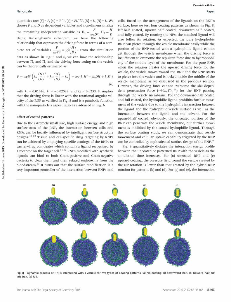
cells. Based on the arrangement of the ligands on the RNP’ssurface, here we test four coating patterns as shown in Fig. 8:left-half coated, upward-half coated, downward-half coated,and fully coated. By rotating the NPs, the attached ligand willalso follow its rotation. As expected, the pure hydrophobicRNP can pierce through the vesicle membrane easily while theportion of the RNP coated with a hydrophilic ligand cannotget through the vesicle membrane when the driving force isinsufficient to overcome the repulsive force due to hydrophobi-city of the middle layer of the membrane. For the pure RNP,when the rotation creates the upward driving force for thevesicle, the vesicle moves toward the RNP and the RNP startsto pierce into the vesicle and is locked inside the middle of thevesicle membrane as we discussed in the previous section.However, the driving force cannot overcome the size-depen-dent penetration force (∼60kBT/rc 54) for the RNP passingthrough the vesicle membrane. For the downward-half coatedand full coated, the hydrophilic ligand prohibits further move-ment of the vesicle due to the hydrophilic interaction betweenthe ligand and the hydrophilic vesicle surface as well as theinteraction between the ligand and the solvent. For theupward-half coated, obviously, the uncoated portion of theRNP can penetrate the vesicle membrane, but further move-ment is inhibited by the coated hydrophilic ligand. Throughthe surface coating study, we can demonstrate that vesiclemovement and cellular uptake capability triggered by the RNPcan be controlled by sophisticated surface design of the RNP.50
Fig. 9 quantitatively dictates the interaction energy profilebetween the uncoated or patterned RNP with the vesicle as thesimulation time increases. For (a) uncoated RNP and (c)upward coating, the pressure field round the vesicle created bythe NP rotation is lower than that created by the hybrid RNProtation for patterns (b) and (d). For (a) and (c), the interaction
Fig. 8 Dynamic process of RNPs interacting with a vesicle for five types of coating patterns. (a) No coating (b) downward-half, (c) upward-half, (d)left-half, (e) full.
Nanoscale Paper
This journal is © The Royal Society of Chemistry 2015 Nanoscale, 2015, 7, 13458–13467 | 13463
Publ
ishe
d on
19
June
201
5. D
ownl
oade
d by
Uni
vers
ity o
f G
eorg
ia o
n 06
/08/
2015
20:
24:2
8.
View Article Online
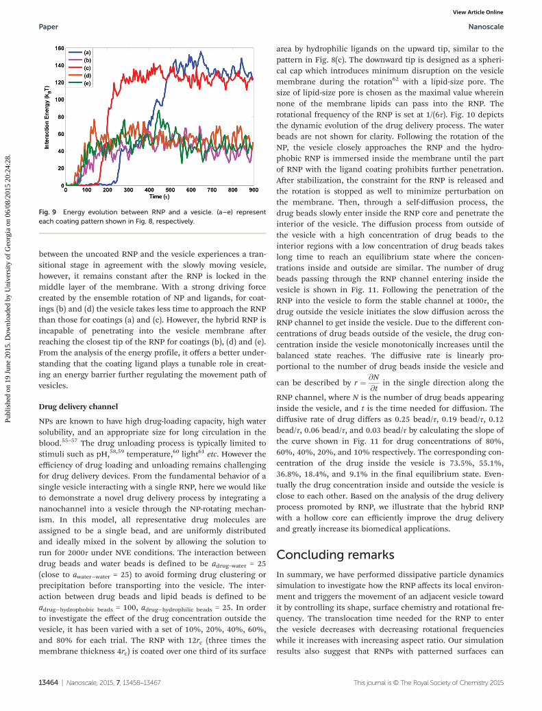
between the uncoated RNP and the vesicle experiences a tran-sitional stage in agreement with the slowly moving vesicle,however, it remains constant after the RNP is locked in themiddle layer of the membrane. With a strong driving forcecreated by the ensemble rotation of NP and ligands, for coat-ings (b) and (d) the vesicle takes less time to approach the RNPthan those for coatings (a) and (c). However, the hybrid RNP isincapable of penetrating into the vesicle membrane afterreaching the closest tip of the RNP for coatings (b), (d) and (e).From the analysis of the energy profile, it offers a better under-standing that the coating ligand plays a tunable role in creat-ing an energy barrier further regulating the movement path ofvesicles.
Drug delivery channel
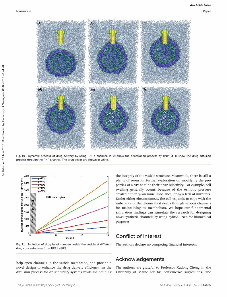
NPs are known to have high drug-loading capacity, high watersolubility, and an appropriate size for long circulation in theblood.55–57 The drug unloading process is typically limited tostimuli such as pH,58,59 temperature,60 light61 etc. However theefficiency of drug loading and unloading remains challengingfor drug delivery devices. From the fundamental behavior of asingle vesicle interacting with a single RNP, here we would liketo demonstrate a novel drug delivery process by integrating ananochannel into a vesicle through the NP-rotating mechan-ism. In this model, all representative drug molecules areassigned to be a single bead, and are uniformly distributedand ideally mixed in the solvent by allowing the solution torun for 2000τ under NVE conditions. The interaction betweendrug beads and water beads is defined to be adrug–water = 25(close to awater−water = 25) to avoid forming drug clustering orprecipitation before transporting into the vesicle. The inter-action between drug beads and lipid beads is defined to beadrug−hydrophobic beads = 100, adrug−hydrophilic beads = 25. In orderto investigate the effect of the drug concentration outside thevesicle, it has been varied with a set of 10%, 20%, 40%, 60%,and 80% for each trial. The RNP with 12rc (three times themembrane thickness 4rc) is coated over one third of its surface
area by hydrophilic ligands on the upward tip, similar to thepattern in Fig. 8(c). The downward tip is designed as a spheri-cal cap which introduces minimum disruption on the vesiclemembrane during the rotation62 with a lipid-size pore. Thesize of lipid-size pore is chosen as the maximal value whereinnone of the membrane lipids can pass into the RNP. Therotational frequency of the RNP is set at 1/(6τ). Fig. 10 depictsthe dynamic evolution of the drug delivery process. The waterbeads are not shown for clarity. Following the rotation of theNP, the vesicle closely approaches the RNP and the hydro-phobic RNP is immersed inside the membrane until the partof RNP with the ligand coating prohibits further penetration.After stabilization, the constraint for the RNP is released andthe rotation is stopped as well to minimize perturbation onthe membrane. Then, through a self-diffusion process, thedrug beads slowly enter inside the RNP core and penetrate theinterior of the vesicle. The diffusion process from outside ofthe vesicle with a high concentration of drug beads to theinterior regions with a low concentration of drug beads takeslong time to reach an equilibrium state where the concen-trations inside and outside are similar. The number of drugbeads passing through the RNP channel entering inside thevesicle is shown in Fig. 11. Following the penetration of theRNP into the vesicle to form the stable channel at 1000τ, thedrug outside the vesicle initiates the slow diffusion across theRNP channel to get inside the vesicle. Due to the different con-centrations of drug beads outside of the vesicle, the drug con-centration inside the vesicle monotonically increases until thebalanced state reaches. The diffusive rate is linearly pro-portional to the number of drug beads inside the vesicle and
can be described by r ¼ @N@t
in the single direction along the
RNP channel, where N is the number of drug beads appearinginside the vesicle, and t is the time needed for diffusion. Thediffusive rate of drug differs as 0.25 bead/τ, 0.19 bead/τ, 0.12bead/τ, 0.06 bead/τ, and 0.03 bead/τ by calculating the slope ofthe curve shown in Fig. 11 for drug concentrations of 80%,60%, 40%, 20%, and 10% respectively. The corresponding con-centration of the drug inside the vesicle is 73.5%, 55.1%,36.8%, 18.4%, and 9.1% in the final equilibrium state. Even-tually the drug concentration inside and outside the vesicle isclose to each other. Based on the analysis of the drug deliveryprocess promoted by RNP, we illustrate that the hybrid RNPwith a hollow core can efficiently improve the drug deliveryand greatly increase its biomedical applications.
Concluding remarks
In summary, we have performed dissipative particle dynamicssimulation to investigate how the RNP affects its local environ-ment and triggers the movement of an adjacent vesicle towardit by controlling its shape, surface chemistry and rotational fre-quency. The translocation time needed for the RNP to enterthe vesicle decreases with decreasing rotational frequencieswhile it increases with increasing aspect ratio. Our simulationresults also suggest that RNPs with patterned surfaces can
Fig. 9 Energy evolution between RNP and a vesicle. (a–e) representeach coating pattern shown in Fig. 8, respectively.
Paper Nanoscale
13464 | Nanoscale, 2015, 7, 13458–13467 This journal is © The Royal Society of Chemistry 2015
Publ
ishe
d on
19
June
201
5. D
ownl
oade
d by
Uni
vers
ity o
f G
eorg
ia o
n 06
/08/
2015
20:
24:2
8.
View Article Online
help open channels in the vesicle membrane, and provide anovel design to enhance the drug delivery efficiency via thediffusion process for drug delivery systems while maintaining
the integrity of the vesicle structure. Meanwhile, there is still aplenty of room for further exploration on modifying the pro-perties of RNPs to tune their drug selectivity. For example, cellswelling generally occurs because of the osmotic pressurecreated either by an ionic imbalance, or by a lack of nutrients.Under either circumstances, the cell expands to cope with theimbalance of the chemicals it needs through various channelsfor maintaining its metabolism. We hope our fundamentalsimulation findings can stimulate the research for designingnovel synthetic channels by using hybrid RNPs for biomedicalpurposes.
Conflict of interest
The authors declare no competing financial interests.
Acknowledgements
The authors are grateful to Professor Xudong Zheng in theUniversity of Maine for his constructive suggestions. The
Fig. 11 Evolution of drug bead numbers inside the vesicle at differentdrug concentrations from 10% to 80%.
Fig. 10 Dynamic process of drug delivery by using RNP’s channel. (a–e) show the penetration process by RNP. (d–f ) show the drug diffusionprocess through the RNP channel. The drug beads are shown in white.
Nanoscale Paper
This journal is © The Royal Society of Chemistry 2015 Nanoscale, 2015, 7, 13458–13467 | 13465
Publ
ishe
d on
19
June
201
5. D
ownl
oade
d by
Uni
vers
ity o
f G
eorg
ia o
n 06
/08/
2015
20:
24:2
8.
View Article Online
authors acknowledge support from the University of Georgia(UGA) start-up fund. The facility support for modeling andsimulations from the UGA Advanced Computing ResourceCenter are greatly appreciated.
References
1 L. Zhang and X. Wang, RSC Adv., 2015, 5, 11776–11785.2 Y. Li, X. Li, Z. Li and H. Gao, Nanoscale, 2012, 4, 3768–
3775.3 Y. Li, H. Yuan, A. von dem Bussche, M. Creighton,
R. H. Hurt, A. B. Kane and H. Gao, Proc. Natl. Acad.Sci. U. S. A., 2013, 110, 12295–12300.
4 C. Huang, Y. Zhang, H. Yuan, H. Gao and S. Zhang, NanoLett., 2013, 13, 4546–4550.
5 R. Vácha, F. J. Martinez-Veracoechea and D. Frenkel, NanoLett., 2011, 11, 5391–5395.
6 T. Yue, X. Zhang and F. Huang, Soft Matter, 2015, 11, 456–465.
7 E. Zhang, M. F. Kircher, M. Koch, L. Eliasson,S. N. Goldberg and E. Renström, ACS Nano, 2014, 8, 3192–3201.
8 C. Wilhelm and F. Gazeau, J. Magn. Magn. Mater., 2009,321, 671–674.
9 M. Bañobre-López, A. Teijeiro and J. Rivas, Rep. Pract.Oncol. Radiother., 2013, 18, 397–400.
10 J. Klostergaard and C. E. Seeney, Maturitas, 2012, 73,33–44.
11 B. Polyak and G. Friedman, Expert Opin. Drug Deliv., 2009,6, 53–70.
12 G. Ciofani, V. Raffa, Y. Obata, A. Menciassi, P. Dario andS. Takeoka, Curr. Nanosci., 2008, 4, 212–218.
13 J. Dobson, Drug Dev. Res., 2006, 67, 55–60.14 R. Singh and J. W. Lillard Jr., Exp. Mol. Pathol., 2009, 86,
215–223.15 F. Gazeau, M. Levy and C. Wilhelm, Nanomedicine, 2008, 3,
831–844.16 C. Sun, J. S. H. Lee and M. Zhang, Adv. Drug Delivery Rev.,
2008, 60, 1252–1265.17 Z. Zhao, D. Huang, Z. Yin, X. Chi, X. Wang and J. Gao,
J. Mater. Chem., 2012, 22, 15717–15725.18 V. Mody, A. Cox, S. Shah, A. Singh, W. Bevins and
H. Parihar, Appl. Nanosci., 2014, 4, 385–392.19 K. Hayashi, K. Ono, H. Suzuki, M. Sawada, M. Moriya,
W. Sakamoto and T. Yogo, ACS Appl. Mater. Interfaces, 2010,2, 1903–1911.
20 J. Geng, K. Kim, J. Zhang, A. Escalada, R. Tunuguntla,L. R. Comolli, F. I. Allen, A. V. Shnyrova, K. R. Cho,D. Munoz, Y. M. Wang, C. P. Grigoropoulos, C. M. Ajo-Franklin, V. A. Frolov and A. Noy, Nature, 2014, 514, 612–615.
21 L. Franceschini, M. Soskine, A. Biesemans and G. Maglia,Nat. Commun., 2013, 4, 2415.
22 R. D. Groot and K. L. Rabone, Biophys. J., 2001, 81, 725–736.23 R. D. Groot and P. B. Warren, J. Chem. Phys., 1997, 107,
4423–4435.
24 X. Li, Y. Liu, L. Wang, M. Deng and H. Liang, Phys. Chem.Chem. Phys., 2009, 11, 4051–4059.
25 J. C. Shillcock and R. Lipowsky, Nat. Mater., 2005, 4, 225–228.
26 S. Plimpton, J. Comput. Phys., 1995, 117, 1–19.27 S. O. Nielsen, B. Ensing, V. Ortiz, P. B. Moore and
M. L. Klein, Biophys. J., 2005, 88, 3822–3828.28 M. Venturoli, B. Smit and M. M. Sperotto, Biophys. J., 2005,
88, 1778–1798.29 N. C. Darnton, L. Turner, S. Rojevsky and H. C. Berg,
J. Bacteriol., 2007, 189, 1756–1764.30 W. R. DiLuzio, L. Turner, M. Mayer, P. Garstecki,
D. B. Weibel, H. C. Berg and G. M. Whitesides, Nature,2005, 435, 1271–1274.
31 L. Zhang, J. J. Abbott, L. Dong, K. E. Peyer, B. E. Kratochvil,H. Zhang, C. Bergeles and B. J. Nelson, Nano Lett., 2009, 9,3663–3667.
32 T. Yue, S. Li, X. Zhang and W. Wang, Soft Matter, 2010, 6,6109–6118.
33 C. M. Hangarter and N. V. Myung, Chem. Mater., 2005, 17,1320–1324.
34 M. Fujiwara, E. Oki, M. Hamada, Y. Tanimoto, I. Mukoudaand Y. Shimomura, J. Phys. Chem. A, 2001, 105, 4383–4386.
35 X. Jia, W. Li, X. Xu, W. Li, Q. Cai and X. Yang, ACS Appl.Mater. Interfaces, 2015, 7, 3170–3179.
36 K. Kim, X. Xu, J. Guo and D. L. Fan, Nat. Commun., 2014, 5,3632.
37 A. Ghosh and P. Fischer, Nano Lett., 2009, 9, 2243–2245.
38 F. M. White, Viscous Fluid Flow, McGraw-Hill, New York,1991.
39 J. Feng, H. Ding, C. Ren and Y. Ma, Nanoscale, 2014, 6,13606–13612.
40 J. Howard, Mechanics of Motor Proteins and the Cytoskeleton,Palgrave Macmillan, London, 2001.
41 E. C. Wang and A. Z. Wang, Integr. Biol., 2014, 6, 9–26.42 H. Gao, J. Mech. Phys. Solids, 2014, 62, 312–339.43 X. Shi, Y. Kong and H. Gao, Acta Mech. Sin., 2008, 24, 161–
169.44 S. Zhang, J. Li, G. Lykotrafitis, G. Bao and S. Suresh, Adv.
Mater., 2009, 21, 419–424.45 H. Gao, W. Shi and L. B. Freund, Proc. Natl. Acad. Sci.
U. S. A., 2005, 102, 9469–9474.46 T. Yue, X. Zhang and F. Huang, Soft Matter, 2014, 10, 2024–
2034.47 X. Chen, F. Tian, X. Zhang and W. Wang, Soft Matter, 2013,
9, 7592–7600.48 A. Verma, O. Uzun, Y. Hu, Y. Hu, H.-S. Han, N. Watson,
S. Chen, D. J. Irvine and F. Stellacci, Nat. Mater., 2008, 7,588–595.
49 A. E. Nel, L. Madler, D. Velegol, T. Xia, E. M. V. Hoek,P. Somasundaran, F. Klaessig, V. Castranova andM. Thompson, Nat. Mater., 2009, 8, 543–557.
50 L. Zhang, M. Becton and X. Wang, J. Phys. Chem. B, 2015,119, 3786–3794.
Paper Nanoscale
13466 | Nanoscale, 2015, 7, 13458–13467 This journal is © The Royal Society of Chemistry 2015
Publ
ishe
d on
19
June
201
5. D
ownl
oade
d by
Uni
vers
ity o
f G
eorg
ia o
n 06
/08/
2015
20:
24:2
8.
View Article Online
51 A. K. Gupta, C. Berry, M. Gupta and A. Curtis, IEEE Trans.Nanobiosci., 2003, 2, 255–261.
52 K. E. Scarberry, E. B. Dickerson, J. F. McDonald andZ. J. Zhang, J. Am. Chem. Soc., 2008, 130, 10258–10262.
53 J.-J. Lee, K. J. Jeong, M. Hashimoto, A. H. Kwon, A. Rwei,S. A. Shankarappa, J. H. Tsui and D. S. Kohane, Nano Lett.,2013, 14, 1–5.
54 K. Yang and Y.-Q. Ma, Nat. Nanotechnol., 2010, 5, 579–583.55 Z. Ahmad, A. Shah, M. Siddiq and H. B. Kraatz, RSC Adv.,
2014, 4, 17028–17038.56 N. Nasongkla, E. Bey, J. Ren, H. Ai, C. Khemtong,
J. S. Guthi, S.-F. Chin, A. D. Sherry, D. A. Boothman andJ. Gao, Nano Lett., 2006, 6, 2427–2430.
57 K. Miyata, R. J. Christie and K. Kataoka, React. Funct.Polym., 2011, 71, 227–234.
58 H. Ding and Y. Ma, Sci. Rep., 2013, 3, 2804.59 K. Huh, H. Kang, Y. Lee and Y. Bae, Macromol. Res., 2012,
20, 224–233.60 A. A. Manzoor, L. H. Lindner, C. D. Landon, J. Y. Park,
A. J. Simnick, M. R. Dreher, S. Das, G. Hanna, W. Park,A. Chilkoti, G. A. Koning, T. L. ten Hagen, D. Needham andM. W. Dewhirst, Cancer Res., 2012, 72, 5566–5575.
61 S. Kumar, J.-F. Allard, D. Morris, Y. L. Dory, M. Lepage andY. Zhao, J. Mater. Chem., 2012, 22, 7252–7257.
62 X. Shi, A. von dem Bussche, R. H. Hurt, A. B. Kane andH. Gao, Nat. Nanotechnol., 2011, 6, 714–719.
Nanoscale Paper
This journal is © The Royal Society of Chemistry 2015 Nanoscale, 2015, 7, 13458–13467 | 13467
Publ
ishe
d on
19
June
201
5. D
ownl
oade
d by
Uni
vers
ity o
f G
eorg
ia o
n 06
/08/
2015
20:
24:2
8.
View Article Online