cme the montreal definition and classification of ...vakil).pdf · the montreal definition and...
TRANSCRIPT
American Journal of Gastroenterology ISSN 0002-9270C© 2006 by Am. Coll. of Gastroenterology doi: 10.1111/j.1572-0241.2006.00630.xPublished by Blackwell Publishing
The Montreal Definition and Classification ofGastroesophageal Reflux Disease: A GlobalEvidence-Based Consensus
CME
Nimish Vakil, M.D., F.A.C.G.,1 Sander V. van Zanten, M.D.,2 Peter Kahrilas, M.D.,3 John Dent, M.D.,4 RogerJones, M.D.,5 and the Global Consensus Group1University of Wisconsin School of Medicine and Public Health, Madison, Wisconsin and Marquette UniversityCollege of Health Sciences, Milwaukee, Wisconsin; 2Dalhousie University, Halifax, Nova Scotia, Canada;3Northwestern University, Chicago, Illinois; 4University of Adelaide, Adelaide, Australia; and 5Kings College,London, United Kingdom
OBJECTIVES: A globally acceptable definition and classification of gastroesophageal reflux disease (GERD) isdesirable for research and clinical practice. The aim of this initiative was to develop a consensusdefinition and classification that would be useful for patients, physicians, and regulatory agencies.
METHODS: A modified Delphi process was employed to reach consensus using repeated iterative voting. Aseries of statements was developed by a working group of five experts after a systematic review ofthe literature in three databases (Embase, Cochrane trials register, Medline). Over a period of 2 yr,the statements were developed, modified, and approved through four rounds of voting. The votinggroup consisted of 44 experts from 18 countries. The final vote was conducted on a 6-point scaleand consensus was defined a priori as agreement by two-thirds of the participants.
RESULTS: The level of agreement strengthened throughout the process with two-thirds of the participantsagreeing with 86%, 88%, 94%, and 100% of statements at each vote, respectively. At the final vote,94% of the final 51 statements were approved by 90% of the Consensus Group, and 90% ofstatements were accepted with strong agreement or minor reservation. GERD was defined as acondition that develops when the reflux of stomach contents causes troublesome symptoms and/orcomplications. The disease was subclassified into esophageal and extraesophageal syndromes.Novel aspects of the new definition include a patient-centered approach that is independent ofendoscopic findings, subclassification of the disease into discrete syndromes, and the recognition oflaryngitis, cough, asthma, and dental erosions as possible GERD syndromes. It also proposes a newdefinition for suspected and proven Barrett’s esophagus.
CONCLUSIONS: Evidence-based global consensus definitions are possible despite differences in terminology andlanguage, prevalence, and manifestations of the disease in different countries. A global consensusdefinition for GERD may simplify disease management, allow collaborative research, and makestudies more generalizable, assisting patients, physicians, and regulatory agencies.
(Am J Gastroenterol 2006;101:1900–1920)
INTRODUCTION
A number of guidelines and recommendations for the di-agnosis and management of gastroesophageal reflux disease(GERD) have been published in different countries, but a uni-versally accepted definition of GERD and its various symp-toms and complications is lacking (1–9). Reflux symptomsare common in primary care and GERD is frequently diag-nosed based on symptoms alone, but there is no consensuson the distinction of GERD from dyspepsia, so that theseterms may lead to confusion in primary care settings. This
To access a continuing medical education exam for this article, please visitwww.acg.gi.org/journalcme.
has led some authorities to combine these entities in pri-mary care management strategies (10). There is also uncer-tainty about the extraesophageal manifestations of GERD,coupled with an expanding list of putative extraesophagealdisorders, resulting in both over- and underdiagnosisof the disease. Finally, the definition of Barrett’s esophagusvaries in different regions of the world, causing confusionin the assessment of risk and the appropriate use of surve-illance.
The aim of this international Consensus Group was to de-velop a global definition and classification of GERD, us-ing rigorous methodology, that could be used clinically byprimary care physicians and that embraces the needs of
1900
The Montreal Definition and Classification of GERD 1901
physicians, patients, researchers, and regulatory bodies fromdifferent parts of the world.
METHODS
A modified Delphi process was used to develop the con-sensus definition of GERD (11–13). The Delphi pro-cess is a method for developing consensus that has beenused for complex problems in medicine and industry. Anovel aspect of this endeavor was the combination of theprinciples of evidence-based medicine, supported by sys-tematic literature reviews, with the Delphi process. A keyelement of the Delphi process is the use of anonymous vot-ing, which allows a change of views from a previously heldposition without embarrassment, together with controlledfeedback regulated by a nonvoting chairman that preventsthe process from being hijacked by a vocal minority. Sys-tematic literature reviews were chosen to support the ev-idence base as this orientates the consensus process awayfrom clinical opinion to methodologically sound evidence.Multiple iterations of the statements that make up the def-inition and classification were created until consensus wasreached.
The principal steps in the process were: (1) Selection ofthe Consensus Group and development of draft statements bya Working Group; (2) Systematic literature reviews to iden-tify the evidence to support each statement; (3) Grading ofthe evidence; (4) Voting discussion and repeated anonymousvoting on a series of iterations of the statements until a con-sensus was reached. Each of these steps is described in moredetail below.
Consensus Group SelectionMembers of the Consensus Group were selected using severalcriteria:
1. Demonstrated knowledge/expertise in GERD by publi-cation/research or participation in national or regionalGERD consensus guidelines or an interest in guidelinedevelopment and dissemination.
2. Geographical considerations: individuals who met the cri-teria under (1) were then invited to provide broad repre-sentation of different regions of the world (North America,South America, Asia, Europe, Australia) that have differ-ences in prevalence and manifestation.
3. Diversity of views and expertise related to GERD (includ-ing experts in Barrett’s esophagus, surgeons, and primarycare physicians).
The Consensus Group was led by a nonvoting chairman(NV). The Working Group, who are the primary authors ofthis article, developed the initial statements and prepared andreviewed the evidence to support the statements that were pre-sented to the Consensus Group. The Consensus Group, whichincluded the Working Group, consisted of 44 experts from18 countries: Argentina, Australia, Belgium, Brazil, Canada,
China, Denmark, France, Germany, Hong Kong, Italy, Japan,Mexico, Netherlands, Peru, Sweden, United Kingdom, andthe United States.
Systematic SearchesSystematic literature reviews, with defined inclusion andexclusion criteria, were conducted to identify and gradethe available evidence to support each statement. Literaturesearches were conducted of English language publications inMedline, Embase, and the Cochrane trials register, in humansubjects from 1980 onwards. Searches of meeting abstracts(American College of Gastroenterology, American Gastroen-terological Association, British Society of Gastroenterology,United European Gastroenterology Week) and review arti-cles were limited to the preceding 2 yr. A number of searchstrings were used that are too numerous to list in the arti-cle. A complete list of the search strings may be obtained bycommunicating with the lead author of this article. Due to thelarge number of citations retrieved on each of the topics, theprimary reviewer reviewed each of the abstracts and selectedarticles and meeting abstracts for further review. The reviewwas qualitative and the primary reviewer reached an assess-ment on the grade assigned to the statement that was thenreviewed in the Working Group. Quantitative meta-analyseswere not performed. The references cited in this article area fraction of the articles reviewed in each area and were se-lected to amplify the statements and the discussion in theWorking Group.
Grades of EvidenceAssignment of the grade of evidence for each statement,where applicable, employed the GRADE system, which takesinto account the type of evidence while increasing or decreas-ing the grade depending on the quality of the study and data(14). The final grade provides a practical indication of thelikely impact of further research on confidence in the esti-mate of effect. The grading of evidence is as follows:
� High: Further research is unlikely to change our confidencein the estimate of effect.
� Moderate: Further research is likely to have an importantimpact on our confidence in the estimate of effect and maychange the estimate.
� Low: Further research is likely to have an important impacton our confidence in the estimate of effect and is very likelyto change the estimate.
� Very low: Any estimate of effect is uncertain.
An initial assessment of grade was made by the primaryreviewer of the topic from within the Working Group. The as-signed grade was then discussed within the Working Groupand a final determination of grade was made. Assignmentof grade was not voted upon in the broader ConsensusGroup. A grade of not applicable was chosen for defini-tions or statements that cannot be influenced by research. For
1902 Vakil et al.
example a cluster of symptoms that is defined as a syndromeis an arbitrary designation and cannot be altered by re-search.
VotingThe entire process lasted 2 yr and the Consensus Group votedon four iterations of the statements. Between each of thefour votes, statements were revised by the Working Groupbased on feedback from the Consensus Group and additionalliterature reviews. All votes were anonymous.
1. A first vote (baseline) was conducted for the entire Con-sensus Group electronically (by e-mail), without expla-nation or justification of the statements, and the resultswere collated (Vote 1). Feedback on the statements wassolicited.
2. A meeting of the entire Consensus Group was held todiscuss suggested modifications based on feedback fromthe first vote and to review and discuss the evidence tosupport specific statements. Subsequently, a second votewas held, using electronic keypads to ensure anonymity(Vote 2).
3. Focus subgroups were created within the ConsensusGroup to address controversies in Barrett’s esophagusand extraesophageal syndromes. Statements were againrevised, this time with input from the focus subgroups. Athird electronic vote was conducted by e-mail (Vote 3).
4. A final Consensus Group meeting was held and the com-plete results of the previous votes were reviewed, followedby an open discussion of all statements, including focusedpresentations on those statements where there was stilllack of consensus. This culminated in the fourth and finalvote, using keypads (Vote 4).
Regulatory agencies were invited to the initiative and theEuropean Medicines Agency was represented by a nonvotingobserver at the final Consensus Group meeting.
For the first two votes, a simple 2-point scale(agree/disagree) was used in order to rapidly identify areaswhere consensus/lack of consensus existed. For the third andfourth votes, a 6-point Likert scale was used: 1, agree strongly(A+); 2, agree with minor reservation (A); 3, agree withmajor reservation (A−); 4, disagree with major reservation(D−); 5, disagree with minor reservation (D); 6, disagreestrongly (D+). Agreement with a statement (A+, A, or A−)by two-thirds (i.e., ≥67%) of the group was defined a priorias consensus. The level of agreement in the final vote is givenfor each statement, expressed as the percentage vote at eachpoint on the Likert scale.
Funding SourcesThe process was funded by an unrestricted grant from Astra-Zeneca Research and Development. The European MedicinesAgency was responsible for the costs of their observer.
Endorsement by the World Organization ofGastroenterologyThe final document was endorsed by the World Organizationof Gastroenterology (WGO-OMGE) as “an important devel-opment in a critical area of gastroenterology worldwide.”“Montreal” is in the title because the results of the study werefirst presented at the World Congress of Gastroenterology inMontreal.
RESULTS AND DISCUSSION
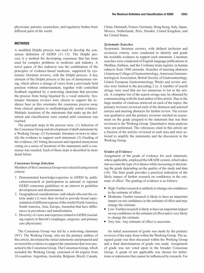
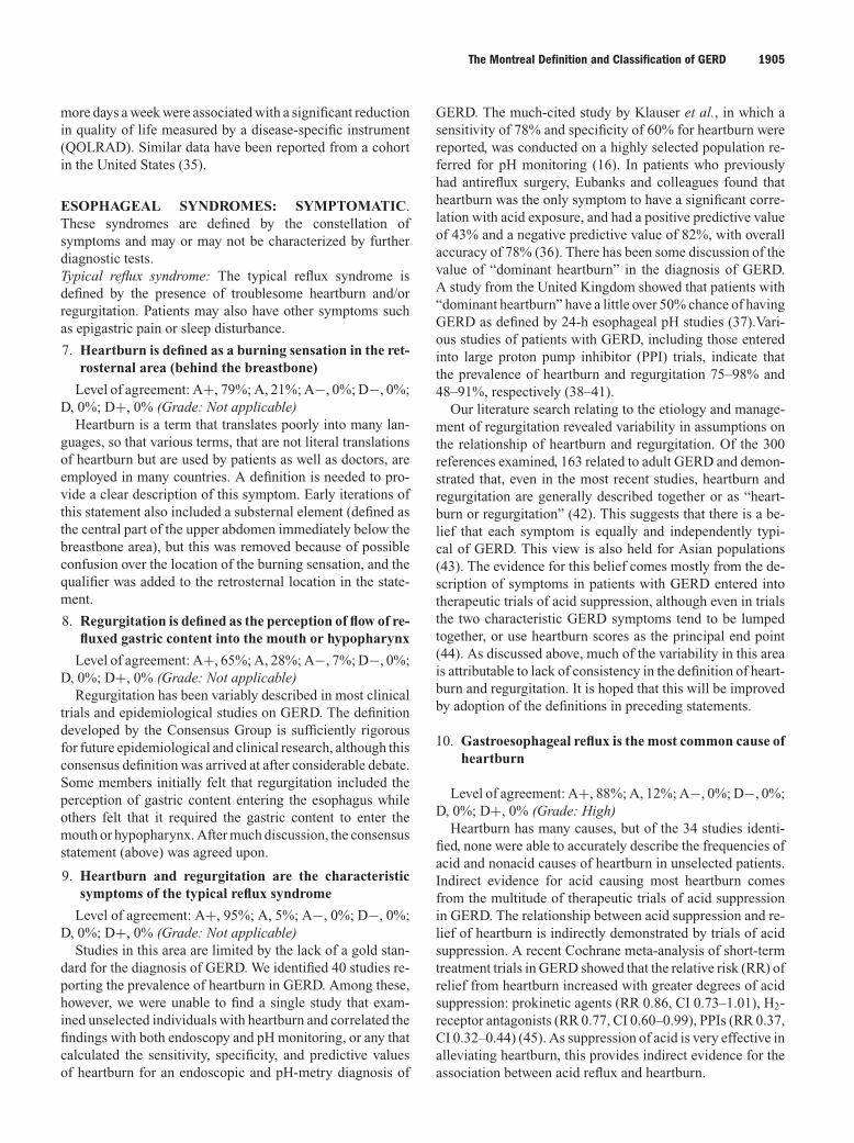
Overview of the Voting on StatementsA total of 57 statements were presented for the baseline Vote1 and, following discussion of the supporting evidence, forVote 2. The statements were subsequently revised and consol-idated, providing 53 statements for Vote 3. Further discussionand modification at the final Consensus Group meeting re-sulted in 51 statements for the final Vote 4.
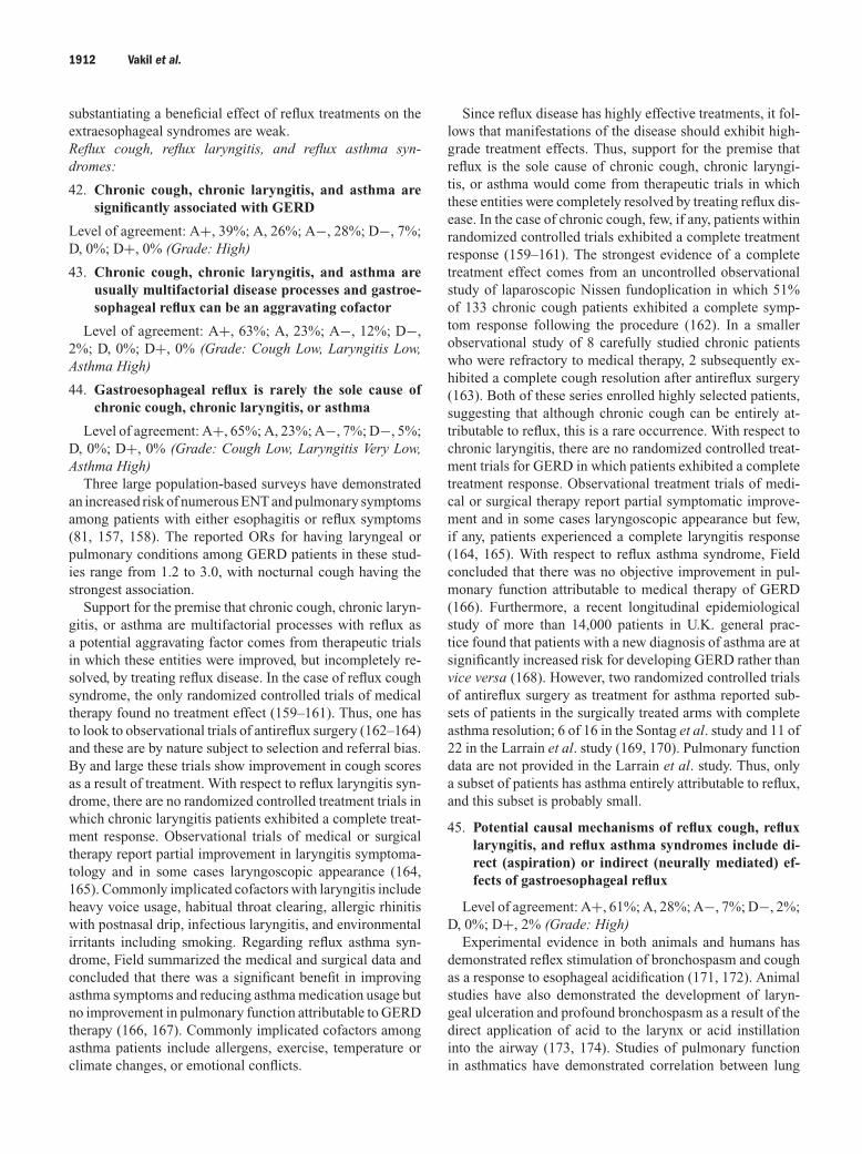
The level of consensus increased with each round of vot-ing, with a high level of consensus in the fourth and finalvote (Fig. 1). At each of the four votes, there was consen-sus (agreement by ≥67% of the group) on 86%, 88%, 94%,and finally 100% of statements, respectively. Over 90% ofthe group agreed with 94% (48) of the 51 final statements.Moreover the strength of agreement was very high by the finalvote, as illustrated by the average percentage vote across thefinal 51 statements at each level of the 6-point Likert scale(Table 1). Following the final vote it became apparent thatone statement had become redundant as it was already ad-dressed in a preceding statement. Consequently, statementsand accompanying commentary are given for 50 rather than51 statements.
Voting on the Process and Sponsor InfluenceAnonymous votes were also obtained on the Delphi processand the influence of the sponsor on the outcome. Ninety per-cent of participants agreed that the voting process was fairand that they had a chance to input adequately. Ninety-two
0
10
20
30
40
50
60
70
80
90
100
Vote 1 Vote 2 Vote 3 Vote 4
>67% agreement>75% agreement>85% agreement>90% agreement
%
Figure 1. Percentage of statements at each level of agreement ateach vote.
The Montreal Definition and Classification of GERD 1903
Table 1. The Average Percentage for the Final Vote, Across the Final51 Statements, at Each Level of the 6-Point Scale
A+ Agree strongly 67.2%A Agree with minor reservation 23.4%A− Agree with major reservation 6.7%D− Disagree with major reservation 1.5%D Disagree with minor reservation 0.9%D+ Disagree strongly 0.3%
percent of the participants agreed that the sponsor had not,in any way, influenced their voting
THE GLOBAL DEFINITION OF GERD.
1. GERD is a condition which develops when the re-flux of stomach contents causes troublesome symp-toms and/or complications
Level of agreement: A+, 81%; A, 14%; A−, 5%; D−, 0%;D, 0%; D+, 0% (Grade: Not applicable)
We used the general definition of a disease to arrive at adefinition of GERD, i.e., a disease is defined as a morbidentity characterized usually by at least two of these criteria:(1) recognized etiologic agent(s); (2) identifiable group ofsigns and symptoms; (3) consistent anatomic alterations (15).We considered a number of descriptive terms before choosing“troublesome” because it satisfactorily describes the negativeaspects of the symptoms from a patient’s standpoint, allowsitself to be translated into a number of languages, and rec-ognizes the variability in how symptoms impact individualpatients. The group recognized that the characteristic symp-toms of GERD are retrosternal burning (often labeled heart-burn) and regurgitation, and the most common manifestationof esophageal injury is reflux esophagitis (16–18).
The language of the definition is designed to allow asymp-tomatic patients with complications such as Barrett’s esoph-agus to be included in the case-definition of GERD, and beindependent of technology used to achieve a diagnosis. Forexample, patients may be diagnosed based on typical symp-toms alone or on the basis of investigations that demonstratereflux of stomach contents (e.g., pH testing, impedence mon-itoring) or the injurious effects of the reflux (endoscopy, his-tology, electron microscopy), in the presence of typical oratypical symptoms or complications (19, 20). The new def-inition also recognizes that the refluxate causing symptomsmay be weakly acidic or gaseous and these patients also meetthe case-definition of GERD.
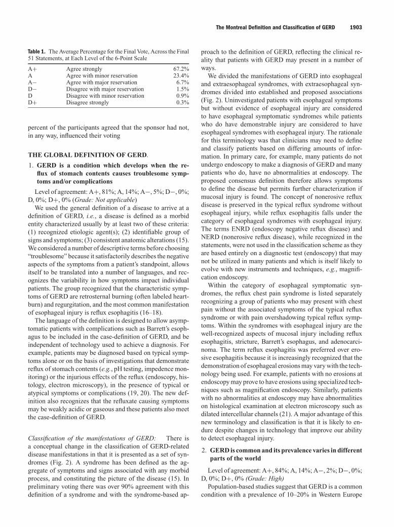
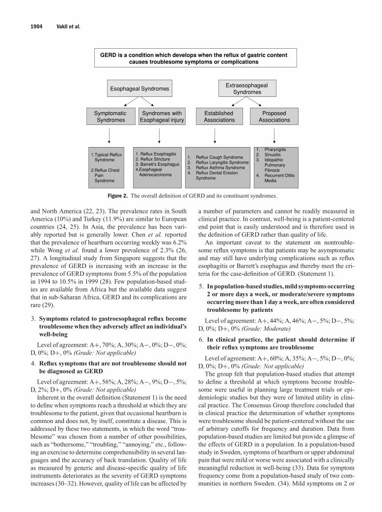
Classification of the manifestations of GERD: There isa conceptual change in the classification of GERD-relateddisease manifestations in that it is presented as a set of syn-dromes (Fig. 2). A syndrome has been defined as the ag-gregate of symptoms and signs associated with any morbidprocess, and constituting the picture of the disease (15). Inpreliminary voting there was over 90% agreement with thisdefinition of a syndrome and with the syndrome-based ap-
proach to the definition of GERD, reflecting the clinical re-ality that patients with GERD may present in a number ofways.
We divided the manifestations of GERD into esophagealand extraesophageal syndromes, with extraesophageal syn-dromes divided into established and proposed associations(Fig. 2). Uninvestigated patients with esophageal symptomsbut without evidence of esophageal injury are consideredto have esophageal symptomatic syndromes while patientswho do have demonstrable injury are considered to haveesophageal syndromes with esophageal injury. The rationalefor this terminology was that clinicians may need to defineand classify patients based on differing amounts of infor-mation. In primary care, for example, many patients do notundergo endoscopy to make a diagnosis of GERD and manypatients who do, have no abnormalities at endoscopy. Theproposed consensus definition therefore allows symptomsto define the disease but permits further characterization ifmucosal injury is found. The concept of nonerosive refluxdisease is preserved in the typical reflux syndrome withoutesophageal injury, while reflux esophagitis falls under thecategory of esophageal syndromes with esophageal injury.The terms ENRD (endoscopy negative reflux disease) andNERD (nonerosive reflux disease), while recognized in thestatements, were not used in the classification scheme as theyare based entirely on a diagnostic test (endoscopy) that maynot be utilized in many patients and which is itself likely toevolve with new instruments and techniques, e.g., magnifi-cation endoscopy.
Within the category of esophageal symptomatic syn-dromes, the reflux chest pain syndrome is listed separatelyrecognizing a group of patients who may present with chestpain without the associated symptoms of the typical refluxsyndrome or with pain overshadowing typical reflux symp-toms. Within the syndromes with esophageal injury are thewell-recognized aspects of mucosal injury including refluxesophagitis, stricture, Barrett’s esophagus, and adenocarci-noma. The term reflux esophagitis was preferred over ero-sive esophagitis because it is increasingly recognized that thedemonstration of esophageal erosions may vary with the tech-nology being used. For example, patients with no erosions atendoscopy may prove to have erosions using specialized tech-niques such as magnification endoscopy. Similarly, patientswith no abnormalities at endoscopy may have abnormalitieson histological examination at electron microscopy such asdilated intercellular channels (21). A major advantage of thisnew terminology and classification is that it is likely to en-dure despite changes in technology that improve our abilityto detect esophageal injury.
2. GERD is common and its prevalence varies in differentparts of the world
Level of agreement: A+, 84%; A, 14%; A−, 2%; D−, 0%;D, 0%; D+, 0% (Grade: High)
Population-based studies suggest that GERD is a commoncondition with a prevalence of 10–20% in Western Europe
1904 Vakil et al.
GERD is a condition which develops when the reflux of gastric contentcauses troublesome symptoms or complications
Esophageal SyndromesExtraesophageal
Syndromes
SymptomaticSyndromes
Syndromes withEsophageal injury
EstablishedAssociations
ProposedAssociations
1.Typical RefluxSyndrome
2.Reflux ChestPainSyndrome
1. Reflux Esophagitis2. Reflux Stricture3. Barrett’s Esophagus4.Esophageal
Adenocarcinoma
1. Reflux Cough Syndrome2. Reflux Laryngitis Syndrome3. Reflux Asthma Syndrome4. Reflux Dental Erosion
Syndrome
1. Pharyngitis2. Sinusitis3. Idiopathic
PulmonaryFibrosis
4. Recurrent OtitisMedia
Figure 2. The overall definition of GERD and its constituent syndromes.
and North America (22, 23). The prevalence rates in SouthAmerica (10%) and Turkey (11.9%) are similar to Europeancountries (24, 25). In Asia, the prevalence has been vari-ably reported but is generally lower. Chen et al. reportedthat the prevalence of heartburn occurring weekly was 6.2%while Wong et al. found a lower prevalence of 2.3% (26,27). A longitudinal study from Singapore suggests that theprevalence of GERD is increasing with an increase in theprevalence of GERD symptoms from 5.5% of the populationin 1994 to 10.5% in 1999 (28). Few population-based stud-ies are available from Africa but the available data suggestthat in sub-Saharan Africa, GERD and its complications arerare (29).
3. Symptoms related to gastroesophageal reflux becometroublesome when they adversely affect an individual’swell-being
Level of agreement: A+, 70%; A, 30%; A−, 0%; D−, 0%;D, 0%; D+, 0% (Grade: Not applicable)
4. Reflux symptoms that are not troublesome should notbe diagnosed as GERD
Level of agreement: A+, 56%; A, 28%; A−, 9%; D−, 5%;D, 2%; D+, 0% (Grade: Not applicable)
Inherent in the overall definition (Statement 1) is the needto define when symptoms reach a threshold at which they aretroublesome to the patient, given that occasional heartburn iscommon and does not, by itself, constitute a disease. This isaddressed by these two statements, in which the word “trou-blesome” was chosen from a number of other possibilities,such as “bothersome,” “troubling,” “annoying,” etc., follow-ing an exercise to determine comprehensibility in several lan-guages and the accuracy of back translation. Quality of lifeas measured by generic and disease-specific quality of lifeinstruments deteriorates as the severity of GERD symptomsincreases (30–32). However, quality of life can be affected by
a number of parameters and cannot be readily measured inclinical practice. In contrast, well-being is a patient-centeredend point that is easily understood and is therefore used inthe definition of GERD rather than quality of life.
An important caveat to the statement on nontrouble-some reflux symptoms is that patients may be asymptomaticand may still have underlying complications such as refluxesophagitis or Barrett’s esophagus and thereby meet the cri-teria for the case-definition of GERD. (Statement 1).
5. In population-based studies, mild symptoms occurring2 or more days a week, or moderate/severe symptomsoccurring more than 1 day a week, are often consideredtroublesome by patients
Level of agreement: A+, 44%; A, 46%; A−, 5%; D−, 5%;D, 0%; D+, 0% (Grade: Moderate)
6. In clinical practice, the patient should determine iftheir reflux symptoms are troublesome
Level of agreement: A+, 60%; A, 35%; A−, 5%; D−, 0%;D, 0%; D+, 0% (Grade: Not applicable)
The group felt that population-based studies that attemptto define a threshold at which symptoms become trouble-some were useful in planning large treatment trials or epi-demiologic studies but they were of limited utility in clini-cal practice. The Consensus Group therefore concluded thatin clinical practice the determination of whether symptomswere troublesome should be patient-centered without the useof arbitrary cutoffs for frequency and duration. Data frompopulation-based studies are limited but provide a glimpse ofthe effects of GERD in a population. In a population-basedstudy in Sweden, symptoms of heartburn or upper abdominalpain that were mild or worse were associated with a clinicallymeaningful reduction in well-being (33). Data for symptomfrequency come from a population-based study of two com-munities in northern Sweden. (34). Mild symptoms on 2 or
The Montreal Definition and Classification of GERD 1905
more days a week were associated with a significant reductionin quality of life measured by a disease-specific instrument(QOLRAD). Similar data have been reported from a cohortin the United States (35).
ESOPHAGEAL SYNDROMES: SYMPTOMATIC.These syndromes are defined by the constellation ofsymptoms and may or may not be characterized by furtherdiagnostic tests.Typical reflux syndrome: The typical reflux syndrome isdefined by the presence of troublesome heartburn and/orregurgitation. Patients may also have other symptoms suchas epigastric pain or sleep disturbance.
7. Heartburn is defined as a burning sensation in the ret-rosternal area (behind the breastbone)
Level of agreement: A+, 79%; A, 21%; A−, 0%; D−, 0%;D, 0%; D+, 0% (Grade: Not applicable)
Heartburn is a term that translates poorly into many lan-guages, so that various terms, that are not literal translationsof heartburn but are used by patients as well as doctors, areemployed in many countries. A definition is needed to pro-vide a clear description of this symptom. Early iterations ofthis statement also included a substernal element (defined asthe central part of the upper abdomen immediately below thebreastbone area), but this was removed because of possibleconfusion over the location of the burning sensation, and thequalifier was added to the retrosternal location in the state-ment.
8. Regurgitation is defined as the perception of flow of re-fluxed gastric content into the mouth or hypopharynx
Level of agreement: A+, 65%; A, 28%; A−, 7%; D−, 0%;D, 0%; D+, 0% (Grade: Not applicable)
Regurgitation has been variably described in most clinicaltrials and epidemiological studies on GERD. The definitiondeveloped by the Consensus Group is sufficiently rigorousfor future epidemiological and clinical research, although thisconsensus definition was arrived at after considerable debate.Some members initially felt that regurgitation included theperception of gastric content entering the esophagus whileothers felt that it required the gastric content to enter themouth or hypopharynx. After much discussion, the consensusstatement (above) was agreed upon.
9. Heartburn and regurgitation are the characteristicsymptoms of the typical reflux syndrome
Level of agreement: A+, 95%; A, 5%; A−, 0%; D−, 0%;D, 0%; D+, 0% (Grade: Not applicable)
Studies in this area are limited by the lack of a gold stan-dard for the diagnosis of GERD. We identified 40 studies re-porting the prevalence of heartburn in GERD. Among these,however, we were unable to find a single study that exam-ined unselected individuals with heartburn and correlated thefindings with both endoscopy and pH monitoring, or any thatcalculated the sensitivity, specificity, and predictive valuesof heartburn for an endoscopic and pH-metry diagnosis of
GERD. The much-cited study by Klauser et al., in which asensitivity of 78% and specificity of 60% for heartburn werereported, was conducted on a highly selected population re-ferred for pH monitoring (16). In patients who previouslyhad antireflux surgery, Eubanks and colleagues found thatheartburn was the only symptom to have a significant corre-lation with acid exposure, and had a positive predictive valueof 43% and a negative predictive value of 82%, with overallaccuracy of 78% (36). There has been some discussion of thevalue of “dominant heartburn” in the diagnosis of GERD.A study from the United Kingdom showed that patients with“dominant heartburn” have a little over 50% chance of havingGERD as defined by 24-h esophageal pH studies (37).Vari-ous studies of patients with GERD, including those enteredinto large proton pump inhibitor (PPI) trials, indicate thatthe prevalence of heartburn and regurgitation 75–98% and48–91%, respectively (38–41).
Our literature search relating to the etiology and manage-ment of regurgitation revealed variability in assumptions onthe relationship of heartburn and regurgitation. Of the 300references examined, 163 related to adult GERD and demon-strated that, even in the most recent studies, heartburn andregurgitation are generally described together or as “heart-burn or regurgitation” (42). This suggests that there is a be-lief that each symptom is equally and independently typi-cal of GERD. This view is also held for Asian populations(43). The evidence for this belief comes mostly from the de-scription of symptoms in patients with GERD entered intotherapeutic trials of acid suppression, although even in trialsthe two characteristic GERD symptoms tend to be lumpedtogether, or use heartburn scores as the principal end point(44). As discussed above, much of the variability in this areais attributable to lack of consistency in the definition of heart-burn and regurgitation. It is hoped that this will be improvedby adoption of the definitions in preceding statements.
10. Gastroesophageal reflux is the most common cause ofheartburn
Level of agreement: A+, 88%; A, 12%; A−, 0%; D−, 0%;D, 0%; D+, 0% (Grade: High)
Heartburn has many causes, but of the 34 studies identi-fied, none were able to accurately describe the frequencies ofacid and nonacid causes of heartburn in unselected patients.Indirect evidence for acid causing most heartburn comesfrom the multitude of therapeutic trials of acid suppressionin GERD. The relationship between acid suppression and re-lief of heartburn is indirectly demonstrated by trials of acidsuppression. A recent Cochrane meta-analysis of short-termtreatment trials in GERD showed that the relative risk (RR) ofrelief from heartburn increased with greater degrees of acidsuppression: prokinetic agents (RR 0.86, CI 0.73–1.01), H2-receptor antagonists (RR 0.77, CI 0.60–0.99), PPIs (RR 0.37,CI 0.32–0.44) (45). As suppression of acid is very effective inalleviating heartburn, this provides indirect evidence for theassociation between acid reflux and heartburn.
1906 Vakil et al.
11. Heartburn can have a number of nonreflux relatedcauses. The prevalence of these is unknown
Level of agreement: A+, 65%; A, 31%; A−, 2%; D−, 0%;D, 2%; D+, 0% (Grade: Moderate)
Most studies examining the relevance of nonacidic orweakly acidic causes of heartburn have been conducted inpatients with persistent/refractory symptoms, in selected sec-ondary or tertiary care populations, or in postoperative pa-tients (46). The importance of heartburn in this setting hasoften been emphasized and discussed but infrequently quan-tified (47, 48). A careful study of poorly responsive heartburnpatients, using pH monitoring and Bilitec monitoring duringPPI therapy, found that duodenogastric reflux played a rolein the genesis of symptoms (49). Using impedance and pHrecordings, it has been found that gas reflux, with and with-out drops in pH, particularly in patients with reflux-attributedlaryngeal lesions, coincided with symptoms (50). What re-mains unclear is the extent to which nonacid or weakly acidreflux plays a role in the genesis of GERD symptoms in un-treated patients, although it is clear from these and other stud-ies that acid reflux is far more common than nonacid reflux,but that this pattern changes when PPI treatment is initiated(51).
12. The typical reflux syndrome can be diagnosed on thebasis of the characteristic symptoms, without diag-nostic testing
Level of agreement: A+, 79%; A, 16%; A−, 5%; D−, 0%;D, 0%; D+, 0% (Grade: Moderate)
13. Nonerosive reflux disease is defined by the presenceof troublesome reflux-associated symptoms and theabsence of mucosal breaks at endoscopy
Level of agreement: A+, 81%; A, 12%; A−, 7%; D−, 0%;D, 0%; D+, 0% (Grade: Not applicable)
Because the typical reflux syndrome is defined symptomat-ically, it can be diagnosed on the basis of a clinical history,without the need for diagnostic testing. This is supported by arecent systematic evaluation of approaches to symptom eval-uation in GERD (52). Furthermore, patients with character-istic reflux symptoms but no esophageal injury at endoscopymeet the criteria of the typical reflux syndrome. The absenceof visible erosions is reported in over 50% of patients pre-senting with reflux symptoms in primary care, but if theirsymptoms are troublesome, they have the typical reflux syn-drome (53–57). These statements have a strong message forprimary care physicians, faced with the need to make a clin-ical diagnosis and to minimize expensive investigation, andare supported by the meta-analysis of treatment trials referredto earlier (45).
14. Epigastric pain can be the major symptom of GERD
Level of agreement: A+, 49%; A, 28%; A−, 14%; D−,2%; D, 7%; D+, 0% (Grade: Moderate)
Perfusion of dilute acid into the distal esophagus has beenshown to cause epigastric pain, but there are few data on theprevalence of epigastric pain in reflux disease (58). Some au-
thors have suggested that most patients with pain or discom-fort centered in the upper abdomen (dyspepsia) who respondto acid suppression have acid reflux when they undergo pHtesting (59). In two large randomized controlled trials of acidinhibition in nonerosive reflux disease, 69% of patients hadepigastric pain in addition to symptoms of heartburn (60). Allpatients had undergone endoscopy to rule out the presenceof significant mucosal disease of the esophagus so that thisis a selected population. Acid-suppressive therapy resolvedheartburn and epigastric pain in these patients and there wasa strong correlation between the resolution of heartburn andthe resolution of epigastric pain. As most endoscopic testsin patients with epigastric pain do not reveal any signifi-cant abnormalities, studies such as these raise the question ofwhether the typical reflux syndrome is the principal cause ofepigastric pain in nonulcer dyspepsia as well. A recent studyhas identified a subset of heartburn-negative functional dys-pepsia patients, with moderate to severe epigastric pain, whoalso have pathological esophageal acid exposure (61).
15. GERD is frequently associated with sleep disturbance
Level of agreement: A+, 44%; A, 37%; A−, 15%; D−,2%; D, 0%; D+, 2% (Grade: Moderate, as fully publisheddata are as yet limited)
This statement is supported by a large general populationsurvey that found that heartburn occurred during the sleepperiod in 25% of 15,314 respondents and also by surveys ofpatients with reflux disease that have reported a prevalence ofsleep disturbance ascribed to heartburn and/or regurgitationranging from 23% to 81% (62–66). Similar data come fromclinical trials that examine sleep disturbance prior to the startof therapy for reflux disease (67–69). The methods used toassess sleep disturbance have varied from polysomnographyto fully validated questionnaires and single questions of un-certain validity. The increase of interest in this area meansthat several important studies are not yet fully reported.
16. Night-time heartburn and sleep disturbance reportedby patients with GERD are substantially improved byPPI therapy or antireflux surgery
Level of agreement: A+, 51%; A, 36%; A−, 11%; D−,2%; D, 0%; D+, 0% (Grade: Moderate, as fully publisheddata are as yet limited)
Therapeutic studies of reflux disease provide the most ex-tensive data to support a causal link between reflux diseaseand sleep disturbance. One large scale placebo-controlledtrial of acid suppression in reflux disease patients providesthe most rigorous support of the statement (70). Other lesswell-controlled or smaller studies are also supportive (66–69,71).
17. Physical exercise may induce troublesome symptomsof GERD in patients who have no/minimal symptomsat other times (exercise-induced gastroesophagealreflux)
Level of agreement: A+, 65%; A, 30%; A−, 5%; D− 0%;D, 0%; D+, 0% (Grade: Low)
The Montreal Definition and Classification of GERD 1907
Symptoms of GERD can develop with physical exer-cise. Exercise-induced gastroesophageal reflux is a well-recognized condition, that has been studied in the laboratoryand in controlled environments. However, little community-based or epidemiological data are presently available. Sta-tionary cycling, running, and weight training can producereflux in healthy volunteers (72). Experiments using gradedexercise in athletes have revealed reductions in the duration,amplitude, and frequency of esophageal contractions, accom-panied by increases in the number of gastroesophageal refluxepisodes and the duration of acid exposure during exercise,particularly at the most intense levels of exercise (73, 74).These physiological changes appear to be dependent bothon the nature of the exercise and its intensity. Similar re-sults have been obtained in untrained subjects. More recently,these data have been replicated for other activities, althoughresearch in trained cyclists has suggested that the physicalagitation and movement of the body may be more importantthan the exercise per se in producing these symptoms (75, 76).There appears to be no correlation between gastroesophagealreflux and exercise-induced bronchoconstriction or asthma(77). In a small study of 14 subjects with heartburn stud-ied during exercise, only a minority of symptomatic episodeswere associated with reflux episodes. Exercise worsened re-flux by pH-metry, and PPI therapy decreased reflux episodesas measured by pH studies. However, symptoms improvedonly in patients with a symptom index >50% (78). Exercise-induced gastroesophageal reflux is not characterized by anyspecific signs or complications. Furthermore, the impor-tant and potentially confusing links with exercise-inducedchest pain and ischemic heart disease need to be borne inmind.
Reflux chest pain syndrome:
18. Chest pain indistinguishable from ischemic cardiacpain can be caused by GERD
Level of agreement: A+, 79%; A, 14%; A−, 7%; D−, 0%;D, 0%; D+, 0% (Grade: High)
19. Gastroesophageal reflux can cause episodes of chestpain that resemble ischemic cardiac pain, without ac-companying heartburn or regurgitation
Level of agreement: A+, 74%; A, 19%; A− 5%; D−, 2%;D, 0%; D+, 0% (Grade: Moderate)
We found 178 articles on “noncardiac chest pain” andGERD. Few were based in the community or in primary care,and these were generally of cross-sectional design. In a studyusing the general practice research database (GPRD), a co-hort of 13,740 patients with new onset chest pain in 1996 wasidentified and compared with an age- and sex-matched sam-ple of 20,000 nonchest pain patients (79). At 1-yr follow-upthe odds ratio (OR) for a diagnosis of GERD was 3.0, fordyspepsia 2.7, and for peptic ulcer disease 3.0. The ORs forischemic heart disease and heart failure were 14.9 and 4.7, re-spectively. Richards and colleagues in Glasgow showed that
in a large community sample of chest pain sufferers, withan overall prevalence of chest pain of approximately 15%,noncardiac pain was more common than angina in men andwomen in the more affluent social strata, but that the preva-lence of cardiac pain exceeded that of noncardiac pain inboth men and women in lower socioeconomic groups (80).A number of studies have reported population prevalences ofnoncardiac chest pain of up to 25% (81–83).
A more recent Australian population-based study found aprevalence of noncardiac chest pain of 32% in men and 39%in women (84). The prevalence of diagnosed ischemic heartdisease was 7%, while heartburn and acid regurgitation wereboth significantly and independently associated. A study inHong Kong, using similar methodology to Richards et al.,found a population prevalence of chest pain of 20.6%, andthat GERD was present in 51% of subjects with noncardiacchest pain, which was also associated with higher levels ofdepression and anxiety (80, 85).
In their Swedish primary care follow-up study, Nilsson andcolleagues examined 38,075 general practitioner consulta-tions, of which 577 (1.5%) were for chest pain (86). Ischemicheart disease was diagnosed in 8% of these and excluded in83%, of which the majority were thought to have a muscu-loskeletal cause. An esophageal cause was suspected in 10%although the nonischemic heart disease patients were not in-vestigated by endoscopy or pH-metry. More research into therelationship between chest pain and GERD is necessary toclarify some of these issues.
20. Esophageal motor disorders can cause pain that re-sembles ischemic cardiac pain by a mechanism sepa-rate from gastroesophageal reflux
Level of agreement: A+, 77%; A, 23%; A−, 0%; D−, 0%;D, 0%; D+, 0% (Grade: Moderate)
21. Gastroesophageal reflux is more frequently a cause ofchest pain than esophageal motor disorders
Level of agreement: A+, 77%; A, 21%; A−, 2%; D−, 0%;D, 0%; D+, 0% (Grade: Moderate)
The importance of gastroesophageal reflux, compared withesophageal motor disorders, in causing noncardiac chest painis demonstrated both by analysis of treatment trials of acidsuppression in noncardiac chest pain, summarized in a recentmeta-analysis, and by the relative infrequency with whichmotor abnormalities are found in noncardiac chest pain, ex-cept when associated with significant dysphagia (87–89). Ina study of 140 patients undergoing esophageal manometryfor noncardiac chest pain, manometry was normal in 70% ofpatients and the most frequent abnormality was a hypotensivelower esophageal sphincter (61% of abnormal studies). Spas-tic motility disorders, such as nutcracker esophagus (10%),hypertensive lower esophageal sphincter (10%), and diffuseesophageal spasm (2%), were much less common (89).
1908 Vakil et al.
ESOPHAGEAL SYNDROMES: SYNDROMES WITHESOPHAGEAL INJURY.
22. Esophageal complications of gastroesophageal refluxdisease are reflux esophagitis, hemorrhage, stricture,Barrett’s esophagus, and adenocarcinoma
Level of agreement: A+, 42%; A, 26%; A−, 16%; D−,9%; D, 7%; D+, 0% (Grade: High)
In clinical practice, endoscopic esophagitis is seen in lessthan 50% of patients with typical GERD symptoms (90–93). Esophageal erosions, i.e., reflux esophagitis, thereforerepresent the most common consequence of esophageal in-jury rather than the principal manifestation of GERD. Refluxesophagitis is the most common manifestation of esophagealinjury. The advantage of the term reflux esophagitis is that itcan be easily documented during endoscopy and provides anobjective criterion for diagnosis. Healing of reflux esophagi-tis can also be used as a reliable end point for success oftherapy and correlates well with improvement of symptoms.Indeed, the fact that acid inhibition heals reflux esophagitissupports the notion that it is a manifestation of GERD.
Esophagitis may also be found at histopathology. Micro-scopic changes of the esophageal mucosa can be present inpatients who do not have endoscopically visible esophagitisbut the reliability of histology in making a diagnosis of GERDhas been questioned (94). Histological abnormalities includean increase in polymorphonuclear and mononuclear whitecells, basal cell hyperplasia, and elongation of the papilla(95). Electron microscopic abnormalities, such as dilated in-tercellular spaces, have been described in nonerosive refluxdisease (96).
Other less common complications of GERD are hemor-rhage, stricture, Barrett’s esophagus, and adenocarcinoma ofthe distal esophagus (97, 98). Bleeding due to GERD is rareand is mainly seen in patients who have esophageal ulcers(99). The other manifestations of esophageal injury listedabove are addressed in more detail in subsequent statements.
Reflux esophagitis:
23. Reflux esophagitis is defined endoscopically by visiblebreaks of the distal esophageal mucosa
Level of agreement: A+, 93%; A, 7%; A−, 0%; D−, 0%; D,0%; D+, 0% (Grade: Not applicable)
Reflux esophagitis is diagnosed by endoscopy when visiblebreaks are seen in the esophageal mucosa at or immediatelyabove the GE junction. Various classification systems havebeen published to grade the severity of endoscopic esophagi-tis. Over the last 10 yr the Los Angeles classification hasgained general acceptance (100–102). There is strong evi-dence that visible breaks in the mucosa are the most reliableendoscopic sign of esophagitis (100–104). Other findingssuch as erythema at the GE junction or an irregular Z-linehave proven not to be reliable findings for a diagnosis ofreflux esophagitis (103, 104).
24. Mucosal breaks may be intermittently present in pa-tients with the reflux esophagitis syndrome
Level of agreement: A+, 65%; A, 28%; A−, 5%; D−, 2%;D, 0%; D+, 0% (Grade: Low)
25. Over a 20-yr period, the severity of reflux esophagitisdoes not increase in most patients
Level of agreement: A+, 12%; A, 44%; A−, 37%; D−,5%; D, 2%; D+, 0% (Grade: Low)
Data on the natural course of GERD are sparse. Few stud-ies have specifically investigated whether severity of symp-toms or severity of complications, especially reflux esophagi-tis, change over time. Large studies of the natural history ofGERD are unlikely to be conducted, as the majority of pa-tients will be treated for their symptoms. The available lim-ited evidence suggests that the severity of GERD symptoms,both on and off treatment, does not change over time in mostpatients (91, 105–110). There is also evidence that in mostpatients GERD is a chronic condition and that symptoms willpersist (105–110). Consequently many of these patients willrequire long-term treatment either continuously or intermit-tently. In this statement “a 20-yr period” was added becausethere are no published data beyond this time frame. It is likelythat slow progression will occur in a proportion of patients.The data showing that older individuals have more severeesophagitis, and that the prevalence of complications suchas Barrett’s esophagus and cancer increases with age, sup-port this notion (111). A very limited number of studies haveevaluated whether endoscopic findings, such as presence orabsence of reflux esophagitis or grade of esophagitis, are sta-ble over time. A few studies suggest that mucosal breaks maybe intermittently present in patients who were previously di-agnosed with reflux esophagitis (105–107, 109). Similarly,reflux esophagitis will be seen in a proportion of patients inwhom an earlier endoscopy did not reveal endoscopic abnor-malities, suggesting that progression may take place at a slowrate in a subset of patients (105–107, 109). One problem thatis frequently encountered in practice is that many patients arealready receiving, or have recently received, treatment whenthey come for endoscopy. This will make it difficult to makedefinitive statements about whether the patient ever had refluxesophagitis.
26. Although heartburn frequency and intensity corre-late with the severity of mucosal injury, neither willaccurately predict the severity of mucosal injury inthe individual patient
Level of agreement: A+, 65%; A, 21%; A−, 9%; D−, 5%;D, 0%; D+, 0% (Grade: Moderate)
Factors that predict the presence of esophagitis are the fre-quency and duration of reflux episodes, occurrence of dayand night time reflux episodes, and the presence of a hia-tus hernia (112–116). Although the frequency and intensityof symptoms have been shown to have a moderate correla-tion with severity of endoscopic findings in several studies,generally symptoms will not accurately predict what the en-doscopic findings will be in an individual patient (93, 112–116). Furthermore, for elderly patients there are data to sug-gest that despite evidence of more severe esophagitis, the
The Montreal Definition and Classification of GERD 1909
intensity of heartburn symptoms was less when comparedto younger patients (111). By relying on heartburn severityone may therefore underestimate the severity of esophagitisin elderly patients (111). Similarly, there is also some evi-dence that patients with Barrett’s esophagus may report lessfrequent or less severe symptoms (117).Reflux stricture:
27. A reflux stricture is defined as a persistent luminalnarrowing of the esophagus caused by GERD
Level of agreement: A+, 93%; A, 7%; A−, 0%; D−, 0%; D,0%; D+, 0% (Grade: Not applicable)
28. The characteristic symptom of a stricture is persistenttroublesome dysphagia
Level of agreement: A+, 96%; A, 2%; A−, 2%; D− 0%;D, 0%; D+, 0% (Grade: High)
29. Dysphagia is a perceived impairment of the passageof food from the mouth into the stomach
Level of agreement: A+, 84%; A, 11%; A−, 5%; D−, 0%;D, 0%; D+, 0% (Grade: Not applicable)
A reflux stricture can develop as a result of severe re-flux disease, when inflammation results in narrowing of theesophageal lumen so that passage of food is impaired. Thisis seen in <5% of GERD patients (91). Usually patients whohave a reflux stricture will complain of persistent and trouble-some dysphagia. Often such patients will require endoscopicdilatation in addition to acid suppressive therapy to obtainimprovement in dysphagia symptoms.
The term dysphagia should be limited to the sensation ofimpeded passage of solid food or liquids through the esoph-agus, while oropharyngeal dysphagia is difficulty with themovement of solids or liquids from the mouth into the esoph-agus, which is unrelated to GERD. Odynophagia is defined aspainful swallowing and is a common symptom in infectiousesophagitis (e.g., candida, herpes).
An important component of the new GERD definition isthat symptoms are troublesome. Troublesome dysphagia ismore related to solids than liquids. Nontroublesome dyspha-gia is common in GERD. In a combined analysis of 11,495patients with erosive esophagitis, 37% reported dysphagiawhen a symptom checklist was used. Dysphagia resolved inmost patients (83%) following treatment with a PPI (118).
30. Troublesome dysphagia is present when patients needto alter eating patterns or report food impaction
Level of agreement: A+, 75%; A, 19%; A−, 2%; D−, 2%;D, 2%; D+, 0% (Grade: Not applicable)
31. Dysphagia is troublesome in a small proportion ofpatients with GERD
Level of agreement: A+, 70%; A, 28%; A−, 2%; D−, 0%;D, 0%; D+, 0% (Grade: Low)
32. Persistent, progressive, or troublesome dysphagia is awarning symptom for stricture or cancer of the esoph-agus and warrants investigation
Level of agreement: A+, 88%; A, 10%; A−, 0%; D−, 0%;D, 2%; D+, 0% (Grade: High)
Troublesome dysphagia is present when patients need toalter their eating patterns or have symptoms of solid foodgetting impacted. Dysphagia is troublesome only in a minor-ity of GERD patients. There is agreement that troublesomeand worsening dysphagia, especially for solids, is an alarmsymptom. It warrants investigation, as it could be indicativeof more serious pathology, such as a peptic stricture or cancerof the esophagus. Recent reviews have confirmed that havingdysphagia increases the risk (OR 3–4) of having an upper GImalignancy (119, 120).Barrett’s esophagus:
33. The term Barrett’s esophagus is variably interpretedat the present time and lacks the clarity needed forclinical and scientific communication about columnarmetaplasia of the esophageal mucosa
Level of agreement: A+, 63%; A, 19%; A−, 11%; D−, 7%;D, 0%; D+, 0% (Grade: Not applicable)
There is a universal agreement that the core component ofall of the varying definitions of Barrett’s esophagus is the par-tial replacement, from the gastroesophageal junction proxi-mally, of esophageal squamous epithelium with metaplas-tic columnar epithelium. The term “Barrett’s esophagus” iscurrently confusing and ambiguous because the spectrum ofwhat is currently referred to as “Barrett’s esophagus” rangesfrom some clinicians making this diagnosis solely on thebasis of endoscopic appearances of any extent, to the re-quirement that intestinal-type esophageal columnar metapla-sia be proven histologically before this diagnosis is made(121, 122). A recent study in clinical practice in Munichshowed that the consistency of endoscopic and histologi-cal findings between an index endoscopy and one performed2 yr later was poor, with similar results obtained in only one-third of patients (123). In patients in whom the endoscopyinitially suggested Barrett’s esophagus but the biopsy wasnot confirmatory of intestinal metaplasia, 42% of patientsdid not have Barrett’s esophagus at endoscopy or on histol-ogy at a subsequent examination. Thus, there appears to be avariability in the endoscopic diagnosis of Barrett’s esophagusas well. Some of these results may be explained by biopsysampling error or the demonstration of gastric metaplasia atbiopsy. These differing usages were acknowledged as a prob-lem by the Consensus Group. At a recent workshop, 72% ofthe 18 physicians reviewing the data on Barrett’s esophagusagreed that esophageal intestinal metaplasia documented byhistology was a prerequisite for the diagnosis of Barrett’sesophagus, while 16% had major reservations with this re-quirement for the definition and 12% rejected this concept(124). A subsequent study examining the conformity betweenpracticing U.S. gastroenterologists and the workshop groupfound further disparities in opinion. Only 72% of practicingU.S. gastroenterologists agreed that intestinal metaplasia wasa prerequisite for the diagnosis of Barrett’s esophagus (125).These data suggest the notion that intestinal metaplasia is a
1910 Vakil et al.
prerequisite for the diagnosis of Barrett’s esophagus is notuniformly accepted even in the United States where this con-cept originated.
34. Neither the frequency nor the severity of heartburn isuseful for prediction of the presence, type, or extentof esophageal columnar metaplasia
Level of agreement: A+, 84%; A, 12%; A−, 2%; D−, 0%;D, 2%; D+, 0% (Grade: Moderate)
The qualifier “useful” in this statement was taken to meanthe ability to recognize individual patients with esophagealcolumnar metaplasia on the basis of heartburn severity andfrequency alone. It was readily agreed that these criteria arenot discriminatory (126–128). It has also been shown that5.6–25% of older people free of troublesome heartburn haveevidence of esophageal columnar metaplasia (19, 129). Forpatients with reflux disease, detailed analysis of factors suchas age, gender, and duration and pattern of reflux symp-toms can identify individuals at an increased risk of havingesophageal columnar metaplasia (127, 128, 130).
35. Endoscopically suspected esophageal metaplasia(ESEM) describes endoscopic findings consistentwith Barrett’s esophagus that await histological eval-uation
Level of agreement: A+, 72%; A, 24%; A−, 2%; D−, 0%;D, 2%; D+, 0% (Grade: Not applicable)
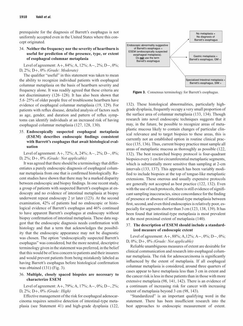
It was agreed that there should be a terminology that differ-entiates a purely endoscopic diagnosis of esophageal colum-nar metaplasia from one that is confirmed histologically. Re-cent studies have shown that there may be a marked disparitybetween endoscopic and biopsy findings. In one recent study,a group of patients with suspected Barrett’s esophagus at en-doscopy and no evidence of intestinal metaplasia at biopsyunderwent repeat endoscopy 2 yr later (123). At the secondexamination, 42% of patients had no endoscopic or histo-logical evidence of Barrett’s esophagus and 46% continuedto have apparent Barrett’s esophagus at endoscopy withoutbiopsy confirmation of intestinal metaplasia. These data sug-gest that the endoscopic diagnosis needs confirmation withhistology and that a term that acknowledges the possibil-ity that the endoscopic appearance may not be diagnosticwas chosen. The option “endoscopically suspected Barrett’sesophagus” was considered, but the more neutral, descriptiveterminology given in the statement was preferred, in the beliefthat this would be of less concern to patients and their insurersand would prevent patients from being mistakenly labeled ashaving Barrett’s esophagus before histological confirmationwas obtained (131) (Fig. 3).
36. Multiple, closely spaced biopsies are necessary tocharacterize ESEM
Level of agreement: A+, 79%; A, 17%; A−, 0%; D−, 2%;D, 2%; D+, 0% (Grade: High)
Effective management of the risk for esophageal adenocar-cinoma requires sensitive detection of intestinal-type meta-plasia (see Statement 41) and high-grade dysplasia (122,
evitseggus ytilamronba cipocsodnEs =ugahpose s’tterraB fo
( MESE yllacipocsodne detcepsus laegahpose aisalpatem .)
oD ton mret eht esut's esophagusterraB
oN aisalpatem = fo sisongaid oN
sugahpose s’tterraB
cirtsaG aisalpatem =+ MG ,sugahpose s’tterraB
Specialized Intestinal metaplasia =+ MIS ,sugahpose s’tterraB
Figure 3. Consensus terminology for Barrett’s esophagus.
132). These histological abnormalities, particularly high-grade dysplasia, frequently occupy a very small proportion ofthe surface area of columnar metaplasia (133, 134). Thoughresearch into novel endoscopic techniques suggests that itmay, in the future, be possible to recognize areas of meta-plastic mucosa likely to contain changes of particular clin-ical relevance and to target biopsies to these areas, this iscurrently not an established option in routine clinical prac-tice (135, 136). Thus, current biopsy practice must sample allareas of metaplastic mucosa as thoroughly as possible (122,132). The best researched biopsy protocol is four-quadrantbiopsies every 1 cm for circumferential metaplastic segments,which is substantially more sensitive than sampling at 2-cmintervals (133, 137). This approach has been variably modi-fied to include biopsies at the top of tongue-like metaplasticextensions. These onerous and usually expensive protocolsare generally not accepted as best practice (122, 132). Evenwith the use of such protocols, there is still evidence of signifi-cant sampling inaccuracies, since concordance for the findingof presence or absence of intestinal-type metaplasia betweenfirst, second, and even third endoscopies is relatively poor, es-pecially for segments shorter than 3 cm (123, 138, 139). It hasbeen found that intestinal-type metaplasia is most prevalentat the most proximal extent of metaplasia (140).
37. The description of ESEM should include a standard-ized measure of endoscopic extent
Level of agreement: A+, 88%; A,12%; A−, 0%; D−, 0%;D, 0%; D+, 0% (Grade: Not applicable)
Reliable unambiguous measures of extent are desirable forclinical communication and research into esophageal colum-nar metaplasia. The risk for adenocarcinoma is significantlyinfluenced by the extent of metaplasia. If all esophagealcolumnar metaplasia is considered, around three quarters ofcases appear to have metaplasia less than 3 cm in extent andthe cancer risk is less in these patients than in those with moreextensive metaplasia (98, 141, 142). There is an evidence ofa continuum of increasing risk for cancer with increasingextent of metaplasia beyond 3 cm (98, 143).
“Standardized” is an important qualifying word in thestatement. There has been insufficient research into thebest approaches to endoscopic measurement of extent.
The Montreal Definition and Classification of GERD 1911
Accordingly, differing approaches have been used in pub-lished studies, with resultant difficulties in making compar-isons among studies or with pooling their data. The lack of avalidated, standard approach to measurement of extent meansthat this clinically relevant variable is often poorly describedand in terms that are open to interpretation. Recently, how-ever, an international working group has developed standardcriteria that may aid further research (144).
38. When biopsies of ESEM show columnar epithelium itshould be called Barrett’s esophagus and the presenceor absence of intestinal-type metaplasia specified
Level of agreement: A+, 49%; A, 28%; A−, 9%; D−, 5%;D, 2%; D+, 7% (Grade: Not applicable)
This statement is the final product of the most controver-sial topic of the workshop. In the early phase of discussionit was decided that the eponymous term “Barrett” shouldbe retained in any definition because it would be futile andcounterproductive to try to remove such an embedded wordfrom general use. Pragmatism aside, opinion was dividedas to whether Barrett’s original scientific contribution war-ranted continued use of his name as a label, but this was notput to formal discussion and voting. With retention of theword “Barrett” decided, there was eventual consensus thatall types of histologically proven esophageal columnar meta-plasia should be included under this umbrella word, with theimportant added descriptors of either “intestinal-type meta-plasia positive” or “negative” (see statement above). One ma-jor reason for the statement and the voting on it was the farfrom perfect sensitivity of even a rigorous biopsy protocolfor detection of intestinal-type metaplasia (see Statement 36),let alone the less rigorous biopsy protocols used in routineclinical practice in every part of the world where practicehas been surveyed (145–147). This is not just a problem oftissue sampling, since the staining techniques and interpre-tation of biopsies can influence the sensitivity of detectionof intestinal-type metaplasia (148, 149). A literature searchfailed to reveal any systematic review or meta-analysis of therisk for esophageal adenocarcinoma of definite esophagealcolumnar metaplasia in which intestinal metaplasia had notbeen shown to be present, despite careful biopsy sampling(Fig. 3).
Adenocarcinoma:
39. Adenocarcinoma of the esophagus is a complicationof GERD
Level of agreement: A+, 67%; A, 26%; A−, 7%; D−, 0%;D, 0%; D+, 0% (Grade: Moderate)
40. The risk of adenocarcinoma of the esophagus riseswith increasing frequency and duration of heartburn
Level of agreement: A+, 47%; A, 42%; A−, 7%; D−, 0%;D, 2%; D+, 2% (Grade: Moderate)
There is strong epidemiological evidence, especially fromcase-control studies in Sweden, that esophageal adenocarci-noma is a complication of GERD and that chronic GERDsymptoms increase the risk of esophageal adenocarcinoma
(97, 98). In the study by Lagergren et al. the risk of esophagealadenocarcinoma was increased (OR 7.7) in patients sufferingfrom longstanding reflux symptoms (97). Higher frequency(greater than 3 times per week) and long duration (greater than10–20 yr) of symptoms further increased the OR to 16.4 and20. Over the last 25 yr there has been a remarkable change inthe epidemiology of esophageal cancer in Western countries.The incidence of esophageal adenocarcinoma has been risingsubstantially although the absolute lifetime risk of developingadenocarcinoma is <1% (97, 150–152). In addition, until re-cently, the incidence of esophageal squamous cell carcinomaused to be much higher than esophageal adenocarcinoma. Ac-cording to recent data from the United States, the incidence ofadenocarcinoma of the esophagus now has surpassed the rateof squamous carcinoma (151). The rise in adenocarcinomaincidence is in keeping with the rising GERD prevalence inother parts of the world. For example in Japan, where the dis-ease used to be rare, the prevalence of GERD is increasing,as are Barrett’s esophagus and esophageal adenocarcinoma(153, 154).
41. Long-segment Barrett’s esophagus with intestinal-type metaplasia is the most important identified riskfactor for esophageal adenocarcinoma
Level of agreement: A+, 67%; A, 21%; A−, 12%; D−,0%; D, 0%; D+, 0% (Grade: High)
A wealth of consistently supportive data resulted in promptconsensus on this statement (see Statement 37) (97, 132, 155,156). Wang et al. have tabulated the reported experience onadenocarcinoma development from endoscopic surveillancestudies (132). A large scale prospective Swedish study of pa-tients with the diagnosis of esophageal adenocarcinoma pro-vides the most definitive data (97). It is unclear what propor-tion of the esophageal columnar metaplasia-negative casesnoted in this study were accounted for either by destructionof esophageal columnar metaplasia by cancer or by misclassi-fication of adenocarcinoma of the gastric cardia as esophagealadenocarcinoma (97).
EXTRAESOPHAGEAL SYNDROMES: ESTABLISHEDASSOCIATIONS. Although a great amount has been pub-lished on the extraesophageal GERD syndromes, little ofthis represents high-level original work. This realizationprompted an evolution in the statements regarding the ex-traesophageal syndromes, as paucity of evidence supportingthe initial versions became apparent. Thus, whereas in theinitial iterations, the statements strongly suggested causal-ity between reflux and cough, laryngitis, asthma, and dentalerosions, the final iterations were much more restrained, em-phasizing (1) the existence of an association between thesesyndromes and GERD, (2) the rarity of extraesophageal syn-dromes occurring in isolation without a concomitant mani-festations of the typical esophageal syndrome, (3) that thesesyndromes are usually multifactorial with GERD as one ofthe several potential aggravating cofactors, and (4) that data
1912 Vakil et al.
substantiating a beneficial effect of reflux treatments on theextraesophageal syndromes are weak.Reflux cough, reflux laryngitis, and reflux asthma syn-dromes:
42. Chronic cough, chronic laryngitis, and asthma aresignificantly associated with GERD
Level of agreement: A+, 39%; A, 26%; A−, 28%; D−, 7%;D, 0%; D+, 0% (Grade: High)
43. Chronic cough, chronic laryngitis, and asthma areusually multifactorial disease processes and gastroe-sophageal reflux can be an aggravating cofactor
Level of agreement: A+, 63%; A, 23%; A−, 12%; D−,2%; D, 0%; D+, 0% (Grade: Cough Low, Laryngitis Low,Asthma High)
44. Gastroesophageal reflux is rarely the sole cause ofchronic cough, chronic laryngitis, or asthma
Level of agreement: A+, 65%; A, 23%; A−, 7%; D−, 5%;D, 0%; D+, 0% (Grade: Cough Low, Laryngitis Very Low,Asthma High)
Three large population-based surveys have demonstratedan increased risk of numerous ENT and pulmonary symptomsamong patients with either esophagitis or reflux symptoms(81, 157, 158). The reported ORs for having laryngeal orpulmonary conditions among GERD patients in these stud-ies range from 1.2 to 3.0, with nocturnal cough having thestrongest association.
Support for the premise that chronic cough, chronic laryn-gitis, or asthma are multifactorial processes with reflux asa potential aggravating factor comes from therapeutic trialsin which these entities were improved, but incompletely re-solved, by treating reflux disease. In the case of reflux coughsyndrome, the only randomized controlled trials of medicaltherapy found no treatment effect (159–161). Thus, one hasto look to observational trials of antireflux surgery (162–164)and these are by nature subject to selection and referral bias.By and large these trials show improvement in cough scoresas a result of treatment. With respect to reflux laryngitis syn-drome, there are no randomized controlled treatment trials inwhich chronic laryngitis patients exhibited a complete treat-ment response. Observational trials of medical or surgicaltherapy report partial improvement in laryngitis symptoma-tology and in some cases laryngoscopic appearance (164,165). Commonly implicated cofactors with laryngitis includeheavy voice usage, habitual throat clearing, allergic rhinitiswith postnasal drip, infectious laryngitis, and environmentalirritants including smoking. Regarding reflux asthma syn-drome, Field summarized the medical and surgical data andconcluded that there was a significant benefit in improvingasthma symptoms and reducing asthma medication usage butno improvement in pulmonary function attributable to GERDtherapy (166, 167). Commonly implicated cofactors amongasthma patients include allergens, exercise, temperature orclimate changes, or emotional conflicts.
Since reflux disease has highly effective treatments, it fol-lows that manifestations of the disease should exhibit high-grade treatment effects. Thus, support for the premise thatreflux is the sole cause of chronic cough, chronic laryngi-tis, or asthma would come from therapeutic trials in whichthese entities were completely resolved by treating reflux dis-ease. In the case of chronic cough, few, if any, patients withinrandomized controlled trials exhibited a complete treatmentresponse (159–161). The strongest evidence of a completetreatment effect comes from an uncontrolled observationalstudy of laparoscopic Nissen fundoplication in which 51%of 133 chronic cough patients exhibited a complete symp-tom response following the procedure (162). In a smallerobservational study of 8 carefully studied chronic patientswho were refractory to medical therapy, 2 subsequently ex-hibited a complete cough resolution after antireflux surgery(163). Both of these series enrolled highly selected patients,suggesting that although chronic cough can be entirely at-tributable to reflux, this is a rare occurrence. With respect tochronic laryngitis, there are no randomized controlled treat-ment trials for GERD in which patients exhibited a completetreatment response. Observational treatment trials of medi-cal or surgical therapy report partial symptomatic improve-ment and in some cases laryngoscopic appearance but few,if any, patients experienced a complete laryngitis response(164, 165). With respect to reflux asthma syndrome, Fieldconcluded that there was no objective improvement in pul-monary function attributable to medical therapy of GERD(166). Furthermore, a recent longitudinal epidemiologicalstudy of more than 14,000 patients in U.K. general prac-tice found that patients with a new diagnosis of asthma are atsignificantly increased risk for developing GERD rather thanvice versa (168). However, two randomized controlled trialsof antireflux surgery as treatment for asthma reported sub-sets of patients in the surgically treated arms with completeasthma resolution; 6 of 16 in the Sontag et al. study and 11 of22 in the Larrain et al. study (169, 170). Pulmonary functiondata are not provided in the Larrain et al. study. Thus, onlya subset of patients has asthma entirely attributable to reflux,and this subset is probably small.
45. Potential causal mechanisms of reflux cough, refluxlaryngitis, and reflux asthma syndromes include di-rect (aspiration) or indirect (neurally mediated) ef-fects of gastroesophageal reflux
Level of agreement: A+, 61%; A, 28%; A−, 7%; D−, 2%;D, 0%; D+, 2% (Grade: High)
Experimental evidence in both animals and humans hasdemonstrated reflex stimulation of bronchospasm and coughas a response to esophageal acidification (171, 172). Animalstudies have also demonstrated the development of laryn-geal ulceration and profound bronchospasm as a result of thedirect application of acid to the larynx or acid instillationinto the airway (173, 174). Studies of pulmonary functionin asthmatics have demonstrated correlation between lung
The Montreal Definition and Classification of GERD 1913
resistance and the occurrence of spontaneous gastroe-sophageal reflux (175).
46. In the absence of heartburn or regurgitation, unex-plained asthma and laryngitis are unlikely to be re-lated to GERD
Level of agreement: A+, 37%; A, 33%; A−, 14%; D−,7%; D, 9%; D+, 0% (Grade: Laryngitis Low, Asthma High)
This statement implies that individuals with conclusivereflux laryngitis and reflux asthma syndromes usually haveesophageal manifestations of reflux as well. Since the onlypatients in whom these diagnoses can be confidently estab-lished are those that convincingly responded to reflux treat-ment, it is the responders who must be evaluated with respectto whether or not they had frequent heartburn. With respect toreflux laryngitis syndrome, the only randomized controlledtrials demonstrating a treatment effect were on patients withclear-cut reflux disease in addition to the laryngitis, whereasthe recent trial that excluded patients with frequent heart-burn demonstrated no benefit of a PPI over placebo in treat-ing the laryngitis (176–178). With respect to asthma, mostasthmatics have objective evidence of reflux disease as well asreflux symptoms (179). A recent randomized controlled studyof 770 asthmatics evaluated twice-daily PPI therapy and onlythe patient group with both nocturnal respiratory and GERDsymptoms responded to the PPI better than to placebo in theprimary study outcome measure (morning peak expiratoryflow) (180). In the two randomized controlled trials of an-tireflux surgery that showed treatment benefit with respectto asthma, objective evidence of reflux was either an entrycriterion for the study or objectively demonstrated in almostall patients (169, 170).
47. Medical and surgical treatment trials aimed at im-proving presumed reflux cough, reflux laryngitis, andreflux asthma syndromes by treating GERD are as-sociated with uncertain and inconsistent treatmenteffect
Level of agreement: A+, 51%; A, 40%; A−, 7%; D−,0%; D, 0%; D+, 2% (Grade: Cough Very Low, LaryngitisModerate, Asthma High)
In the case of reflux cough syndrome, two small random-ized controlled trials have evaluated the effects of PPI treat-ment on chronic cough. One of these found no significantimprovement in cough between the PPI and placebo groups(12% vs 0%) with only 1 of 8 patients randomized to thePPI showing a response (159). However, during subsequent,open-label treatment 5 of the 9 placebo-treated patients, all ofwhom had markedly abnormal pH studies, responded dramat-ically. The other randomized controlled PPI trial was com-promised by a crossover design that the authors concludedresulted in treatment effect from the first period carryingover to the second. When the analysis was restricted to thegroup randomized to initial placebo therapy (N = 13), a sig-nificant reduction in cough score was demonstrated whenthey crossed over to PPI (160). Crossover studies are prone
to overestimating treatment effect and these studies shouldbe viewed with caution. One randomized controlled trial ofH2-receptor antagonist therapy for chronic cough showed notherapeutic benefit (161). Several uncontrolled trials on H2-receptor antagonists, with or without prokinetics, have re-ported improvement in cough in 70–100% of treated patients(176, 177, 181, 182). With respect to treatment of suspectedreflux cough syndrome with antireflux surgery, there are nocontrolled trials. There are, however, consistently positive re-sults from uncontrolled studies suggesting benefit in a subsetof chronic cough patients but these studies have the usuallimitations in that they overestimate treatment effect (164,183).
For reflux laryngitis there are four published randomizedcontrolled trials using twice-daily PPI therapy for 8–12 wkencompassing a total of 88 patients (176, 177, 181, 182). Oneadditional study of 88 patients has thus far been publishedonly in abstract form (178). One trial showed a significantdifference between the PPI and placebo in resolution of laryn-geal symptoms and one other for hoarseness and throat clear-ing (159, 177). No significant difference in laryngoscopichealing was found between placebo and PPI-treated groups inany of the trials. There are substantial inconsistencies amongthe trials in laryngoscopic criteria for defining reflux laryn-gitis, pH-monitoring protocols, and most importantly, inclu-sion of patients with concomitant heartburn. The trial withthe best therapeutic result enrolled patients with-high-grade,unequivocal laryngoscopic findings and markedly abnormalesophageal pH-monitoring studies (161). The large treatmenttrial finding no PPI benefit enrolled patients with low-gradelaryngoscopic findings and excluded patients with frequentheartburn (178).
With respect to reflux asthma syndrome, Field concludedthat there was a significant benefit in improving asthma symp-toms and reducing asthma medication usage but no objectiveimprovement in pulmonary function attributable to GERDtherapy (166, 170, 184–190). A recent large study, using es-omeprazole 40 mg twice daily, enrolled a total of 770 patientsand subdivided asthmatics into those with only nocturnal res-piratory symptoms, only nocturnal GERD symptoms, or bothnocturnal respiratory and GERD symptoms. The primaryoutcome variable was the change in morning peak expiratoryflow. Of the three patient groups, only those with both noctur-nal respiratory and GERD symptoms responded to esomepra-zole better than to placebo with a mean difference in morningpeak expiratory flow of 8.7 L/min (180). A difference of 20L/min is generally considered the threshold for clinical sig-nificance. Also of interest is a recent study analyzing a subsetof asthmatic patients with cough and reflux (191). This un-controlled treatment trial demonstrated substantial improve-ment in cough, pulmonary function, asthma symptoms, andreflux symptoms (when present) after 3 months of PPI ther-apy (esomeprazole 40 mg once daily). In a complementaryanalysis of the effects of antireflux surgery on asthma, therewere only two controlled trials again showing improvement
1914 Vakil et al.
in asthma symptoms and medication use but no improve-ment in pulmonary function (167, 169, 170). Similar to thecase with the laryngitis studies, there are substantial incon-sistencies among trials in asthma definition and in whetheror not patients with well-defined or symptomatically evidentGERD were included. Of particular note, the largest placebo-controlled trial of surgical therapy was a three-armed trialinvolving 90 patients conducted by a single group of inves-tigators (170). This trial, which reported the best therapeuticresults in both the medical and surgical domain, excluded pa-tients with “allergic” asthma and required that they had refluxsymptoms.Reflux dental erosion syndrome:
48. The prevalence of dental erosions, especially on thelingual and palatal tooth surfaces, is increased in pa-tients with GERD
Level of agreement: A+, 42%; A, 35%; A−, 19%; D−, 2%;D, 0%; D+, 2%(Grade: High)
In a prospective consecutive series, 253 patients were di-vided into two groups based on reflux symptoms: 181 re-fluxers and 72 controls (192). The percentage with dentalerosions was significantly higher among the reflux group(47.5% vs 12.5% p < 0.001) but there were no differences inother clinical or dental parameters. A similar analysis amongintellectually disabled individuals found 19 of 29 individuals(65.5%) with dental erosions to have pH-monitoring criteriafor GERD compared to only 9 of 34 without dental erosions(26.5%) (p = 0.04) (193). Another study found a positivecorrelation between esophageal acid exposure measured bypH monitoring and dental erosion score among 30 patientswith and without GERD (194).
EXTRAESOPHAGEAL SYNDROMES: PROPOSED AS-SOCIATIONS.
49. It is unclear whether gastroesophageal reflux is a sig-nificant causal or exacerbating factor in the patho-genesis of sinusitis, pulmonary fibrosis, pharyngitis,or recurrent otitis media
Level of agreement: A+, 91%; A, 9%; A−, 0%; D−, 0%;D, 0%; D+, 0% (Grade: Low, reflecting lack of authoritativemechanistic or therapeutic studies)
The generally low quality, uncontrolled published studiesrelevant to this statement have been reviewed recently (195,196). Epidemiological studies have shown a modestly in-creased OR for sinusitis in the U.S. military veterans with re-flux esophagitis of 1.6 (1.51–1.70) (157). This risk is slightlyhigher at 2.34 (1.72–3.19) for children with GERD (197).Adequate evidence of causal linkage is lacking. U.S. militaryveterans with reflux esophagitis have a slightly increased riskfor idiopathic pulmonary fibrosis, with an OR of 1.36 (1.25–1.48) (157). There is no persuasive evidence of causal link-age. There are no authoritative, confirmed data that indicate
GERD is a clinically significant contributor to pharyngitis orotitis media.
50. It is unclear whether gastroesophageal reflux playsa role in triggering apneic episodes in patients withobstructive sleep apnea
Level of agreement: A+, 74%; A, 21%; A−, 5%; D−,0%; D, 0%; D+, 0% (Grade: Low, because of lack of directmechanistic study data)
An increased prevalence of GERD has been found con-sistently in obstructive sleep apnea patients, but uncertaintyremains whether reflux episodes are true precipitants of ap-neic episodes (198–204). The argument that lack of correla-tion of severity of reflux-induced symptoms with severity ofobstructive sleep apnea is an evidence against precipitationof apneic episodes by reflux episodes is unconvincing (205).More definitive mechanistic data are required.
CONCLUSIONS
In conclusion, a new definition and classification of GERDhas been developed by an International Consensus Group.It provides a basis for universally accepted terminology thatbridges cultures and countries and may simplify disease man-agement, allow collaborative research, and make studies moregeneralizable, assisting patients, physicians, and regulatoryagencies. Coupling evidence-based medicine with modernconsensus development techniques allows a broad consensusamong different regions of the world. For practicing physi-cians, this definition and classification clarify the criteria nec-essary for a diagnosis of GERD, simplify the classificationof suspected and proven Barrett’s esophagus, and define thestate of our incomplete knowledge in extraesophageal disor-ders. For patients, the consensus statement provides clarityon a diagnosis that is based on a patient-centered definition oftroublesome symptoms and may help to prevent patients frombeing inappropriately labeled as having Barrett’s esophagus.Clarification of the role of GERD in patients with cough andhoarseness may also help the management of patients withthese difficult conditions. Finally, regulators may benefit froma uniform terminology and classification to use with clinicaltrial data submissions.
ACKNOWLEDGMENTS
Other members of the Consensus Group: Lars Agreus (Stock-holm, Sweden), David Armstrong (Hamilton, Canada),Stephen Attwood (Manchester, UK), Jean-Francois Bergman(Paris, France), Marc-Andre Bigard (Vandoeuvre-les-Nancy,France), Patricia Blount (Seattle, WA), Peter Bytzer(Glostrup, Denmark), Tsutomu Chiba (Kyoto, Japan), Gi-anfranco Delle Fave (Rome, Italy), Hashem El-Serag (Hous-ton, TX), Daiming Fan (Xi’an, China), Ronnie Fass (Tucson,AZ), Nigel Flook (Edmonton, Canada), Jean-Paul Galmiche(Nantes, France), Michio Hongo (Sendai, Japan), Francisco
The Montreal Definition and Classification of GERD 1915
Huerta Iga (Coahuila, Mexico), Janusz Jankowski (Leicester,UK), David Johnson (Norfolk, VR), Folke Johnsson (Lund,Sweden), Peter Katelaris (Concord, Australia), Yoshi Ki-noshita (Izumo, Japan), Elly Klinkenberg-Knol (Amsterdam,Netherlands), Joachim Labenz (Siegen, Germany), HarleyLiker (Beverly Hills, CA), Peter Malfertheiner (Magdeburg,Germany), Hiroto Miwa (Tokyo, Japan), Paul Moayyedi(Hamilton, Canada), Fabio Pace (Milan, Italy), JoachimPrado (Sao Paulo, Brazil), Joel Richter (Philadelphia, PA),Graciela Salis (Buenos Aires, Argentina), Lin San-Ren (Bei-jing, China), Prateek Sharma (Kansas City, MO), VincenzoStanghellini (Bologna, Italy), Jan Tack (Leuven, Belgium),Nicholas Talley (Rochester, MN), Benjamin Wong (HongKong, China), Yuan Yaozong (Shanghai, China), Carlos Za-pata (Lima, Peru).
The Barrett’s esophagus subgroup comprised StephenAttwood, Patricia Blount, Peter Malfertheiner, and NicholasTalley.
The extraesophageal reflux syndromes subgroup com-prised Peter Bytzer, Hashem El-Serag, Fabio Pace, and JoelRichter.
Reprint requests and correspondence: Nimish Vakil M.D.,F.A.C.G., ASMC, 945 North 12th Street, Room 4040, Milwaukee,WI 53233.
Received December 10, 2005; accepted February 7, 2006.
REFERENCES
1. Jian R, Chassany O, Douet MC, et al. French-Belgian con-sensus conference on adult gastro-oesophageal reflux dis-ease ’diagnosis and treatment’: Report of a meeting heldin Paris, France, 21–22 January 1999. Eur J GastroenterolHepatol 2000;12:129–37.
2. Koop H, Schepp W, Mueller-Lissner S, et al. Consensusconference of the DGVS on gastro-esophageal reflux. ZGastroenterol 2005;43:163–93.
3. Hongo M. GERD guideline workshop report 2002. TherRes 2003;24:830–5.
4. DeVault KR, Castell DO. American College of Gastroen-terology. Updated guidelines for the diagnosis and treat-ment of gastro-esophageal reflux disease. Am J Gastroen-terol 2005;100:190–200.
5. National Institute for Clinical Excellence. Managementof dyspepsia in adults in primary care. Available at:http://www.nice.org Accessed 7 October 2005.
6. Moraes-Filho JPP, Cecconello I, Gama-Rodrigues J, et al.Brazilian consensus on gastro-esophageal reflux disease:Proposals for assessment, classification, and management.Am J Gastroenterol 2002;97:241–8.
7. Katelaris P, Holloway R, Talley N, et al. Gastro-oesophageal reflux disease in adults: Guidelines for clini-cians. J Gastroenterol Hepatol 2002;17:825–33.
8. Fock K, Talley N, Hunt R, et al. Report of the Asia-Pacificconsensus on the management of gastro-esophageal refluxdisease. J Gastroenterol Hepatol 2004;19:357–67.
9. Armstrong D, Marshall JK, Chiba N, et al. Canadian asso-ciation of gastroenterology GERD consensus group. Cana-dian consensus conference on the management of gastro-esophageal reflux disease in adults—update 2004. Can JGastroenterol 2005;19:15–35.
10. Veldhuyzen van Zanten SJO, Bradette M, Chiba N, etal. Evidence-based recommendations for short- and long-term management of uninvestigated dyspepsia in primarycare: An update of the Canadian dyspepsia working group(CanDys) clinical management tool. Can J Gastroenterol2005;19:285–303.
11. Linstone H, Turoff M. The Delphi method: Tech-niques and applications. URL: http://www.is.njit.edu/pubs/delphibook/.
12. Dalkey N. An experimental study of group opinion: TheDelphi method. Futures 1969;September:408–26.
13. Lomaz J. Words without action? The production, dissem-ination and impact of consensus recommendations. AnnuRev Public Health 1991;12:41–65.
14. The GRADE Working Group. Grading quality of evidenceand strength of recommendations. BMJ 2004;328:1490–4.
15. Stedman’s online medical dictionary. Available at:http://www.stedmans.com. Accessed 7 October 2005.
16. Klauser AG, Schindlbeck NE, Muller-Lissner SA.Symptoms in gastro-oesophageal reflux disease. Lancet1990;335:205–8.
17. Berstad A, Hatlebakk JG. The predictive value of symp-toms in gastro-oesophageal reflux disease. Scand J Gas-troenterol Suppl 1995;211:1–4.
18. Cicala M, Emerenziani S, Caviglia R, et al. Intra-oesophageal distribution and perception of acid reflux inpatients with non-erosive gastro-oesophageal reflux dis-ease. Aliment Pharmacol Ther 2003;18:605–13.
19. Rex DK, Cummings OW, Shaw M, et al. Screening for Bar-rett’s esophagus in colonoscopy patients with and withoutheartburn. Gastroenterology 2003;125:1670–7.
20. Venables TL, Newland RD, Patel AC, et al. Omeprazole10 milligrams once daily, omeprazole 20 milligrams oncedaily, or ranitidine 150 milligrams twice daily, evaluatedas initial therapy for the relief of symptoms of gastro-oesophageal reflux disease in general practice. Scand JGastroenterol 1997;32:965–73.
21. Caviglia R, Ribolsi M, Maggiano N, et al. Dilated intercel-lular spaces of esophageal epithelium in nonerosive refluxdisease patients with physiological esophageal acid expo-sure. Am J Gastroenterol 2005;100:543–8.
22. Stanghellini V. Relationship between upper gastrointesti-nal symptoms and lifestyle, psychosocial factors and co-morbidity in the general population: Results from the do-mestic/international gastroenterology surveillance study(DIGEST). Scand J Gastroenterol Suppl 1999;231:29–37.
23. Dent J, El-Serag HB, Wallander MA, et al. Epidemiologyof gastro-oesophageal reflux disease: A systematic review.Gut 2005;54:710–7.
24. Prado J, Moraes-Filho P. Gastro-esophageal reflux disease:Prevalence and management in Brazil. Best Pract Res ClinGastroenterol 2004;18:23–6.
25. Bor S, Mandiracioglu A, Kitapcioglu G, et al. Gastro-esophageal reflux disease in a low-income region in Turkey.Am J Gastroenterol 2005;100:759–65.
26. Chen M, Xiong L, Chen H, et al. Prevalence, risk factorsand impact of gatroesophageal reflux disease symptoms: Apopulation based study in South China. Scand J Gastroen-terol 2005;40:750–67.
27. Wong WM, Lai KC, Lam KF, et al. Prevalence, clinicalspectrum and health care utilization of gastro-esophagealreflux disease in a Chinese population: A population-basedstudy. Aliment Pharmacol Ther 2003;18:595–604.
28. Lim SL, Goh WT, Lee JM, et al. Community medicineGI studygroup. Changing prevalence of gastro-esophagealreflux with changing time: Longitudinal study in an
1916 Vakil et al.
Asian population. J Gastroenterol Hepatol 2005;20:995–1001.
29. Segal I. The gastro-esophageal reflux disease complex insub-Saharan Africa. Eur J Cancer Prev 2001;10:209–12.
30. Revicki DA, Wood M, Maton PN, et al. The impact ofgastro-esophageal disease on health-related quality of life.Am J Med 1998;104:252–8.
31. Revicki DA, Wood M, Wiklund I, et al. Reliability andvalidity of the gastrointestinal symptom rating scale in pa-tients with gastro-esophageal reflux disease. Qual Life Res1998;1:75–83.
32. Talley NJ, Fullerton S, Junghard O, et al. Quality of lifein patients with endoscopy—negative heartburn: Reliabil-ity and sensitivity of disease-specific instruments. Am JGastroenterol 2001;96:1998–2004.
33. Wiklund I, Carlsson J, Vakil N. Gastro-esophageal refluxsymptoms and well-being in a random sample of the gen-eral population of a Swedish community. Am J Gastroen-terol 2006;101:18–28.
34. Aro P, Ronkainen J, Storskrubb T, et al. Quality of life ina general adult population with gastro-esophageal refluxsymptoms and/or esophagitis: A report from the Kalixandastudy. Gastroenterology 2003;124:A168.
35. Shaw MJ, Adlis S, Beebe T, et al. When does heartburnbecome a disease? Gastroenterology 1998;114: G1166.
36. Eubanks TR, Omelanczuk P, Richards C, et al. Out-comes of laparoscopic anti-reflux procedures. Am J Surg2000;179:391–5.
37. Moayyedi P, Axon ATR. The usefulness of the likelihoodratio in the diagnosis of dyspepsia and gastro-esophagealreflux disease. Am J Gastroenterol 1999;94:3122–5.
38. Lin M, Gerson LB, Lascar R, et al. Features of gastro-esophageal reflux disease in women. Am J Gastroenterol2004;99:1442–7.
39. Vakil NB, Lauritsen K, Junghard O, et al. Esomeprazoleprovides more effective maintenance therapy followinghealing of erosive esophagitis in patients with dysphagiaat baseline compared with lansoprazole. Gastroenterology2003;124(4 suppl 1):A-542-3.
40. Jones RH, Hungin APS, Phillips J, et al. Gastro-oesophageal reflux disease in primary care in Europe: Clin-ical presentation and endoscopic findings. Eur J Gen Pract1995;1:149–54.
41. Wu JC, Sung JJ, Ng EK, et al. Prevalence and distributionof Helicobacter pylori in gastro-esophageal reflux disease:A study from the East. Am J Gastroenterol 1999;4:1790–4.
42. Nilsson M, Johnsen R, Ye W, et al. Prevalence of gastro-oesophageal reflux symptoms and the influence of age andsex. Scand J Gastroenterol 2004;39:1040–5.
43. Kusano M. Diagnosis and investigation of gastro-oesophageal reflux disease in Japanese patients. AlimentPharmacol Ther 2004;29(suppl 8):14–8.
44. Bochenek WJ, Mack ME, Fraga PD, et al. Panto-prazole provides rapid and sustained relief in patientstreated for erosive oesophasitis. Aliment Pharmacol Ther2004;20:1105–14.
45. Van Pinxteren B, Numan ME, Bonis PA, et al. Short-term treatment with proton pump inhibitors, H2-receptorantagonists and prokinetics for gastro-oesophageal refluxdisease-like symptoms and endoscopy negative reflux dis-ease. Cochrane Database Syst Rev 2004;(4):CD002095.
46. Marshall RE, Anggiansah A, Owen WA, et al. Investiga-tion of oesophageal reflux symptoms after gastric surgerywith combined pH and bilirubin monitoring. Br J Surg1999;86:271–5.
47. Lin KM, Ueda RK, Hinder RA, et al. Etiology and
importance of alkaline oesophageal reflux. Am J Surg1991;162:553–7.
48. Vaezi MF. ‘Refractory GERD’: Acid, nonacid or notGERD? Am J Gastroenterol 2004;99:989–90.
49. Tack J, Koek G, Demedts I, et al. Gastro-esophageal refluxdisease poorly responsive to single-dose proton pump in-hibitors in patients without Barrett’s oesophagus: Acid re-flux, bile reflux or both? Am J Gastroenterol 2004;99:981–8.
50. Kawamura O, Aslam M, Rittmann T, et al. Physican andpH properties of gastroesophago-pharyngeal refluxate: A24-hour simultaneous ambulatory impedance and pH mon-itoring study. Am J Gastroenterol 2004;99:1000–10.
51. Sifrim D, Holloway R, Silny J, et al. Acid, nonacid and gasreflux in patients with gastro-esophageal reflux disease dur-ing ambulatory 24-hour pH-impedance recordings. Gas-troenterology 2001;120:1588–98.
52. Dent J, Armstrong D, Delaney B, et al. Symptom evaluationin reflux disease: Workshop background, processes, ter-minology, recommendations, and discussion outputs. Gut2004;53(suppl 4):iv1–24.
53. Johansson KE, Ask P, Boeryd B, et al. Oesophagitis, signsof reflux, and gastric acid secretion in patients with symp-toms of gastro-oesophageal reflux disease. Scand J Gas-troenterol 1986;21:837–47.
54. Johnsson F, Joelsson B, Gudmundsson K, et al. Symp-toms and endoscopic findings in the diagnosis ofgastro-esophageal reflux disease. Scand J Gastroenterol1987;22:714–8.
55. Joelsson B, Johnsson F. Heartburn—the acid test. Gut1989;30:1523–5.
56. Robinson M, Earnest D, Rodriguez-Stanley S, et al. Heart-burn requiring frequent antacid use may indicate significantillness. Arch Intern Med 1998;158:2373–6.
57. Tefera L, Fein M, Ritter MP, et al. Can the combination ofsymptoms and endoscopy confirm the presence of gastro-esophageal reflux disease? Am Surg 1997;63:933–6.
58. Earlam RJ. Further experience with epigastric pain re-production test in duodenal ulceration. BMJ 1972;2:683–5.
59. Farup P, Hovde O, Torp R, et al. Patients with functionaldyspepsia responding to omeprazole have a characteristicgastro-esophageal reflux pattern. Scand J Gastroenterol1999;34:575–9.
60. Vakil N, Katz P, Traxler B, et al. Resolution of epigas-tric pain accompanies resolution of hearturn in endoscopy-negative GERD. Gastroenterology 2003;124:A-542.
61. Tack J, Caenepeel P, Arts J, et al. Prevalence of acid refluxin functional dyspepsia and its association with symptomprofile. Gut 2005;54:1370–6.
62. Fass R, Quan SF, O’Connor GT, et al. Predictors of heart-burn during sleep in a large prospective cohort study. Chest2005;127:1658–66.
63. Farup C, Kleinman L, Sheldon S, et al. The impact of noc-turnal symptoms associated with gastro-esophageal refluxdisease on health-related quality of life. Arch Intern Med2001;161:45–52.
64. Shaker R, Castell DO, Schoenfeld PS, et al. Nighttimeheartburn is an under-appreciated clinical problem that im-pacts sleep and daytime function: The results of a Gallupsurvey conducted on behalf of the American Gastroentero-logical Association. Am J Gastroenterol 2003;98:1487–93.
65. Elewaut A, Mainguet P, Imschoot J. The impact of sever-ity of pyrosis and regurgitation on quality of sleep (QOS)in patients with esophagitis. Gastroenterology 2004;A-310:M1324.
The Montreal Definition and Classification of GERD 1917
66. Chand N, Johnson DA, Tabangin M, et al. Sleep dys-function in patients with gastro-esophageal reflux disease:Prevalence and response to GERD therapy, a pilot study.Aliment Pharmacol Ther 2004;20:969–74.
67. Cohen JA, Arain A, Harris PA, et al. Surgical trial in-vestigating nocturnal gastro-esophageal reflux and sleep(STINGERS). Surg Endosc 2003;17:394–400.
68. Nilsson G, Wenner J, Larsson S, et al. Randomized clinicaltrial of laparoscopic versus open fundoplication for gastro-esophageal reflux. Br J Surg 2004;91:552–9.
69. Leodolter A, Kulig M, Nocon M, et al. Esomepra-zole therapy improves sleep disorders in patients withgastro-esophageal reflux disease (GERD): A reportfrom the ProGerd study. Gastroenterology 2004;A-226:S1585.
70. Johnson DA, Orr WC, Crawley JA, et al. Effect of es-omeprazole on nighttime heartburn and sleep quality inpatients with GERD: A randomized, placebo-controlledtrial. Am J Gastroenterol 2005;100:1914–22.
71. Pratha V, Wan GJ, Soffer E, et al. Pantoprazole positivelyimpacts nighttime heartburn symptoms and quality of sleepin patients with gastro-esophageal reflux disease. Am JGastroenterol 2003;(suppl 9):S234:A708.
72. Clark CS, Kraus BB, Sinclair J, et al. Gastro-esophagealreflux induced by exercise in health volunteers. JAMA1989;261:3599–601.
73. Soffer EE, Merchant RK, Duethman G, et al. Ef-fect of graded exercise on esophageal motility andgastro-esophageal reflux in trained athletes. Dig Dis Sci1993;38:220–4.
74. Soffer EE, Wilson J, Duethman G, et al. Effect of gradedexercise on esophageal motility and gastro-esophageal re-flux in nontrained subjects. Dig Dis Sci 1994;39:193–8.
75. Yazaki E, Shawdon A, Beasley I, et al. The effect of dif-ferent types of exercise on gastro-esophageal reflux. AustJ Sci Med Sport 1996;28:93–6.
76. Van Nieuwenhoven MA, Brouns F, Brummer RJ. The ef-fect of physical exercise on parameters of gastrointestinalfunction. Neurogastroenterol Motil 1999;11:431–9.
77. Weiner P, Konson N, Sternberg A, et al. Is gastro-esophageal reflux a factor in exercise-induced asthma?Respir Med 1998;92:1071–5.
78. Peters HP, DeKort AF, van Krevelen H, et al. The ef-fect of omeprazole on gastro-esophageal reflux and symp-toms during strenuous exercise. Aliment Pharmacol Ther1999;13:1015–22.
79. Garcia-Rodriguez LA, Wallander M, Johansson S, et al.Natural history of chest pain in GERD. Gut 2005;54(supplVII):A75, OP-G-325.
80. Richards H, McConnachie A, Morrison C, et al. Socialand gender variation in the prevalence, presentation andgeneral practitioner provisional diagnosis of chest pain. JEpidemiol Community Health 2000;54:714–8.
81. Locke GR 3rd, Talley NJ, Fett SL, et al. Prevalence and clin-ical spectrum of gastro-esophageal reflux: A population-based study in Olmsted county, Minnesota. Gastroenterol-ogy 1997;112:1448–56.
82. Brattberg G, Parker MG, Thorslund M. A longitudinalstudy of pain: Reported pain from middle age to old age.Clin J Pain 1997;13:144–9.
83. Lampe FC, Whincup PH, Wannamethee SG, et al. Chestpain on questionnaire and prediction of major ischaemicheart disease events in men. Eur Heart J 1998;19:63–73.
84. Eslick GD, Jones M, Talley NJ. Non-cardiac chestpain: Prevalence, risk-factors, impact and consulting—a population-based study. Aliment Pharmacol Ther2003;17:1115–24.
85. Wong WM, Lam KF, Cheng C, et al. Population basedstudy of non cardiac chest pain in southern China: Preva-lence, psychosocial factors and health care utilization.World J Gastroenterol 2004;10:707–12.
86. Nilsson S, Scheike M, Engblom D, et al. Chest pain andischaemic heart disease in primary care. Br J Gen Pract2003;53:378–82.
87. Cremonini F, Wise J, Moayyedi P, et al. Diagnosticand therapeutic use of proton pump inhibitors in non-cardiac chest pain: A meta-analysis. Am J Gastroenterol2005;100:1226–32.
88. Ciriza de los Rios C, Garcia Menendez L, Diez HernandezA, et al. Role of stationary esophageal manometry in clin-ical practice. Manometric results in patients with gastro-esophageal reflux, dysphagia or non-cardiac chest pain.Rev Esp Enferm Dig 2004;96:606–11.
89. Dekel R, Pearson T, Wendel C, et al. Assessment of oe-sophageal motor function in patients with dysphagia orchest pain—the clinical outcomes research initiative expe-rience. Aliment Pharmacol Ther 2003;18:1083–9.
90. Dent J, Brun J, Fendrick AM, et al. on behalf of the Gen-val Working Group. An evidence-based appraisal of refluxdisease management—the Genval workshop report. Gut1999;44(suppl 2):S1–16.
91. Sonnenberg A, El-Serag HB. Clinical epidemiology andnatural history of gastro-esophageal reflux disease. Yale JBiol Med 1999;72:81–92.
92. El-Serag HB, Sonnenberg A. Associations between dif-ferent forms of gastro-esophageal reflux disease. Gut1997;41:594–9.
93. Thomson ABR, Barkun AN, Armstrong D, et al. Theprevalence of clinically significant upper gastrointestinalfindings in primary care patients with uninvestigated dys-pepsia: The Canadian adult dyspepsia empiric treatment—prompt endoscopy (CADET-PE) study. Aliment Pharma-col Ther 2003;17:1481–91.
94. Nandurkar S, Talley NJ, Martin CJ, et al. Esophagealhistology does not provide additional useful informa-tion over clinical assessment in identifying reflux patientspresenting for esophagogastroduodenoscopy. Dig Dis Sci2000;45:217–24.
95. Zentilin P, Savarino V, Mastracci L, et al. Reassessment ofthe diagnostic value of histology in patients with GERD,using multiple biopsy sites and an appropriate controlgroup. Am J Gastroenterol 2005;100:2299–306.
96. Solcia E, Villani L, Luinetti O, et al. Altered intercel-lular glycoconjugates and dilated intercellular spaces ofesophageal epithelium in reflux disease. Virchows Arch2000;436:207–16.
97. Lagergren J, Bergstrom R, Lindgren A, et al. Symptomaticgastro-esophageal reflux as a risk factor for esophagealadenocarcinoma. N Engl J Med 1999;340:825–31.
98. Avidan B, Sonnenberg A, Schnell TG, et al. Hiatal herniasize, Barrett’s length, and severity of acid reflux are all riskfactors for esophageal adenocarcinoma. Am J Gastroen-terol 2002;97:1930–6.
99. Niguchi D, Sugawa C, Shah SH, et al. Etiology, treat-ment, and outcome of esophageal ulcers: A 10-year ex-perience in an urban emergency hospital. J GastrointestSurg 2003;7:836–42.
100. Armstrong D, Bennett JR, Blum AL, et al. The endoscopicassessment of esophagitis: A progress report on observeragreement. Gastroenterology 1996;111:85–92.
101. Lundell LR, Dent J, Bennett JR, et al. Endoscopic assess-ment of oesophagitis: Clinical and functional correlatesand further validation of the Los Angeles classification.Gut 1999;45:172–80.
1918 Vakil et al.
102. Pandolfino JE, Vakil NB, Kahrilas PJ. Comparison of inter-and intraobserver consistency for grading of esophagitisby expert and trainee endoscopists. Gastrointest Endosc2002;56:639–43.
103. Vieth M, Haringsma J, Delarive J, et al. Red streaks inthe oesophagus in patients with reflux disease: Is therea histomorphological correlate? Scand J Gastroenterol2001;36:1123–7.
104. Bytzer P, Havelund T, Hansen JM. Interobserver variationin the endoscopic diagnosis of reflux esophagitis. Scand JGastroenterol 1993;28:119–25.
105. Isolauri J, Luostarinen M, Isolauri E, et al. Natural courseof gastro-esophageal reflux disease: 17–22 year follow-upof 60 patients. Am J Gastroenterol 1997;92:37–41.
106. McDougall NI, Johnston BT, Collins JS, et al. Three-to 4.5-year prospective study of prognostic indicators ingastro-esophageal reflux disease. Scand J Gastroenterol1998;33:1016–22.
107. Pace F, Bollani S, Molteni P, et al. Natural history of gastro-esophageal reflux disease without oesophagitis (NERD)—a reappraisal 10 years on. Dig Liver Dis 2004;36:111–5.
108. Schindlbeck NE, Klauser AG, Berghammer G, et al. Threeyear follow up of patients with gastrooesophageal refluxdisease. Gut 1992;33:1016–9.
109. Manabe N, Yoshihara M, Sasaki A, et al. Clinical character-istics and natural history of patients with low-grade refluxesophagitis. J Gastroenterol Hepatol 2002;17:949–54.
110. Garrido Serrano A, Guerrereo Igea F, Lepe Jiminez J,et al. Clinical features and endoscopic progression ofgastro-esophageal reflux disease. Rev Esp Enferm Dig2003;95:712–6.
111. Johnson DA, Fennerty MB. Heartburn severity under-estimates erosive esophagitis severity in elderly patientswith gastro-esophageal reflux disease. Gastroenterology2004;126:660–4.
112. Rokkas T, Sladen GE. Ambulatory esophageal pH record-ing in gastro-esophageal reflux: Relevance to the devel-opment of esophagitis. Am J Gastroenterol 1988;83:629–32.
113. Saraswat VA, Dhiman RK, Mishra A, et al. Correlationof 24-hr esophageal pH patterns with clinical features andendoscopy in gastro-esophageal reflux disease. Dig Dis Sci1994;39:199–205.
114. Eisen GM, Sandler RS, Murray S, et al. The relation-ship between gastro-esophageal reflux disease and its com-plications with Barrett’s esophagus. Am J Gastroenterol1997;92:27–31.
115. El-Serag HB, Johanson JF. Risk factors for the severityof erosive esophagitis in Helicobacter pylori-negative pa-tients with gastro-esophageal reflux disease. Scand J Gas-troenterol 2002;37:899–904.
116. Avidan B, Sonnenberg A, Schnell TG, et al. There are noreliable symptoms for erosive oesophagitis and Barrett’sesophagus: Endoscopic diagnosis is still essential. AlimentPharmacol Ther 2002;16:735–42.
117. Brandt MG, Darling GE, Miller L. Symptoms, acid expo-sure and motility in patients with Barrett’s esophagus. CanJ Surg 2004;47:47–51.
118. Vakil NB, Traxler B, Levine D. Dysphagia in patients witherosive esophagitis: Prevalence, severity, and response toproton pump inhibitor treatment. Clin Gastroenterol Hep-atol 2004;2:665–8.
119. Kapoor N, Bassi A, Sturgess R, et al. Predictive value ofalarm features in a rapid access upper gastrointestinal can-cer service. Gut 2005;54:40–5.
120. Fransen GA, Janssen MJ, Muris JW, et al. Meta-analysis: The diagnostic value of alarm symptoms for up-
per gastrointestinal malignancy. Aliment Pharmacol Ther2004;20:1045–52.
121. Spechler SJ, Goyal RK. The columnar-lined esophagus, in-testinal metaplasia, and Norman Barrett. Gastroenterology1996;110:614–21.
122. Sampliner RE, The Practice Parameters Committee of theAmerican College of Gastroenterology. Updated guide-lines for the diagnosis, surveillance, and therapy ofBarrett’s esophagus. Am J Gastroenterol 2002;97:1888–95.
123. Meining A, Ott R, Becker I, et al. The Munich Barrettfollow-up study: Suspicion of Barrett’s esophagus basedon either endoscopy or histology only—what is the clinicalsignificance? Gut 2004;53:1402–7.
124. Sharma P, McQuaid K, Dent J, et al. A critical reviewof the diagnosis and management of Barrett’s esoph-agus: The AGA Chicago workshop. Gastroenterology2004;127:310–30.
125. Howden C, Metz D, Chey W, et al. Degree of conformitybetween US Gastroenterologists and experts concerningBarrett’s esophagus. Gastroenterology 2005;128:A-148.
126. Winters C, Spurling TJ, Chobanian SJ, et al. Barrett’sesophagus. A prevalent, occult complication of gastro-esophageal reflux disease. Gastroenterology 1987;92:118–24.
127. Locke GR, Zinsmeister AR, Talley NJ. Can symptoms pre-dict endoscopic findings in GERD? Gastrointest Endosc2003;58:661–70.
128. Lieberman DA, Oehlke M, Helfand M, GORGE Consor-tium. Risk factors for Barrett’s esophagus in community-based practice. Am J Gastroenterol 1997;92:1293–7.
129. Gerson LB, Shetler K, Triadafilopoulos G. Prevalenceof Barrett’s esophagus in asymptomatic individuals. Gas-troenterology 2002;123:461–7.
130. Gerson LB, Edson R, Lavori PW, et al. Use of a simplesymptom questionnaire to predict Barrett’s esophagus inpatients with symptoms of gastro-esophageal reflux. Am JGastroenterol 2001;96:2005–12.
131. Shaheen NJ, Dulai GS, Ascher B, et al. Effect of a newdiagnosis of Barrett’s esophagus on insurance status. AmJ Gastroenterol 2005;100:577–80.
132. Wang KK, Wongkeesong M, Buttar NS. American Gas-troenterological Association technical review on the roleof the gastroenterologist in the management of esophagealcarcinoma. Gastroenterology 2005;128:1471–505.
133. Reid BJ, Weinstein WM, Lewin KJ, et al. Endoscopicbiopsy can detect high-grade dysplasia or early adenocarci-noma in Barrett’s esophagus without grossly recognizableneoplastic lesions. Gastroenterology 1988;94:81–90.
134. Thompson JJ, Zinsser KR, Etnerline HT. Barrett’s meta-plasia and adenocarcinoma of the esophagus and gastro-esophageal junction. Hum Pathol 1983;14:42–61.
135. Sharma P, Weston AP, Topalovski M, et al. Magnificationchromoendoscopy for the detection of intestinal metaplasiaand dysplasia in Barrett’s esophagus. Gut 2003;52:24–7.
136. Hamamoto Y, Endo T, Nosho K, et al. Usefulness ofnarrow-band imaging endoscopy for diagnosis of Barrett’sesophagus. J Gastroenterol 2004;39:14–20.
137. Reid BJ, Blount PL, Feng Z, et al. Optimizing endoscopicbiopsy detection of early cancers in Barrett’s high-gradedysplasia. Am J Gastroenterol 2000;95:3089–96.
138. Kim SL, Waring JP, Spechler SJ, et al, The Department ofVeterans Affairs Gastro-esophageal Reflux Study Group.Diagnostic inconsistencies in Barrett’s esophagus. Gas-troenterology 1994;107:945–9.
139. Jones TF, Sharma P, Daaboul B, et al. Yield of intesti-nal metaplasia in patients with suspected short-segment
The Montreal Definition and Classification of GERD 1919
Barrett’s esophagus (SSBE) on repeat endoscopy. Dig DisSci 2002;27:2108–11.
140. Chandrasoma PT, Der R, Dalton P, et al. Distribution andsignificance of epithelial types in columnar-lined esopha-gus. Am J Surg Pathol 2001;25:1188–93.
141. Rudolph RE, Vaughan TL, Storer BE, et al. Effect of seg-ment length on risk for neoplastic progression in patientswith Barrett esophagus. Ann Intern Med 2000;132:612–20.
142. Csendes A, Smok G, Burdiles P, et al. Prevalence of intesti-nal metaplasia according to the length of the specializedcolumnar epithelium lining the distal esophagus in patientswith gastro-esophageal reflux. Dis Esophagus 2003;16:24–8.
143. Weston AP, Badr AS, Hassanein RS. Prospective multivari-ate analysis of clinical, endoscopic, and histological factorspredictive of the development of Barrett’s multifocal high-grade dysplasia or adenocarcinoma. Am J Gastroenterol1999;94:3413–9.
144. Armstrong D. Review article: Towards consistency in theendoscopic diagnosis of Barrett’s esophagus and columnarmetaplasia. Aliment Pharmacol Ther 2004;20(suppl 5):40–7.
145. Stein HJ, Panel of Experts. Esophageal cancer: Screeningand surveillance. Results of a consensus conference heldat the VIth world congress of the international society fordiseases of the esophagus. Dis Esophagus 1996;9(suppl1):3–19.
146. MacNeil-Covin L, Casson AG, Malatjalian D, et al. A sur-vey of Canadian gastroenterologists about the managementof Barrett’s esophagus. Can J Gastroenterol 2003;17:313–7.
147. Gorlet I, Bruley des Varannes S, Le Rhun M, et al. Eval-uation du diagnostic endoscopique de l’endobra-chyoesophage. Gastroenterol Clin Biol 2003;27:700–7.
148. Nandurkar S, Talley NJ, Martin CJ, et al. Short segmentBarrett’s esophagus: Prevalence, diagnosis and associa-tions. Gut 1997;40:710–5.
149. Alikhan M, Rex D, Dhan A, et al. Variable pathologicinterpretation of columnar lined esophagus by generalpathologists in community practice. Gastrointest Endosc1999;50:23–6.
150. Blot WJ, Devesa SS, Kneller RW, et al. Rising incidenceof adenocarcinoma of the esophagus and gastric cardia.JAMA 1991;265:1287–9.
151. Devesa SS, Blot WJ, Fraumeni JF Jr. Changing patterns inthe incidence of esophageal and gastric carcinoma in theUnited States. Cancer 1998;83:2049–53.
152. El-Serag HB, Mason AC, Petersen N, et al. Epidemiologi-cal differences between adenocarcinoma of the esophagusand adenocarcinoma of the gastric cardia in the USA. Gut2002;50:368–72.
153. Fujimoto K. Review article: Prevalence and epidemiologyof gastro-esophageal reflux disease in Japan. Aliment Phar-macol Ther 2004;20(suppl 8):5–8.
154. Hongo M. Review article: Barrett’s esophagus and carci-noma in Japan. Aliment Pharmacol Ther 2004;20(suppl8):50–4.
155. Jankowski JA, Provenzale D, Moayyedi P. Esophageal ade-nocarcinoma arising from Barrett’s metaplasia has regionalvariations in the west. Gastroenterology 2002;122:588–95.
156. Solaymani-Dodaran M, Logan RFA, West J, et al. Riskof oesophageal cancer in Barrett’s esophagus and gastro-esophageal reflux disease. Gut 2004;53:1070–4.
157. El-Serag HB, Sonnenberg A. Comorbid occurrence of la-ryngeal or pulmonary disease with esophagitis in United
States military veterans. Gastroenterology 1997;113:755–60.
158. Gislason T, Janson C, Vermeire P, et al. Respiratory symp-toms and nocturnal gastro-esophageal reflux: A populationbased study of young adults in three European countries.Chest 2002;121:158–63.
159. Ours TM, Kavuru MS, Schilz RJ, et al. A prospectiveevaluation of esophageal testing and a double-blind, ran-domized study of omeprazole in a diagnostic and thera-peutic algorithm for chronic cough. Am J Gastroenterol1999;94:3131–8.
160. Kiljander TO, Salomaa ERM, Hietanen EK, et al. Chroniccough and gastro-esophageal reflux: A double-blindplacebo-controlled study with omeprazole. Eur Respir J2000;16:633–8.
161. Ing AJ, Ngu MC, Breslin ABX. A randomised doubleblind placebo controlled crossover study of ranitidine inpatients with chronic persistent cough (CPC) associatedwith gastro-esophageal reflux (GOR). Am Rev Respir Dis1992;145:A11.
162. Allen CJ, Anvari M. Gastro-esophageal reflux relatedcough and its response to laparoscopic fundoplication.Thorax 1998;53:963–8.
163. Irwin RS, Zawacki JK, Wilson MM, et al. Chronic coughdue to gastro-esophageal reflux disease: Failure to re-solve despite total/near-total elimination of esophagealacid. Chest 2002;121:1132–40.
164. So JB, Zeitels SM, Rattner DW. Outcomes of atypicalsymptoms attributed to gastro-esophageal reflux treated bylaparoscopic fundoplication. Surgery 1998;124:28–32.
165. Kamel PL, Hanson D, Kahrilas PJ. Prospective trial ofomeprazole in the treatment of posterior laryngitis. Am JMed 1994;96:321–6.
166. Field SK, Sutherland LR. Does medical antirefluxtherapy improve asthma in asthmatics with gastro-esophageal reflux? A critical review of the literature. Chest1998;114:275–83.
167. Field SK. The effects of antireflux surgery on asthmat-ics with gastro-esophageal reflux. Chest 1999;116:766–74.
168. Ruigomez A, Rodrıguez G, Wallander M, et al. Gastro-esophageal reflux disease and asthma. A longitudi-nal study in UK general practice. Chest 2005;128:85–93.
169. Sontag SJ, O’Connell S, Khandelwal S, et al. Asthmaticswith gastro-esophageal reflux: Long term results of a ran-domized trial of medical and surgical antireflux therapies.Am J Gastroenterol 2003;98:987–99.
170. Larrain A, Carrasco E, Galleguillos F, et al. Medi-cal and surgical treatment of nonallergic asthma associ-ated with gastro-esophageal reflux. Chest 1991;99:1330–5.
171. Field SK, Evans JA, Price LM. The effects of acid perfusionof the esophagus on ventilation and respiratory sensation.Am J Respir Crit Care Med 1998;157:1058–62.
172. Ing AJ, Ngu MC, Breslin AB. Pathogenesis of chronicpersistent cough associated with gastro-esophageal reflux.Am J Respir Crit Care Med 1994;149:160–7.
173. Adhami T, Goldblum JR, Richter JE, et al. The role ofgastric and duodenal agents in laryngeal injury: An exper-imental canine model. Am J Gastroenterol 2004;99:2098–106.
174. Tuchman DN, Boyle JT, Pack AI, et al. Comparison ofairway responses following tracheal or esophageal acidifi-cation in the cat. Gastroenterology 1984;87:872–81.
175. Cuttitta G, Cibella F, Visconti A, et al. Spontaneousgastro-esophageal reflux and airway patency during the
1920 Vakil et al.
night in adult asthmatics. Am J Respir Crit Care Med2000;161:177–81.
176. El-Serag HB, Lee P, Buchner A, et al. Lansoprazole treat-ment of patients with chronic idiopathic laryngitis: Aplacebo-controlled trial. Am J Gastroenterol 2001;96:979–83.
177. Noordzij JP, Khidr A, Evans BA, et al. Evaluation ofomeprazole in the treatment of reflux laryngitis: A prospec-tive, placebo-controlled, randomized, double-blind study.Laryngoscope 2001;111:2147–51.
178. Vaezi MF, Richter JE, Stasney CR, et al. A randomized,double-blind, placebo-controlled study of acid suppressionfor the treatment of suspected laryngopharyngeal reflux.Gastroenterology 2004;126(suppl 2):A22.
179. Sontag S, O’Connell S, Khandelwal S, et al. Mostasthmatics have gastro-esophageal reflux with or withoutbronchodilator therapy. Gastroenterology 1990;99:613–20.
180. Harding S, Kiljander T, Field S, et al. Efficacy of esomepra-zole 40 mg twice daily for 16 weeks in adults with asthma:A randomized placebo-controlled trial. Gastroenterology2005;128 (suppl 2):A-61.
181. Havas T, Huang S, Levy M, et al. Posterior phayrn-golaryngitis, double-blind randomized placebo-controlledtrial of proton pump inhibitor therapy. Aust J Otolaryngol1999;3:243–6.
182. Eherer AJ, Habermann W, Hammer HF, et al. Effect ofpantoprazole on the course of reflux-associated laryngitis:A placebo-controlled double-blind crossover study. ScandJ Gastroenterol 2003;38:462–7.
183. Allen CJ, Anvari M. Does laparoscopic fundoplication pro-vide long-term control of gastro-esophageal reflux relatedcough? Surg Endosc 2004;18:633–7.
184. Ekstrom T, Lindgren BR, Tibbling L. Effects of ranitidinetreatment on patients with asthma and a history of gastro-esophageal reflux: A double blind crossover study. Thorax1989;44:19–23.
185. Goodall RJR, Earis JE, Cooper DN, et al. Relationshipbetween asthma and gastro-esophageal reflux. Thorax1981;36:116–21.
186. Nagel RA, Brown P, Perks WH, et al. Ambulatory pHmonitoring of gastro-esophageal reflux in ’morning dipper’asthma. BMJ 1988;297:1371–3.
187. Gustafsson PM, Kjellman N-IM, Tibbling L. A trial ofranitidine in asthmatic children and adolescents with orwithout pathological gastro-esophageal reflux. Eur RespirJ 1992;5:201–6.
188. Ford GA, Oliver PS, Prior JS, et al. Omeprazole in the treat-ment of asthmatics with nocturnal symptoms and gastro-esophageal reflux: A placebo controlled crossover study.Postgrad Med J 1994;70:350–4.
189. Teichtahl H, Kronborg IJ, Yeomans ND, et al. Adult asthmaand gastro-esophageal reflux: The effect of omeprazoletherapy on asthma. Aust N Z J Med 1996;26:671–6.
190. Meier JH, McNally PR, Punja M, et al. Does omepra-zole (Prilosec) improve respiratory function in asthmaticswith gastro-esophageal reflux? A double-blind placebo-controlled crossover study. Dig Dis Sci 1994;39:2127–33.
191. Bocskei C, Viczian M, Bocskel R, et al. The influenceof gastro-esophageal reflux disease and its treatment onasthmatic cough. Lung 2005;183:53–62.
192. Munoz JV, Herreros B, Sanchiz V, et al. Dental and pe-riodontal lesions in patients with gastro-esophageal refluxdisease. Dig Liver Dis 2003;35:461–7.
193. Bohmer CJ, Klinkenberg-Knol EC, Niezen-de Boer MC,et al. Dental erosions and gastro-esophageal reflux disease
in institutionalized intellectually disabled individuals. OralDis 1997;3:272–5.
194. Schroeder PL, Filler SJ, Ramirez B, et al. Dental erosionsand acid reflux disease. Ann Intern Med 1995;122:809–15.
195. Weaver EM. Association between gastro-esophageal re-flux and sinusitis, otitis media, and laryngeal malig-nancy: A systematic review of the evidence. Am J Med2003;115:81S–9S.
196. Groen JN, Smout AJPM. Supra-oesophageal manifesta-tions of gastro-esophageal reflux disease. Eur J Gastroen-terol Hepatol 2003;15:1339–50.
197. El-Serag HB, Gilger M, Kuebeler M, et al. Extrae-sophageal associations of gastro-esophageal reflux diseasein children without neurologic defects. Gastroenterology2001;121:1294–9.
198. Graf K-I, Karaus M, Heinemann S, et al. Gastro-esophageal reflux in patients with sleep apnea syndrome.Z Gastroenterol 1995;33:689–93.
199. Heinemann S, Graf K-I, Karaus M, et al. Unter-suchungen uber das Auftreten einer obstruktiven schlaf-bezogenen Atmungsstrorung in Zusammenhang miteiner gastro-oesophagealen Refluxerkrankung. Pneumolo-gie 1995;49(suppl 1):S139-41.
200. Teramoto S, Ohga E, Matsui H. Obstructive sleep ap-nea syndrome may be a significant cause of gastro-esophageal reflux disease in older people. J Am GeriatrSoc 1999;47:1273–4.
201. Valipour A, Makker HK, Hardy R, et al. Symptomaticgastro-esophageal reflux in subjects with a breathing sleepdisorder. Chest 2002;121:1748–53.
202. Guda N, Partington S, Vakil N. Symptomatic gastro-esophageal reflux, arousals and sleep quality in patients un-dergoing polysomnography for possible obstructive sleepapnoea. Aliment Pharmacol Ther 2004;20:1153–9.
203. Field SK, Flemons WW. Is the relationship between ob-structive sleep apnea and gastro-esophageal reflux clini-cally important? Chest 2002;121:1730–3.
204. Berg S, Hoffstein V, Gislason T. Acidification of distalesophagus and sleep-related breathing disturbances. Chest2004;125:2001–106.
205. Morse CA, Quan SF, Mays MZ, et al. Is there a relationshipbetween obstructive sleep apnea and gastro-esophageal re-flux disease? Clin Gastroenterol Hepatol 2004;2:761–8.
All member of the consensus group were paid anhonorarium for their participation by Astra -ZenecaResearch and Development.Guarantor of the article: Nimish VakilDevelopment of statements: AllDelphi Process, Global definition and classification:Nimish VakilEsophageal Syndromes, Symptomatic: Roger JonesSyndromes with esophageal injury (reflux esophagitis andstricture): Sender van ZantenSyndromes with esophageal injury (barrets esophagusand adenocarcinoma): John DentIntegration of text and development of figures: NimishVakilFinancial support:Astra-Zeneca research and DevelopmentPotential competing interests: None