cluster k mycobacteriophages: insights into the...
TRANSCRIPT
Cluster K Mycobacteriophages: Insights into theEvolutionary Origins of Mycobacteriophage TM4Welkin H. Pope1, Christina M. Ferreira1, Deborah Jacobs-Sera1, Robert C. Benjamin2, Ariangela J. Davis3,
Randall J. DeJong3, Sarah C. R. Elgin4, Forrest R. Guilfoile1, Mark H. Forsyth5, Alexander D. Harris3,
Samuel E. Harvey5, Lee E. Hughes2, Peter M. Hynes4, Arrykka S. Jackson5, Marilyn D. Jalal2, Elizabeth A.
MacMurray5, Coreen M. Manley2, Molly J. McDonough5, Jordan L. Mosier2, Larissa J. Osterbann3,
Hannah S. Rabinowitz4, Corwin N. Rhyan4, Daniel A. Russell1, Margaret S. Saha5, Christopher D. Shaffer4,
Stephanie E. Simon2, Erika F. Sims4, Isabel G. Tovar2, Emilie G. Weisser4, John T. Wertz3, Kathleen A.
Weston-Hafer4, Kurt E. Williamson5, Bo Zhang4, Steven G. Cresawn6, Paras Jain7, Mariana Piuri1¤,
William R. Jacobs, Jr.7, Roger W. Hendrix1, Graham F. Hatfull1*
1 Pittsburgh Bacteriophage Institute and Department of Biological Sciences, University of Pittsburgh, Pittsburgh, Pennsylvania, United States of America, 2 Department of
Biological Sciences, University of North Texas, Denton, Texas, United States of America, 3 Department of Biology, Calvin College, Grand Rapids , Michigan, United States of
America, 4 Department of Biology, Washington University, St. Louis, Missouri, United States of America, 5 Department of Biology, College of William and Mary,
Williamsburg, Virginia, United States of America, 6 Department of Biology, James Madison University, Harrisonburg , Virginia, United States of America, 7 Department of
Microbiology and Immunology, Albert Einstein College of Medicine, New York, New York, United States of America
Abstract
Five newly isolated mycobacteriophages –Angelica, CrimD, Adephagia, Anaya, and Pixie – have similar genomicarchitectures to mycobacteriophage TM4, a previously characterized phage that is widely used in mycobacterial genetics.The nucleotide sequence similarities warrant grouping these into Cluster K, with subdivision into three subclusters: K1, K2,and K3. Although the overall genome architectures of these phages are similar, TM4 appears to have lost at least twosegments of its genome, a central region containing the integration apparatus, and a segment at the right end. Thissuggests that TM4 is a recent derivative of a temperate parent, resolving a long-standing conundrum about its biology, inthat it was reportedly recovered from a lysogenic strain of Mycobacterium avium, but it is not capable of forming lysogens inany mycobacterial host. Like TM4, all of the Cluster K phages infect both fast- and slow-growing mycobacteria, and all ofthem – with the exception of TM4 – form stable lysogens in both Mycobacterium smegmatis and Mycobacterium tuberculosis;immunity assays show that all five of these phages share the same immune specificity. TM4 infects these lysogenssuggesting that it was either derived from a heteroimmune temperate parent or that it has acquired a virulent phenotype.We have also characterized a widely-used conditionally replicating derivative of TM4 and identified mutations conferringthe temperature-sensitive phenotype. All of the Cluster K phages contain a series of well conserved 13 bp repeatsassociated with the translation initiation sites of a subset of the genes; approximately one half of these contain an additionalsequence feature composed of imperfectly conserved 17 bp inverted repeats separated by a variable spacer. The K1 phagesintegrate into the host tmRNA and the Cluster K phages represent potential new tools for the genetics of M. tuberculosis andrelated species.
Citation: Pope WH, Ferreira CM, Jacobs-Sera D, Benjamin RC, Davis AJ, et al. (2011) Cluster K Mycobacteriophages: Insights into the Evolutionary Origins ofMycobacteriophage TM4. PLoS ONE 6(10): e26750. doi:10.1371/journal.pone.0026750
Editor: Stefan Bereswill, Charite-University Medicine Berlin, Germany
Received August 26, 2011; Accepted October 3, 2011; Published October 28, 2011
Copyright: � 2011 Pope et al. This is an open-access article distributed under the terms of the Creative Commons Attribution License, which permitsunrestricted use, distribution, and reproduction in any medium, provided the original author and source are credited.
Funding: This work was supported by the Howard Hughes Medical Institute and the National Institutes of Health. The funders had no role in study design, datacollection and analysis, decision to publish, or preparation of the manuscript. The Howard Hughes Medical Institute funders did provide assistance in providingannotation tools to some of the authors.
Competing Interests: The authors have declared that no competing interests exist.
* E-mail: [email protected]
¤ Current address: Departamento de Quımica Biologica, Facultad de Ciencias Exactas y Naturales, Universidad de Buenos Aires, Buenos Aires, Argentina
Introduction
Bacteriophages represent a numerical majority of biological
entities in the biosphere although their full genetic diversity remains
ill-defined [1]. Many different virion morphologies have been
described, with the largest group being the Caudovirales, double-
stranded DNA (dsDNA) tailed phages [2]. The complete sequences
of approximately 750 phage genomes have been determined,
although over 70% of the sequenced dsDNA genomes correspond
to just twelve bacterial hosts [1,3]. As many or more prophages also
have been identified in bacterial genome sequencing projects [4,5].
Although phages of different bacterial hosts typically share little
nucleotide sequence similarity, phages of common hosts can also
represent substantial diversity at the nucleotide level [6,7].
Comparative analysis of 80 mycobacteriophage genomes
reveals substantial but not homogenous diversity. Although many
PLoS ONE | www.plosone.org 1 October 2011 | Volume 6 | Issue 10 | e26750
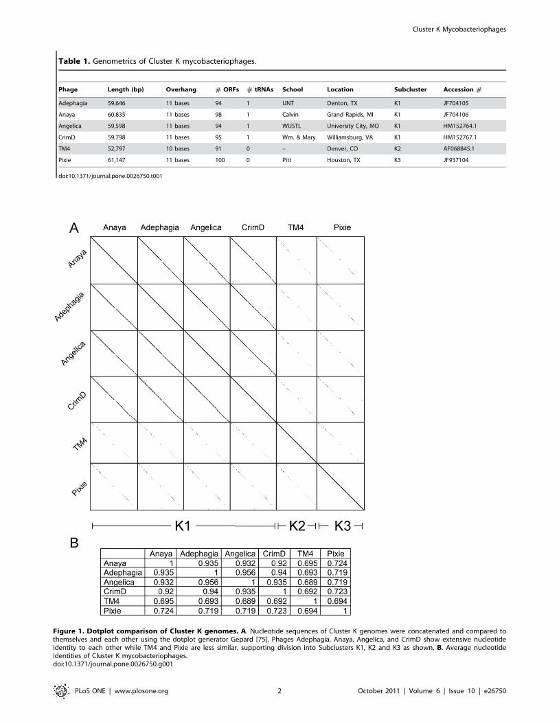
Table 1. Genometrics of Cluster K mycobacteriophages.
Phage Length (bp) Overhang # ORFs # tRNAs School Location Subcluster Accession #
Adephagia 59,646 11 bases 94 1 UNT Denton, TX K1 JF704105
Anaya 60,835 11 bases 98 1 Calvin Grand Rapids, MI K1 JF704106
Angelica 59,598 11 bases 94 1 WUSTL University City, MO K1 HM152764.1
CrimD 59,798 11 bases 95 1 Wm. & Mary Williamsburg, VA K1 HM152767.1
TM4 52,797 10 bases 91 0 – Denver, CO K2 AF068845.1
Pixie 61,147 11 bases 100 0 Pitt Houston, TX K3 JF937104
doi:10.1371/journal.pone.0026750.t001
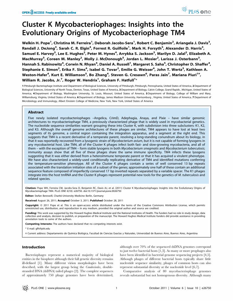
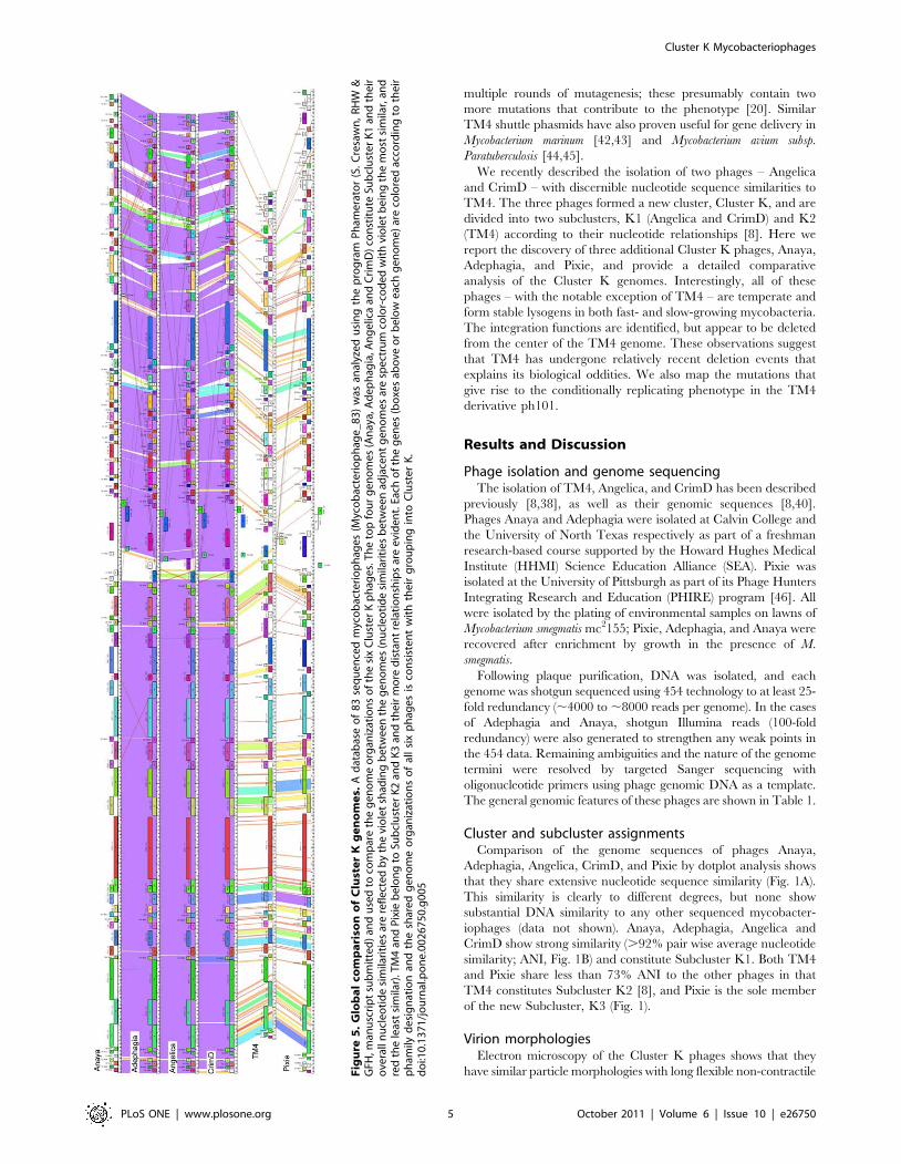
Figure 1. Dotplot comparison of Cluster K genomes. A. Nucleotide sequences of Cluster K genomes were concatenated and compared tothemselves and each other using the dotplot generator Gepard [75]. Phages Adephagia, Anaya, Angelica, and CrimD show extensive nucleotideidentity to each other while TM4 and Pixie are less similar, supporting division into Subclusters K1, K2 and K3 as shown. B. Average nucleotideidentities of Cluster K mycobacteriophages.doi:10.1371/journal.pone.0026750.g001
Cluster K Mycobacteriophages
PLoS ONE | www.plosone.org 2 October 2011 | Volume 6 | Issue 10 | e26750
phages have little or no sequence similarity to each other,
examples of genomes with substantial nucleotide sequence
similarities have also been documented [7,8]. To facilitate
analysis, the phages have been sorted into clusters on the basis
of gross DNA relationships using the cluster metrics described
previously [6,9], and a total of eleven Clusters (A–K) have been
described [8]. The diversity among phages within clusters varies
greatly. At one extreme, the four genomes of Cluster G – Angel,
BPs, Halo and Hope – differ in only few nucleotide positions and
at the variable position of the Mycobacteriophage Mobile
Element (MPME) [8,10]. At the other extreme, the current
members of both Clusters A and B can be subdivided into four
subclusters (A1–A4, B1–B4), with subclustered genomes having
common genomic architectures but relatively low levels of
nucleotide sequence similarity [8]. Five of the 80 completely
sequenced mycobacteriophage genomes – Corndog, Giles,
Figure 2. Virion morphologies of Cluster K phages. Particles of Cluster K phages were put on 400 mesh coated copper grids and stained with1% uranyl acetate. Virions were imaged using a Morgagni transmission electron microscope. All the cluster K phages exhibit a flexible non-contractiletailed morphology with short side tail fibers. Virion capsids are ,55 nm in diameter and tails average ,190 nm in length.doi:10.1371/journal.pone.0026750.g002
Figure 3. All Cluster K phages infect M. tuberculosis. Lysates of Cluster K phages were serially diluted with phage buffer and dilutions werespotted onto lawns of M. tuberculosis mc27000. All cluster K phages infect M. smegmatis mc2155 (data not shown) and M. tuberculosis mc27000 withequal efficiency. TM4 forms clear plaques whereas all other Cluster K phages form turbid plaques.doi:10.1371/journal.pone.0026750.g003
Cluster K Mycobacteriophages
PLoS ONE | www.plosone.org 3 October 2011 | Volume 6 | Issue 10 | e26750
LeBron, Omega, and Wildcat – are ‘singletons’, having no close
relatives [8]. There are a total of 26 different subcluster and
singleton genomes, a remarkably large number for a collection of
phages that infect a common bacterial host strain, M. smegmatis
mc2155. Like most phage genomes, mycobacteriophages have
mosaic genomic architectures [9,11] with illegitimate recombi-
nation predicted to play a key role in the exchange of modules
amongst phage types [12].
Mycobacteriophages provide extremely useful tools for the
study and manipulation of their hosts. Many mycobacteriophages
were isolated originally for uses in phage typing of clinical
bacterial isolates [13,14] but have since proven to be workhorses
for developing mycobacterial genetics. A landmark achievement
was the construction of shuttle phasmids – chimeric vectors that
replicate in Escherichia coli as cosmids and upon transfection of M.
smegmatis yield mycobacteriophage particles that can deliver
foreign DNA to Bacillus Calmette Guerin (BCG) or M. tuberculosis
[15]. Incorporation of a drug resistance marker into a temperate
shuttle phasmid led to the development of the first transformation
systems [16], and addition of reporter genes such as firefly
luciferase or green fluorescent protein enabled construction of
tools for rapid diagnosis and drug susceptibility testing of
M. tuberculosis [17,18,19]. Other applications include the efficient
delivery of transposons to generate transposon libraries
[20,21,22], and for targeted gene replacement [23] or transfer
of point mutations [24] by specialized transduction. Mycobacter-
iophages have also been adapted for diagnostic applications in
amplification assays [25,26,27] and exploited for the development
of integration-proficient vectors [28,29], non-antibiotic selectable
markers [30], and recombineering systems [31,32,33,34].
Mycobacteriophage TM4 plays a central role in mycobacterial
genetics, being the first phage to be used for shuttle phasmid
construction [15] and still widely employed for efficient gene
delivery to M. tuberculosis. It has also been useful for understanding
the role of phage-encoded WhiB proteins [35], lysis systems [36],
and the role of conserved peptidoglycan hydrolyzing motifs in
tapemeasure proteins [37]. The phage was initially recovered from
a putative lysogenic strain of M. avium [38], although following
purification the original host strain was not immune to TM4
superinfection [38]. It has a broad host range infecting both fast-
and slow-growing mycobacteria [39] but does not appear to form
stable lysogens in any strain [15,17]. Timme et al., (1984) suggest
that either the original strain became cured of its prophage, or the
prophage is present in a pseudolysogenic state, such that a
majority of cells remain susceptible to infection. The complete
sequence of the TM4 genome [40] shows that it is 52,797 bp in
length and contains 92 predicted protein-coding genes and no
tRNA genes [40]. There is no evidence for a phage repressor,
but because repressors encompass considerable sequence
diversity [30] they cannot always be readily identified bioinfor-
matically. It is clear that TM4 does not encode a serine- or
tyrosine-integrase, partitioning functions, or recognizable trans-
posases that might indicate a temperate life-style. Thus the
relationship of TM4 to its parent strain M. avium 8/9 serovar 4
remains unresolved.
A potential disadvantage of the use of shuttle phasmids for gene
delivery purposes is that infection typically results in phage
replication and lysis of the infected host. To circumvent this,
conditionally replicating derivatives of TM4 have been isolated
that grow at 30uC but fail to replicate at 37uC [20,41]. To ensure
that reversion to temperature resistance does not interfere with
recovery of derivatives at 37uC that require low frequency events
(such as transposon or recombination), conditionally replicating
derivatives such as TM4 derivative ph101 were generated using
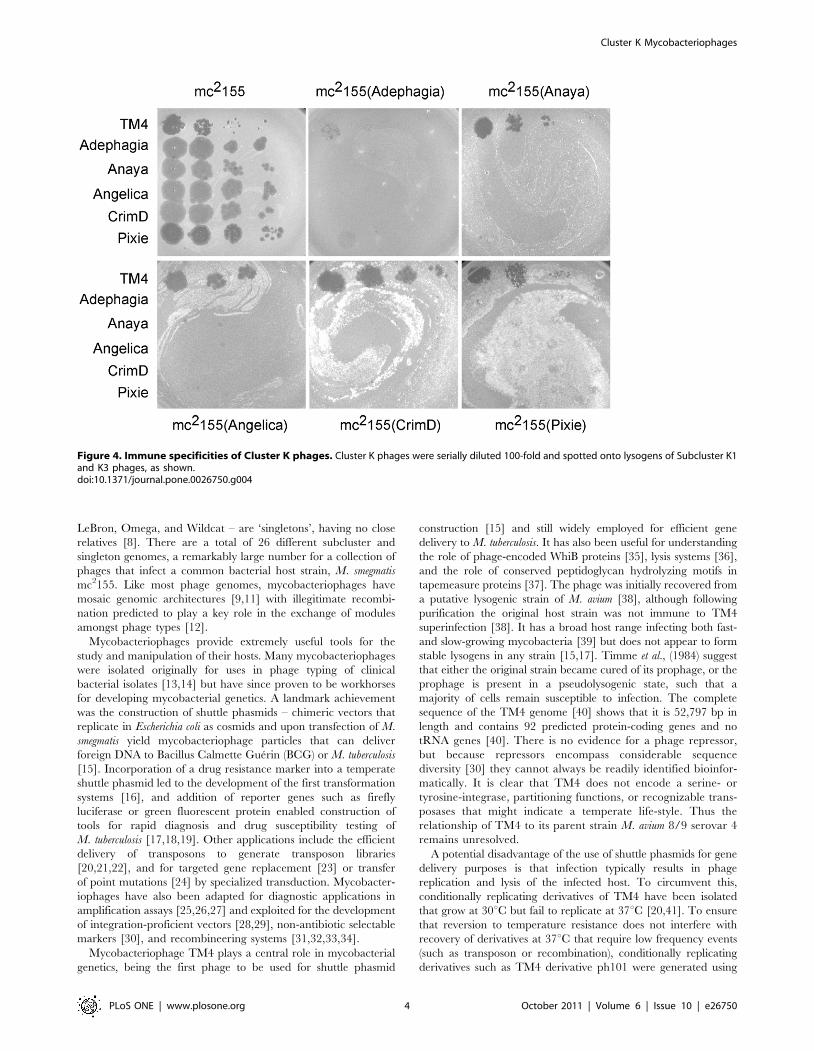
Figure 4. Immune specificities of Cluster K phages. Cluster K phages were serially diluted 100-fold and spotted onto lysogens of Subcluster K1and K3 phages, as shown.doi:10.1371/journal.pone.0026750.g004
Cluster K Mycobacteriophages
PLoS ONE | www.plosone.org 4 October 2011 | Volume 6 | Issue 10 | e26750
multiple rounds of mutagenesis; these presumably contain two
more mutations that contribute to the phenotype [20]. Similar
TM4 shuttle phasmids have also proven useful for gene delivery in
Mycobacterium marinum [42,43] and Mycobacterium avium subsp.
Paratuberculosis [44,45].
We recently described the isolation of two phages – Angelica
and CrimD – with discernible nucleotide sequence similarities to
TM4. The three phages formed a new cluster, Cluster K, and are
divided into two subclusters, K1 (Angelica and CrimD) and K2
(TM4) according to their nucleotide relationships [8]. Here we
report the discovery of three additional Cluster K phages, Anaya,
Adephagia, and Pixie, and provide a detailed comparative
analysis of the Cluster K genomes. Interestingly, all of these
phages – with the notable exception of TM4 – are temperate and
form stable lysogens in both fast- and slow-growing mycobacteria.
The integration functions are identified, but appear to be deleted
from the center of the TM4 genome. These observations suggest
that TM4 has undergone relatively recent deletion events that
explains its biological oddities. We also map the mutations that
give rise to the conditionally replicating phenotype in the TM4
derivative ph101.
Results and Discussion
Phage isolation and genome sequencingThe isolation of TM4, Angelica, and CrimD has been described
previously [8,38], as well as their genomic sequences [8,40].
Phages Anaya and Adephagia were isolated at Calvin College and
the University of North Texas respectively as part of a freshman
research-based course supported by the Howard Hughes Medical
Institute (HHMI) Science Education Alliance (SEA). Pixie was
isolated at the University of Pittsburgh as part of its Phage Hunters
Integrating Research and Education (PHIRE) program [46]. All
were isolated by the plating of environmental samples on lawns of
Mycobacterium smegmatis mc2155; Pixie, Adephagia, and Anaya were
recovered after enrichment by growth in the presence of M.
smegmatis.
Following plaque purification, DNA was isolated, and each
genome was shotgun sequenced using 454 technology to at least 25-
fold redundancy (,4000 to ,8000 reads per genome). In the cases
of Adephagia and Anaya, shotgun Illumina reads (100-fold
redundancy) were also generated to strengthen any weak points in
the 454 data. Remaining ambiguities and the nature of the genome
termini were resolved by targeted Sanger sequencing with
oligonucleotide primers using phage genomic DNA as a template.
The general genomic features of these phages are shown in Table 1.
Cluster and subcluster assignmentsComparison of the genome sequences of phages Anaya,
Adephagia, Angelica, CrimD, and Pixie by dotplot analysis shows
that they share extensive nucleotide sequence similarity (Fig. 1A).
This similarity is clearly to different degrees, but none show
substantial DNA similarity to any other sequenced mycobacter-
iophages (data not shown). Anaya, Adephagia, Angelica and
CrimD show strong similarity (.92% pair wise average nucleotide
similarity; ANI, Fig. 1B) and constitute Subcluster K1. Both TM4
and Pixie share less than 73% ANI to the other phages in that
TM4 constitutes Subcluster K2 [8], and Pixie is the sole member
of the new Subcluster, K3 (Fig. 1).
Virion morphologiesElectron microscopy of the Cluster K phages shows that they
have similar particle morphologies with long flexible non-contractileFig
ure
5.
Glo
ba
lco
mp
ari
son
of
Clu
ste
rK
ge
no
me
s.A
dat
abas
eo
f8
3se
qu
en
ced
myc
ob
acte
rio
ph
age
s(M
yco
bac
teri
op
hag
e_
83
)w
asan
alyz
ed
usi
ng
the
pro
gra
mP
ham
era
tor
(S.
Cre
saw
n,
RH
W&
GFH
,man
usc
rip
tsu
bm
itte
d)
and
use
dto
com
par
eth
eg
en
om
eo
rgan
izat
ion
so
fth
esi
xC
lust
er
Kp
hag
es.
Th
eto
pfo
ur
ge
no
me
s(A
nay
a,A
de
ph
agia
,An
ge
lica
and
Cri
mD
)co
nst
itu
teSu
bcl
ust
er
K1
and
the
iro
vera
lln
ucl
eo
tid
esi
mila
riti
es
are
refl
ecte
db
yth
evi
ole
tsh
adin
gb
etw
een
the
ge
no
me
s(n
ucl
eoti
de
sim
ilari
tie
sb
etw
een
adja
cen
tg
en
om
esar
esp
ect
rum
colo
r-co
de
dw
ith
vio
let
be
ing
the
mo
stsi
mila
r,an
dre
dth
ele
ast
sim
ilar)
.TM
4an
dP
ixie
be
lon
gto
Sub
clu
ste
rK
2an
dK
3an
dth
eir
mo
red
ista
nt
rela
tio
nsh
ips
are
evi
den
t.Ea
cho
fth
eg
en
es(b
oxe
sab
ove
or
be
low
eac
hg
en
om
e)
are
colo
red
acco
rdin
gto
the
irp
ham
ilyd
esi
gn
atio
nan
dth
esh
are
dg
en
om
eo
rgan
izat
ion
so
fal
lsi
xp
hag
es
isco
nsi
ste
nt
wit
hth
eir
gro
up
ing
into
Clu
ste
rK
.d
oi:1
0.1
37
1/j
ou
rnal
.po
ne.
00
26
75
0.g
00
5
Cluster K Mycobacteriophages
PLoS ONE | www.plosone.org 5 October 2011 | Volume 6 | Issue 10 | e26750
tails and isometric heads (Fig. 2). The heads of all six phages are
approximately 55 nm in diameter and the tails are 185–200 nm
long. The Cluster K phages are thus classified morphologically as
members of the Siphoviridae. Short side tail fibers at the tip of the tail
can be seen on many of the particles.
Host-range of Cluster K phagesThe host-range of TM4 has been described previously
[38,39,45]; it is reported to infect fast-growing mycobacteria such
as M. smegmatis as well as the slow-growing M. tuberculosis H37Rv and
M. ulcerans. However, these reports differ in regards to the infection
of M. avium by TM4, with substrains M. avium 701; 6, M. avium 702;
7, M. avium 3746/02 being resistant to infection [39], whereas
substrains M. avium Bridge, serovar 2, M. avium 158, serovar 2, M.
avium TMC 1419, serovar 2, and M. avium TMC 1461, serovar 2 are
sensitive [45]. Timme et al (1984) report that TM4 infects nine M.
avium strains, all of different serovars. Rybniker et al (2006) postulate
that because TM4 was derived from a putative lysogenic strain of M.
avium 6/8 serovar 4, the failure to infect some substrains of M. avium
may be due to superinfection immunity conferred by resident
prophages.
We tested phages Adephagia, Anaya, Angelica and CrimD as
examples of Subcluster K1 as well as TM4 and Pixie for plaque
formation on M. tuberculosis mc27000, M. bovis BCG strain Pasteur,
M. avium 104, and M. marinum strains M and 927. All six phages
infected M. tuberculosis mc27000 efficiently, albeit with different
plaque morphologies (Fig. 3); TM4 yields larger clear plaques
while Angelica, CrimD, and Pixie produce smaller, turbid plaques.
Adephagia and Anaya produce large turbid plaques, although
Anaya only produces plaques when incubated at or below 33uC.
Only TM4 showed infectivity on M. bovis BCG, although we
observed a reduction of efficiency of plating relative to M. smegmatis
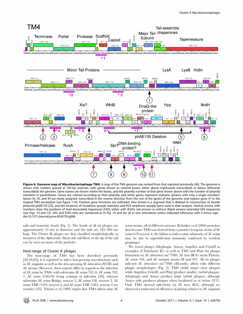
Figure 6. Genome map of Mycobacteriophage TM4. A map of the TM4 genome was revised from that reported previously [40]. The genome isshown with markers spaced at 100 bp intervals, with genes shown as colored boxes, either above (rightwards transcribed) or below (leftwardstranscribed) the genome. Gene names are shown within the boxes, and the phamily number of that gene shown above with the number of phamilymembers in parentheses. Genes are colored according to their phamily, and white genes represent orphams (phams with only a single member).Genes 92, 93, and 94 are newly assigned, transcribed in the reverse direction from the rest of the genes of the genome, and replace gene 41 in theoriginal TM4 annotation (see figure 11A). Putative gene functions are indicated. Also shown is a segment that is deleted in construction of shuttlephasmid phAE159 [23], and the locations of mutations (purple asterisks) and PCR amplicons (purple bars) used in their analysis. Vertical arrows withnumbers show the positions of Start-Associated Sequences (SAS), either with (ESAS; red arrows) or without (black arrows) extended SAS sequences(see Figs. 14 and 15). SAS and ESAS sites are numbered as in Fig. 14 and are all in one orientation unless indicated otherwise with a minus sign.doi:10.1371/journal.pone.0026750.g006
Cluster K Mycobacteriophages
PLoS ONE | www.plosone.org 6 October 2011 | Volume 6 | Issue 10 | e26750
by between five and six orders of magnitude. No infectivity of
M. avium 104 was observed with any of the Cluster K phages tested
here. Adephagia, Anaya, Angelica and CrimD showed no
infection of either M. marinum strain, although both TM4 and
Pixie did, albeit at a greatly reduced efficiency of plating (data not
shown). Plaques picked from these plates and re-spotted on lawns
of M. marinum did not show an increased ability to infect either M.
marinum strain over the parent phages. These plaques were only
observed using 0.35% top agar and incubating at room
temperature. We do not yet know the basis for these observed
reductions in plating efficiencies, although it could be the result of
restriction, CRISPR’s, abortive infection, or the need for
mutations that would expand the host range.
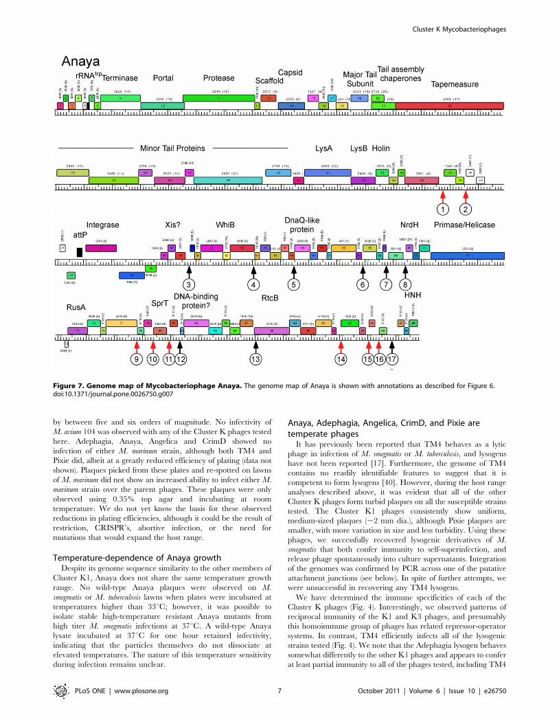
Temperature-dependence of Anaya growthDespite its genome sequence similarity to the other members of
Cluster K1, Anaya does not share the same temperature growth
range. No wild-type Anaya plaques were observed on M.
smegmatis or M. tuberculosis lawns when plates were incubated at
temperatures higher than 33uC; however, it was possible to
isolate stable high-temperature resistant Anaya mutants from
high titer M. smegmatis infections at 37uC. A wild-type Anaya
lysate incubated at 37uC for one hour retained infectivity,
indicating that the particles themselves do not dissociate at
elevated temperatures. The nature of this temperature sensitivity
during infection remains unclear.
Anaya, Adephagia, Angelica, CrimD, and Pixie aretemperate phages
It has previously been reported that TM4 behaves as a lytic
phage in infection of M. smegmatis or M. tuberculosis, and lysogens
have not been reported [17]. Furthermore, the genome of TM4
contains no readily identifiable features to suggest that it is
competent to form lysogens [40]. However, during the host range
analyses described above, it was evident that all of the other
Cluster K phages form turbid plaques on all the susceptible strains
tested. The Cluster K1 phages consistently show uniform,
medium-sized plaques (,2 mm dia.), although Pixie plaques are
smaller, with more variation in size and less turbidity. Using these
phages, we successfully recovered lysogenic derivatives of M.
smegmatis that both confer immunity to self-superinfection, and
release phage spontaneously into culture supernatants. Integration
of the genomes was confirmed by PCR across one of the putative
attachment junctions (see below). In spite of further attempts, we
were unsuccessful in recovering any TM4 lysogens.
We have determined the immune specificities of each of the
Cluster K phages (Fig. 4). Interestingly, we observed patterns of
reciprocal immunity of the K1 and K3 phages, and presumably
this homoimmune group of phages has related repressor-operator
systems. In contrast, TM4 efficiently infects all of the lysogenic
strains tested (Fig. 4). We note that the Adephagia lysogen behaves
somewhat differently to the other K1 phages and appears to confer
at least partial immunity to all of the phages tested, including TM4
Figure 7. Genome map of Mycobacteriophage Anaya. The genome map of Anaya is shown with annotations as described for Figure 6.doi:10.1371/journal.pone.0026750.g007
Cluster K Mycobacteriophages
PLoS ONE | www.plosone.org 7 October 2011 | Volume 6 | Issue 10 | e26750
and Pixie (Fig. 4). These observations are especially revealing
about TM4 and its previously characterized properties. A simple
explanation is that TM4 is a relatively recent derivative of a
temperate phage that was heteroimmune with other Cluster K
phages, but which has lost its immunity functions. When this event
may have happened is unclear, it could have occurred during
passage of the phage between its isolation in 1984 and genome
sequencing in 1998, during the process of isolation, or at some
prior time as a naturally occurring event. This is discussed in
greater detail below. We note the obvious parallels to the
relationship between D29 and Cluster A phages such as L5
[47,48]. In D29, a 3.6 kbp deletion removes a segment that in L5
contains the repressor, and although D29 is lytic in nature, it is
homoimmune with L5 immunity [30].
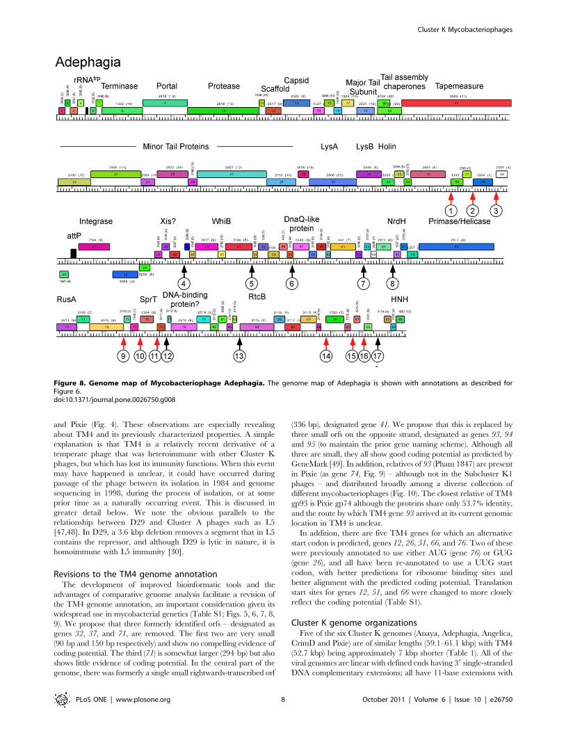
Revisions to the TM4 genome annotationThe development of improved bioinformatic tools and the
advantages of comparative genome analysis facilitate a revision of
the TM4 genome annotation, an important consideration given its
widespread use in mycobacterial genetics (Table S1; Figs. 5, 6, 7, 8,
9). We propose that three formerly identified orfs – designated as
genes 32, 37, and 71, are removed. The first two are very small
(90 bp and 150 bp respectively) and show no compelling evidence of
coding potential. The third (71) is somewhat larger (294 bp) but also
shows little evidence of coding potential. In the central part of the
genome, there was formerly a single small rightwards-transcribed orf
(336 bp), designated gene 41. We propose that this is replaced by
three small orfs on the opposite strand, designated as genes 93, 94
and 95 (to maintain the prior gene naming scheme). Although all
three are small, they all show good coding potential as predicted by
GeneMark [49]. In addition, relatives of 93 (Pham 1847) are present
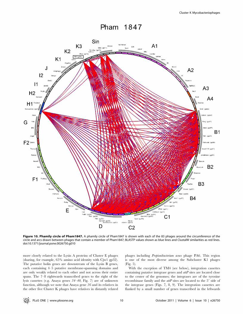
in Pixie (as gene 74, Fig. 9) – although not in the Subcluster K1
phages – and distributed broadly among a diverse collection of
different mycobacteriophages (Fig. 10). The closest relative of TM4
gp93 is Pixie gp74 although the proteins share only 53.7% identity,
and the route by which TM4 gene 93 arrived at its current genomic
location in TM4 is unclear.
In addition, there are five TM4 genes for which an alternative
start codon is predicted, genes 12, 26, 51, 66, and 76. Two of these
were previously annotated to use either AUG (gene 76) or GUG
(gene 26), and all have been re-annotated to use a UUG start
codon, with better predictions for ribosome binding sites and
better alignment with the predicted coding potential. Translation
start sites for genes 12, 51, and 66 were changed to more closely
reflect the coding potential (Table S1).
Cluster K genome organizationsFive of the six Cluster K genomes (Anaya, Adephagia, Angelica,
CrimD and Pixie) are of similar lengths (59.1–61.1 kbp) with TM4
(52.7 kbp) being approximately 7 kbp shorter (Table 1). All of the
viral genomes are linear with defined ends having 39 single-stranded
DNA complementary extensions; all have 11-base extensions with
Figure 8. Genome map of Mycobacteriophage Adephagia. The genome map of Adephagia is shown with annotations as described forFigure 6.doi:10.1371/journal.pone.0026750.g008
Cluster K Mycobacteriophages
PLoS ONE | www.plosone.org 8 October 2011 | Volume 6 | Issue 10 | e26750

the exception of TM4, which is reported to have a 10-base extension
[40]. The genomes contain between 90 and 100 predicted protein-
coding genes and the four Cluster K1 genomes – Anaya,
Adephagia, Angelica and CrimD – all encode a single tRNAtrp
near their left end (see Tables S1, S2, S3, S4, S5, S6).
To facilitate comparative genomic analysis, the three newly
sequenced genomes were added to the 80 previously described
mycobacteriophage genomes to create a database (Mycobacter-
iophage_83) for the genome comparison program, Phamerator
[50]. The total of 9,308 predicted protein-coding genes were
compared with each other using ClustalW and BlastP, and
assembled into 2,667 phamilies using previously published param-
eters (manuscript submitted). Of these, 1,120 (47.3%) are orphams
(phams containing only a single gene member). The mean pham
size is 3.932.
An overview of the relationships between the six cluster K
phages is shown in Fig. 5, and several patterns emerge. First, the
extent of nucleotide sequence similarities between the genomes are
clearly illustrated, and emphasizes the close similarity among the
Cluster K1 phages, and the more distant relationships between
these and the subcluster K2 and K3 phages. The left parts of the
Cluster K1 genomes are especially closely related, with greater
deviations in the right parts (Fig. 5). Secondly, the overall genome
architecture is shared by all six phages with a substantial number
of shared genes, as seen from the commonality of the color-coded
pham assignments (Fig. 5). Thirdly, the basis for the smaller size of
the TM4 genome compared to both its subcluster K1 and K3
relatives is apparent, with reductions in size near the left end, in
the middle, and at the extreme right end (Fig. 5; see below).
Genome maps of Anaya, Adephagia, TM4 and Pixie are shown
in Figures 6, 7, 8, 9 [Angelica and CrimD were reported recently
[8] and maps are provided as Figs. S1 and S2; the TM4 map
(Fig. 6) is a revision of that reported previously [40]. In all of the
Cluster K phages the virion structure and assembly genes occupy
the leftmost 22–24 kbp and are transcribed rightwards. There is
considerable departure among the genomes at their extreme left
ends, with a variable number of small genes of no known function
between the terminase large subunit gene and the left physical end.
All of the K1 phages, but neither TM4 nor Pixie, contain a
tRNAtrp gene in this region. Within the virion structure and
assembly genes there are a few notable differences between the
genomes. First, the putative capsid assembly proteases of the K1
and K3 phages are larger than that of TM4 (Figs. 6, 7, 8, 9) due to
a central insertion of about 1.1 kbp. This central portion does not
appear to be related to inteins, homing endonucleases, or other
mobile elements, but does have weak sequence similarity to parts
of methyl-accepting chemotaxis proteins of several bacteria
including Planctomyces limnophilus and Chromobacterium violaceum;
however, it is compositionally biased (rich in alanine) which could
account for the weak sequence similarity. The tapemeasure
proteins are similar in length with the exception of Pixie gp20,
which is 114 amino acid residues longer than the others; Pixie has
a correspondingly longer tail than the other Cluster K phages
(Fig. 2). To the right of the tail genes are the lysis cassettes, each of
which contains a Lysin A gene, a Lysin B gene, and a putative
holin gene. However, there is substantial diversity among the
Cluster K phages in these genes. For example, the Lysin A of Pixie
(gp31) is unrelated to the other Cluster K Lysin A proteins, and is
Figure 9. Genome map of Mycobacteriophage Pixie. The genome map of Pixie is shown with annotations as described for Figure 6.doi:10.1371/journal.pone.0026750.g009
Cluster K Mycobacteriophages
PLoS ONE | www.plosone.org 9 October 2011 | Volume 6 | Issue 10 | e26750
more closely related to the Lysin A proteins of Cluster E phages
(sharing, for example, 65% amino acid identity with Cjw1 gp32).
The putative holin genes are downstream of the Lysin B genes,
each containing 4–5 putative membrane-spanning domains and
are only weakly related to each other and not across their entire
spans. The 7–8 rightwards transcribed genes to the right of the
lysis cassettes (e.g. Anaya genes 34–40, Fig. 7) are of unknown
function, although we note that Anaya gene 36 and its relatives in
the other five Cluster K phages have relatives in distantly related
phages including Propionibacterium acnes phage PA6. This region
is one of the most diverse among the Subcluster K1 phages
(Fig. 5).
With the exception of TM4 (see below), integration cassettes
containing putative integrase genes and attP sites are located close
to the center of the genomes; the integrases are of the tyrosine
recombinase family and the attP sites are located to the 59 side of
the integrase genes (Figs. 7, 8, 9). The integration cassettes are
flanked by a small number of genes transcribed in the leftwards
Figure 10. Phamily circle of Pham1847. A phamily circle of Pham1847 is shown with each of the 83 phages around the circumference of thecircle and arcs drawn between phages that contain a member of Pham1847; BLASTP values shown as blue lines and ClustalW similarities as red lines.doi:10.1371/journal.pone.0026750.g010
Cluster K Mycobacteriophages
PLoS ONE | www.plosone.org 10 October 2011 | Volume 6 | Issue 10 | e26750
direction, whose function is unknown. Putative Xis genes encoding
proteins with MerR-like DNA binding domains are located to the
right within an apparently long rightwards-transcribed operon that
extends to the right end of the genomes. This region contains
WhiB-related proteins, e.g. TM4 gp49, a protein that has been
shown to be non-essential for TM4 growth [35] although it is
well-conserved among the Cluster K phages. Other genes whose
functions can be predicted from database similarity searches are
those related to SprT (e.g. Pixie gp78), RusA (e.g. Adephagia
gp75), HNH homing proteins (e.g. TM4 gp92), glutaredoxin-like
NrdH proteins (e.g. TM4 gp67) and a large Primase/Helicase
protein (e.g. TM4 gp70). The Subcluster K1 genomes also encode
Figure 11. Putative deletions giving rise to phage TM4. Comparison of the TM4 genome to the other Cluster K genomes reveals twosegments that appear to have been lost from TM4, and which may contribute to its non-temperate phenotype. A. The central parts of the CrimD,TM4, and Pixie genomes are aligned, with the colored shading reflecting the presence of genes of shared phamilies (i.e. homologues; note thisshading does not reflect nucleotide sequence similarity as in Fig. 5). Although nucleotide sequence similarity is minimal, the alignment of sharedgenes suggests the loss of about 3.5 kbp form TM4 compared to its relatives. B. Alignment of the right ends of the CrimD, TM4 and Pixie genomessuggesting loss of ,3.3 kbp from TM4 compared to its relatives; shading is as described for A.doi:10.1371/journal.pone.0026750.g011
Cluster K Mycobacteriophages
PLoS ONE | www.plosone.org 11 October 2011 | Volume 6 | Issue 10 | e26750
relatives of RtcB (e.g. Anaya gp88), a putative RNA ligase
component [51]. Because only the Subcluster K1 genomes encode
both tRNA and the RtcB proteins, we speculate that these phage-
encoded RtcB proteins play a role in protection against a host-
mediated tRNA cleavage defense against viral infection [52]. The
remainder of the proteins encoded in these regions are of unknown
function, and we note that about 30% of the Pixie genes in this
region are orphams, reflecting its high genomic diversity from all
other mycobacteriophages.
TM4 is a derivative of a temperate parentTM4 was originally isolated by recovery from a strain of M.
avium, although understanding its origin is complicated by the
observations that it is able to infect the original M. avium strain and
does not appear to be temperate in any mycobacterial host [39]
(Fig. 3). Because the related Cluster K phages are all temperate, we
have investigated potential genes that are deleted in TM4 and that
could contribute to a temperate lifestyle.
Because none of the other phages are closely related to TM4 at
the nucleotide sequence level (Fig. 5), the most informative
comparisons emerge from comparing shared genes with amino
acid sequence similarity (Fig. 11). We have focused on two regions
of the genomes. The first is at the center of the genomes where the
integration cassettes are found in the Subcluster K1 and K3
phages (Fig. 11A). TM4 genes 40 and 42 correspond to CrimD
genes 38 and 44 such that the three leftwards transcribed TM4
genes, 93, 94, and 95, occupy the location corresponding to
CrimD genes 39 and 44 (Fig. 11A). Thus a simple explanation is
that TM4 has lost a DNA segment approximately 3.5 kbp in
length from a temperate parent that included the integrase gene
and attP site. Interestingly, TM4 retains the predicted Xis
function encoded by gene 43, consistent with this interpretation
(Fig. 11A).
The second region of interest is at the right end. The
comparison between CrimD and TM4 is perhaps the most
informative. CrimD contains homologues of TM4 gp84 and gp85
(CrimD gp83 and gp90), but they are separated by a 3.3 kbp DNA
segment containing six predicted open reading frames (Fig. 11B).
This suggests that TM4 has undergone a deletion of approxi-
mately 3.3 kbp between genes 84 and 85 from its putative
temperate parent. It is plausible that one of the lost genes
corresponds to a phage repressor, consistent with TM4’s clear
plaque phenotype. We note that the L5 repressor (gp71) is
encoded near the right end of its genome, so this is a not an
unusual genomic position for a repressor gene. Although none
of the genes in these regions of the Cluster K1 or K3 genomes
have sequence similarity to known repressors, all the K1 and K3
phages are homoimmune and are thus expected to share similar
repressors. Pixie is quite different from the K1 genomes in this
region, and there is only a single gene that they share in this
interval, corresponding to Pixie gp85, Anaya gp90, Adephagia
gp86, CrimD gp87, and Angelica gp84. However, prelimi-
nary analysis suggests that expression of CrimD gp87 from a
plasmid in the host cell does not confer immunity to any of
Cluster K phages and it is therefore an unlikely repressor
candidate.
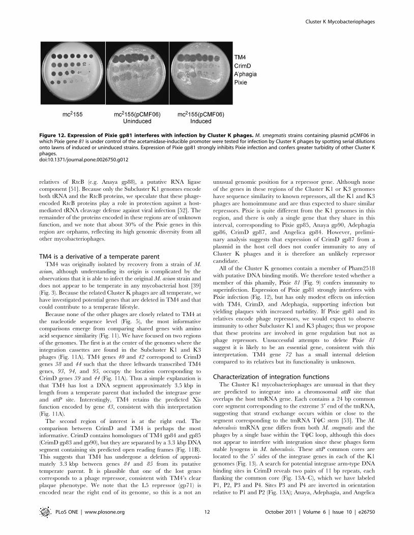
All of the Cluster K genomes contain a member of Pham2518
with putative DNA binding motifs. We therefore tested whether a
member of this phamily, Pixie 81 (Fig. 9) confers immunity to
superinfection. Expression of Pixie gp81 strongly interferes with
Pixie infection (Fig. 12), but has only modest effects on infection
with TM4, CrimD, and Adephagia, supporting infection but
yielding plaques with increased turbidity. If Pixie gp81 and its
relatives encode phage repressors, we would expect to observe
immunity to other Subcluster K1 and K3 phages; thus we propose
that these proteins are involved in gene regulation but not as
phage repressors. Unsuccessful attempts to delete Pixie 81
suggest it is likely to be an essential gene, consistent with this
interpretation. TM4 gene 72 has a small internal deletion
compared to its relatives but its functionality is unknown.
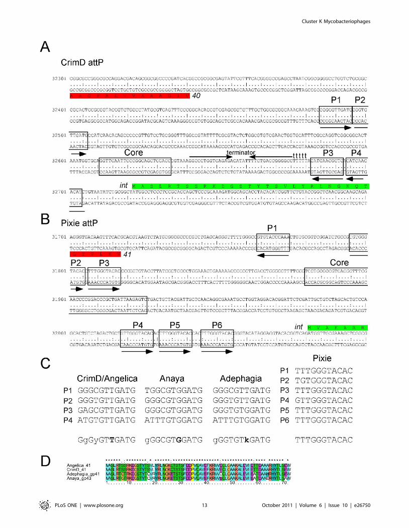
Characterization of integration functionsThe Cluster K1 mycobacteriophages are unusual in that they
are predicted to integrate into a chromosomal attB site that
overlaps the host tmRNA gene. Each contains a 24 bp common
core segment corresponding to the extreme 39 end of the tmRNA,
suggesting that strand exchange occurs within or close to the
segment corresponding to the tmRNA TyC stem [53]. The M.
tuberculosis tmRNA gene differs from both M. smegmatis and the
phages by a single base within the TyC loop, although this does
not appear to interfere with integration since these phages form
stable lysogens in M. tuberculosis. These attP common cores are
located to the 59 sides of the integrase genes in each of the K1
genomes (Fig. 13). A search for potential integrase arm-type DNA
binding sites in CrimD reveals two pairs of 11 bp repeats, each
flanking the common core (Fig. 13A–C), which we have labeled
P1, P2, P3 and P4. Sites P3 and P4 are inverted in orientation
relative to P1 and P2 (Fig. 13A); Anaya, Adephagia, and Angelica
Figure 12. Expression of Pixie gp81 interferes with infection by Cluster K phages. M. smegmatis strains containing plasmid pCMF06 inwhich Pixie gene 81 is under control of the acetamidase-inducible promoter were tested for infection by Cluster K phages by spotting serial dilutionsonto lawns of induced or uninduced strains. Expression of Pixie gp81 strongly inhibits Pixie infection and confers greater turbidity of other Cluster Kphages.doi:10.1371/journal.pone.0026750.g012
Cluster K Mycobacteriophages
PLoS ONE | www.plosone.org 12 October 2011 | Volume 6 | Issue 10 | e26750
Cluster K Mycobacteriophages
PLoS ONE | www.plosone.org 13 October 2011 | Volume 6 | Issue 10 | e26750
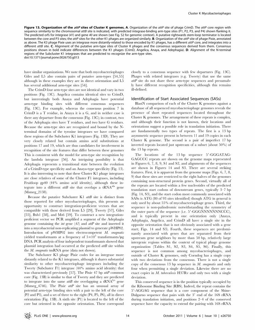
have similar organizations. We note that both mycobacteriophages
Giles and L5 also contain pairs of putative arm-types [54,55]
although in these examples they are in direct orientation and L5
has several additional arm-type sites [54].
The CrimD four arm-type sites are not identical and vary in two
positions (Fig. 13C). Angelica contains identical sites to CrimD,
but interestingly both Anaya and Adephagia have potential
arm-type binding sites with different consensus sequences
(Fig. 13C). For example, whereas the consensus position 7 in
CrimD is a T residue, in Anaya it is a G, and in neither case is
there any departure from the consensus (Fig. 13C); in contrast, two
of the Adephagia sites have T residues, and two have G residues.
Because the arm-type sites are recognized and bound by the N-
terminal domains of the tyrosine integrases we have compared
these regions of the Subcluster K1 integrases (Fig. 13D). They are
very closely related but contain amino acid substitutions at
positions 17 and 19, which are thus candidates for involvement in
recognition of the site features that differ between these genomes
This is consistent with the model for arm-type site recognition by
the lambda integrase [56]. An intriguing possibility is that
Adephagia represents a transitional state between the evolution
of a CrimD-type specificity and an Anaya-type specificity (Fig. 13).
It is also interesting to note that these Cluster K1 phage integrases
are close relatives of some of the Cluster F1 integrases, including
Fruitloop gp40 (44% amino acid identity), although these in-
tegrate into a different attB site that overlaps a tRNAala gene
(Msmeg_2138).
Because the putative Subcluster K1 attB site is distinct from
those reported for other mycobacteriophages, this presents an
opportunity to construct integration-proficient vectors that are
compatible with those derived from L5 [29], Tweety [57], Giles
[55], Bxb1 [58], and Ms6 [59]. To construct a new integration-
proficient vector we PCR amplified a segment of the Adephagia
genome containing the int gene (41) and attP site and inserted it
into a mycobacterial non-replicating plasmid to generate pWHP02.
Introduction of pWHP02 into electrocompetent M. smegmatis
yielded transformants at a frequency of 56105 transformants/mg
DNA. PCR analysis of four independent transformants showed that
plasmid integration had occurred at the predicted attB site within
the M. smegmatis tmRNA gene (data not shown).
The Subcluster K3 phage Pixie codes for an integrase more
distantly related to the K1 integrases, although it shares substantial
similarity to other mycobacteriophage integrases including the
Tweety (Subcluster F1) integrase (44% amino acid identity) that
was characterized previously [57]. The Pixie 47 bp attP common
core (Fig. 13B) is similar to that of Tweety and they are predicted
to integrate into the same attB site overlapping a tRNAlys gene
(Msmeg_4746). The Pixie attP site has an unusual array of
potential arm-type binding sites with a pair to the left of the core
(P2 and P3), and a set of three to the right (P4, P5, P6), all in direct
orientation (Fig. 13B). A sixth site (P1) is located to the left of the
core but oriented in the opposite orientation. These correspond
closely to a consensus sequence with few departures (Fig. 13C).
Phages with related integrases (e.g. Tweety) that use the same
attP site do not share these arm-type sequences and presumab-
ly have different recognition specificities, although this remains
ill-defined.
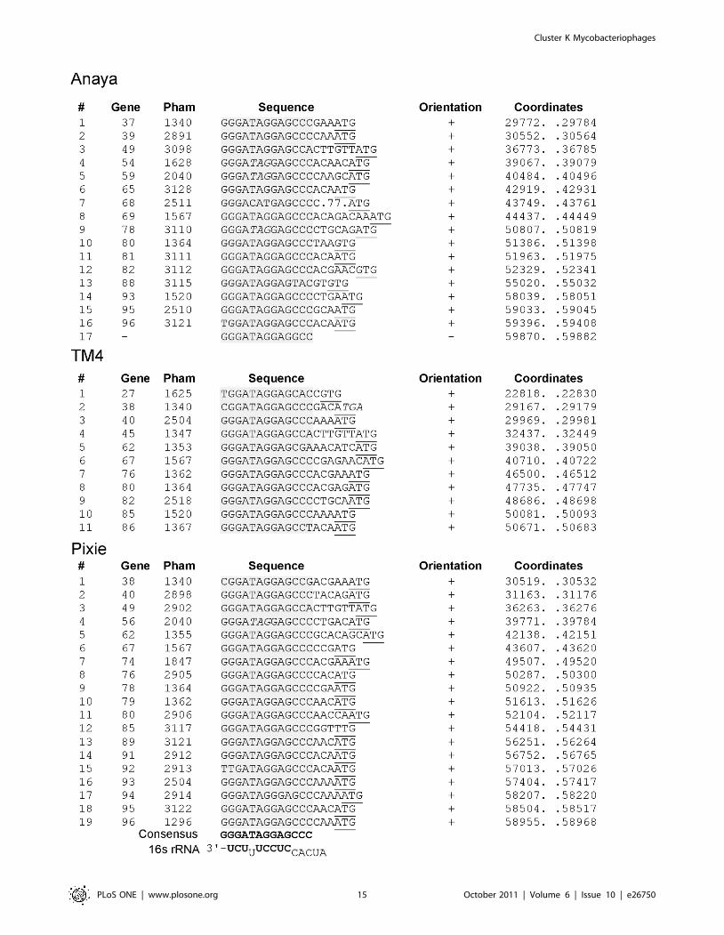
Identification of Start Associated Sequences (SASs)BlastN comparison of each of the Cluster K genomes against a
database of all sequenced mycobacteriophage genomes reveals the
presence of short repeated sequences located throughout the
Cluster K genomes. The arrangement of these repeats is complex,
and although their function is not known, their locations and
orientations suggest a possible role in translation initiation. There
are fundamentally two types of repeats. The first is a 13 bp
asymmetric sequence present in between 11 and 19 copies in each
Cluster K genome. The second is a pair of imperfect 17 bp
inverted repeats located just upstream of a subset (about 50%) of
the 13 bp repeats.
The locations of the 13 bp sequence 59-GGGATAG-
GAGCCC repeats are shown on the genome maps represented
in Figures 6, 7, 8, 9, S1 and S2, and alignments of the sequences
are shown in Figures 14 and S3. There are several striking
features. First, it is apparent from the genome maps (Figs. 6, 7, 8,
9) that these sites are restricted to the right halves of the genomes
containing non-structural protein genes. Second, virtually all of
the repeats are located within a few nucleotides of the predicted
translation start codons of downstream genes, typically 3–7 bp
(Fig. 14, S3), and the start codon most commonly associated with
SASs is ATG (80 of 93 sites identified) though ATG in general is
only used by about 55% of mycobacteriophage genes. Third, the
sequence is non-palindromic notwithstanding the symmetry of
the outer parts of the sequence (i.e. 59-GGGNNNNNNNCCC),
and is typically present in one orientation only (Anaya,
Adephagia, Angelica, and CrimD all have a single site in the
opposite orientation that is not obviously associated with a gene
start; Figs. 14 and S3). Fourth, these sequences are predomi-
nantly associated with genes that are separated from their
upstream gene neighbors by more than 50 bp, relatively large
intergenic regions within the context of typical phage genome
organization (Tables S1, S2, S3, S4, S5, S6). Finally, this
sequence is not common among mycobacteriophages, and
outside of Cluster K genomes, only Corndog has a single copy
with two deviations from the consensus. There is not a single
copy of the consensus 13 bp sequence in M. smegmatis and only
four when permitting a single deviation. Likewise there are no
exact copies in M. tuberculosis H37Rv and only two with a single
deviation.
This conserved sequence is in the position typically occupied by
the Ribosome Binding Site (RBS). Indeed, the repeat contains the
59-AGGAG sequence that is a core component of the Shine-
Dalgarno sequence that pairs with the 39 end of the 16S rRNA
during translation initiation, and positions 2–4 of the conserved
sequence have the capacity to extend the pairing with 16S rRNA
Figure 13. Organization of the attP sites of Cluster K genomes. A. Organization of the attP site of phage CrimD. The attP core region withsequence similarity to the chromosomal attB site is indicated, with predicted integrase-binding arm-type sites (P1, P2, P3, and P4) shown flanking it.The predicted orfs for integrase (41) and gene 40 are shown (see Fig. S2 for genomic context). A putative rightwards stem-loop terminator is locatedbetween the core and P3 sites; the attP sites for the other K1 phages are organized similarly. B. Organization of the attP site of phage Pixie, annotatedas above. The K3 phage Pixie uses an integrase more distantly-related to those encoded by K1 phages, has a different attP core, and integrates into adifferent attB site. C. Alignment of the putative arm-type sites of Cluster K phages and the consensus sequences derived from them. Consensuspositions shown in bold indicate differences between the K1 phages (CrimD, Angelica, Anaya, and Adephagia). D. Alignment of the N-terminalregions of the Subcluster K1 integrases that are predicted to recognize the arm-type sites.doi:10.1371/journal.pone.0026750.g013
Cluster K Mycobacteriophages
PLoS ONE | www.plosone.org 14 October 2011 | Volume 6 | Issue 10 | e26750
Cluster K Mycobacteriophages
PLoS ONE | www.plosone.org 15 October 2011 | Volume 6 | Issue 10 | e26750
(Fig. 14). However, it seems unlikely that this repeat simply
corresponds to just a favorable translation initiation site. First, the
starting base of the sequence is extremely well conserved (Fig. 14)
but has no corresponding base to pair with in 16S rRNA. Second,
positions 10–13 are also highly conserved, but do not have pairing
potential with rRNA (Fig. 14). Nonetheless, the positioning of
these repeats suggests a role in translation initiation – in contrast to
the 13 bp stoperator sequences in L5 and other Cluster A phages
that play a role in transcription regulation [60] – and we therefore
propose that they be called Start Associated Sequences (SASs).
Whether these act independently or represent binding sites for
either a host- or phage-encoded gene product (either RNA or
protein) remains to be determined. The conservation of these sites
across the three subclusters – often associated with genes of
different phamilies (Table 2) – strongly suggests that they play
important roles for these phages.
Approximately one half of the genes with an SAS also contain a
second sequence feature composed of imperfect 17 bp inverted
repeats (IRs) separated by a variable spacer (Figs. 15, S4). Because
these are tightly associated with SASs, we refer to these as
extended SASs (ESAS); in one notable exception the inverted
repeat upstream of TM4 gene 79 does not appear to be associated
with an SAS (Fig. 15A). For each genome a consensus sequence
can be derived (Fig. 15B) from the left and right IRs, although the
left IRs typically have a closer correspondence to the consensus
than the right IRs (Figs. 15, S4); the spacer region between the IRs
is variable, but is 4–13 bp in the vast majority of sites (Figs. 15A,
S4). Interestingly, the consensus sequence of the IRs is different for
phages of the three subclusters. The four Subcluster K1 phages
have very similar IR consensus sequences (Figs. 15, S4), but differ
from those of the Subcluster K2 (TM4) and K3 (Pixie) at positions
11, 12 and 13. For example, at position 11, there is predominantly
a C in Anaya (in 15 of 16 IRs), but a T in both Pixie and TM4 (16
of 18 and 10 of 12 IRs respectively). At position 12, the C residue
is strongly conserved in both Pixie and TM4, with no departures in
any of the 30 constituent IRs, but this site is predominantly an A
residue in Anaya (two of the 16 IRs have a C). At position 13 Pixie
and TM4 have a consensus A residue, with no departures in any of
the 30 IRs, whereas in Anaya this site is predominantly a T (two
IRs have a G, and one has a A) (Fig. 15).
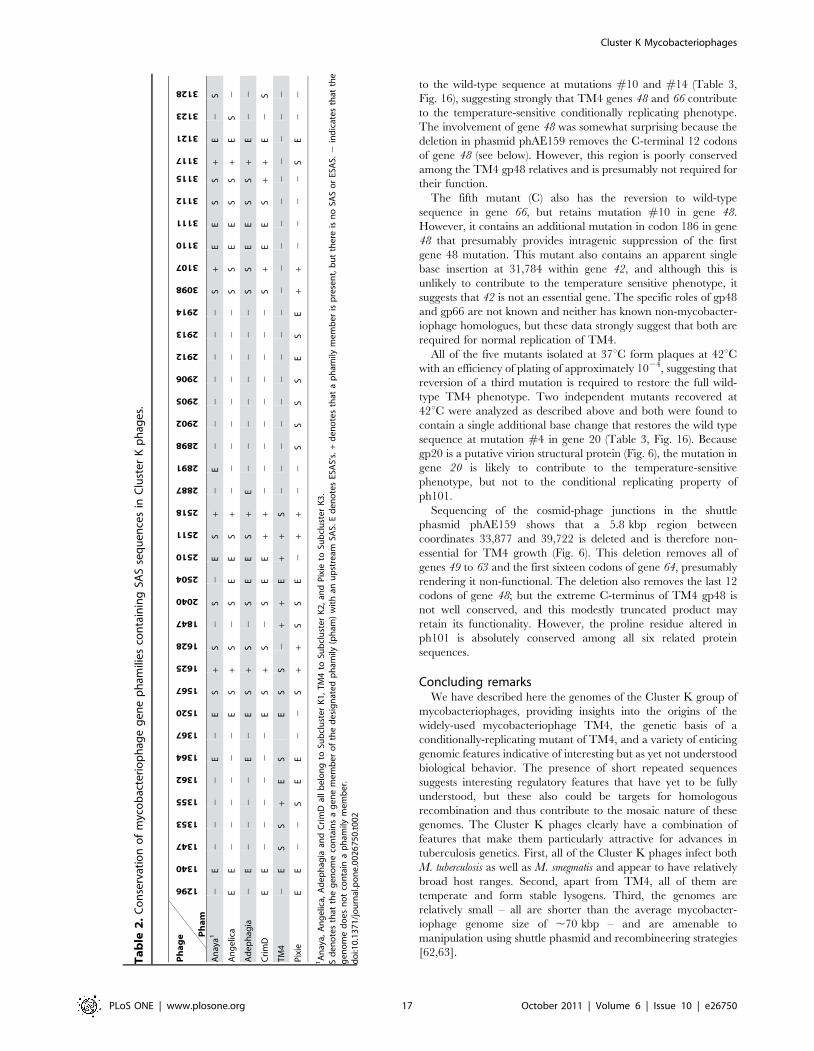
The ESAS sites are well conserved among the Cluster K
genomes, in that if a gene of a particular phamily is associated with
an ESAS in one genome, then other Cluster K genomes
containing a gene member of that phamily also have an
associated ESAS (Table 2). A notable exception is TM4 gene
80 (Pham 1364), which lacks an ESAS (it has an SAS), whereas all
other phamily members have an ESAS (Table 2). Inspection of
the TM4 sequence shows that the site is completely lacking,
rather than having more highly diverged but related IRs. The
conservation of these sites strongly suggests that they serve
important functions for the phages, although it is not clear what
they are. Because these are closely linked with the SASs that in
turn are associated with translation initiation sites, it is tempting
to assume that they also play a role in translation initiation.
However, there is little support for the possibility that the two IRs
form hairpin structures in mRNA, in that departures in the left
and right IRs do not generally support RNA base-pairing.
Therefore, it seems more likely that these represent binding sites
for DNA-binding proteins and that the differences in consensus
sequences represent different specificities in the three subclusters.
One possible role might be in transcription initiation (i.e.
promoters), but alternatively they could be operator sites for
phage repressors. This latter explanation is attractive except that
the K1 and K2 phages are homoimmune (Fig. 4), which is not
consistent with the consensus differences. Furthermore, it is
unclear why in virtually every occurrence the IRs are closely
associated with translation initiation signals if they are operator
sites. Finally, we note that in Pixie and TM4 each 17 bp IR itself
has a symmetrical character, and can be considered as a 6 bp half
site (59-TGTTGA) separated by a 4 bp spacer from the inverse
complement (Fig. 15B). However, this is not true for the
Subcluster K1 phages because of the consensus differences at
positions 11–13 (Fig. 15B, S4), as discussed above.
Characterization of a conditionally-replicating mutant ofTM4
Bardarov et al. (1997) described a conditionally replicating
mutant of TM4 that fails to form plaques and fails to kill infected
cells at temperatures of 37uC or above. This mutant – ph101 – is
the basis for the construction of conditionally replicating shuttle
phasmids used for delivery of reporter genes, transposons, and
allelic exchange substrates to mycobacterial hosts [20,23,61]. The
mutant was isolated using two rounds of hydroxylamine
mutagenesis with the goal of isolating mutants that revert only at
very low frequencies [20]. Because the functions of so few TM4
genes are known, we characterized the mutations in ph101.
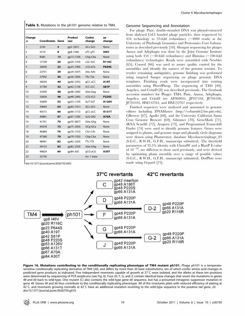
Sequencing of the complete ph101 genome reveals a total of 23
differences (Table 3). One of these is a one base insertion in a non-
coding region at the extreme right end of the genome; the others
are all base substitution transitions, consistent with the mutagenic
spectrum of hydroxylamine (Table 3). The large number of
mutations reflects the heavy mutagenesis employed to recover the
non-reverting mutants. Twelve of the base substitutions do not
alter the predicted coding sequences, whereas the other ten do and
are therefore candidates for contributing to the temperature-
sensitive phenotype. Because the reversion frequency of ph101 is
low (,1028) it is likely that more than one mutation contributes to
this phenotype. Three of the affected genes are predicted virion
structure genes (8, 20, 23) and are unlikely to be involved in DNA
replication (Fig. 6).
To gain insight into which of the mutations contribute to the
temperature sensitive phenotype we isolated five independent
revertant mutants (C, D, F, G, and J) that are able to grow at
37uC, followed by PCR amplification and sequencing of the
regions containing the ten non-synonymous mutations (Fig. 16).
Revertants D, F, G, and J each contains nucleotide changes back
Figure 14. Location of Start Associated Sequences (SASs). Repeated sequences were identified in Cluster K phages through BLASTNcomparisons with other mycobacteriophages, followed by scanning for the presence of the sequence 59-GGGATAGGAGCCC, allowing for up to twodeviations from the scanned sequence. (Pixie site #19 has three departures from the consensus but is included in the list because it is associated withan Extended SAS, see Fig. 15). Sites for Angelica, Adephagia, and CrimD are shown in Fig. S3. The sequence is asymmetric and most copies areorientated in one direction as indicated. With the rare exception of those sites in the opposite orientation (e.g. Anaya site #17), all are immediatelyupstream of gene start sites (Anaya site #7 is a notable exception.) The gene immediately downstream is listed along with its phamily (Pham)designation; the putative translation initiation codons are underlined; where the termination codon of the upstream gene lies within the conservedsequence it is italicized. The consensus sequence is shown in bold and the positions of the sites are shown by the colored highlighting. The extreme39 end of the 16S rRNA is shown with bases predicted to contribute to pairing with mRNA shown in bold. The genomic locations of these SASs areshown by numbered vertical arrows in Figs. 6, 7, 8, 9, S1 and S2).doi:10.1371/journal.pone.0026750.g014
Cluster K Mycobacteriophages
PLoS ONE | www.plosone.org 16 October 2011 | Volume 6 | Issue 10 | e26750
to the wild-type sequence at mutations #10 and #14 (Table 3,
Fig. 16), suggesting strongly that TM4 genes 48 and 66 contribute
to the temperature-sensitive conditionally replicating phenotype.
The involvement of gene 48 was somewhat surprising because the
deletion in phasmid phAE159 removes the C-terminal 12 codons
of gene 48 (see below). However, this region is poorly conserved
among the TM4 gp48 relatives and is presumably not required for
their function.
The fifth mutant (C) also has the reversion to wild-type
sequence in gene 66, but retains mutation #10 in gene 48.
However, it contains an additional mutation in codon 186 in gene
48 that presumably provides intragenic suppression of the first
gene 48 mutation. This mutant also contains an apparent single
base insertion at 31,784 within gene 42, and although this is
unlikely to contribute to the temperature sensitive phenotype, it
suggests that 42 is not an essential gene. The specific roles of gp48
and gp66 are not known and neither has known non-mycobacter-
iophage homologues, but these data strongly suggest that both are
required for normal replication of TM4.
All of the five mutants isolated at 37uC form plaques at 42uCwith an efficiency of plating of approximately 1024, suggesting that
reversion of a third mutation is required to restore the full wild-
type TM4 phenotype. Two independent mutants recovered at
42uC were analyzed as described above and both were found to
contain a single additional base change that restores the wild type
sequence at mutation #4 in gene 20 (Table 3, Fig. 16). Because
gp20 is a putative virion structural protein (Fig. 6), the mutation in
gene 20 is likely to contribute to the temperature-sensitive
phenotype, but not to the conditional replicating property of
ph101.
Sequencing of the cosmid-phage junctions in the shuttle
phasmid phAE159 shows that a 5.8 kbp region between
coordinates 33,877 and 39,722 is deleted and is therefore non-
essential for TM4 growth (Fig. 6). This deletion removes all of
genes 49 to 63 and the first sixteen codons of gene 64, presumably
rendering it non-functional. The deletion also removes the last 12
codons of gene 48; but the extreme C-terminus of TM4 gp48 is
not well conserved, and this modestly truncated product may
retain its functionality. However, the proline residue altered in
ph101 is absolutely conserved among all six related protein
sequences.
Concluding remarksWe have described here the genomes of the Cluster K group of
mycobacteriophages, providing insights into the origins of the
widely-used mycobacteriophage TM4, the genetic basis of a
conditionally-replicating mutant of TM4, and a variety of enticing
genomic features indicative of interesting but as yet not understood
biological behavior. The presence of short repeated sequences
suggests interesting regulatory features that have yet to be fully
understood, but these also could be targets for homologous
recombination and thus contribute to the mosaic nature of these
genomes. The Cluster K phages clearly have a combination of
features that make them particularly attractive for advances in
tuberculosis genetics. First, all of the Cluster K phages infect both
M. tuberculosis as well as M. smegmatis and appear to have relatively
broad host ranges. Second, apart from TM4, all of them are
temperate and form stable lysogens. Third, the genomes are
relatively small – all are shorter than the average mycobacter-
iophage genome size of ,70 kbp – and are amenable to
manipulation using shuttle phasmid and recombineering strategies
[62,63].
Ta
ble
2.
Co
nse
rvat
ion
of
myc
ob
acte
rio
ph
age
ge
ne
ph
amili
es
con
tain
ing
SAS
seq
ue
nce
sin
Clu
ste
rK
ph
age
s.
Ph
ag
e Ph
am
1296
1340
1347
1353
1355
1362
1364
1367
1520
1567
1625
1628
1847
2040
2504
2510
2511
2518
2887
2891
2898
2902
2905
2906
2912
2913
2914
3098
3107
3110
3111
3112
3115
3117
3121
3123
3128
An
aya1
2E
22
22
E2
ES
+S
2S
2E
S+
2E
22
22
22
2S
+E
ES
S+
E2
S
An
ge
lica
EE
22
22
22
ES
+S
2S
EE
S+
22
22
22
22
2S
SE
ES
S+
ES
2
Ad
ep
hag
ia2
E2
22
2E
2E
S+
S2
SE
ES
+E
22
22
22
22
SS
EE
SS
+E
22
Cri
mD
EE
22
22
22
ES
+S
2S
EE
++
22
22
22
22
2S
+E
ES
++
E2
S
TM
42
ES
S+
ES
ES
S2
++
E+
+S
22
22
22
22
22
22
22
22
22
2
Pix
ieE
E2
2S
EE
22
S+
+S
SE
2+
+2
2S
SS
SE
SE
++
22
22
SE
22
1A
nay
a,A
ng
elic
a,A
de
ph
agia
and
Cri
mD
all
be
lon
gto
Sub
clu
ste
rK
1,
TM
4to
Sub
clu
ste
rK
2,
and
Pix
ieto
Sub
clu
ste
rK
3.
Sd
en
ote
sth
atth
eg
en
om
eco
nta
ins
ag
en
em
em
be
ro
fth
ed
esi
gn
ate
dp
ham
ily(p
ham
)w
ith
anu
pst
ream
SAS.
Ed
en
ote
sES
AS’
s.+
de
no
tes
that
ap
ham
ilym
em
be
ris
pre
sen
t,b
ut
the
reis
no
SAS
or
ESA
S.2
ind
icat
es
that
the
ge
no
me
do
es
no
tco
nta
ina
ph
amily
me
mb
er.
do
i:10
.13
71
/jo
urn
al.p
on
e.0
02
67
50
.t0
02
Cluster K Mycobacteriophages
PLoS ONE | www.plosone.org 17 October 2011 | Volume 6 | Issue 10 | e26750
Materials and Methods
Bacterial strains and MediaM. smegmatis mc2155, Mycobacterium bovis bacilli Calmette-Guerin
(BCG) (Jacobs et al., 1991; Snapper et al., 1990) and M. tuberculosis
mc27000 have been described previously [64]. M. marinum strains
M and 927 were kind gifts from Dr. Don G. Ennis, University of
Louisiana, Lafayette, LA; M. avium 104 was a gift from Dr.
William R. Bishai, Johns Hopkins School of Medicine, Baltimore,
MD. Media were supplemented with carbenicillin (50 mg ml21)
and/or cycloheximide (10 mg ml21) as required. M. smegmatis and
M. marinum were grown in 7H9 liquid and on 7H10 plates; M.
tuberculosis, M. bovis, and M. avium were grown in 7H9 liquid, and
on 7H11 plates.
Phage Isolation and purificationPhage Pixie was isolated from a dry soil sample obtained from a
yard located in the Northwest Houston Metropolitan Area (TX,
USA). Phage Anaya was isolated from a soil sample obtained from
Grand Rapids, MI (MI, USA). Phage Adephagia was isolated from
a soil sample obtained from Denton, TX (TX, USA). All phages
were isolated by co-plating of soil extracts prepared with phage
buffer (10 mM Tris/HCl pH 7.5, 10 mM MgSO4, 1 mM CaCl2,
68.5 mM NaCl), and M. smegmatis. The soil extract was filtered
through a 0.22 mm filter. For Pixie, 50 ml of this sample was direct
plated with 0.5 mL late-exponential-phase M. smegmatis mc2155 in
4.5 mL 0.35% mycobacterial top agar (MBTA) with 1 mM CaCl2.
The MBTA/phage/bacterial mixture was distributed evenly on a
plate of 7H10 agar (Difco) supplemented with carbenicillin,
cycloheximide, 1 mM CaCl2 and 10% albumin dextrose complex
(ADC). Phages Anaya and Adephagia were isolated by incubating
1 gram of soil with M. smegmatis mc2155 in 50 mL of LB plus 1 mM
CaCl2 at 37uC with shaking for 24 hours. Remaining cells were
then pelleted by centrifugation, and the supernatant was filtered
through a 0.22 mm filter. Fifty microliters of the filtrate was co-
plated on LB plates with 0.5 mL late-exponential phase M. smegmatis
mc2155 in 4.5 mL LB top agar. All phages were incubated at 37uC,
except for Anaya, which was incubated at 30uC. After several
rounds of purification (Sarkis & Hatfull, 1998) high-titer stocks were
prepared and used for subsequent studies.
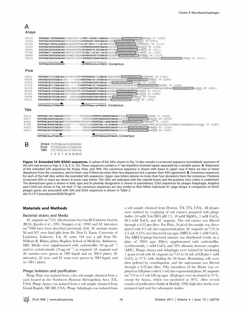
Figure 15. Extended SAS (ESAS) sequences. A subset of the SASs shown in Fig. 14 also contain a conserved sequence immediately upstream ofthe SAS (red arrows in Figs. 6, 7, 8, 9, S1, S2). These sequences contain a 17 bp imperfect inverted repeat separated by a variable spacer. A. Alignmentof the extended SAS sequences for Anaya, Pixie, and TM4. The consensus sequence is shown with bases in upper case if there are two or fewerdepartures from the consensus, and in lower case if there are more than two departures but a greater than 50% agreement. B. Consensus sequencesfor each of the half sites within the extended SAS sequences. Upper case letters denote no more than four deviations from the consensus. Positionsconserved 50% or more are shown in lower case letters. The SASs are indicated with the colored boxes and the putative start codon is underlined.The downstream gene is shown in italic type and its phamily designation is shown in parentheses. ESAS sequences for phages Adephagia, Angelicaand CrimD are shown in Fig. S4; their 17 bp consensus sequences are very similar to their fellow Subcluster K1 page Anaya. A comparison of whichphages genes are associated with SAS and ESAS sequences is shown in Table 2.doi:10.1371/journal.pone.0026750.g015
Cluster K Mycobacteriophages
PLoS ONE | www.plosone.org 18 October 2011 | Volume 6 | Issue 10 | e26750
Genome Sequencing and AnnotationFor phage Pixie, double-stranded DNA was phenol-extracted
from dialyzed CsCl banded phage particles, then sequenced by
454 technology to 25-fold redundancy (,4000 reads) at the
University of Pittsburgh Genomics and Proteomics Core Labora-
tories as described previously [10]. Shotgun sequencing for phages
Anaya and Adephagia was done by the Joint Genome Institute
using both 454 (,30-fold redundancy) and Illumina (,100-fold
redundancy) technologies. Reads were assembled with Newbler
[65]; Consed [66] was used to assure quality control for the
assemblies and identify the natures of the genome termini. To
resolve remaining ambiguities, genome finishing was performed
using targeted Sanger sequencing on phage genomic DNA
templates. Finishing reads were incorporated into existing
assemblies using PhredPhrap. The sequencing of TM4 [40],
Angelica, and CrimD [8] was described previously. The Genbank
accession numbers for Phages TM4, Pixie, Anaya, Adephagia,
Angelica and CrimD are AF068845, JF937104, JF704106,
JF704105, HM152764, and HM152767 respectively.
Finished sequences were analyzed and annotated in genome
editors including DNAMaster (http://cobamide2.bio.pitt.edu),
GBrowse [67], Apollo [68], and the University California Santa
Cruz Genome Browser [69]; Glimmer [70], GeneMark [71],
tRNA ScanSE [72], Aragorn [73], and Programmed Frameshift
Finder [74] were used to identify genome features. Genes were
assigned to phams, and genome maps and phamily circle diagrams
were drawn using Phamerator, database Mycobacteriophage_83
(S.G.C., R.W.H., G.F.H., manuscript submitted). The threshold
parameters of 32.5% identity with ClustalW and a BlastP E-value
of 10250, are different to those used previously, and were derived
by optimizing pham assembly over a range of possible values
(S.G.C., R.W.H., G.F.H., manuscript submitted). DotPlots were
made using Gepard [75].
Table 3. Mutations in the ph101 genome relative to TM4.
Change# Coordinates Gene
Product(aa)
Codonchange
aachange
1 2742 5 gp5 (501) GCc-GCt None
2 5519 8 gp8 (186) aTC-gTC I46V
3 8385 13 gp13 (139) CAg-CAa None
4 17199 20 gp20 (154) cGC-tGC R116C
5 20656 23 gp23 (784) cCG-tCG P644S
6 23751 29 gp29 (547) AAc-AAt None
7 27942 36 gp36 (394) TAc-Tat None
8 30039 40 gp40 (235) gCC-aCC A19T
9 31784 42 gp42 (118) tCC-cCC S81P
10 33509 48 gp48 (246) GAa-Gag None
11 33834 48 gp48 (246) cCG-tCG P220S
12 35899 53 gp53 (129) GcT-GtT A126V
13 39664 63 gp63 (101) GCc-GCt None
14 40515 66 gp66 (172) gCC-aCC A131T
15 40881 67 gp67 (100) GcG-GtG A70A
16 41761 70 gp70 (867) GAa-GAg None
17 45678 75 gp75 (303) GCg-GCa None
18 46889 76 gp76 (153) CAc-CAt None
19 47580 79 gp79 (103) CAg-CAa None
20 48991 82 gp82 (259) TTc-TTt None
21 49153 82 gp82 (259) GAa-GAg None
22 49824 84 gp84 (65) gCG-aCG A30T
23 52756 Ins 1 base
doi:10.1371/journal.pone.0026750.t003
Figure 16. Mutations contributing to the conditionally replicating phenotype of TM4 mutant ph101. Phage ph101 is a temperate-sensitive conditionally replicating derivative of TM4 [20], and differs by more than 20 base substitutions, ten of which confer amino acid changes inpredicted gene products as indicated. Five independent revertants capable of growth at 37uC were isolated, and the alleles at these ten positionswere determined by sequencing of PCR amplicons (see Fig. 6). Four (D, F, G, and J) contain identical base changes that revert the mutations in genes48 and 66 back to wild-type. One mutant (C) also contains the wild-type gene 66 sequence, but has a presumed intragenic suppressor mutation ingene 48. Genes 48 and 66 thus contribute to the conditionally replicating phenotype. All of the revertants plate with reduced efficiency of plating at42uC, and revertants growing normally at 42uC have an additional mutation reverting to the wild-type sequence in the putative tail gene, 20.doi:10.1371/journal.pone.0026750.g016
Cluster K Mycobacteriophages
PLoS ONE | www.plosone.org 19 October 2011 | Volume 6 | Issue 10 | e26750
Lysogen PCR AssaysSite-specific integration between the putative phage attP of
Pixie, Angelica, Anaya, Adephagia, and CrimD with the
corresponding M. smegmatis attB sites was confirmed in lysogens
by PCR amplification of the attL and attB sites. Pelleted, potential
lysogenic cells were suspended in 500 ml of 10 mM Tris (pH 8.0),
1 mM ethylenediaminetetraacetic acid (EDTA), heated for
20 minutes at 95uC and 10 ml was used in PCRs with Pfu
polymerase (Stratagene), 5% Dimethyl sulfoxide (DMSO) and
10 nM dNTPs. Primers CMF1 and CMF2 were used to amplify
bacterial attB of Pixie, primers CMF4 and CMF6 were used to
amplify the bacterial attB of Angelica, CrimD, Adephagia and
Anaya. Primers CMF3 and CMF2 were used to amplify attL of
Pixie, primers CMF5 and CMF6 were used to amplify attL of
Angelica, CrimD and Adephagia, and primers CMF4 and CMF13
were used to amplify attL of Anaya. Primer sequences are listed in
Table S7.
Immunity assaysImmunity to K cluster phages was tested by spotting serial dilutions
of each phage onto lawns of M. smegmatis mc2155, mc2155(Pixie)
lysogens, mc2155(Angelica) lysogens, mc2155(CrimD) lysogens,
mc2155(Anaya) lysogens and mc2155(Adephagia) lysogens.
Plasmid ConstructionsPlasmid pWHP02 was constructed by amplifying the integrase
gene (41) and attP site from Adephagia virions using the primers
Ade-IntF and Ade-IntR. These primers added the restriction sites
SbfI and SacI to flank the target region. The L5-derived integration
vector pMH94 (KanR, oriE) was digested with SbfI and SacI to
remove the L5 attP region, and the amplified, digested Adephagia
attP region was ligated into the vector backbone. The ligation
reaction was transformed into E. coli DH10B, and recovered
colonies were picked into LB-Kan to isolate DNA. Correct
plasmid sequences were confirmed through restriction digestion
and sequencing. Plasmid pWHP02 was then transformed into
electrocompetent M. smegmatis mc2155, and plated on 7H10 Kan.
Recovered colonies were grown in 7H9 Kan, and used as a
template in a PCR amplification to check for insertion into the
tmRNA site using primers CMF4, CMF5 and CMF6.
Plasmid pCMF06 was constructed as follows. Two primers
(CMF18 and CMF19) with NdeI and HpaI restriction sites were
designed and used to amplify Pixie gene 81 from Pixie genomic
DNA. This 899 bp fragment was inserted by sticky-end cloning
into pLAM12. Plasmid pCMF06 contains Pixie gene 81 under the
control of an acetamide inducible promoter, oriE, oriM, and a
kanamycin-resistance gene. Primer sequences are listed in Table
S7. The contributions of all authors are listed in Table S8.
Supporting Information
Figure S1 Genome map of Mycobacteriophage Angeli-ca. A map of the Angelica genome is shown with markers spaced
at 100 bp intervals, with genes shown as colored boxes, either
above (rightwards transcribed) or below (leftwards transcribed) the
genome. Gene names are shown within the boxes, and the
phamily number of that gene shown above with the number of
phamily members in parentheses. Genes are colored according to
their phamily, and white genes represent orphams (phams with
only a single member). Vertical arrows with numbers show the
positions of Start-Associated Sequences (SAS), either with (red
arrows) or without (black arrows) extended SAS sequences (ESAS,
see Figs. 14 and 15).
(PDF)
Figure S2 Genome map of Mycobacteriophage CrimD.A map of the CrimD genome is shown with markers spaced at
100 bp intervals, with genes shown as colored boxes, either above
(rightwards transcribed) or below (leftwards transcribed) the
genome. Gene names are shown within the boxes, and the
phamily number of that gene shown above with the number of
phamily members in parentheses. Genes are colored according to
their phamily, and white genes represent orphams (phams with
only a single member). Vertical arrows with numbers show the
positions of Start-Associated Sequences (SAS), either with (red
arrows) or without (black arrows) extended SAS sequences (ESAS,
see Figs. 14 and 15).
(PDF)
Figure S3 Location of Start Associated Sequences (SASs)in Adephagia, Angelica and CrimD. Repeated sequences
were identified in Adephagia, Angelica and CrimD genomes as
described in Fig. 14. All three genomes contain a single site on the
complementary strand.
(PDF)
Figure S4 Extended SAS (ESAS) sequences in Adephagia,Angelica and CrimD. A. ESAS sequences in the Angelica,
Adephagia and CrimD genomes are shown as described in
Figure 15. B. Consensus sequences for each of the half sites within
the extended SAS sequences. Upper case letters denote no more
than four deviations from the consensus. Positions conserved 50%
or more are shown in lower case letters.
(PDF)
Table S1 Revised gene coordinates for mycobacter-iophage TM4.
(PDF)
Table S2 Gene coordinates for mycobacteriophageAnaya.
(PDF)
Table S3 Gene coordinates for mycobacteriophageAdephagia.
(PDF)
Table S4 Gene coordinates for mycobacteriophagePixie.
(PDF)
Table S5 Gene coordinates for mycobacteriophageAngelica.
(PDF)
Table S6 Gene coordinates for mycobacteriophageCrimD.(PDF)
Table S7 Primers used in this study.
(PDF)
Table S8 Author contributions.
(XLSX)
Author Contributions
Conceived and designed the experiments: CMF GFH WHP. Performed
the experiments: PJ AJD RJDJ ADH LJO JTW MHF SEH ASJ EAM
MJM MSS KEW RCB LEH JLM SES CMF FRG DJ-S MP WHP DAR
PMH HSR CNR EFS EGW KAW-H BZ. Analyzed the data: WRJ PJ
MHF SEH ASJ EAM MJM MSS KEW RCB LEH MDJ CMM SEM IGT
SMF GFH RWH WHP SCRE PMH HSR CNR CDS EFS EGW KAW-
H BZ. Contributed reagents/materials/analysis tools: AJD RJDJ JTW
SGC CMF. Wrote the paper: WRJ GFH RWH WHP.
Cluster K Mycobacteriophages
PLoS ONE | www.plosone.org 20 October 2011 | Volume 6 | Issue 10 | e26750
References
1. Hatfull GF, Hendrix RW (2011) Bacteriophages and their Genomes. Current
Opinions in Virology 1: 298–303.
2. Ackermann HW (1998) Tailed bacteriophages: the order caudovirales. AdvVirus Res 51: 135–201.
3. Hatfull GF (2008) Bacteriophage genomics. Curr Opin Microbiol 11: 447–453.
4. Casjens SR (2005) Comparative genomics and evolution of the tailed-bacteriophages. Curr Opin Microbiol 8: 451–458.
5. Brussow H, Canchaya C, Hardt WD (2004) Phages and the evolution ofbacterial pathogens: from genomic rearrangements to lysogenic conversion.
Microbiol Mol Biol Rev 68: 560–602.
6. Hatfull GF, Jacobs-Sera D, Lawrence JG, Pope WH, Russell DA, et al. (2010)
Comparative Genomic Analysis of 60 Mycobacteriophage Genomes: GenomeClustering, Gene Acquisition, and Gene Size. J Mol Biol 397: 119–143.
7. Hatfull GF (2010) Mycobacteriophages: genes and genomes. Annu RevMicrobiol 64: 331–356.
8. Pope WH, Jacobs-Sera D, Russell DA, Peebles CL, Al-Atrache Z, et al. (2011)Expanding the Diversity of Mycobacteriophages: Insights into Genome
Architecture and Evolution. PLoS ONE 6: e16329.
9. Hatfull GF, Pedulla ML, Jacobs-Sera D, Cichon PM, Foley A, et al. (2006)
Exploring the mycobacteriophage metaproteome: phage genomics as aneducational platform. PLoS Genet 2: e92.
10. Sampson T, Broussard GW, Marinelli LJ, Jacobs-Sera D, Ray M, et al. (2009)Mycobacteriophages BPs, Angel and Halo: comparative genomics reveals a
novel class of ultra-small mobile genetic elements. Microbiology 155:
2962–2977.
11. Pedulla ML, Ford ME, Houtz JM, Karthikeyan T, Wadsworth C, et al. (2003)Origins of highly mosaic mycobacteriophage genomes. Cell 113: 171–182.
12. Hendrix RW, Smith MC, Burns RN, Ford ME, Hatfull GF (1999) Evolutionaryrelationships among diverse bacteriophages and prophages: all the world’s a
phage. Proc Natl Acad Sci U S A 96: 2192–2197.
13. Jones WD, Jr., Good RC, Thompson NJ, Kelly GD (1982) Bacteriophage types
of Mycobacterium tuberculosis in the United States. Am Rev Respir Dis 125:640–643.
14. Snider DE, Jr., Jones WD, Good RC (1984) The usefulness of phage typingMycobacterium tuberculosis isolates. Am Rev Respir Dis 130: 1095–1099.
15. Jacobs WR, Jr., Tuckman M, Bloom BR (1987) Introduction of foreign DNAinto mycobacteria using a shuttle phasmid. Nature 327: 532–535.
16. Snapper SB, Lugosi L, Jekkel A, Melton RE, Kieser T, et al. (1988) Lysogenyand transformation in mycobacteria: stable expression of foreign genes. Proc
Natl Acad Sci U S A 85: 6987–6991.
17. Jacobs WR, Jr., Barletta RG, Udani R, Chan J, Kalkut G, et al. (1993) Rapid
assessment of drug susceptibilities of Mycobacterium tuberculosis by means ofluciferase reporter phages. Science 260: 819–822.
18. Piuri M, Jacobs WR, Jr., Hatfull GF (2009) Fluoromycobacteriophages for rapid,specific, and sensitive antibiotic susceptibility testing of Mycobacterium
tuberculosis. PLoS ONE 4: e4870.
19. Sarkis GJ, Jacobs WR, Jr., Hatfull GF (1995) L5 luciferase reporter
mycobacteriophages: a sensitive tool for the detection and assay of livemycobacteria. Mol Microbiol 15: 1055–1067.
20. Bardarov S, Kriakov J, Carriere C, Yu S, Vaamonde C, et al. (1997)Conditionally replicating mycobacteriophages: a system for transposon delivery
to Mycobacterium tuberculosis. Proc Natl Acad Sci U S A 94: 10961–10966.
21. Sassetti CM, Boyd DH, Rubin EJ (2003) Genes required for mycobacterial
growth defined by high density mutagenesis. Mol Microbiol 48: 77–84.
22. Lamichhane G, Zignol M, Blades NJ, Geiman DE, Dougherty A, et al. (2003) A
postgenomic method for predicting essential genes at subsaturation levels ofmutagenesis: application to Mycobacterium tuberculosis. Proc Natl Acad
Sci U S A 100: 7213–7218.
23. Bardarov S, Bardarov S, Jr., Pavelka MS, Jr., Sambandamurthy V, Larsen M,
et al. (2002) Specialized transduction: an efficient method for generating marked
and unmarked targeted gene disruptions in Mycobacterium tuberculosis, M.bovis BCG and M. smegmatis. Microbiology 148: 3007–3017.
24. Vilcheze C, Wang F, Arai M, Hazbon MH, Colangeli R, et al. (2006) Transferof a point mutation in Mycobacterium tuberculosis inhA resolves the target of
isoniazid. Nat Med.
25. Albert H, Muzaffar R, Mole RJ, Trollip AP (2002) Use of the FASTPlaque test
for TB diagnosis in low-income countries. Int J Tuberc Lung Dis 6: 1130–1131;author reply 1131–1132.
26. Wilson SM, al-Suwaidi Z, McNerney R, Porter J, Drobniewski F (1997)Evaluation of a new rapid bacteriophage-based method for the drug
susceptibility testing of Mycobacterium tuberculosis. Nat Med 3: 465–468.
27. Eltringham IJ, Wilson SM, Drobniewski FA (1999) Evaluation of a bacterio-
phage-based assay (phage amplified biologically assay) as a rapid screen forresistance to isoniazid, ethambutol, streptomycin, pyrazinamide, and ciproflox-
acin among clinical isolates of Mycobacterium tuberculosis. J Clin Microbiol 37:3528–3532.
28. Huff J, Czyz A, Landick R, Niederweis M (2010) Taking phage integration tothe next level as a genetic tool for mycobacteria. Gene 468: 8–19.
29. Lee MH, Pascopella L, Jacobs WR, Jr., Hatfull GF (1991) Site-specificintegration of mycobacteriophage L5: integration-proficient vectors for
Mycobacterium smegmatis, Mycobacterium tuberculosis, and bacille Calm-
ette-Guerin. Proc Natl Acad Sci U S A 88: 3111–3115.
30. Donnelly-Wu MK, Jacobs WR, Jr., Hatfull GF (1993) Superinfection immunity
of mycobacteriophage L5: applications for genetic transformation of mycobac-
teria. Mol Microbiol 7: 407–417.
31. van Kessel JC, Hatfull GF (2007) Recombineering in Mycobacterium tuberculosis.
Nature Methods 4: 147–152.
32. van Kessel JC, Hatfull GF (2008) Efficient point mutagenesis in mycobacteria
using single-stranded DNA recombineering: characterization of antimycobac-terial drug targets. Mol Microbiol 67: 1094–1107.
33. van Kessel JC, Hatfull GF (2008) Mycobacterial recombineering. Methods MolBiol 435: 203–215.
34. van Kessel JC, Marinelli LJ, Hatfull GF (2008) Recombineering mycobacteriaand their phages. Nat Rev Microbiol 6: 851–857.
35. Rybniker J, Nowag A, van Gumpel E, Nissen N, Robinson N, et al. (2010)Insights into the function of the WhiB-like protein of mycobacteriophage TM4–
a transcriptional inhibitor of WhiB2. Mol Microbiol 77: 642–657.
36. Henry M, Begley M, Neve H, Maher F, Ross RP, et al. (2010) Cloning and
expression of a mureinolytic enzyme from the mycobacteriophage TM4. FEMSMicrobiol Lett 311: 126–132.
37. Piuri M, Hatfull GF (2006) A peptidoglycan hydrolase motif within themycobacteriophage TM4 tape measure protein promotes efficient infection of
stationary phase cells. Mol Microbiol 62: 1569–1585.
38. Timme TL, Brennan PJ (1984) Induction of bacteriophage from members of the
Mycobacterium avium, Mycobacterium intracellulare, Mycobacterium scroful-
aceum serocomplex. J Gen Microbiol 130: 2059–2066.
39. Rybniker J, Kramme S, Small PL (2006) Host range of 14 mycobacteriophages
in Mycobacterium ulcerans and seven other mycobacteria including Mycobac-terium tuberculosis–application for identification and susceptibility testing. J Med
Microbiol 55: 37–42.
40. Ford ME, Stenstrom C, Hendrix RW, Hatfull GF (1998) Mycobacteriophage
TM4: genome structure and gene expression. Tuber Lung Dis 79: 63–73.
41. Carriere C, Riska PF, Zimhony O, Kriakov J, Bardarov S, et al. (1997)
Conditionally replicating luciferase reporter phages: improved sensitivity forrapid detection and assessment of drug susceptibility of Mycobacterium
tuberculosis. J Clin Microbiol 35: 3232–3239.
42. Mehta PK, Pandey AK, Subbian S, El-Etr SH, Cirillo SL, et al. (2006)
Identification of Mycobacterium marinum macrophage infection mutants.
Microb Pathog 40: 139–151.
43. Rybniker J, Wolke M, Haefs C, Plum G (2003) Transposition of Tn5367 in
Mycobacterium marinum, using a conditionally recombinant mycobacterioph-age. J Bacteriol 185: 1745–1748.
44. Harris NB, Feng Z, Liu X, Cirillo SL, Cirillo JD, et al. (1999) Development of atransposon mutagenesis system for Mycobacterium avium subsp. paratubercu-
losis. FEMS Microbiol Lett 175: 21–26.
45. Foley-Thomas EM, Whipple DL, Bermudez LE, Barletta RG (1995) Phage
infection, transfection and transformation of Mycobacterium avium complexand Mycobacterium paratuberculosis. Microbiology 141: 1173–1181.
46. Hanauer DI, Jacobs-Sera D, Pedulla ML, Cresawn SG, Hendrix RW, et al.(2006) Inquiry learning. Teaching scientific inquiry. Science 314: 1880–1881.
47. Hatfull GF, Sarkis GJ (1993) DNA sequence, structure and gene expression ofmycobacteriophage L5: a phage system for mycobacterial genetics. Mol
Microbiol 7: 395–405.
48. Ford ME, Sarkis GJ, Belanger AE, Hendrix RW, Hatfull GF (1998) Genome
structure of mycobacteriophage D29: implications for phage evolution. J Mol
Biol 279: 143–164.
49. Besemer J, Borodovsky M (2005) GeneMark: web software for gene finding in
prokaryotes, eukaryotes and viruses. Nucleic Acids Res 33: W451–454.
50. Cresawn S, Bogel MW, Day N, Jacobs-Sera D, Hendrix R, et al. (2011)
Phamerator: A Bioinformatic Tool for Comparative Bacteriophage Genomics.BMC Bioinformatics. In press.
51. Tanaka N, Shuman S (2011) RtcB Is the RNA Ligase Component of anEscherichia coli RNA Repair Operon. J Biol Chem 286: 7727–7731.
52. Labrie SJ, Samson JE, Moineau S (2010) Bacteriophage resistance mechanisms.Nat Rev Microbiol 8: 317–327.
53. Williams KP (2002) Integration sites for genetic elements in prokaryotic tRNAand tmRNA genes: sublocation preference of integrase subfamilies. Nucleic
Acids Res 30: 866–875.
54. Pena CE, Lee MH, Pedulla ML, Hatfull GF (1997) Characterization of the
mycobacteriophage L5 attachment site, attP. J Mol Biol 266: 76–92.
55. Morris P, Marinelli LJ, Jacobs-Sera D, Hendrix RW, Hatfull GF (2008)
Genomic characterization of mycobacteriophage Giles: evidence for phageacquisition of host DNA by illegitimate recombination. J Bacteriol 190:
2172–2182.
56. Fadeev EA, Sam MD, Clubb RT (2009) NMR structure of the amino-terminal
domain of the lambda integrase protein in complex with DNA: immobilization
of a flexible tail facilitates beta-sheet recognition of the major groove. J Mol Biol388: 682–690.
57. Pham TT, Jacobs-Sera D, Pedulla ML, Hendrix RW, Hatfull GF (2007)Comparative genomic analysis of mycobacteriophage Tweety: evolutionary
insights and construction of compatible site-specific integration vectors formycobacteria. Microbiology 153: 2711–2723.
Cluster K Mycobacteriophages
PLoS ONE | www.plosone.org 21 October 2011 | Volume 6 | Issue 10 | e26750
58. Kim AI, Ghosh P, Aaron MA, Bibb LA, Jain S, et al. (2003) Mycobacteriophage
Bxb1 integrates into the Mycobacterium smegmatis groEL1 gene. MolMicrobiol 50: 463–473.
59. Freitas-Vieira A, Anes E, Moniz-Pereira J (1998) The site-specific recombination
locus of mycobacteriophage Ms6 determines DNA integration at the tRNA(Ala)gene of Mycobacterium spp. Microbiology 144: 3397–3406.
60. Brown KL, Sarkis GJ, Wadsworth C, Hatfull GF (1997) Transcriptionalsilencing by the mycobacteriophage L5 repressor. Embo J 16: 5914–5921.
61. Bardarov S, Jr., Dou H, Eisenach K, Banaiee N, Ya S, et al. (2003) Detection
and drug-susceptibility testing of M. tuberculosis from sputum samples usingluciferase reporter phage: comparison with the Mycobacteria Growth Indicator
Tube (MGIT) system. Diagn Microbiol Infect Dis 45: 53–61.62. Marinelli LJ, Piuri M, Swigonova Z, Balachandran A, Oldfield LM, et al. (2008)
BRED: a simple and powerful tool for constructing mutant and recombinantbacteriophage genomes. PLoS ONE 3: e3957.
63. Jacobs WR, Jr., Kalpana GV, Cirillo JD, Pascopella L, Snapper SB, et al. (1991)
Genetic systems for mycobacteria. Methods Enzymol 204: 537–555.64. Sambandamurthy VK, Derrick SC, Hsu T, Chen B, Larsen MH, et al. (2006)
Mycobacterium tuberculosis DeltaRD1 DeltapanCD: a safe and limitedreplicating mutant strain that protects immunocompetent and immunocompro-
mised mice against experimental tuberculosis. Vaccine 24: 6309–6320.
65. Margulies M, Egholm M, Altman WE, Attiya S, Bader JS, et al. (2005) Genomesequencing in microfabricated high-density picolitre reactors. Nature 437:
376–380.
66. Gordon D, Abajian C, Green P (1998) Consed: a graphical tool for sequence
finishing. Genome Res 8: 195–202.
67. Stein LD, Mungall C, Shu S, Caudy M, Mangone M, et al. (2002) The generic
genome browser: a building block for a model organism system database.
Genome Res 12: 1599–1610.
68. Lee E, Harris N, Gibson M, Chetty R, Lewis S (2009) Apollo: a community
resource for genome annotation editing. Bioinformatics 25: 1836–1837.
69. Kent WJ, Sugnet CW, Furey TS, Roskin KM, Pringle TH, et al. (2002) The
human genome browser at UCSC. Genome Res 12: 996–1006.
70. Delcher AL, Harmon D, Kasif S, White O, Salzberg SL (1999) Improved
microbial gene identification with GLIMMER. Nucleic Acids Res 27:
4636–4641.
71. Borodovsky M, McIninch J (1993) GeneMark: Parallel gene recognition for both
DNA strands. Computers & Chemistry 17: 123–133.
72. Lowe TM, Eddy SR (1997) tRNAscan-SE: a program for improved detection of
transfer RNA genes in genomic sequence. Nucleic Acids Res 25: 955–964.
73. Laslett D, Canback B (2004) ARAGORN, a program to detect tRNA genes and
tmRNA genes in nucleotide sequences. Nucleic Acids Res 32: 11–16.
74. Xu J, Hendrix RW, Duda RL (2004) Conserved translational frameshift in
dsDNA bacteriophage tail assembly genes. Mol Cell 16: 11–21.
75. Krumsiek J, Arnold R, Rattei T (2007) Gepard: a rapid and sensitive tool for
creating dotplots on genome scale. Bioinformatics 23: 1026–1028.
Cluster K Mycobacteriophages
PLoS ONE | www.plosone.org 22 October 2011 | Volume 6 | Issue 10 | e26750