clotting, hemostasis, anticoagulation doç.dr.jale Çoban
TRANSCRIPT
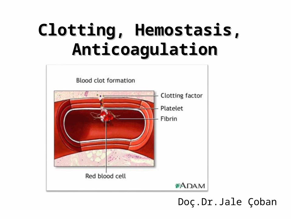
Clotting, Hemostasis, Clotting, Hemostasis, AnticoagulationAnticoagulation
Doç.Dr.Jale Çoban
Topics
1- Hemostasis overview
2- Coagulation
3- Bleeding / thrombosis
4- Laboratory tests
5- Patient samples
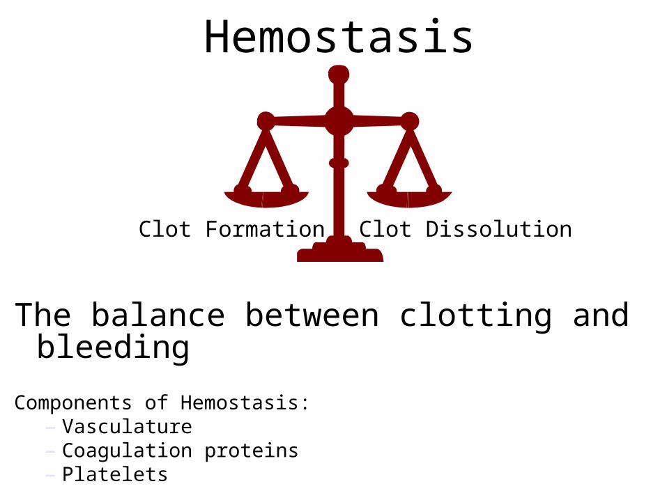
Hemostasis
The balance between clotting and bleeding
Components of Hemostasis:– Vasculature– Coagulation proteins– Platelets
Clot Formation Clot Dissolution
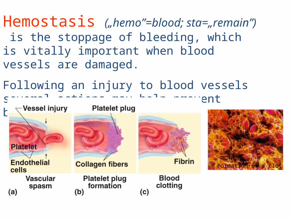
Hemostasis („hemo”=blood; sta=„remain”) is the stoppage of bleeding, which is vitally important when blood vessels are damaged.
Following an injury to blood vessels several actions may help prevent blood loss, including:
Formation of a clot
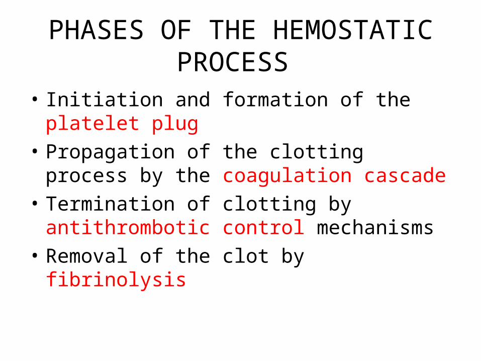
PHASES OF THE HEMOSTATIC PROCESS
• Initiation and formation of the platelet plug • Propagation of the clotting process by the
coagulation cascade• Termination of clotting by antithrombotic
control mechanisms• Removal of the clot by fibrinolysis
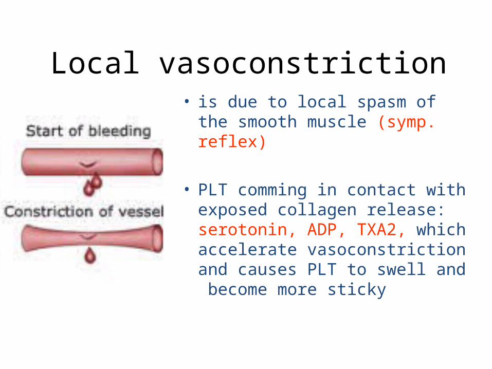
Local vasoconstriction• is due to local spasm of the
smooth muscle (symp. reflex)
• PLT comming in contact with exposed collagen release: serotonin, ADP, TXA2, which accelerate vasoconstriction and causes PLT to swell and become more sticky
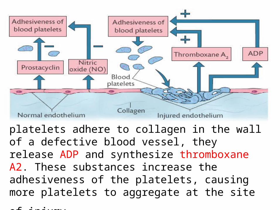
Formation of a platelet plug. When blood platelets adhere to collagen in the wall of a defective blood vessel, they release ADP and synthesize thromboxane A2. These substances increase the adhesiveness of the platelets,
causing more platelets to aggregate at the site of injury.
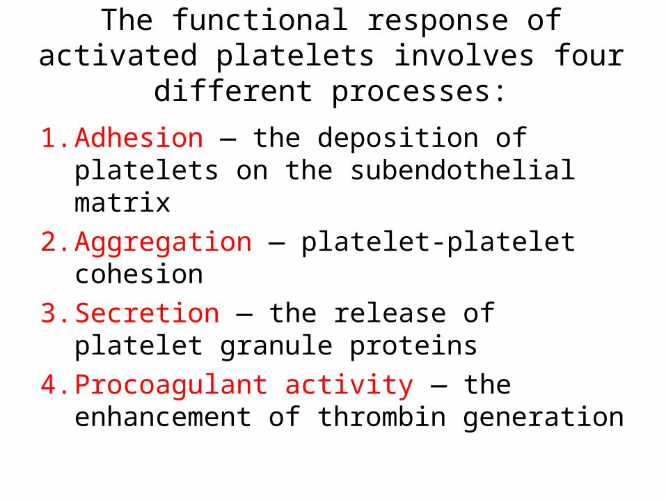
The functional response of activated platelets involves four different processes:
1. Adhesion — the deposition of platelets on the subendothelial matrix
2. Aggregation — platelet-platelet cohesion3. Secretion — the release of platelet granule
proteins4. Procoagulant activity — the enhancement of
thrombin generation

• This process is facilitated by a plasma glycoprotein, von Willebrand factor (vWF). Produced in both endothelial cells and platelets, vWF forms bridges between receptors on the surface of platelets and collagen fibers in the connective tissue.
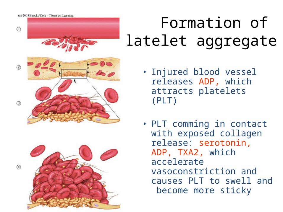
Formation of platelet aggregate
• Injured blood vessel releases ADP, which attracts platelets (PLT)
• PLT comming in contact with exposed collagen release: serotonin, ADP, TXA2, which accelerate vasoconstriction and causes PLT to swell and become more sticky
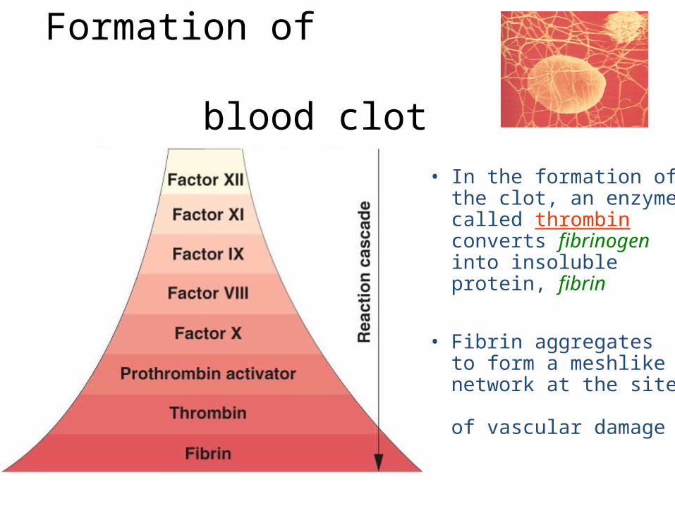
Formation of blood clot
• In the formation of the clot, an enzyme called thrombin converts fibrinogen into insoluble protein, fibrin
• Fibrin aggregates to form a meshlike network at the site of vascular damage
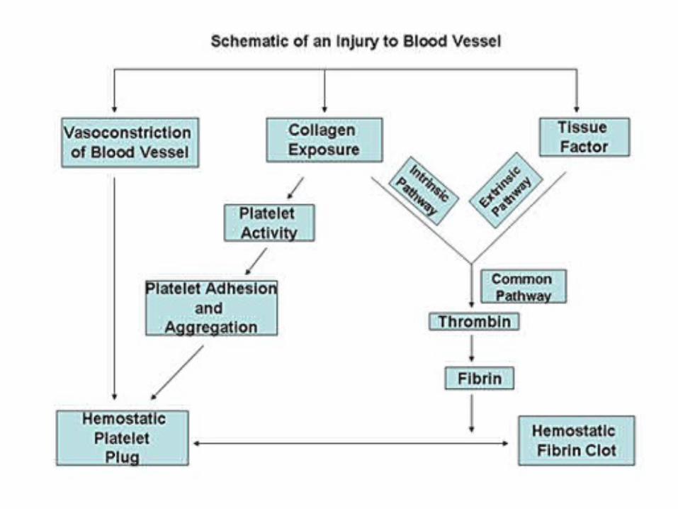
CLOTTING CASCADE AND PROPAGATION OF THE CLOT
The clotting cascade is depicted as consisting of an intrinsic and extrinsic pathway .
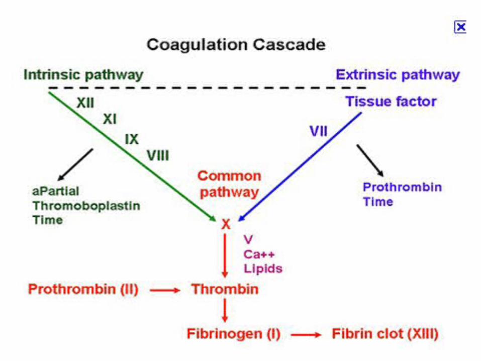
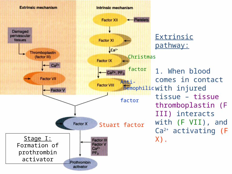
Extrinsic pathway:
1. When blood comes in contact with injured tissue – tissue thromboplastin (F III) interacts with (F VII), and Ca2+ activating (F X).
Stage I: Formation of prothrombin
activator
Ca2+
Stuart factor
Anti- hemophilic factor
Christmas factor
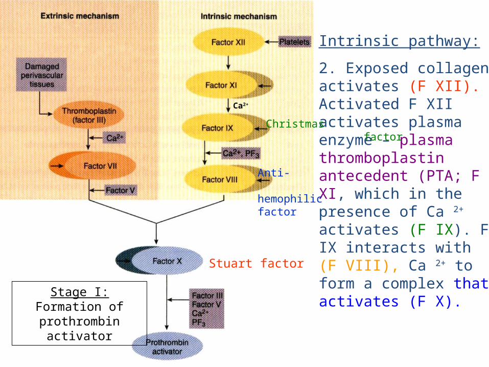
Intrinsic pathway:
2. Exposed collagen activates (F XII). Activated F XII activates plasma enzyme – plasma thromboplastin antecedent (PTA; F XI, which in the presence of Ca 2+ activates (F IX). F IX interacts with (F VIII), Ca 2+ to form a complex that activates (F X).
Stage I: Formation of prothrombin
activator
Ca2+
Christmas factor
Anti- hemophilic factor
Stuart factor

Stage I: Formation of prothrombin
activator
3. Common pathway:
Activated F X in the presence of Ca 2+ forms complexes with accelerin (F V) to form prothrombin activator
Ca2+
Christmas factor
Anti- hemophilic factor
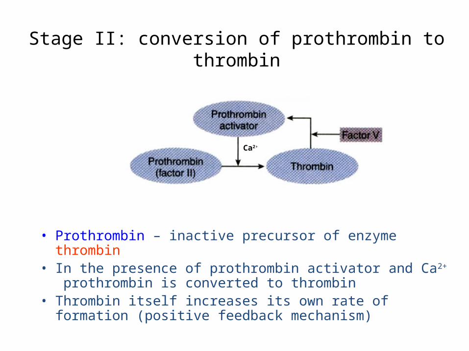
Stage II: conversion of prothrombin to thrombin
• Prothrombin – inactive precursor of enzyme thrombin• In the presence of prothrombin activator and Ca2+
prothrombin is converted to thrombin• Thrombin itself increases its own rate of formation
(positive feedback mechanism)
Ca2+
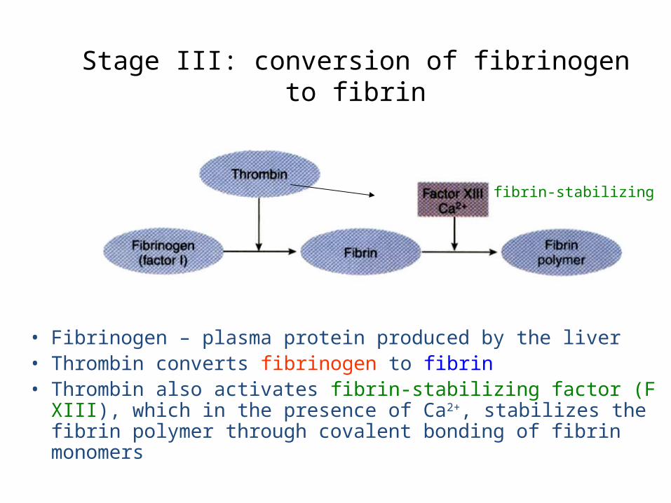
Stage III: conversion of fibrinogen to fibrin
• Fibrinogen – plasma protein produced by the liver• Thrombin converts fibrinogen to fibrin• Thrombin also activates fibrin-stabilizing factor (F XIII),
which in the presence of Ca2+, stabilizes the fibrin polymer through covalent bonding of fibrin monomers
fibrin-stabilizing factor
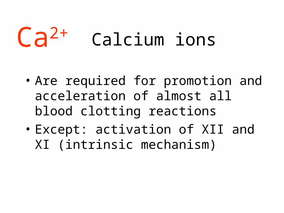
Calcium ions
• Are required for promotion and acceleration of almost all blood clotting reactions
• Except: activation of XII and XI (intrinsic mechanism)
Ca2+

Fibrinolysis
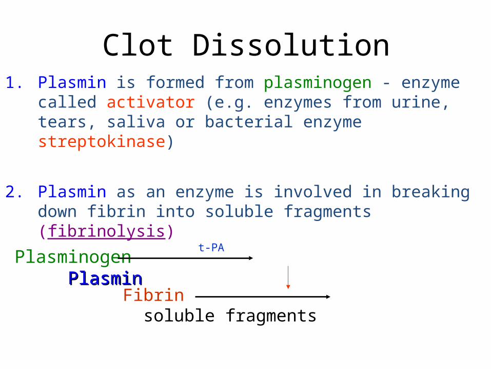
Clot Dissolution1. Plasmin is formed from plasminogen - enzyme called
activator (e.g. enzymes from urine, tears, saliva or bacterial enzyme streptokinase)
2. Plasmin as an enzyme is involved in breaking down fibrin into soluble fragments (fibrinolysis)
Plasminogen PlasminPlasmin
t-PA
Fibrin soluble fragments
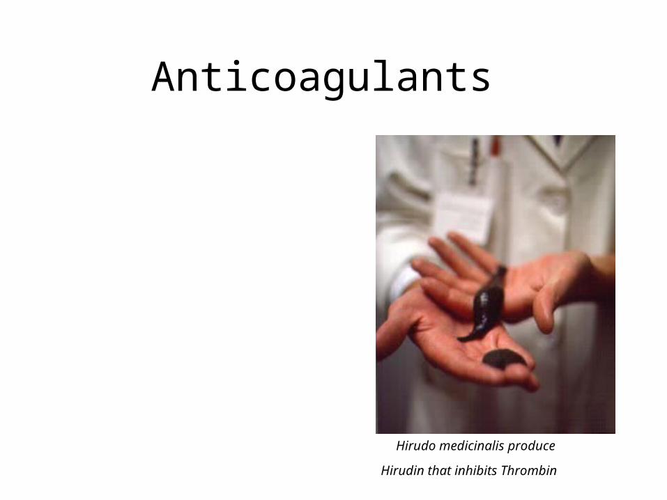
Anticoagulants
Hirudo medicinalis produce
Hirudin that inhibits Thrombin
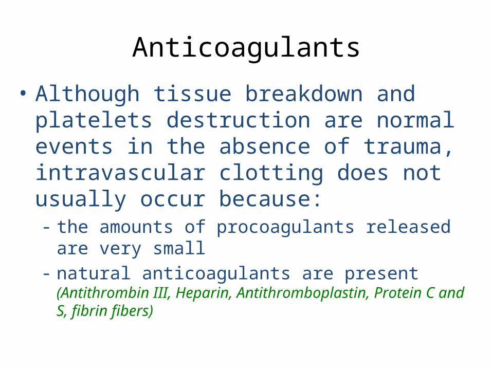
Anticoagulants
• Although tissue breakdown and platelets destruction are normal events in the absence of trauma, intravascular clotting does not usually occur because:- the amounts of procoagulants released are
very small- natural anticoagulants are present
(Antithrombin III, Heparin, Antithromboplastin, Protein C and S, fibrin fibers)
Natural anticoagulants• Antithrombin III – inhibits factor X and thrombin
• Heparin from basophils and mast cells potentiates effects of antithrombin III (together they inhibit IX, X, XI, XII and thrombin)
• Antithromboplastin (inhibits „tissue factors” – tissue thromboplastins)
• Protein C and S – activated by thrombin; degrade factor Va and VIIIa
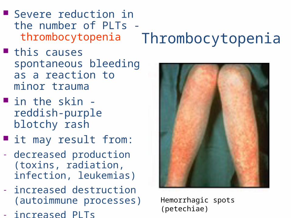
Thrombocytopenia
Severe reduction in the number of PLTs - thrombocytopenia
this causes spontaneous bleeding as a reaction to minor trauma
in the skin - reddish-purple blotchy rash
it may result from:- decreased production
(toxins, radiation, infection, leukemias)
- increased destruction (autoimmune processes)
- increased PLTs consumption (DIC) Hemorrhagic spots (petechiae)
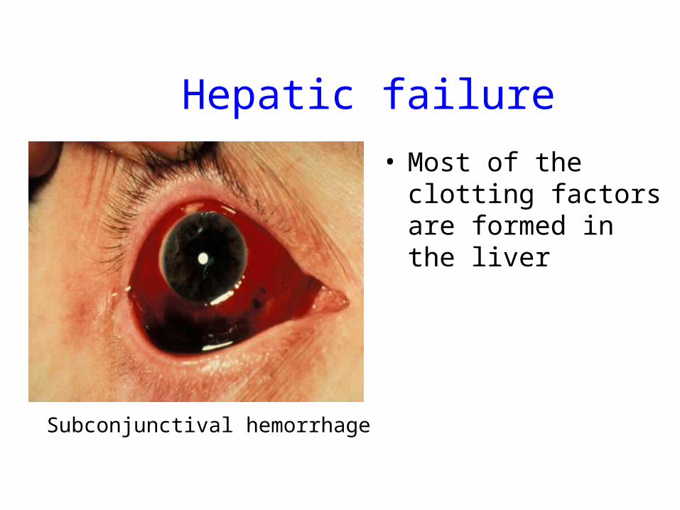
Hepatic failure
• Most of the clotting factors are formed in the liver
Subconjunctival hemorrhage
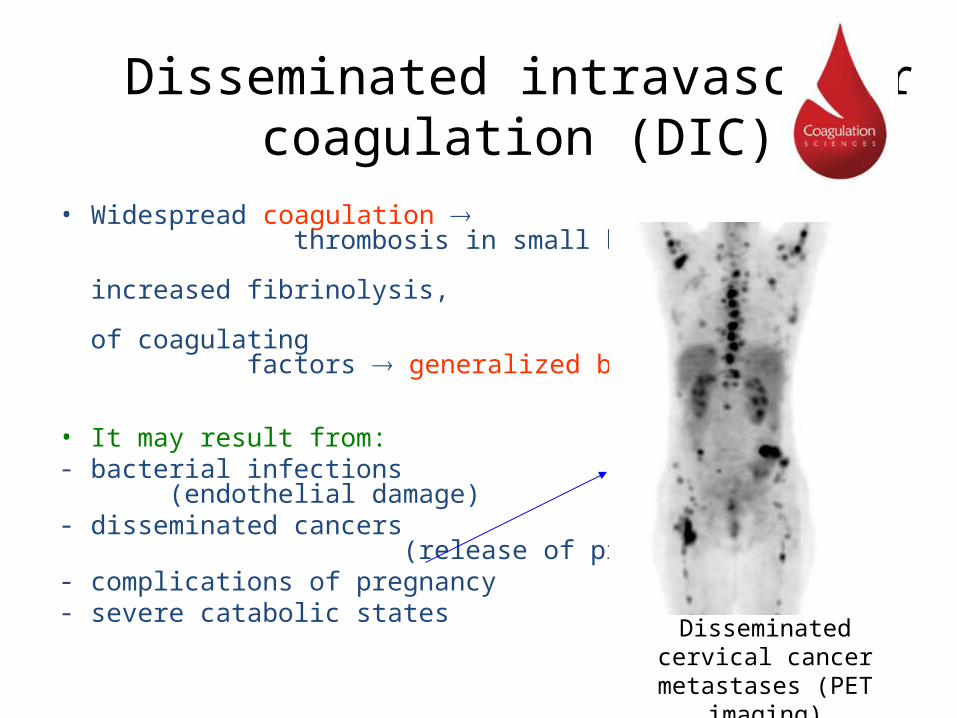
Disseminated intravascular coagulation (DIC)
• Widespread coagulation thrombosis in small blood vessels increased fibrinolysis, and depletion of coagulating factors generalized bleeding
• It may result from:- bacterial infections
(endothelial damage)- disseminated cancers
(release of procoagulants)- complications of pregnancy- severe catabolic states Disseminated cervical
cancer metastases (PET imaging)
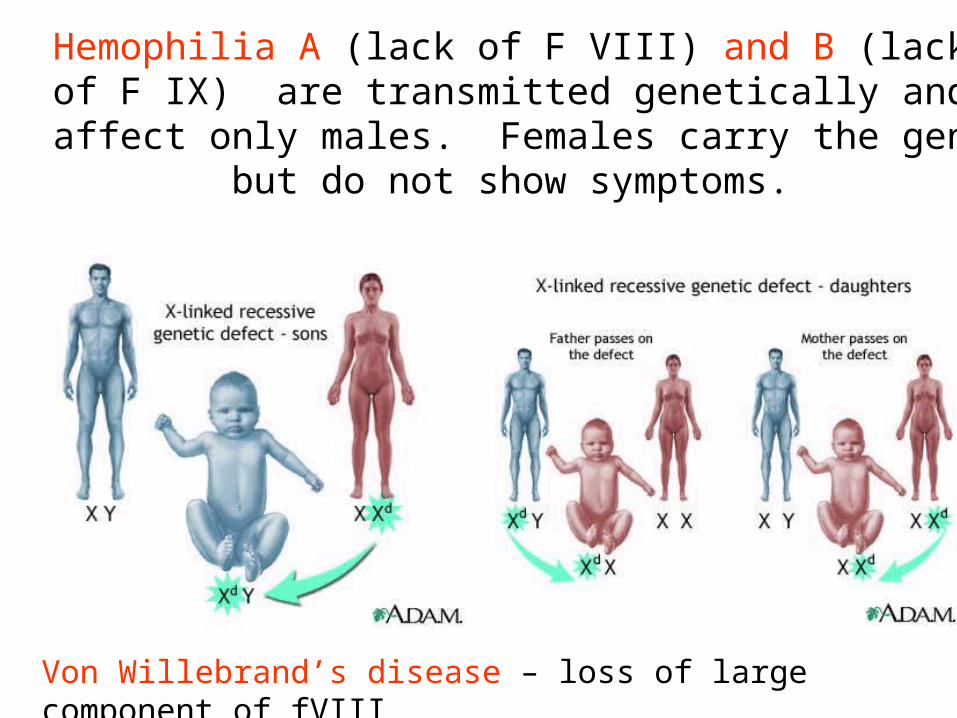
Hemophilia A (lack of F VIII) and B (lack of F IX) are transmitted genetically and affect only males.
Females carry the gen but do not show symptoms.
Von Willebrand’s disease – loss of large component of fVIII
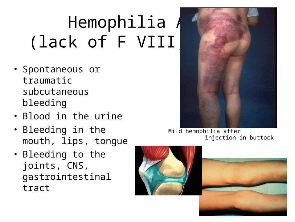
Hemophilia A (lack of F VIII; 85%)
• Spontaneous or traumatic subcutaneous bleeding
• Blood in the urine• Bleeding in the
mouth, lips, tongue• Bleeding to the
joints, CNS, gastrointestinal tract
Mild hemophilia after injection in buttock
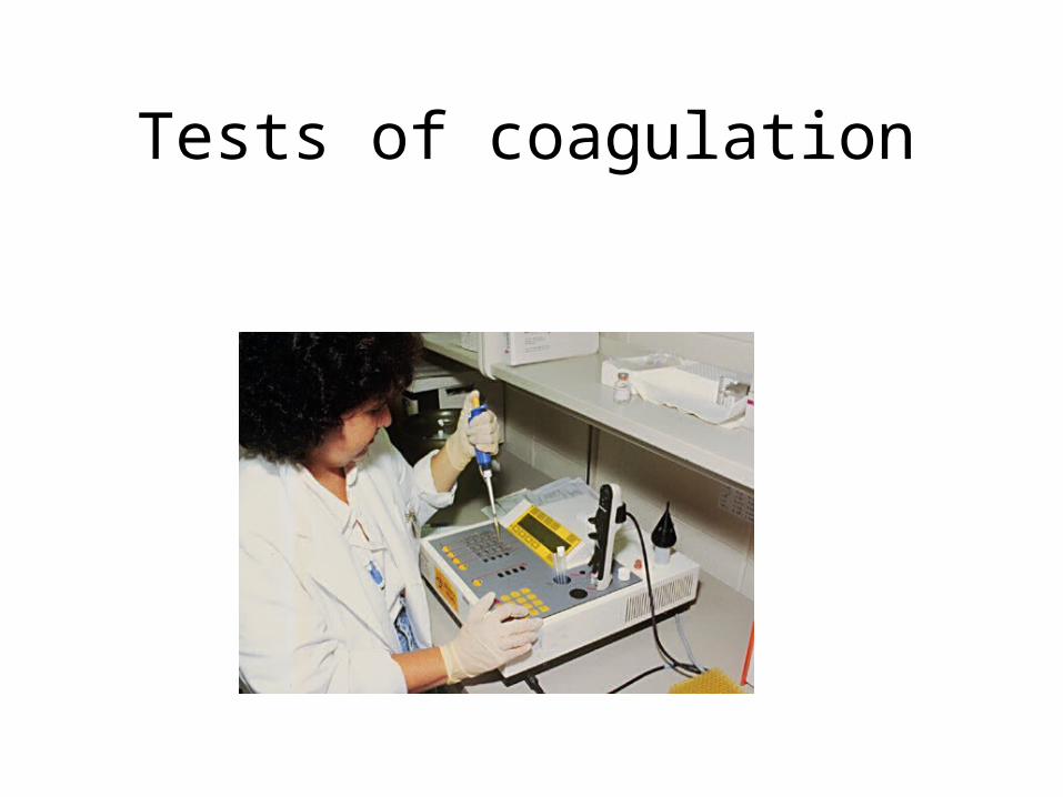
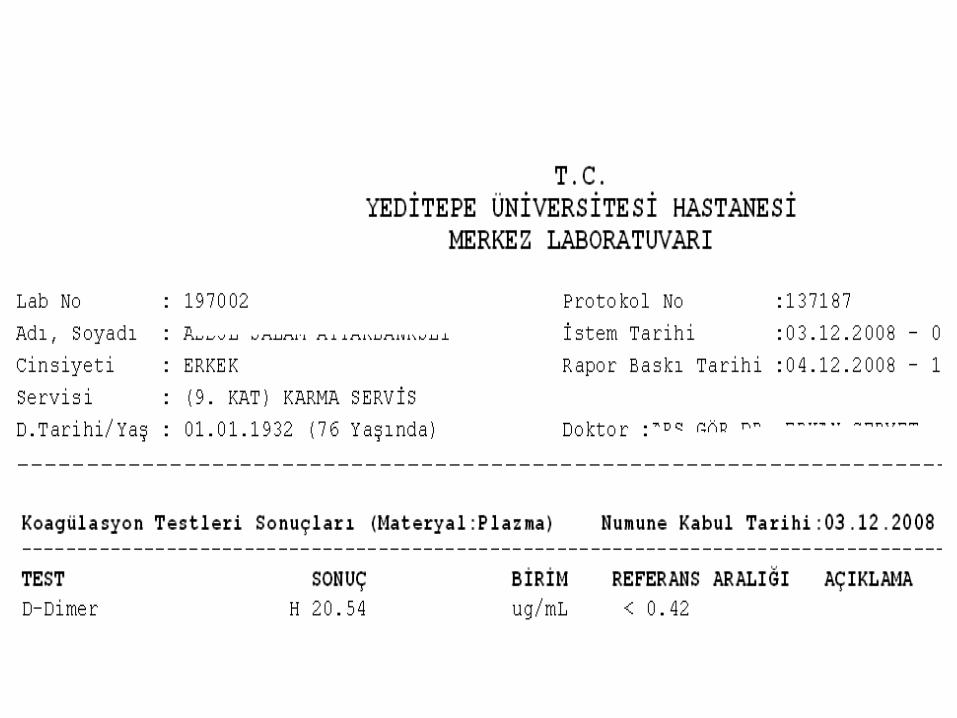
Tests of coagulation
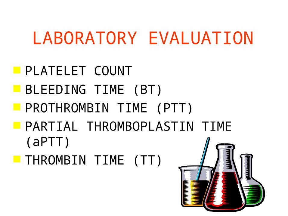
LABORATORY EVALUATION
PLATELET COUNT BLEEDING TIME (BT) PROTHROMBIN TIME (PTT) PARTIAL THROMBOPLASTIN TIME (aPTT) THROMBIN TIME (TT)
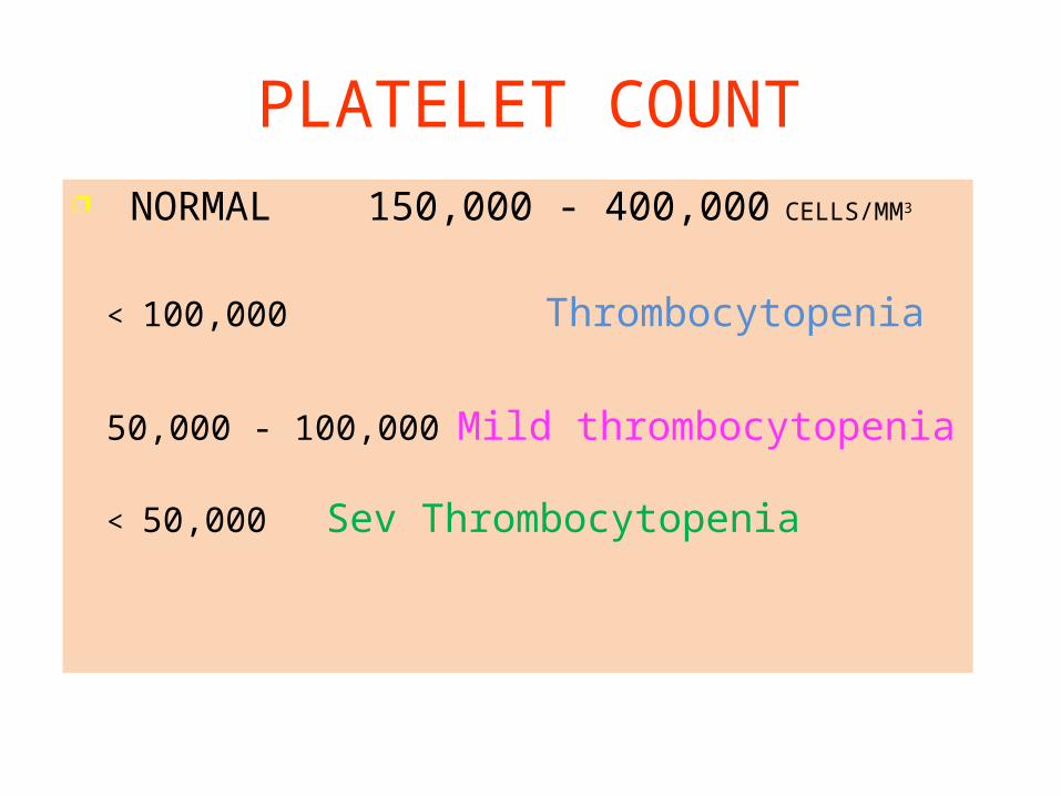
PLATELET COUNT NORMAL 150,000 - 400,000 CELLS/MM3
< 100,000 Thrombocytopenia
50,000 - 100,000 Mild thrombocytopenia
< 50,000 Sev Thrombocytopenia
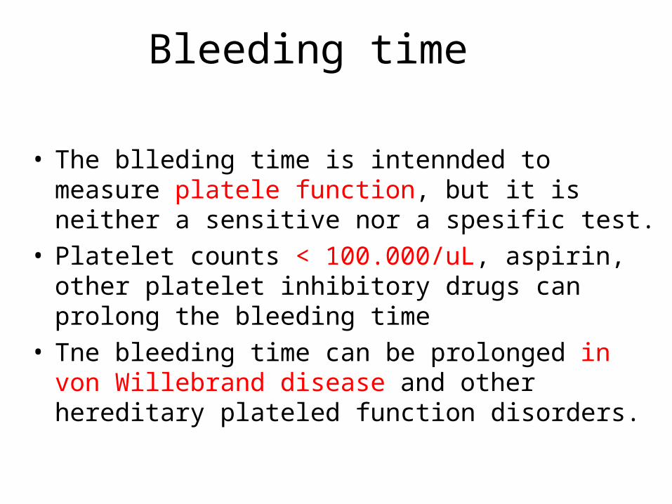
Bleeding time
• The blleding time is intennded to measure platele function, but it is neither a sensitive nor a spesific test.
• Platelet counts < 100.000/uL, aspirin, other platelet inhibitory drugs can prolong the bleeding time
• Tne bleeding time can be prolonged in von Willebrand disease and other hereditary plateled function disorders.
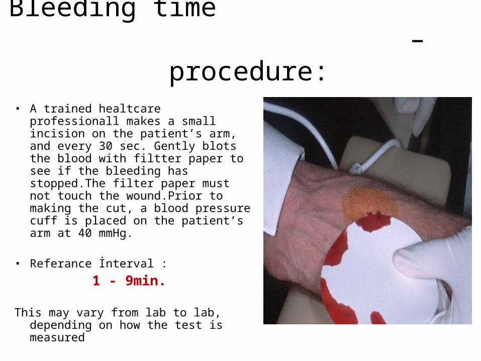
Bleeding time – procedure:
• A trained healtcare professionall makes a small incision on the patient’s arm, and every 30 sec. Gently blots the blood with filtter paper to see if the bleeding has stopped.The filter paper must not touch the wound.Prior to making the cut, a blood pressure cuff is placed on the patient’s arm at 40 mmHg.
• Referance İnterval :
1 - 9min.
This may vary from lab to lab, depending on how the test is measured
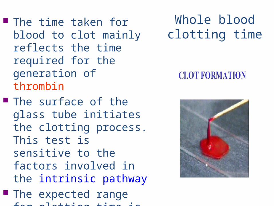
Whole blood clotting time
The time taken for blood to clot mainly reflects the time required for the generation of thrombin
The surface of the glass tube initiates the clotting process. This test is sensitive to the factors involved in the intrinsic pathway
The expected range for clotting time is 4-10 4-10 mins.mins.
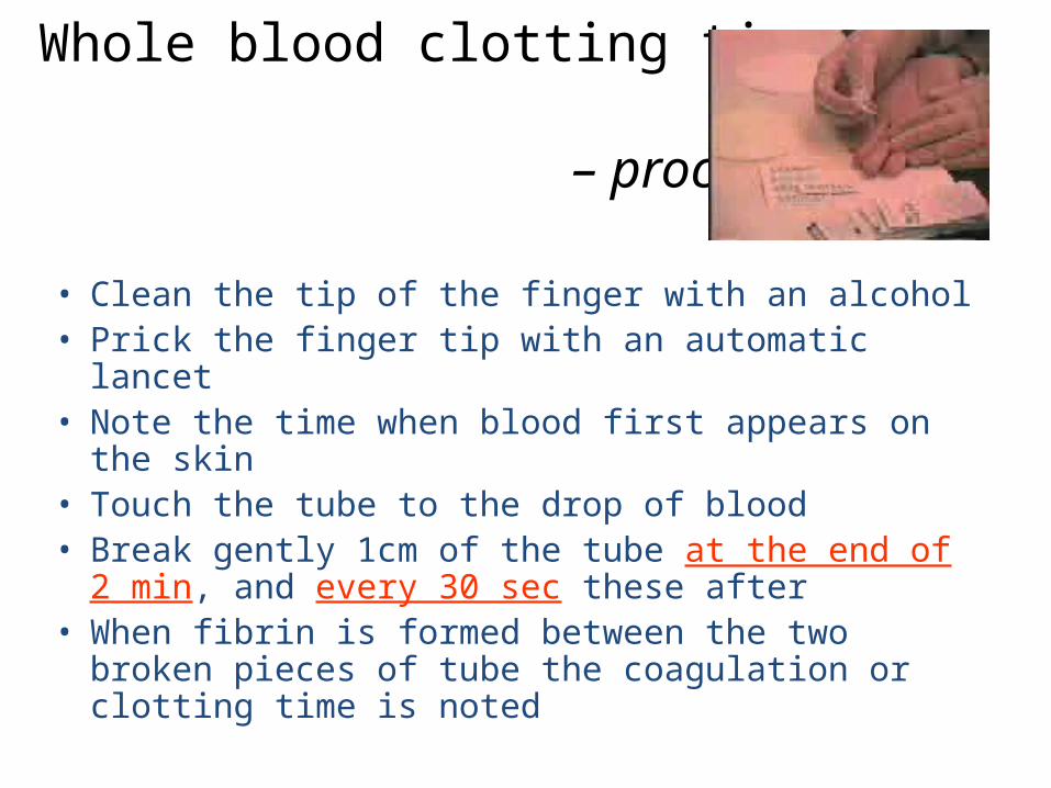
Whole blood clotting time – procedure:
• Clean the tip of the finger with an alcohol • Prick the finger tip with an automatic lancet • Note the time when blood first appears on
the skin • Touch the tube to the drop of blood • Break gently 1cm of the tube at the end of 2
min, and every 30 sec these after • When fibrin is formed between the two
broken pieces of tube the coagulation or clotting time is noted
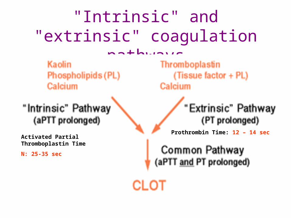
"Intrinsic" and "extrinsic" coagulation pathways
Activated Partial Thromboplastin Time
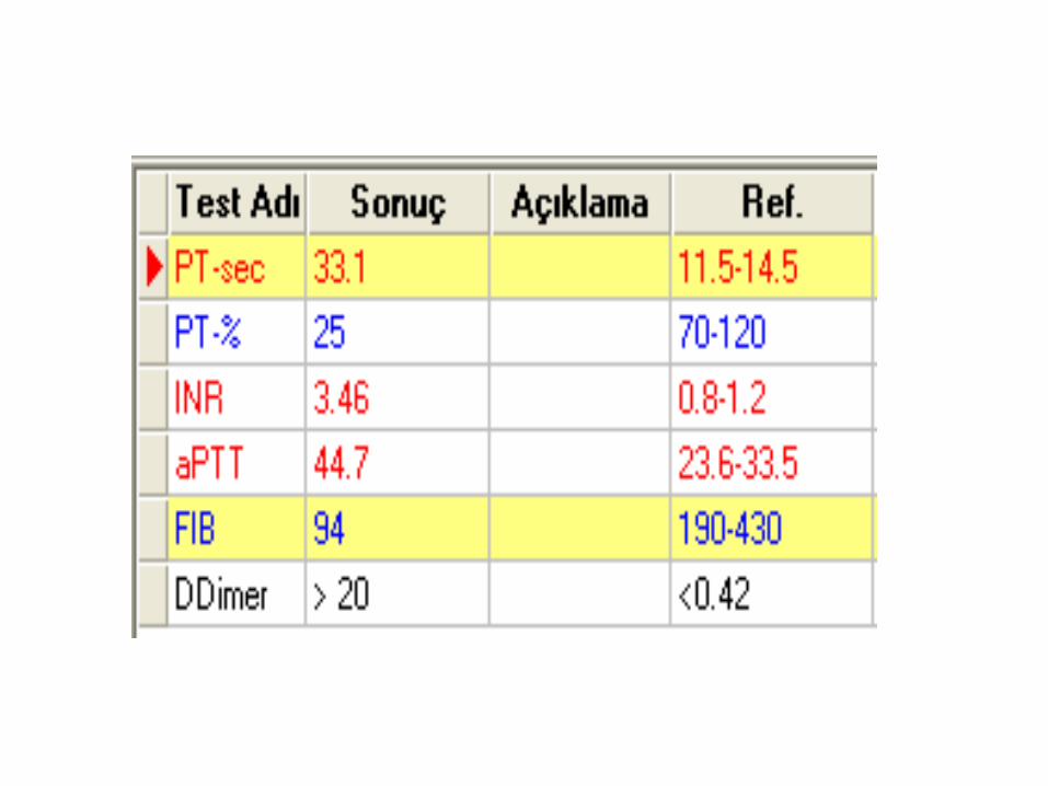
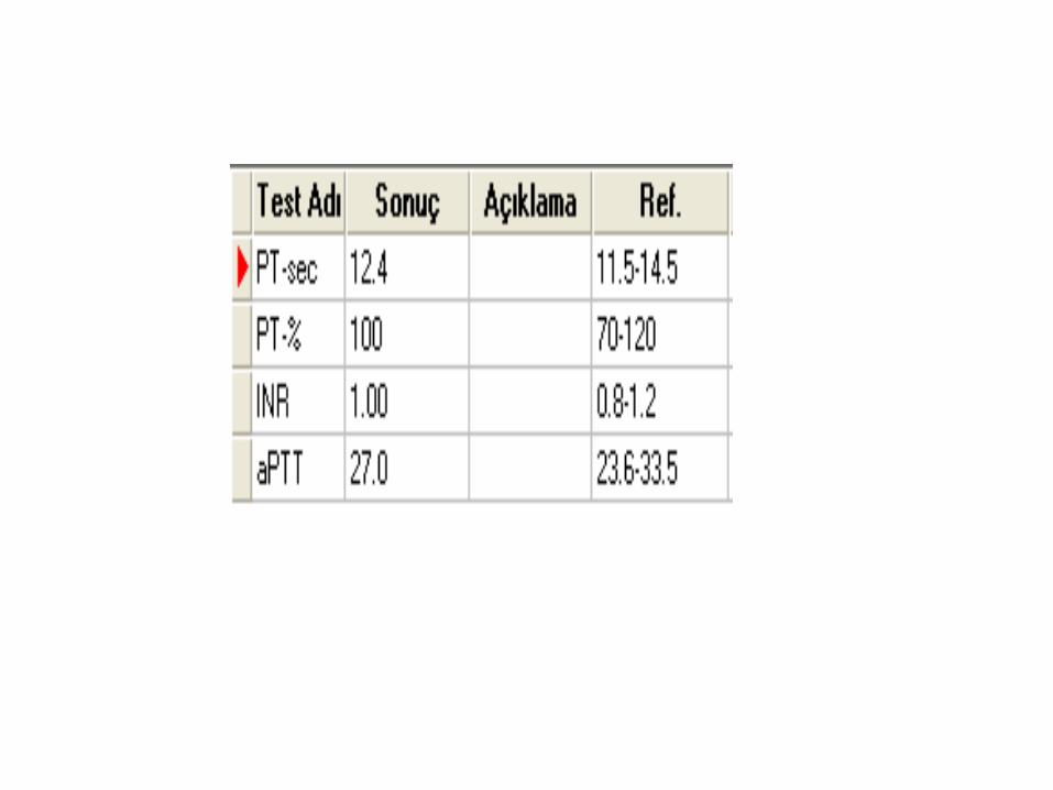
N: 25-35 sec
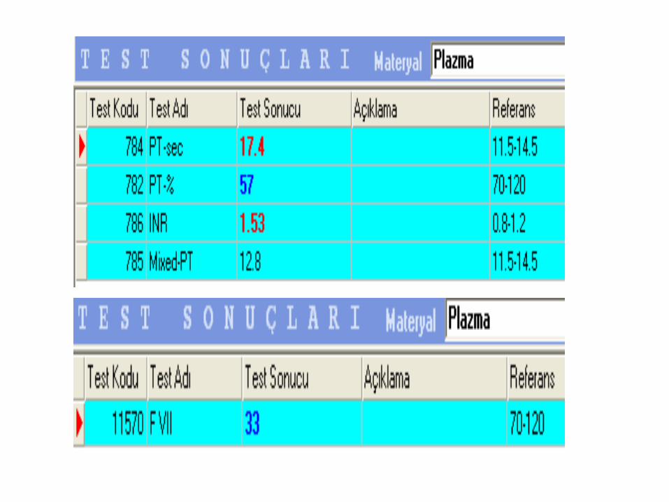
Prothrombin Time: 12 – 14 sec
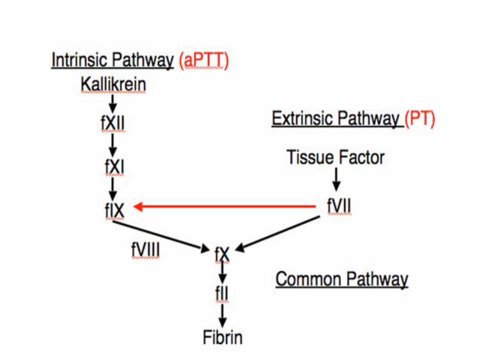
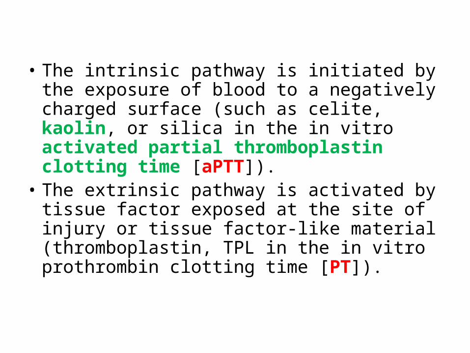
• The intrinsic pathway is initiated by the exposure of blood to a negatively charged surface (such as celite, kaolin, or silica in the in vitro activated partial thromboplastin clotting time [aPTT]).
• The extrinsic pathway is activated by tissue factor exposed at the site of injury or tissue factor-like material (thromboplastin, TPL in the in vitro prothrombin clotting time [PT]).
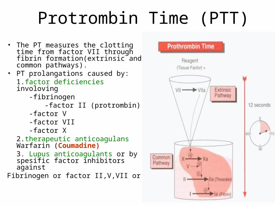
Protrombin Time (PTT)• The PT measures the clotting time from
factor VII through fibrin formation(extrinsic and common pathways).
• PT prolangations caused by: 1.factor deficiencies involoving -fibrinogen
-factor II (protrombin) -factor V -factor VII -factor X2.therapeutic anticoagulans Warfarin (Coumadine)3. Lupus anticoagulants or by spesific factor inhibitors against
Fibrinogen or factor II,V,VII or X

Protrombin Time (PTT)• PT reagents is called thromboplastin (phospholipid with
tissue factor and Ca++). It is added to patient plasma, and the time until clot formation is measured in seconds(Referans interval: 12-14 sec).
• Tissue factor activates the extrinsic pathway.Phospholipid and Ca++ are required cofactors in the coagulation cascade.
• Citrate in the blue top tube prevents clotting by chelating Ca++
• Prolonged PT:- a vitamin K deficiency (vitamin K is a co-factor in the synthesis of
functional factors II prothrombin), VII, IX and X)- liver disease- Warfarin therapy (Coumadin)
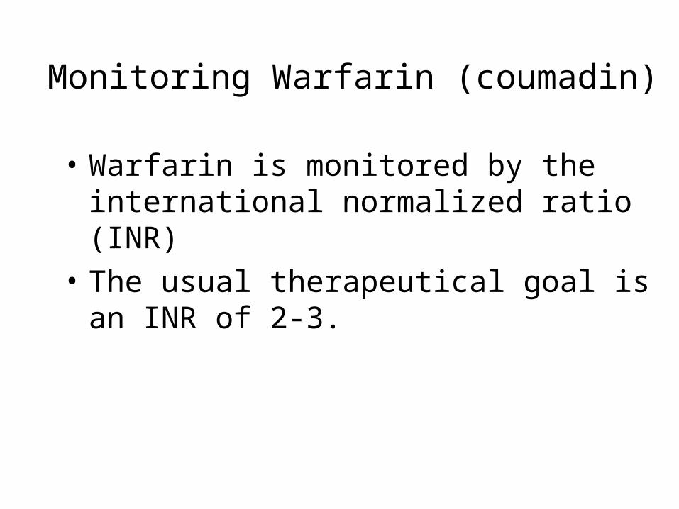
Monitoring Warfarin (coumadin)
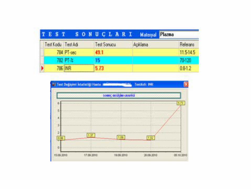
• Warfarin is monitored by the international normalized ratio (INR)
• The usual therapeutical goal is an INR of 2-3.
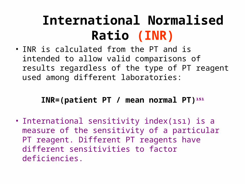
International Normalised Ratio (INR)
• INR is calculated from the PT and is intended to allow valid comparisons of results regardless of the type of PT reagent used among different laboratories:
INR=(patient PT / mean normal PT)ısı
• International sensitivity index(ısı) is a measure of the sensitivity of a particular PT reagent. Different PT reagents have different sensitivities to factor deficiencies.

• A patient’s prothrombin time (PT) test result expressed as a ratio to a normal population control which has been standardized (or normalized) for the potency to the thromboplastin used in the assay.
• It is standardized using a World Health Organization (WHO) international reference thromboplastin preparation, and determined using the equation:
INR = R^ISI,
where R is the PT ratio obtained with the working thromboplastin.
International Normalised Ratio (INR)
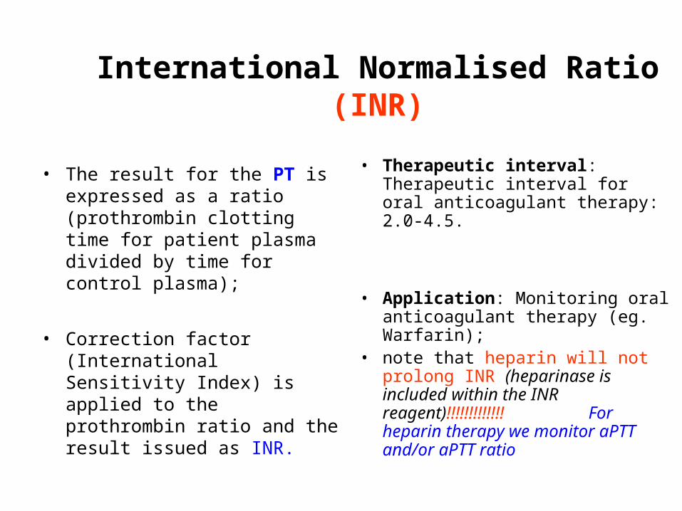
• The result for the PT is expressed as a ratio (prothrombin clotting time for patient plasma divided by time for control plasma);
• Correction factor (International Sensitivity Index) is applied to the prothrombin ratio and the result issued as INR.
• Therapeutic interval: Therapeutic interval for oral anticoagulant therapy: 2.0-4.5.
• Application: Monitoring oral anticoagulant therapy (eg. Warfarin);
• note that heparin will not prolong INR (heparinase is included within the INR reagent)!!!!!!!!!!!!! For heparin therapy we monitor aPTT and/or aPTT ratio
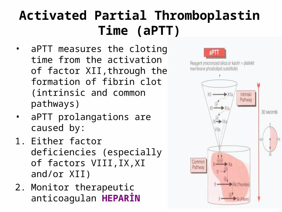
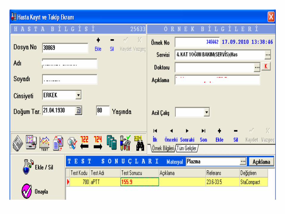
Activated Partial Thromboplastin Time (aPTT)
• aPTT measures the cloting time from the activation of factor XII,through the formation of fibrin clot (intrinsic and common pathways)
• aPTT prolangations are caused by:1. Either factor deficiencies (especially
of factors VIII,IX,XI and/or XII)2. Monitor therapeutic anticoagulan
HEPARİN
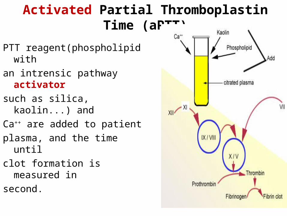
Activated Partial Thromboplastin Time (aPTT)
PTT reagent(phospholipid with an intrensic pathway activator such as silica, kaolin...) and Ca++ are added to patient plasma, and the time until clot formation is measured in second.
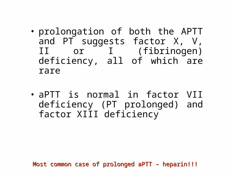
• prolongation of both the APTT and PT suggests factor X, V, II or I (fibrinogen) deficiency, all of which are rare
• aPTT is normal in factor VII deficiency (PT prolonged) and factor XIII deficiency
Most common case of prolonged aPTT – heparin!!!Most common case of prolonged aPTT – heparin!!!
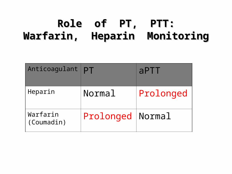
Role of PT, PTT:Role of PT, PTT:Warfarin, Heparin MonitoringWarfarin, Heparin Monitoring
Anticoagulant PT aPTT
Heparin Normal Prolonged
Warfarin (Coumadin)
Prolonged Normal
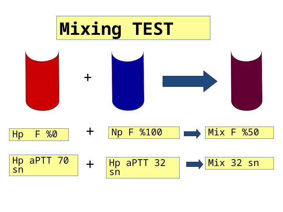
Hp F %0 Np F %100
Mixing TEST
Mix F %50
Hp aPTT 70 sn
Hp aPTT 32 sn
+
+
+ Mix 32 sn
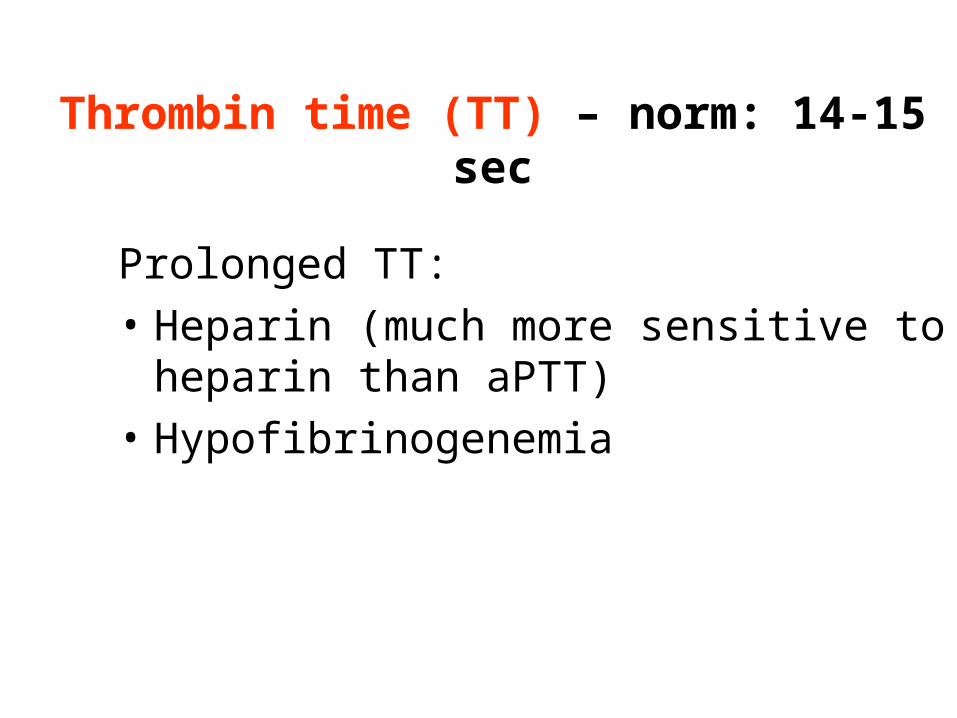
Thrombin time (TT) – norm: 14-15 sec
Prolonged TT:• Heparin (much more sensitive to heparin
than aPTT)• Hypofibrinogenemia
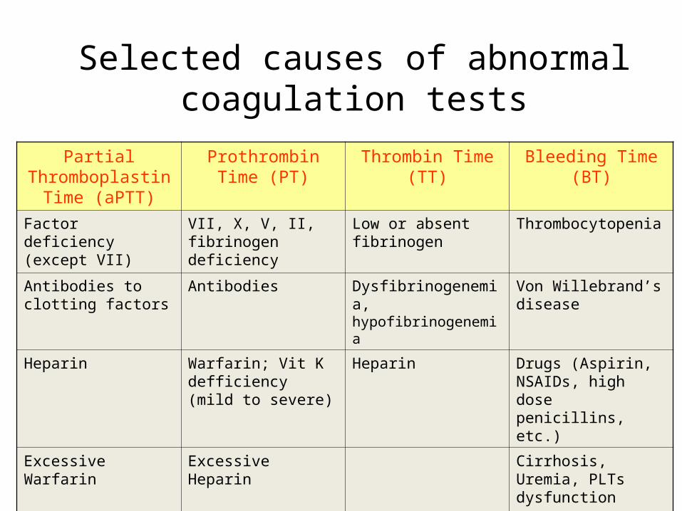
Selected causes of abnormal coagulation tests
Partial Thromboplastin
Time (aPTT)
Prothrombin Time (PT)
Thrombin Time (TT)
Bleeding Time (BT)
Factor deficiency (except VII)
VII, X, V, II, fibrinogen deficiency
Low or absent fibrinogen
Thrombocytopenia
Antibodies to clotting factors
Antibodies Dysfibrinogenemia, hypofibrinogenemia
Von Willebrand’s disease
Heparin Warfarin; Vit K defficiency (mild to severe)
Heparin Drugs (Aspirin, NSAIDs, high dose penicillins, etc.)
Excessive Warfarin Excessive Heparin Cirrhosis, Uremia, PLTs dysfunction
AnticoagulantsAnticoagulants
• Heparin• Warfarin (Coumadin)• Direct thrombin inhibitors• Antiplatelet agents