clostridium difficile enteritis: a new role for an old foe
TRANSCRIPT
ww.sciencedirect.com
t h e s u r g e on x x x ( 2 0 1 4 ) 1e7
Available online at w
ScienceDirectThe Surgeon, Journal of the Royal Colleges
of Surgeons of Edinburgh and Irelandwww.thesurgeon.net
Review
Clostridium difficile enteritis: A new role for an oldfoe
S. Killeen*, S.T. Martin, J. Hyland, P.R. O’ Connell, D.C. Winter
St. Vincent’s University Hospital, Department of Colorectal Surgery, Dublin 4, Ireland
a r t i c l e i n f o
Article history:
Received 25 October 2013
Received in revised form
16 January 2014
Accepted 16 January 2014
Available online xxx
Keywords:
Clostridium difficile
Small bowel
Pathophysiology
Risk factors
Diagnosis and management
* Corresponding author. Tel.: þ353 2214000.E-mail address: [email protected] (S
Please cite this article in press as: Killeenhttp://dx.doi.org/10.1016/j.surge.2014.01.0
1479-666X/$ e see front matter ª 2014 Publinumber SC005317) and Royal College of Surhttp://dx.doi.org/10.1016/j.surge.2014.01.008
a b s t r a c t
Background: Small bowel involvement of Clostridium difficile is increasingly encountered.
Data on many management aspects are lacking.
Aim: To synthesis existing reports and assess the frequency, pathophysiology, outcomes,
risk factors, diagnosis and management of C. difficle enteritis.
Methods: A systematic review of the literature was conducted to evaluate evidence
regarding frequency, pathophysiology, risk factors, optimal diagnosis, management and
outcomes for C. difficle enteritis. Three major databases (PubMed, MEDLINE and the
Cochrane Library) were searched. The review included original articles reporting C. difficle
enteritis from January 1950 to December 2012.
Results: C. difficle enteritis is rare but increasingly encountered. Presentation is variable and
distinct predisposing factors include emergency surgery, white race and increased age.
Diagnosis generally involves a sensitive but often non specific screening test for C. difficile
antigens. Oral metronidazole represents first line therapy and surgery may be required for
complications. Outcomes are inconsistent but may be improving.
Conclusions: A high index of clinical suspicion, early diagnosis and treatment are vital.
Further prospective studies are needed to determine the significance of asymptomatic
small bowel C. difficile infections.
ª 2014 Published by Elsevier Ltd on behalf of Royal College of Surgeons of Edinburgh
(Scottish charity number SC005317) and Royal College of Surgeons in Ireland.
Introduction
The older, immunocompromised and institutionalised patient
population undergoing surgery allied to increased practitioner
awareness has produced a significant rise in all Clostridium
difficile infections.1 Although rare, small bowel involvement of
C. difficile is increasingly identified.2e4
. Killeen).
S, et al., Clostridium dif08
shed by Elsevier Ltd on bgeons in Ireland.
Early reports of small bowel C. difficile infection suggested
significant morbidity and mortality rates.4,5 However the
enhanced recognition of small bowel colonisation post-
operatively suggests a higher than previously thought preva-
lence of a milder or even asymptomatic entity.2,6,7
Notwithstanding this a high index of suspicion is necessary
allied to early intervention to maximise optimum outcomes.
Despite these features the literature on C. difficile enteritis
is sparse comprising mainly of retrospective studies and case
ficile enteritis: A new role for an old foe, The Surgeon (2014),
ehalf of Royal College of Surgeons of Edinburgh (Scottish charity
t h e s u r g e on x x x ( 2 0 1 4 ) 1e72
series. Unlike C. difficile colitis, there are no consensus guide-
lines to assist healthcare practitioners.8
Aim
To synthesis existing reports and assess the frequency,
pathophysiology, outcomes, risk factors, diagnosis and man-
agement of C. difficle enteritis.
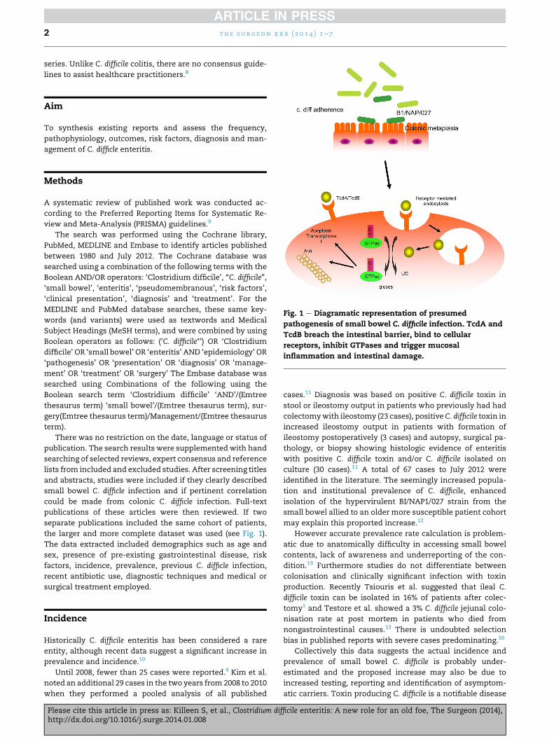
Fig. 1 e Diagramatic representation of presumed
pathogenesis of small bowel C. difficile infection. TcdA and
TcdB breach the intestinal barrier, bind to cellular
receptors, inhibit GTPases and trigger mucosal
inflammation and intestinal damage.
Methods
A systematic review of published work was conducted ac-
cording to the Preferred Reporting Items for Systematic Re-
view and Meta-Analysis (PRISMA) guidelines.9
The search was performed using the Cochrane library,
PubMed, MEDLINE and Embase to identify articles published
between 1980 and July 2012. The Cochrane database was
searched using a combination of the following terms with the
Boolean AND/OR operators: ‘Clostridium difficile’, “C. difficile”,
‘small bowel’, ‘enteritis’, ‘pseudomembranous’, ‘risk factors’,
‘clinical presentation’, ‘diagnosis’ and ‘treatment’. For the
MEDLINE and PubMed database searches, these same key-
words (and variants) were used as textwords and Medical
Subject Headings (MeSH terms), and were combined by using
Boolean operators as follows: (‘C. difficile*’) OR ‘Clostridium
difficile’ OR ‘small bowel’ OR ‘enteritis’ AND ‘epidemiology’ OR
‘pathogenesis’ OR ‘presentation’ OR ‘diagnosis’ OR ‘manage-
ment’ OR ‘treatment’ OR ‘surgery’ The Embase database was
searched using Combinations of the following using the
Boolean search term ‘Clostridium difficile’ ‘AND’/(Emtree
thesaurus term) ‘small bowel’/(Emtree thesaurus term), sur-
gery(Emtree thesaurus term)/Management/(Emtree thesaurus
term).
There was no restriction on the date, language or status of
publication. The search results were supplemented with hand
searching of selected reviews, expert consensus and reference
lists from included and excluded studies. After screening titles
and abstracts, studies were included if they clearly described
small bowel C. difficle infection and if pertinent correlation
could be made from colonic C. difficle infection. Full-text
publications of these articles were then reviewed. If two
separate publications included the same cohort of patients,
the larger and more complete dataset was used (see Fig. 1).
The data extracted included demographics such as age and
sex, presence of pre-existing gastrointestinal disease, risk
factors, incidence, prevalence, previous C. difficle infection,
recent antibiotic use, diagnostic techniques and medical or
surgical treatment employed.
Incidence
Historically C. difficile enteritis has been considered a rare
entity, although recent data suggest a significant increase in
prevalence and incidence.10
Until 2008, fewer than 25 cases were reported.4 Kim et al.
noted an additional 29 cases in the two years from 2008 to 2010
when they performed a pooled analysis of all published
Please cite this article in press as: Killeen S, et al., Clostridium difhttp://dx.doi.org/10.1016/j.surge.2014.01.008
cases.11 Diagnosis was based on positive C. difficile toxin in
stool or ileostomy output in patients who previously had had
colectomywith ileostomy (23 cases), positiveC. difficile toxin in
increased ileostomy output in patients with formation of
ileostomy postoperatively (3 cases) and autopsy, surgical pa-
thology, or biopsy showing histologic evidence of enteritis
with positive C. difficile toxin and/or C. difficile isolated on
culture (30 cases).11 A total of 67 cases to July 2012 were
identified in the literature. The seemingly increased popula-
tion and institutional prevalence of C. difficile, enhanced
isolation of the hypervirulent BI/NAP1/027 strain from the
small bowel allied to an older more susceptible patient cohort
may explain this proported increase.12
However accurate prevalence rate calculation is problem-
atic due to anatomically difficulty in accessing small bowel
contents, lack of awareness and underreporting of the con-
dition.13 Furthermore studies do not differentiate between
colonisation and clinically significant infection with toxin
production. Recently Tsiouris et al. suggested that ileal C.
difficile toxin can be isolated in 16% of patients after colec-
tomy1 and Testore et al. showed a 3% C. difficile jejunal colo-
nisation rate at post mortem in patients who died from
nongastrointestinal causes.13 There is undoubted selection
bias in published reports with severe cases predominating.10
Collectively this data suggests the actual incidence and
prevalence of small bowel C. difficile is probably under-
estimated and the proposed increase may also be due to
increased testing, reporting and identification of asymptom-
atic carriers. Toxin producing C. difficile is a notifiable disease
ficile enteritis: A new role for an old foe, The Surgeon (2014),

t h e s u r g e on x x x ( 2 0 1 4 ) 1e7 3
in many countries and such registries could potentially
furnish more accurate prevalence rates for small bowel C.
difficile if small bowel and colonic aetiology is differentiated
(HPA 2012).14
Pathophysiology
C. difficile is a spore-forming, anaerobic, gram-positive bacillus
that does not invade mucosa, and is the causative agent for
pseudomembranous colitis. It produces two toxins TcdA and
TcdB that breach the intestinal barrier and trigger mucosal
inflammation and intestinal damage. TcdA and TcdB bind to
cellular receptors present onmany cell types and inhibit small
GTPases such as Rho, Rac and Cdc42 that are found in
eukaryotic cells.15 TcdA and TcdB trigger inflammasome-
dependent interleukin (IL)-1beta production, which contrib-
utes to C. difficile induced inflammation and damage in vivo
(see Fig. 1).16 This may involve melanin concentrating factor
(MCH).17
The pathophysiology of small bowel C. difficile infection is
unclear. In vivo rabbit and human post mortem studies
demonstrating comparable pathological findings and the
presence of pseudomembranes suggest a similar pathogen-
esis in small bowel and colon.18
The small intestine undergoes phenotypic changes after
colectomy creating a colon like milieu with colonic-type
metaplasia and partial villous atrophy which may predispose
to C. difficile overgrowth.19 In addition resection of the ileocecal
valve facilitates small bowel colonisation.3,20 (see Fig. 1).
High levels of TcdA may be required for ileal involvement
in C. difficile infections (Testore). The BI/NAP/027 strain pro-
duces higher amounts of TcdA4 and also produces binary
toxin CDT, which is enterotoxic in an ileal loop assay.21
Adhesion of C. difficile to the intestine has been implicated
disease development22 and the BI/NAP/027 strain may colo-
nise the small intestine more easily than other strains.23 This
strain has been implicated in the increase in small bowel C.
difficile cases.4
Polymorphism in the innate immune receptor (NOD2/
CARD15) are involved in the development of C. difficile pou-
chitis24 while patients with Il-8 polymorphisms and a defec-
tive response to TcdA are more likely to acquire C. difficile
enteritis and refractory diarrhoea after colectomy.25
Given that not all cases of small bowel C. difficile occurred
post surgery, another hypothesis postulates that C. difficile is
more frequently involved in small bowel infections than
previously thought, but that limited access to the small bowel
makes anatomical diagnosis difficult.3,26 A 3% C. difficile jeju-
nal colonisation rate at autopsy in patients who died from
nongastrointestinal causes, comparable to stool carriage for C.
difficile in healthy adults suggests the small bowel may act as
reservoir.13
Risk factors
The well established risk factors for C. difficile colitis include
recent antibiotic treatment, hospitalisation, gastrointestinal
surgery, immunosuppressants, chemotherapeutic agents,
Please cite this article in press as: Killeen S, et al., Clostridium difhttp://dx.doi.org/10.1016/j.surge.2014.01.008
gastric acid suppression and inflammatory bowel disease.27 It
has been suggested that these factors also predispose to C.
difficile enteritis.11
The studies by Kim and Holmers demonstrated that over
90% of included patients had recently undergone antibiotic
therapy or in hospital treatment.11,28
In addition surgically altered intestinal continuity, specif-
ically colonic surgery definitely facilitates C. difficile infection,
with the majority of reported cases occurring after colonic
resection.10 Total abdominal colectomy or proctocolectomy is
often necessary in patients with inflammatory bowel disease,
IBD in and of itself may predispose to C. difficle enteritis. Pa-
tients with pre-existing C. difficle infection may develop C.
difficile enteritis after resection or this may be a de novo phe-
nomenon. Previous or concomitant immunosuppression may
further contribute to the development of C. difficile infection
post colonic resection in IBD patients.28,29 C. difficile enteritis
has been reported following ileopouch anal anastomosis
(IPAA) and may occur immediately after ileostomy closure as
part of a staged proctocolectomy.29e31 Cases of C. difficile en-
teritis also occur without previous colonic surgery.12 Patients
with inflammatory bowel disease are particularly susceptible
with the majority of patients appearing to contract C. difficile
as outpatients.32 Concerns have also been raised about inter
species transmission in xenografts.33 A significant number of
patients with small bowel C. difficile were on proton pump
inhibitors or chemotherapeutic agents.28
However a contemporary multivariate retrospective anal-
ysis, the largest cohort assessed to date demonstrated distinct
predisposing factors such as emergency surgery, white race
and increased age by 10 years for C. difficile infection of the
ileum after colectomy.13
Clinical presentation
Patients with clinically significant C. difficile enteritis generally
present with diarrheoa (defined as 3 or more loose stools per
day for 1e2 days), crampy abdominal pain and leukocytosis.34
Highstomaoutputcanbea featureof small bowelC. difficileand
may occur at anytime post surgery (Testore). C. difficile should
also be considered in refractory pouchitis.30 Thus C. difficile
should be sought in any patients with such symptoms. The signifi-
canceofC. difficile toxin identified fromhighoutput ileostomies
inotherwiseasymptomaticpatientspost surgery remains tobe
elucidatedwith some authors recommending treatmentwhile
others support treatment only in the presence of risk fac-
tors.35,36 C. difficile enteritis may also present with profound
sepsis and shock without the classical symptoms mentioned
above.24,35 Sometimes associated ileus prevents attainment of
suitable diagnostic specimens, in addition to the already
documented difficulty of accessing the small bowel.37
Diagnosis
The diagnosis of C. difficile enteritis can be based on toxin
identification, endoscopic, radiological or pathological find-
ings, the same modalities used to identify C. difficile in the
colon.38 The role of the laboratory is to accurately detect the
ficile enteritis: A new role for an old foe, The Surgeon (2014),
t h e s u r g e on x x x ( 2 0 1 4 ) 1e74
presence of virulent (e.g., toxigenic) C. difficile by recovering a
toxin-producing strain using culture or via detection of
toxin(s) or toxin gene(s) in small bowel contents.8
Toxin identification
Testing should only be performed on unformed (liquid) stools
or high output stoma effluent, because a positive result in a
formed stool or normal volume stoma output signifies colo-
nisation only.8 Multiple techniques for toxin identification are
available and the optimal diagnostic algorithm (for sensitivity
and cost) is controversial.39
The detection of C. difficile toxin in a cell-based cytotoxic
assay (CCA) or toxigenic culture of C. difficile obtained from
stool represent the gold standards for diagnosis.40 C. difficile is
usually cultured from stool specimens on a selectivemedium,
CCFA (Cycloserine Cefoxitin Fructose Agar).41 Both stool cul-
ture and CCA have slow turnaround times (>48 h) and CCA is
technically difficult, poorly standardised, and requires
expertise to read.42 Currently, most laboratories use a com-
bination of a sensitive, but not necessarily highly specific,
screening test followed by a more specific test on specimens
that test positive to confirm the presence of toxin.43
Enzyme ImmunoAssays (EIA) to detect C. difficile glutamate
dehydrogenase (GDH) or “common antigen” are frequently
used as screening tests since they are sensitive but not spe-
cific.44 EIA to detect TCDA and TCDB are available but are
relatively insensitive and their positive predictive value is
modest when disease prevalence is low.45 PCR-based
commercially available assays to detect conserved gene tar-
gets within the pathogenicity locus of C. difficile. including
those encoding tcdA and tcdB, and adjacent accessory gene
tcdC (for presumptive identification of PCR-ribotype 027) are
sensitive but not specific. While expensive there are now 11
FDA approved assays.42 Such assays are used within a diag-
nostic algorithm to confirm a positive result on discordant
screening modalities.42
Radiological features
Radiologically the presence of ascites with distended fluid-
filled small bowel (>2.5 cm) and bowel wall thickening
(>0.3 cm) suggests C. difficile enteritis.46 Given that C. difficile
enteritis is a mucosal disease, mesenteric or retroperitoneal
fatty stranding is a non specific sign.47
Endosocpy
Small bowel pseudomembranes, inflammation and copious
mucous at endoscopy are pathognomic C. difficile enteritis.48
However the presence of ileal pseudomembranes is
frequently not documented endoscopically and endoscopy
was only performed in 8 of the reported cases to date.4
Endoscopy may facilitate differentiation between IBD enteri-
tis, pouchitis, and C. difficile enteritis in patient post surgery.28
Pathology
C. difficile can be identified from small bowel resection speci-
mens following emergency surgery or at autopsy by
Please cite this article in press as: Killeen S, et al., Clostridium difhttp://dx.doi.org/10.1016/j.surge.2014.01.008
macroscopic evidence of pseudomembranes.11 Microscopi-
cally small bowel mucosa may show a spectrum from mild
inflammation to extensive ulceration with deposits of mucin,
fibrin and polymorphs.8 Findings need to be assessed in the
appropriate clinical scenario.
Treatment
Guidelines for treatment of C. difficile enteritis have yet to be
established and there is no evidence based concensus
regarding optimum therapy.
Ideally therapy with the inciting antimicrobial agent(s)
should be discontinued as soon as possible, as this may in-
fluence the risk of CDI recurrence.2 The significance of C.
difficile toxin in normal consistency ileal effluent and the ne-
cessity for mandatory treatment has recently been
questioned.2
The majority of reported studies utilised oral metronida-
zole as a first line antibiotic for 14 days.48 In their algorithim
Holmer et al. suggested an escalation to oral vancomycin
(given its reliable therapeutic small bowel concentrations via
the enteral route) and IV metronidazole for severe cases. This
should again be continued for 14 days in responsive cases.28
Vancomycin enemas and vancomycin via nasogastric or
nasojejunal tube could be utilise in patients who fail to
improve.50
Other than one reported case which utilised a combination
metronidazole, vancomycin and rifampicin, there is little data
on second line antibiotics such as tigecycline, fusidic acid,
Rifaximin or Tolevamer in C. difficile enteritis.10 However
Fidaxomicin is a first-in-class macrocyclic antibacterial that is
approved in several countries for the treatment of adult pa-
tients with Clostridium difficile-associated diarrhoea. A rando-
mised controlled trial including patients post colectomy was
noninferior to vancomycin treatment with regard to clinical
cure rates and was associated with statistically significantly
lower C. difficile infection recurrence rates and statistically
significantly higher global cure rates than vancomycin.51
Evidence for the use of human immunoglobulins and anti-
TcdA and TcdB antibodies is also lacking in this setting.52
Furthermore there is no data the frequency and manage-
ment of recurrent small bowel C. difficile.
These patients frequently require level 1 care and in a
recent review over 60% of patients required admission to
intensive care.11 Ultimately the patientmay require surgery to
resect involved bowel segments if complications such as
perforations arise.52 A recent study demonstrated duodenal
infusion for recurrent C. difficile colitis has promise. Although
not assessed in small bowel disease this modality may well be
applicable in C. difficile enteritis.53
Outcomes
Initially reported mortality rates for small bowel C. difficile
enteritis were high (60%e80%).54 This may be secondary to
enteric necrosis with perforation or delayed diagnosis and
treatment.53 Also small bowelmucosa has a high permeability
which favours bacterial and toxin translocation with
ficile enteritis: A new role for an old foe, The Surgeon (2014),
t h e s u r g e on x x x ( 2 0 1 4 ) 1e7 5
subsequent septicaemia.55 In addition among patients infec-
ted with the BI/NAP1/027 strain, there was a higher case-
fatality ratio after adjustment for confounding factors such
as age and the burden of chronic co-morbidities.4,23
However mortality rates depend on the numerator and
denominator (WPS).26 Thus if the prevalence of mild disease
is high, mortality rate reduce.56 C. difficile enteritis may be
more frequent than previously reported with only severe
cases (i.e. those requiring surgery or found at autopsy) being
treated and published, resulting in high reported mortality
rates.13 Indeed recent studies suggest a mortality rate of
approximately 30%11 while Tsiouris who identified C. difficile
in the stoma effluent of16% of post colectomy patients had
no reported mortality.2
Data on C. difficile enteritis associated morbidity, length of
stay, cost implications (to the patient and service provider)
and quality of life assessments is lacking. Undoubtedly C.
difficile enteritis will impact on all these variables but this
needs further investigation.
Discussion
Small bowel C. difficile involvement is increasingly identified.
This may be due to a combination of true increase in preva-
lence, enhanced awareness, more sensitive assays and
increased reporting. C. difficile enteritis shares a number of risk
factors with C. difficile colitis including antibiotic therapy,
hospitalisation, immunocompromise and old age.7,11 Howev-
er emergency surgery, white race and increased age by 10
years may be distinct risk factors for C. difficile infection of the
ileum after colectomy and clinicians need to be cognisant of
this when assessing suspected cases.2,7,13
Caregivers need to be especially coniscent of patients with
inflammatory bowel disease as they are at increased risk of
developing C. difficile infection (CDI), have worse outcomes of
CDI-including higher rates of colectomy and death. IBD pa-
tients with C. difficile tend be younger, have less prior antibi-
otic exposure, and most cases of C. difficile in these patients
represent outpatient acquired infections.
Thus a high index of suspicion is necessary allied to allow
early intervention and maximise optimum outcomes. How-
ever it may not be necessary to treat asymptomatic carriers of
C. difficile.2
Analogous to colonic infection, antibiotics such as metro-
nidazole and vancomycin are the corner stone of treatment.49
Immunoglobulins have a limited role and novel antimicro-
bials remain to be rigorously analysed in this setting.51 Ulti-
mately these patients may require operative intervention to
deal with complications such as perforation or bleeding.52
Despite these features, the literature on C. difficile enteritis
is sparse comprising mainly of retrospective studies and case
series. Unlike C. difficile colitis, there are no consensus guide-
lines to assist healthcare practitioners.
Unfortunately this review incorporated predominantly
retrospective heterogenous studies involving a spectrum of
case severities. Overall reported numbers are small and few
studies differentiate between colonisation and infection.
Diagnostic and treatment modalities vary. This collectively
limits conclusions and prohibits any formal meta-analysis.
Please cite this article in press as: Killeen S, et al., Clostridium difhttp://dx.doi.org/10.1016/j.surge.2014.01.008
However this review may provide a useful framework for
more structured recommendations.
Conclusion
A high index of clinical suspicion and early diagnosis is vital.
Patients with ileal CDI should be treated with oral metroni-
dazole. Further prospective studies are needed to determine
the significance of asymptomatic small bowel C. difficile
infections.
Author contribution
Mr S. Killeen: (Study conception and design, Acquisition of
data, Analysis and interpretation of data, Writing
manuscript).
Prof PR O Connell: (Analysis and interpretation of data,
Writing manuscript).
Prof J Hyland: (Analysis and interpretation of data, Writing
manuscript).
Prof. DC Winter: (Study conception and design, Analysis
and interpretation of data, Writing manuscript).
Mr STMartin: (Study conception and design, Acquisition of
data, Analysis and interpretation of data, Writing
manuscript).
r e f e r e n c e s
1. Lessa FC, Gould CV, McDonald LC. Current status ofClostridium difficile infection epidemiology. Clin Infect Dis2012 Aug;55(Suppl. 2):S65e70. http://dx.doi.org/10.1093/cid/cis319. Review. PubMed PMID: 22752867.
2. Tsiouris A, Neale JA, Reickert CA, Times M. Clostridiumdifficile of the ileum following total abdominal colectomy,with or without proctectomy: who is at risk? Dis Colon Rectum2012 Apr;55(4):424e8. PubMed PMID: 22426266.
3. Tsutaoka B, Hansen J, Johnson D, Holodniy M. Antibiotic-associated pseudomembranous enteritis due to Clostridiumdifficile. Clin Infect Dis 1994 Jun;18(6):982e4. PubMed PMID:8086563.
4. Lavallee C, Laufer B, Pepin J, Mitchell A, Dube S, Labbe AC.Fatal Clostridium difficile enteritis caused by the BI/NAP1/027strain: a case series of ileal C. difficile infections. Clin MicrobiolInfect 2009 Dec;15(12):1093e9 [Epub 2009 Jul 22]. Review.PubMed PMID: 19681954.
5. Vaishnavi C. Established and potential risk factors forClostridium difficile infection. Indian J Med Microbiol2009;27:289e300.
6. Gagandeep D, Ira S. Clostridium difficile enteritis 9 years aftertotal proctocolectomy: a rare case report. Am J Gastroenterol2010;105:962e3.
7. Williams RN, Hemingway D, Miller AS. Enteral Clostridiumdifficile, an emerging cause for high-output ileostomy. J ClinPathol 2009;62:951e3.
8. Cheng AC, Ferguson JK, Richards MJ, Robson JM, Gilbert GL,McGregor A, et al., Australasian Society for InfectionsDiseases. Australasian Society for Infectious Diseasesguidelines for the diagnosis and treatment of Clostridiumdifficile infection. Med J Aust 2011 Apr 4;194(7):353e8.
ficile enteritis: A new role for an old foe, The Surgeon (2014),
t h e s u r g e on x x x ( 2 0 1 4 ) 1e76
9. Moher D, Liberati A, Tetzlaff J, Altman DG, PRISMA Group.Preferred reporting items for systematic reviews and meta-analyses: the PRISMA statement. Ann Intern Med2009;151:264e9. W64.
10. Causey MW, Spencer MP, Steele SR. Clostridium difficileenteritis after colectomy. Am Surg 2009 Dec;75(12):1203e6.
11. Kim JH, Muder RR. Clostridium difficile enteritis: a review andpooledanalysis of the cases. Anaerobe 2011 Apr;17(2):52e5.http://dx.doi.org/10.1016/j.anaerobe.2011.02.002 [Epub 2011Feb 18]. Review. PubMed PMID: 21334446.
12. Wiggelinkhuizen M, Gerrits MA. Clostridium difficile-inducednecrotizingenteritis. Ned Tijdschr Geneeskd 2011;155(49):A2414.Dutch. PubMed PMID: 22166175.
13. Testore GP, Nardi F, Babudieri S, Giuliano M, Di Rosa R,Panichi G. Isolation of Clostridium difficile from humanjejunum: identification of a reservoir for disease? J Clin Pathol1986;39:861e2.
14. http://www.hpa.org.uk/Topics/InfectiousDiseases/InfectionsAZ/ClostridiumDifficile/ [accessed 17.12.12].
15. Voth DE, Ballard JD. Clostridium difficile toxins: mechanismof action and role in disease. Clin Microbiol Rev 2005;18:247e63.
16. Ng J, Hirota SA, Gross O, Li Y, Ulke-Lemee A, Potentier MS,et al. Clostridium difficile toxin-induced inflammation andintestinal injury are mediated by the inflammasome.Gastroenterology 2010 Aug;139(2):542e52. 552.e1e3. [Epub 2010Apr 13]. PubMed PMID: 20398664.
17. Kokkotou E, Espinoza DO, Torres D, Karagiannides I,Kosteletos S, Savidge T, et al. Melanin-concentrating hormone(MCH)modulates Cdifficile toxinA-mediated enteritis inmice.Gut 2009 Jan;58(1):34e40 [Epub 2008 Sep 29]. PubMed PMID:18824554; PubMed Central PMCID: PMC3058236.
18. Kelly CP, Becker S, Linevsky JK, et al. Neutrophil recruitmentin Clostridium difficile toxin a enteritis in the rabbit. J ClinInvest 1994;93:1257e65.
19. Neut C, Bulois P, Desreumaux P, Membre JM, Lederman E,Gambiez L, et al. Changes in the bacterial flora of theneoterminal ileum after ileocolonic resection for crohn’sdisease. Am J Gastroenterol 2002 Apr;97(4):939e46.
20. Kralovich KA, Sacksner J, Karmy-Jones RA, Eggenberger JC.Pseudomembranous colitis with associated fulminant ileitisin the defunctionalized limb of a jejunal-ileal bypass: reportof a case. Dis Colon Rectum 1997;40:622e4.
21. Geric B, Carman RJ, Rupnik M, et al. Binary toxin-producing,large clostridial toxin-negative Clostridium difficile strainsare enterotoxic but do not cause disease in hamsters. J InfectDis 2006;193:1143e50.
22. Keel MK, Songer JG. The distribution and density ofClostridium difficile toxin receptors on the intestinal mucosaof neonatal pigs. Vet Pathol 2007;44:814e22.
23. Warny M, Pepin J, Fang A, et al. Toxin production by anemerging strain of clostridium difficile associated withoutbreaks of severe disease in North America and Europe.Lancet 2005;366:1079e84.
24. Meier CB, Hegazi RA, Aisenberg J, et al. Innate immunereceptor genetic polymorphisms in pouchitis: is CARD15 asusceptibility factor? Inflamm Bowel Dis 2005;11:965e71.
25. Jiang ZD, Garey KW, Price M, et al. Association of interleukin-8 polymorphism and immunoglobulin G anti-toxin A inpatients with Clostridium difficile-associated diarrhea. ClinGastroenterol Hepatol 2007;5:964e8.
26. Yee Jr HF, Brown Jr RS, Ostroff JW. Fatal Clostridium difficileenteritis after total abdominal colectomy. J Clin Gastroenterol1996;22:45e7.
27. Kachrimanidou M, Malisiovas N. Clostridium difficileinfection: a comprehensive review. Crit Rev Microbiol 2011Aug;37(3):178e87 [Epub 2011 May 24]. Review. PubMed PMID:21609252.
Please cite this article in press as: Killeen S, et al., Clostridium difhttp://dx.doi.org/10.1016/j.surge.2014.01.008
28. Holmer C, Zurbuchen U, Siegmund B, Reichelt U, Buhr HJ,Ritz JP. Clostridium difficile infection of the small bowel e twocase reports with a literature survey. Int J Colorectal Dis 2011Feb;26(2):245e51. http://dx.doi.org/10.1007/s00384-010-1001-y[Epub 2010 Jul 14]. PubMed PMID: 20628882.
29. Wood MJ, Hyman N, Hebert JC, Blaszyk H. CatastrophicClostridium difficile enteritis in a pelvic pouch patient: reportof a case. J Gastrointest Surg 2008 Feb;12(2):350e2 [Epub 2007Dec 11]. PubMed PMID: 18071831.
30. Shen B, Remzi FH, Fazio VW. Fulminant Clostridium difficile-associated pouchitis with a fatal outcome. Nat RevGastroenterol Hepatol 2009 Aug;6(8):492e5. http://dx.doi.org/10.1038/nrgastro.2009.105. Review. PubMed PMID: 19654602.
31. Suzuki H, Ogawa H, Shibata C, Haneda S, Watanabe K,Takahashi K, et al. The long-term clinical course of pouchitisafter total proctocolectomy and IPAA for ulcerative colitis. DisColon Rectum 2012 Mar;55(3):330e6. PubMed PMID: 22469801.
32. Issa M, Ananthakrishnan AN, Binion DG. Clostridium difficileand inflammatory bowel disease. Inflamm Bowel Dis 2008Oct;14(10):1432e42. http://dx.doi.org/10.1002/ibd.20500.Review. PubMed PMID: 18484669.
33. Bakri MM, Sutherland AD, Brown DJ, Vesely P, Crossan C,Scobie L. Assessment of the potential risk of infectionassociated with Clostridium difficile from porcine xenografts.Xenotransplantation 2009 NoveDec;16(6):472e6. PubMed PMID:20042046.
34. Hsu J, Abad C, Dinh M, Safdar N. Prevention of endemichealthcare associated Clostridium difficile infection: reviewingthe evidence. Am J Gastroenterol 2010;105:2327e39.
35. Malkan AD, Pimiento JM, Maloney SP, Palesty JA, Scholand SJ.Unusual manifestations of Clostridium difficile infection.Surg Infect (Larchmt) 2010 Jun;11(3):333e7. http://dx.doi.org/10.1089/sur.2008.099. PubMed PMID: 19795991.
36. Khan MS, Levy D, Mann S. Clostridium difficile infection inthe absence of a colon. BMJ Case Rep 2010 Oct 21;2010. http://dx.doi.org/10.1136/bcr.02.2010.2728. pii:bcr0220102728.PubMed PMID: 22791474; PubMed Central PMCID:PMC3027549.
37. Yafi FA, Selvasekar CR, Cima RR. Clostridium difficile enteritisfollowing total colectomy. Tech Coloproctol 2008Mar;12(1):73e4.
38. Shetty N, Wren MW, Coen PG. The role of glutamatedehydrogenase for the detection of Clostridium difficile infaecal samples: a meta-analysis. J Hosp Infect 2011Jan;77(1):1e6 [Epub 2010 Dec 8].
39. Cohen SH, Gerding DN, Johnson S, Kelly CP, Loo VG,McDonald LC, et al., Society for Healthcare Epidemiology ofAmerica, Infectious Diseases Society of America. Clinicalpractice guidelines for Clostridium difficile infection in adults:2010 update by the society for healthcare epidemiology ofAmerica (SHEA) and the infectious diseases society of America(IDSA). Infect Control Hosp Epidemiol 2010 May;31(5):431e55.http://dx.doi.org/10.1086/651706. PubMed PMID: 20307191.
40. Crobach MJ, Dekkers OM, Wilcox MH, Kuijper EJ. EuropeanSociety of Clinical Microbiology and Infectious Diseases(ESCMID): data review and recommendations for diagnosingClostridium difficile infection (CDI). Clin Microbiol Infect2009;15:1053e66.
41. Jousimies-Somer H, Summanen P, Citron D, Baron E,Wexler H, Finegold S. Anaerobic bacteriology manual. StarPublishing Company; 2002.
42. Swindells J, Brenwald N, Reading N, Oppenheim B. Evaluationof diagnostic tests for Clostridium difficile infection. J ClinMicrobiol 2010;48:606e8.
43. Burnham CA, Carroll KC. Diagnosis of Clostridium difficileinfection: an ongoing conundrum for clinicians and forclinical laboratories. Clin Microbiol Rev 2013;26(3):604e30.
ficile enteritis: A new role for an old foe, The Surgeon (2014),
t h e s u r g e on x x x ( 2 0 1 4 ) 1e7 7
44. Planche T, Aghaizu A, Holliman R, et al. Diagnosis ofClostridium difficile infection by toxin detection kits: asystematic review. Lancet Infect Dis 2008;8:777e84.
45. Peterson LR, Mehta MS, Patel PA, Hacek DM, Harazin M,Nagwekar PP, et al. Laboratory testing for Clostridium difficileinfection: light at the end of the tunnel. Am J Clin Pathol 2011Sep;136(3):372e80.
46. Wee B, Poels JA, McCafferty IJ, Taniere P, Olliff J. A descriptionof CT features of Clostridium difficile infection of the smallbowel in four patients and a review of literature. Br J Radiol2009 Nov;82(983):890e5 [Epub 2009 Jul 20]. Review. PubMedPMID: 19620176.
47. Lundeen SJ, Otterson MF, Binion DG, Carman ET,Peppard WJ. Clostridium difficile enteritis: an earlypostoperative complication in inflammatory bowel diseasepatients after colectomy. J Gastrointest Surg2007;11:138e42.
48. Boland E, Thonpson JS. Fulminant Clostridium difficileenteritis after proctocolectomy and ileal pouch-analanastamosis. Gastroenterol Res Pract 2008;2008:985658. http://dx.doi.org/10.1155/2008/985658 [Epub 2009 Feb 1].
49. Follmar KE, Condron SA, Turner II, Nathan JD, Ludwig KA.Treatment ofmetronidazole-refractory Clostridium difficile
Please cite this article in press as: Killeen S, et al., Clostridium difhttp://dx.doi.org/10.1016/j.surge.2014.01.008
enteritis with vancomycin. Surg Infect (Larchmt) 2008Apr;9(2):195e200. Review.
50. Thomas K, Taylor J, Everitt L, Nelson R. Clostridium difficiledoes not only affect the colon: a case series. Colorectal Dis 2011Jun;13(6):e156e7. http://dx.doi.org/10.1111/j.1463-1318.2010.02315.x [Epub 2010 May 17].
51. Louie TJ, Miller MA, Mullane KM, et al. Fidaxomicin versusvancomycin for Clostridium difficile infection. N Engl J Med2011;364(5):422e31.
52. Lowy I, Molrine DC, Leav BA, et al. Treatment withmonoclonal antibodies against Clostridium difficile toxins. NEngl J Med 2010;362:197e205.
53. vanNood E,VriezeA,NieuwdorpM, Fuentes S, Zoetendal EG, deVosWM, et al. Duodenal infusion of donor feces for recurrentClostridium difficile. N Engl J Med 2013 Jan 31;368(5):407e15.
54. Hayetian FD, Read TE, Brozovich M, Garvin RP, Caushaj PF.Ileal perforation secondary to Clostridium difficile enteritis:report of 2 cases. Arch Surg 2006 Jan;141(1):97e9.
55. Vesoulis Z, Williams G, Matthews B. Pseudomembranousenteritis after proctocolectomy: report of a case. Dis ColonRectum 2000 Apr;43(4):551e4. PubMed PMID: 10789757.
56. World Population Prospect (WPS), the 2008 revision UN data.http://data.un.org/Data [accessed 18.12.12].
ficile enteritis: A new role for an old foe, The Surgeon (2014),