cloning, sequencing, and expression of a gene …jb.asm.org/content/173/9/2776.full.pdfcloning,...
TRANSCRIPT
Vol. 173, No. 9
Cloning, Sequencing, and Expression of a Gene Encoding a 100-Kilodalton Mosquitocidal Toxin from Bacillus sphaericus SSII-1THIRUMARAN THANABALU, JOHN HINDLEY, JEANNETTE JACKSON-YAP, AND COLIN BERRY*
Institute of Molecular and Cell Biology, National University of Singapore, 10 Kent Ridge Crescent,Singapore 0511, Republic of Singapore
Received 17 January 1991/Accepted 22 February 1991
A cosmid library was prepared from a partial BamHJ digest of total DNA from Bacillus sphaericus SSII-1.Two hundred fifty Escherichia coli clones were screened for toxicity against larvae of the mosquito Culexquinquefasciatus. One toxic clone, designated pKF2, was chosen for further study. Two toxic subclones,designated pXP33 and pXP34, obtained by ligating PstI-derived fragments of pKF2 into pUC18, contained thesame 3.8-kb fragment, but in opposite orientations. Sequence analysis revealed the presence of an open readingframe corresponding to a 100-kDa protein and the 3' end of a further open reading frame having significanthomology to open reading frames of transposons TnSOl and Tn2l. The sequence of the SSII-1 toxin wascompared with those of known toxins and was found to show regional homology to those of ADP-ribosyltransferase toxins. The distribution of the toxin gene among other B. sphaericus strains was examined.
Bacillus sphaericus is an aerobic spore-forming Bacillusspecies, several strains of which are pathogenic for mosquitolarvae (13). B. sphaericus SSII-1 was isolated in 1973 frominfected mosquito larvae collected in India (36). This strainwas considerably more toxic than the other B. sphaericusstrains known at that time. Early studies indicated that thetoxin was retained within or attached to the bacterial cellsand that the toxic activity was markedly unstable. Storage ofcells under refrigeration or heating at 80°C resulted in theloss of activity. Toxin production occurred predominantly inthe vegetative phase of growth before the onset of sporula-tion (27, 29).
Further studies on B. sphaericus SSII-1 declined rapidlywith the discovery of strains, such as 1593 (36a), 2362 (40),and 2297 (41), which had higher toxicities. In contrast tostrain SSII-1, these strains develop relatively stable, hightoxicity at the onset of sporulation (7). To date, two types oftoxin have been characterized in the highly toxic strains: apair of proteins of 51.4 and 41.9 kDa associated with theparasporal crystal (3, 5, 19) and a toxin of 110 kDa related toa 125-kDa surface layer protein (5, 6). Genes encoding the51.4- and 41.9-kDa proteins were shown to be absent fromstrain SSII-i (4). In the same study, Baumann et al. showedthat sequences inserted in their group C clones were alsoabsent from strain SSII-1. The group C clones were latershown to contain sequences corresponding to the geneencoding the 125-kDa protein (6). Davidson (12) described atoxin of 100 kDa isolated from the cytoplasm of sporulatingB. sphaericus 1593 cells. The distribution of this toxin amongother B. sphaericus strains has not been studied. Its rela-tionship, if any, to the 110-kDa toxin of Baumann et al. (5)has not been established.
In the present study, toxicity against second-instar larvaeof the mosquito Culex quinquefasciatus was used to identifya clone carrying a mosquitocidal toxin gene from B. sphaeri-cus SSII-1 in a cosmid library. A 3.8-kb PstI fragment wasinserted into pUC18, and the recombinant was shown toexpress toxicity after transformation into Escherichia coli
* Corresponding author.
HB101. The 3.8-kb fragment was sequenced, and the distri-bution of the SSII-1 toxin gene among other strains of B.sphaericus was examined.
MATERIALS AND METHODS
Bacterial strains and vectors. B. sphaericus SSII-1, ATCC7054, and ATCC 7055 were obtained from E. W. Davidson;strains Kellen Q, 2315, and 31 were obtained from A. A.Yousten; strain BSE18 was obtained from F. Priest; strain1593M was obtained from J. Szulmajster; and all otherBacillus strains were obtained from H. de Barjac.
Construction of the cosmid library. To prepare total ge-nomic DNA from B. sphaericus SSII-1, we lysed cells in thepresence of 1% sodium dodecyl sulfate and 0.1 mg ofproteinase K per ml. DNA was purified from the lysate bythe method of Ausubel et al. (2). In brief, hexadecyltrimethylammonium bromide was added, and the mixture was incu-bated at 65°C for 20 min prior to extraction with chloroform-isoamyl alcohol. DNA was precipitated with ethanol fromthe aqueous phase and purified by banding on a cesiumchloride gradient coqtaining ethidium bromide. DNA waspartially digested with BamHI under conditions found toyield a predominance of fragments in the range of 20 to 40kb. Typically, 0.12 U of enzyme was used for each micro-gram of DNA in a 15-min incubation at 37°C. Fragmentsfrom the partial digest were ligated into BamHI-cut, phos-phatase-treated cosmid pHC79 (20). The cosmids were pack-aged with the Packagene in vitro packaging system(Promega) in accordance with the manufacturer's instruc-tions, transfected into E. coli HB101, and plated onto LBagar containing 60 jig of ampicillin per ml.
Isolation of toxic clones. Two hundred fifty colonies wereindividually picked and stored. Pooled groups of 10 colonieseach were grown overnight and assayed for toxicity. Tensecond-instar C. quinquefasciatus larvae were placed in 5 mlof water, to which the cells for assay were added. Mortalitywas assessed after 24 h of incubation at 30°C. One poolexhibited toxicity. The individual clones in this pool wereseparately assayed against C. quinquefasciatus larvae. A
2776
JOURNAL OF BACTERIOLOGY, May 1991, p. 2776-27850021-9193/91/092776-10$02.00/0Copyright © 1991, American Society for Microbiology
on July 7, 2018 by guesthttp://jb.asm
.org/D
ownloaded from
B. SPHAERICUS SSII-1 TOXIN 2777
ORF A
l - - 1020 1207mtx
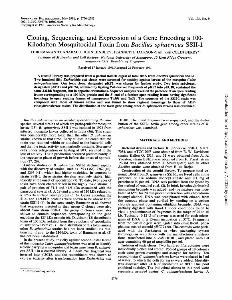
I I III II I . ... . . .................................. . .........................S P Sp K CSt BE X E X HH H2 Hp/H2CPFIG. 1. Restriction map of the sequence presented in Fig. 2. Shaded boxes represent the mtx (mosquitocidal toxin) gene and upstream
open reading frame (ORF). Arrows show gene orientation. Nucleotide numbers correspond to those shown in Fig. 2, beginning at the firstnucleotide of the Sau3AI site. Restriction enzymes: S, Sau3AI (not unique); P, PstI; Sp, SphI; K, KpnI; C, ClaI; St, StuI; B, BamHI; E,EcoRI; X, XmnI; H, Hindlll; H2, Hindll; Hp, HpaI.
3817
single toxin-producing clone, designated pKF2, was identi-fied and used as the parent clone for the experiments.
Subcloning of the toxin-encoding fragment. Subclones wereproduced from cosmid clone pKF2 to focus on the region ofthe inserted DNA encoding toxicity. At each step, subcloneswere assayed for larvicidal activity, and those exhibitingtoxicity were chosen for further manipulation.Cosmid clone pKF2 was subjected to partial digestion
with Sau3AI (0.2 ,ug of cosmid DNA, 0.1 U of Sau3AI, 15min, 37°C), and the resulting fragments were ligated intoBamHI-cut, phosphatase-treated pUC18. Four toxic cloneswere produced, and restriction mapping revealed that theycontained overlapping inserts. The plasmid designatedpKG19 contained the shortest inserted sequence (approxi-mately 9 kb) and was chosen for further subcloning. Thisplasmid was digested with XbaI to remove an approximately4.5-kb fragment. Religation of the remaining insert yieldedclone pKG19X, which retained full toxicity. A PstI fragmentof 3.8 kb derived from pKG19X was removed from pKG19Xand recloned into PstI-cut, phosphatase-treated pUC18.Two toxic clones, pXP33 and pXP34, which contained thesame PstI fragment in opposite orientations relative to thepUC18 vector were produced.DNA sequencing. Double-stranded DNA sequencing was
carried out by the method of Heinrich (18) with plasmidDNA as the template. Subclones from plasmids pXP33 andpXP34 were made in pUC18 with the restriction sites shownin Fig. 1. A sequence further upstream was obtained fromclone pKG19 (nucleotides [nt] 1 to 312). DNA was se-quenced with the pUC-M13 forward and reverse primers. Inaddition, further oligonucleotide primers were synthesizedto ensure complete sequencing of both strands.
Sequence analyses. Sequence analyses and manipulationswere performed with the following software: DNASTARpackage (DNASTAR Inc.), PC/GENE package (IntelliGe-netics), and UWGCG package (University of Wisconsin).Initial protein sequence alignments done with the DNAS-TAR package were based on the method of Lipman andPearson (22). Multiple alignments were created with theGapzero shell (31a), which uses the programs of theUWGCG package (14) run on the SEQNET central com-puter at the SERC Laboratory, Daresbury, United King-dom. Hydropathy predictions were made with the PC/GENE package by the method of Kyte and Doolittle (21).
Distribution of the toxin gene. Total DNA was preparedfrom 16 strains ofB. sphaericus as described above. Approx-imately 5 jig of each preparation was digested to completionwith ClaI and subjected to electrophoresis in a Tris-borate-EDTA (TBE)-buffered 1% agarose gel (24). DNA was blot-ted onto a Hybond-N membrane (Amersham International)by the method of Southern (37). The EcoRI fragment (nt1395 to 1815; Fig. 2) derived from the SSII-1 toxin gene wasradiolabeled by the nick translation method of Rigby et al.(34) and used as a probe for the toxin-encoding sequence.
Nucleotide sequence accession number. The nucleotidesequence shown in Fig. 2 was submitted to the GenBankdatabase (nucleotide sequence accession number M60446).
RESULTS
Features of the sequence. Figure 2 shows a sequence of4,133 nt containing two open reading frames along with thededuced amino acid sequence. There are similarities be-tween the restriction site pattern of this sequence and thatpublished by Souza et al. (38) for their clone pAS377PT.Their clone was derived from DNA isolated from B. sphaeri-cus 1593M and may represent the equivalent toxin from thisstrain.Sequence of the toxin. The toxicity of recombinants con-
taining sequences corresponding to nt 960 to 4133 (StuI-PstI)and nt 307 to 3864 (PstI-HpaI) indicates that the openreading frame commencing at nt 1207 and terminating at nt3817 represents the toxin gene. We have designated this genethe mtx (mosquitocidal toxin) gene. This gene encodes aprotein of 100.557 kDa and 870 amino acids in length.The deduced protein has features characteristic of signal
peptides of gram-positive bacteria, namely, (i) positivelycharged residues in the N terminus followed by (ii) a stretchof residues with a neutral or hydrophobic character and (iii)a cleavage site (often after an alanine residue) at the Cterminus of the signal sequence (10). The N terminus of thededuced protein contains three lysine residues, an isoleu-cine-rich stretch of neutral and hydrophobic residues, and apotential cleavage site following the alanine residue at posi-tion 30, forming a potential signal sequence. However, thetoxic activity of our SSII-1 cultures was found to be associ-ated with the cell pellet and not with the medium.
Figure 3 shows the hydropathy profile for the deducedtoxin sequence. Two interesting regions of hydrophobicitycan be seen. The first is the potential signal sequencedescribed above. The second, from positions 43 to 60 (-AASLTWLMDMSSLLYQLI-), was predicted to be a potentialtransmembrane helix by the method of Rao and Argos (33).As the mechanism of action of this toxin is unknown, wecannot assess whether this region plays an important role intoxicity.The deduced amino acid sequence of the 100-kDa toxin
was compared with protein sequences in the NBRF-PIRdatabase with the algorithm of Lipman and Pearson (22).Whereas the overall homology of the toxin with proteins inthe database was low, a strong regional homology was foundbetween the 100-kDa toxin and the S1 subunit of pertussistoxin (31). The homologous sequence corresponds in part toa region of the pertussis toxin (residues 8 to 15 of the S1subunit) found to be essential for activity (11) and previouslyobserved to be related to the A subunits of both choleratoxin and E. coli heat-labile toxin (23, 31). These three toxinsact by ADP-ribosylation of G proteins. Figure 4 shows the
VOL. 173, 1991
on July 7, 2018 by guesthttp://jb.asm
.org/D
ownloaded from

2778 THANABALU ET AL.
50 100
D H A Y L E K M L S S L R D S F V Q P F T I H G Q I T F V Q M S L G
150 . . . 200GAATCGCAAGTTATCCAATTGATGGAGTGACTCAAGAAGAGCTTCTCAAACATGCTGATATCGCCATGTATAAAGCGAAAGAATTAGGGGGGAATAACCA
50I A S Y P I D G V T Q E E L L K H A D I A M Y K A K E L G G N N H
250 . . . . 300TCGTTTTTTTGACGAAAAGATGAATGAACTCGTTATCAAAAAGGACCAAATAGAGCGAATGATACGTCTAGCGCTAGAAAGAAATGAATTTAGCGTCCAT
100R F F D E K M N E L V I K K D Q I E R M I R L A L E R N E F S V H
350 . . . 400TATCAACTGCAGATAGAAGCCACTACTGGAAAGATCCGAGGGTTTGAGGCGTTAGTGCGCTGGAAAAGCCCTGAGCTTGGACTTGTTTCTCCAGAAGATT
Y Q L Q I E A T T G K I R G F E A L V R W K S P E L G L V S P E D F
450 500
I P I A E K T G L I T Q I D E W V M Y Q A C L K N V E L Q H Q F G
550 600
200Y P F L M S V N I S A L Q L G R A D F V D K V Q L I L N E T K M K
650 . . 700CCGGAACATTTAGAAATTGAGATTACTGAGAGTATCTTAGTTGAGTCATTTGAAAGCTCTATATGCATACTGCGAAAATTAAAGAACCTAGGGGTGAAAA
P E H L E I E I T E S I L V E S F E S S I C I L R K L K N L G V K I
750 . . . 800TTGCCCAGGATGATTTTGGTACCGGTTATTCTTCACTTAACTACTTAACATTATTGCCGATTCATACCCTTAAAATTGATAGATCCCTTATTCAAAATAT
250A Q D D F G T G Y S S L N Y L T L L P I H T L K I D R S L I Q N M
850 900
300T S A T A E K T I I E S I I H L A H K L G H D V V A E G V E T K E
950 . . . . 1000CAGTATATCTTGCTCAAAGAATGGAACTGCGATTTTGTCCAGGGATACTATTTCAGTAGGCCTGTTTCGTCAGACATACTTGTCGAATGTCTGACAGGGG
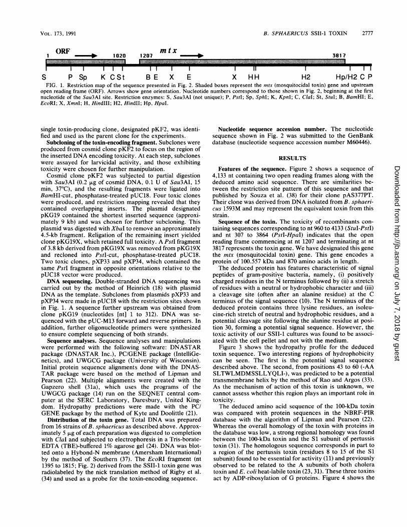
Q Y I L L K E W N C D F V Q G Y Y F S R P V S S D I L V E C L T G EFIG. 2. Nucleotide sequence of 4,133 bp containing the gene encoding the 100-kDa toxin of B. sphaericus SSII-1 and the 3' end of another
open reading frame. The deduced amino acid sequence is shown below the nucleotide sequence. Lines above the nucleotide sequence denoteinverted repeats; single underlining indicates putative promoter sequences; and double underlining indicates the ribosome-binding site.
homologies between these toxins and the 100-kDa toxin fromB. sphaericus SSII-1. A further sequence homology betweenthe 100-kDa toxin and ADP-ribosyltransferase toxins wasfound in the sequence Glu-Ile-Val-Arg-Ile-Trp (amino acids219 to 224). The sequence Glu-X-X-X-X-Trp has been found
in exotoxin A from Pseudomonas aeruginosa and in the Asubunit of diphtheria toxin (9). These Glu and Trp residuesof diphtheria toxin have been shown to be important inbinding to the nicotinamide moiety of NAD (25). No signif-icant homology was found between the 100-kDa toxin and
J. BACTERIOL.
on July 7, 2018 by guesthttp://jb.asm
.org/D
ownloaded from
VOL. 173, 1991 B. SPHAERICUS SSII-1 TOXIN 2779
. . . 1050 . . . . 1100
AAAAGAACTCCACAAATCAGTAAAAAATAAAGCTCTGTAATTACAAAAACAAACAACCATGCCAATTTAGATTATAAATTATTGTTAGAAATTCACACTA
K N S T N Q *
. . . 1150 . _ . . . 1200TCATCCCTTTCAAAATAAATATATAACTATTATTTTTATTTAATGTTATTAAATACATAATTTAATAACATTAAATAAATAAATACCAAAAAGAGGTGCA
* . . . 1250 . . . 1300ATTGATATGGCTATAAAAAAAGTATTAAAAATAATTTTAGCAATAATTATTATAATTAGTTGCCAACTACCACTGAATCAAAAAACTGTTTATGCTTCAC
M A I K K V L K I I L A I I I I I S C Q L P L N Q K T V Y A S P
* . . . 1350 . . . . 1400
CTAATTCTCCAAAAGATAACACTTGGATCCAAGCTGCTTCACTTACATGGTTAATGGATATGTCGAGTTTATTGTATCAACTGATTTCAACAAGAATTCC50
N S P K D N T W I Q A A S L T W L M D M S S L L Y Q L I S T R I P
* . . . 1450 . . . . 1500
CTCTTTTGCTTCTCCAAATGGATTACATATGAGAGAACAAACTATTGATAGCAATACTGGACAAATACAAATAGATAATGAGCATAGGCTATTAAGATGG
S F A S P N G L H M R E Q T I D S N T G Q I Q I D N E H R L L R W
. . 1550 . . . . 1600
GATAGGCGACCGCCTAATGATATTTTTTTAAATGGATTTATACCTAGAGTAACAAACCAAAATCTATCTCCAGTAGAAGACACACATCTTCTAAATTATT100
D R R P P N D I F L N G F I P R V T N Q N L S P V E D T H L L N Y L
* . . . 1650 . . . . 1700
TAAGAACAAATTCTCCATCCATTTTTGTTTCCACAACTAGAGCTAGATACAATAATTTAGGTTTAGAAATAACACCTTGGACACCTCATAGTGCTAATAA150
R T N S P S I F V S T T R A R Y N N L G L E I T P W T P H S A N N
* . . . 1750 . . . . 1800
CAATATAATTTACAGATATGAAATTTTTGCTCCAGGTGGTATTGATATTAATGCAAGTTTTTCAAGAAACCACAATCCCTTTCCTAACGAGGATGAAATA
N I I Y R Y E I F A P G G I D I N A S F S R N H N P F P N E D E I
* . . . 1850 . . . . 1900
ACTTTTCCAGGGGGAATTCGTCCTGAGTTTATACGTTCAACATATGAATATCACAATGGTGAAATTGTTAGAATATGGATTAACCCTAATTTTATTAATC200
T F P G G I R P E F I R S T Y E Y H N G E I V R I W I N P N F I N P
* . . . 1950 . . . . 2000
CATCAACTCTAAATGATGTATCAGGACCCTCTAATATAAGCAAAGTATTTTGGCATGAAAATCATTCTGAAGGAAATAATATGGATTCTAAAGGTTTTAT250
S T L N D V S G P S N I S K V F W H E N H S E G N N M D S K G F I
* . . . 2050 . . . . 2100
ACTAGATTTAGATTATAATCAAGATTTTGACATGTTTGCCCCCAATGGAGAAATACCTAATAATAATTTATTAAATAATAATAGTTTAAATGTTATACAA
L D L D Y N Q D F D M F A P N G E I P N N N L L N N N S L N V I QFIG. 2.-Continued.
other B. sphaericus toxins, namely, the 51.4- and 41.9-kDa the toxin gene, AAAAAGAGGTG (nt 1188 to 1198), showstoxins (3, 19) and the 125-kDa surface layer protein, from a 10-base homology to the 3' terminus ofBacillus subtilis 16Swhich the 110-kDa toxin is derived (6). rRNA (26). The free energy of binding for these two se-Upstream elements. The putative ribosome-binding site for quences, calculated by the method of Tinoco et al. (39), is
on July 7, 2018 by guesthttp://jb.asm
.org/D
ownloaded from
2780 THANABALU ET AL.
2150 . . . . 2200AACTCAGAATATCAAATCAAAAATAAGAAGGATAGGAATATTGTTGTAACGTTAGATTCTGATTATGGAGGAAGTCCAGTAGAGTCGTATAAGAATTTTG300
N S E Y Q I K N K K D R N I V V T L D S D Y G G S P V E S Y K N F G
2250 . . . . 2300GTTTTGAAAATCAAAAATGGAACATCAAATACGATAGCAAAAAAAATGCTTATAAAATCTACAATAGAGAAACCCCTACTTTACTATTAAGTTGGAATAG
350
F E N Q K W N I K Y D S K K N A Y K I Y N R E T P T L L L S W N S
2350 2400
N S S N G E Q V I R G Y T E S G S N N Q Y W T I E K N V N G F Y K
2450 . . . . 2500TTTAGAAATCTTTCTGACCCTAGCAAAATATTAGACTTAAAAGATGGTAACACTCTAAATAAAACTCCTTTAGTCGTTTCAAGTGAGAACAGTAGCTCAT400
F R N L S D P S K I L D L K D G N T L N K T P L V V S S E N S S S S
2550 2600
450Q E W L I E K T N Y Q T V K D G T Y Q V S S K L N E N K V I E Q I
2650 2700
S T N K V H I F S N S D K E N Q V W N L I Y N P I L K A Y K I K S
* . . . 2750 . . . 2800TTAAAGTATCCTAACTATTCATTGGCTTGGGATAGTAATAATACTAGAACAATTGTAGCAGCTACAGGGGATTATAATGATCAATATTGGTTAATAGAAA500
L K Y P N Y S L A W -D S N N T R T I V A A T G D Y N D Q Y W L I E R
* . . . 2850 . . . . 2900GAAACGAGGATAATACTTATATAATTAGAAATTATGAAAATAGAAAAATTGTTTTAGATTTATCAAATGGTAGTACTACTGATGGCAATGGATTATTAGG
550N E D N T Y I I R N Y E N R K I V L D L S N G S T T D G N G L L G
* . . . 2950 . . . . 3000
ATTTGAATTTCATGGGGGAATAAATCAAAGATGGATCATTAAGCCTTTTTCATTTAATTCTATTCAAGATGGGATTTATCAATTTATGACCGTAATTAAT
F E F H G G I N Q R W I I K P F S F N S I Q D G I Y Q F M T V I N
* . . . 3050 . . . . 3100CAGGATTTAATTGCTGACTTGACGACTAATAATTATACAATTGCTACGAAAACAAATAACTATTCTAGTAATCAAAAATGGACAGTAACTTATAATGATA600
Q D L I A D L T T N N Y T I A T K T N N Y S S N Q K W T V T Y N D KFIG. 2.-Continued.
-15.4 kcal (ca. -64.4 U) per mol. Similar ribosome-bindingsites have been proposed for other B. sphaericus genes, forexample, GAAAGGGG for the BspRI modification methyl-ase gene of strain R (32) and GGAGG for the 125-kDasurface layer protein gene of strain 2362 (6).
It is possible to identify several sequences in the regionupstream of the toxin gene which resemble promoters. The
putative promoter sequences -10 TATAAC (nt 1122 to1127) and -35 TTCACA (nt 1092 to 1097) show homology tothe consensus sequences for the u"55 vegetative promoter ofB. subtilis (-10 TATAAA and -35 TTGACA).
Located between the -10 region and the ribosome-bindingsite for the toxin gene is an A+T-rich inverted repeatsequence (nt 1135 to 1179) capable of forming a hairpin loop
J. BACTERIOL.
on July 7, 2018 by guesthttp://jb.asm
.org/D
ownloaded from
B. SPHAERICUS SSII-1 TOXIN 2781
* . . . 3150 . . . . 3200
AAAAACGTGCCTATAAAATTAGGAATTTACAACATGCTCATCTCAGTTTAGCATGGGATAGCAATCACTCAGATAAAATTTTTGGCGCTACCGGAGATTA650
K R A Y K I R N L Q H A H L S L A W D S N H S D K I F G A T G D Y
* . . . 3250 . . . . 3300
TGATGATCAATATTGGATTCCTATTTTGCAAACAGATGGATCATTTATTTTTAGGAATTATAAAAATCCAAATAAGATATTTGGAACAAATGGTCAACCC
D D Q Y W I P I L Q T D G S F I F R N Y K N P N K I F G T N G Q P
* . . . 3350 . . . . 3400
ATTAATGATATTCCGTTAAAGGCTCAAGATGTAACTGGACAAAATAATCAAAAGTGGTATTTAAGACATTTAAATTCTTCCAATAATTTTACTGGATACT700
I N D I P L K A Q D V T G Q N N Q K W Y L R H L N S S N N F T G Y F
3450 . . . . 3500
TTAACATATCCAGTAAAAAGAATTTTAATAAAATTATTACTATGAACTCAAATAAAACTCAAGCTGTTATTTTTGACAATATTGGAATTAACAATCAGTC750
N I S S K K N F N K I I T M N S N K T Q A V I F D N I G I N N Q S
3550 3600
W K L K Y N D N K N A Y Q I H I L D N F L Y F Q G G H N I V A T M
3650 . . 3700CGAAATGTTACTAATGATGATTTAAGAAGTTATTGGTATGTAGAATATAACTTTAATAAAGATGGGTTTATAATAAGAAACGCATTTGATACAAGCTACG800
R N V T N D D L R S Y W Y V E Y N F N K D G F I I R N A F D T S Y V
3750 . . . . 3800TACTTGATGTATTTCAAGGAAATTTTGCTAACAATAGACCTATCATCACTTATCAAAACTATTTAAACGACAACCAGTTATGGAATTTTATACCTTCATT
850L D V F Q G N F A N N T P I I T Y Q N Y L N D N Q L W N F I P S L
* . . . 3850 . . . . 3900
AGGTGTAGAACCTAGATAGTAATGACTGTTTTATGCATAGAAGACAAGCAAAAAAGAGGTTAACAGAGATATAATGAAATTATAATAACATCTAAATTGT
G V E P R * * *
_________________. ____________ _. .3950 . . . . 4000
AAATAAAGGGTGATACATATCGTAATATGTATCACCCTTTTTACTTTTGTACAACAATCACTACCGATTGTAGGTTAAAATAAAGTTAGATTTTTTTCTG
.. . . ~~~~~~4050.... 4100
TGATCTAATCTCCAATAAAAACCTTTCCCTTTTCCTCTCTTAAAATGCAGGTATCGATTTAGTAGCCTTCTTAAATTTTTAAAAAAGATGAATTATCAAT
4030TTGATTTACCGTTAAAATTTATTTATACTGCAG
FIG. 2.--Continued.
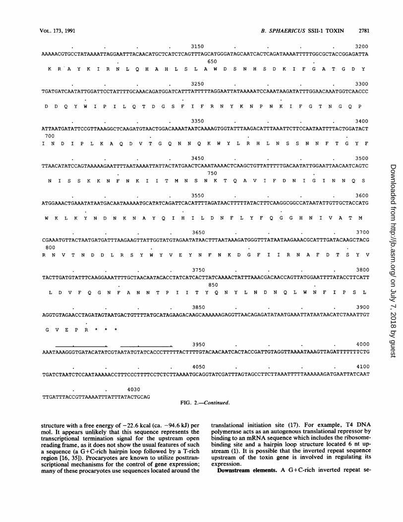
structure with a free energy of -22.6 kcal (ca. -94.6 kJ) permol. It appears unlikely that this sequence represents thetranscriptional termination signal for the upstream openreading frame, as it does not show the usual features of sucha sequence (a G+C-rich hairpin loop followed by a T-richregion [16, 35]). Procaryotes are known to utilize posttran-scriptional mechanisms for the control of gene expression;many of these procaryotes use sequences located around the
translational initiation site (17). For example, T4 DNApolymerase acts as an autogenous translational repressor bybinding to an mRNA sequence which includes the ribosome-binding site and a hairpin loop structure located 6 nt up-stream (1). It is possible that the inverted repeat sequenceupstream of the toxin gene is involved in regulating itsexpression.Downstream elements. A G+C-rich inverted repeat se-
VOL. 173, 1991
on July 7, 2018 by guesthttp://jb.asm
.org/D
ownloaded from

2782 THANABALU ET AL.
58
48
30
28
l8
-28
-18
-30
-48
1L 108 200 300 400 588 680 700 800
FIG. 3. Hydropathy profile for the amino acid sequence of the 100-kDa toxin. Amino acid numbers are marked on the horizontal axis, andunits of hydropathicity (calculated by the algorithm of Kyte and Doolittle [21]) are marked on the vertical axis. The dotted line at -5 divideshydrophobic regions above from hydrophilic regions below.
quence capable of forming a hairpin loop structure with afree energy of -27.6 kcal (ca. -115.5 kJ) per mol begins 86nt downstream of the translational stop codon of the toxingene. This inverted repeat sequence is followed by a T-richsequence and thus has the characteristics of a transcriptionaltermination signal.Upstream open reading frame. The sequence presented in
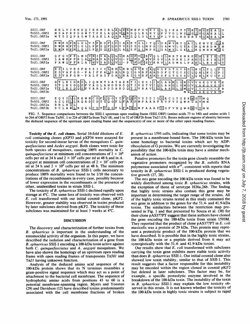
Fig. 2 also contains the 3' end of another open reading frame(nt 1 to 1020). Beginning after methionine residue 74 in oursequence there was significant homology to open readingframes ORF2 from TnSOJ and ORF2a from Tn2J (8). TheTn2J homology continues after residue 290 in our sequencein ORF2b (15) (Fig. 5). The function of these open readingframes of the mercury resistance-conferring transposonsTnSOJ and Tn2J is not known. B. sphaericus SSII-1 wastested for mercury resistance by two methods. The ability ofthe bacteria to grow on LB agar containing 10 jig of mercuricacetate per ml and the ability of the bacteria to volatilizemercuric chloride were tested by the method of Nakamuraand Nakahara (30). No colonies were formed on mercuricacetate plates after incubation at 30°C for 72 h, and no
SSII-TOX
PT-Si
CT-A
LT-A
evidence of mercuric chloride volatilization was seen. Theseresults indicate that the upstream open reading frame is notassociated with mercury resistance in B. sphaericus SSII-1.
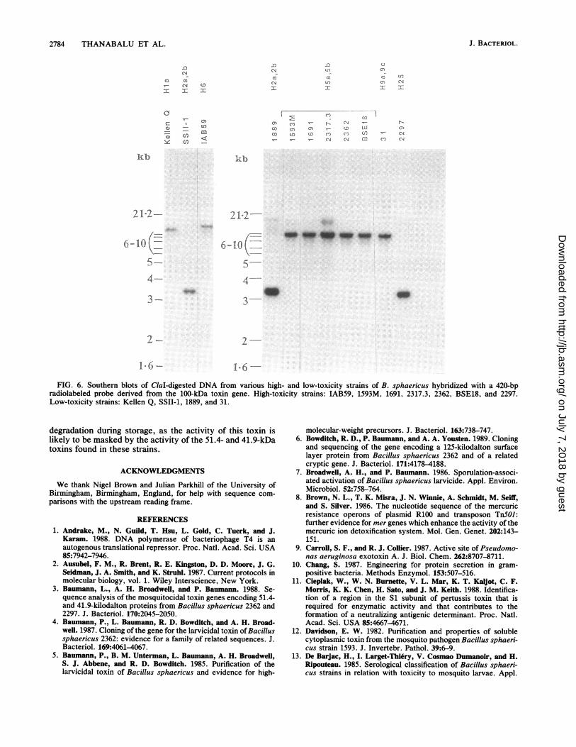
Distribution of the toxin sequence. Figure 6 shows theresults of Southern hybridization of a toxin gene probe toClal-digested DNA from various B. sphaericus strains. Themtx gene is widely distributed among both high- and low-toxicity strains. The sizes of the major hybridizing bandsappeared to vary among serotypes, with estimated sizes ofapproximately 11 kb in the Hla strain, 3.2 kb in H2a,2bstrains, 10 kb in H5a,5b strains, 13 kb in the H6 strain, 8.4 kbin the H9a.,9c strain, and 2.9 kb in the H25 strain. Additionalweakly hybridizing bands were also seen in some strains.These bands are currently under investigation. In addition toDNA from the strains shown in Fig. 6, Clal-digested DNAfrom the weakly toxic strains 2173 and 2315 (serotypeH26a,26b) and the nontoxic strains BM2 (serotypeH26a,26b) and ATCC 7054 and ATCC 7055 (serotype H9)was used in similar hybridization experiments. No hybrid-ization of the toxin gene probe was observed (results notshown).
E9L R W D R R P PNql-s[; i E - - - V T NE-
V Y R Y D S R P P E D V F QHN G - F T - A - - - - - - - W G N N - D -
L Y R A D S R P P D E I K Q S G G L M P R G Q S E Y F D R G T Q M N I
L Y R A D S R P P D E I K R S G G L M P R G H N E Y F D R G T Q M N I
SSII-TOX S P HELTE L N Y L - R --N - P I FV S T T R A[ Y
PT-Si N L D H L T G R S C Q V G- S N HAF V S T S S SHRCT-A N L Y D H A R G -T -Q T G F V R H D D G Y V S T S I S L R
LT-A N TLY H A G -T Q T G F V R Y D D G Y V S T S L S L R
FIG. 4. Sequence comparisons between the 100-kDa toxin and other bacterial toxins. The deduced amino acid sequence from amino acids95 to 148 of the 100-kDa toxin from B. sphaericus SSII-1 (SSII-TOX) is aligned with amino acids 7 to 58 of the S1 subunit of pertussis toxin(PT-Si) (31) and amino acids 5 to 67 of the A subunit of cholera toxin (CT-A) and the A subunit of E. coli heat-labile toxin (LT-A) (42). Boxesindicate regions of identity between the sequence of the 100-kDa toxin and the sequence(s) of one or more of the other toxins.
J. BACTERIOL.
on July 7, 2018 by guesthttp://jb.asm
.org/D
ownloaded from
B. SPHAERICUS SSII-1 TOXIN 2783
SSII.ORF N E L V I K K D Q I E R M I R L A L E R N E F S V H Y Q L Q I E A T T G K I R G F E A L VTn5Ol.ORF2 M S A F R P D G W T T P E L A Q A V E R G Q L E L H Y Q P V V D L R SS G WV G A E A L LTn2l.ORF2a M T S S Q P A G W T A A E L A Q A A A R G Q L D L H Y Q P L V D L R D H R T V G A E A L M
SSII.ORF R W K S P E L G L V S P E D F I P I A E K T G L I T Q I D E W V M Y Q A C L K N V E L HTn5Ol.ORF2 R W R H P T L GL L P P G Q F L P V V E|S S G L M P E I G A W V L G E A C R Q M R D W R MTn2l.ORF2a R W R H R L PP G Q FL P L A SF G L M P E G A W V L G E A CR Q M H K WM G
SSII.ORF Q F G Y P F L M S V N I S A L Q L G R A|D F V[K V Q L I L N E T K M K P E H|L E I E|ImTn5Ol.ORF2 L A W R P F R L A V N A S A S Q V G - P [ F|D G WIVIK G V|L|A D A E L P A|EIY|L E I E|LT|Tn2l.ORF2a P A W Q PIFR L A I NJV AJ V G - P T F D[EE V K R V L A D M A L P A E L L E I E|L T
SSII.ORF E I L V E S F E S S I C I R K L K N L|G V|K I A Q D D F G T G Y s L N Y L T L L P ITnSOl.ORF2 E SIV A F G D - P A I F P A WD A|L R Q I|G V R FIAI AD D F G T G Y S C L|Q HILIK C CIP ITn2l.ORF2a [ V A F G N - P A L F A S F D A R A I|G R F A|D D F G T G Y S|C L Q H [ K C C[
SSII.ORF H T K I D|R S L I Q N M T S A T A E K[T ITI E S I I H[L AHFKKITGTH[DVV A E G V ETT KTn5Ol.ORF2 SIT L K IDIQISIF V A G L A N D R R D QIT I|V H T VII IQIL A HIGIL GI M DVV A E G V E T STn2l.ORF2a T|T L K I D Q S F-V A R L P D D A R D Q|T I|V R A V I Q[L A H|G|L G|M D VI-F RTn2l.ORF2b M E|V|V A E G V P
SSII.ORF E Q Y I L K E W N CTD F E Q G|Y Y[ St V S S D I Ln E C L T G E K[ ]S T N QTn5Ol.ORF2 A S L D L L R Q A D|C D T G Q G|F LIF A KIP M P A A A F A V F V S Q W R G A TIM NTn2l.ORF2a R R L H QILI I 4 4 IITn2l.ORF2b D C L A WL R Q A G|C D|T MW§GF L F A m P|M P A A A F[2JG F V N Q W R[EJT T M N
FIG. 5. Sequence comparisons between the SSII-1 upstream open reading frame (SSII.ORF) (amino acids 75 to 340) and amino acids 1to 264 of ORF2 from Tn501, 1 to 224 of ORF2a from Tn21 (8), and 1 to 52 of ORF2b from Tn21 (15). Boxes indicate regions of identity betweenthe deduced sequence of the upstream open reading frame and the sequence(s) of one or more of the other open reading frames.
Toxicity of the E. coli clones. Serial 10-fold dilutions of E.coli containing clones pXP33 and pXP34 were assayed fortoxicity for second-instar larvae of the mosquitoes C. quin-quefasciatus and Aedes aegypti. Both clones were toxic forboth species of mosquitoes, causing 100% mortality in C.quinquefasciatus at minimum cell concentrations of 2 x 10⁶cells per ml at 24 h and 2 x 10⁵ cells per ml at 48 h and in A.aegypti at minimum cell concentrations of 2 x 10⁷ cells perml at 24 h and 2 x 10⁶ cells per ml at 48 h. The minimumconcentrations of B. sphaericus SSII-1 cells necessary toproduce 100% mortality were found to be 1/10 the concen-trations of the recombinants in every case, possibly becauseof lower expression in the recombinants or the presence ofother, unidentified toxins in strain SSII-1.The toxicity of B. sphaericus SSII-1 declined rapidly upon
storage at 4°C. The same loss of toxicity was observed withE. coli transformed with our initial cosmid clone, pKF2.However, greater stability was observed in toxins producedby later subclones derived from pKF2. The toxicity of thesesubclones was maintained for at least 3 weeks at 4°C.
DISCUSSION
The discovery and characterization of further toxins fromB. sphaericus is important in the understanding of theentomocidal activity of the organism. In this paper, we havedescribed the isolation and characterization of a gene fromB. sphaericus SSII-1 encoding a 100-kDa toxin active againstboth C. quinquefasciatus and A. aegypti mosquitoes. Wehave also shown the homology of an upstream open readingframe with open reading frames of transposons TnSOJ andTn21 having unknown function.
Analysis of the deduced amino acid sequence of the100-kDa protein shows that its N terminus resembles agram-positive signal sequence which may act as a point ofattachment to the bacterial cell membrane. The sequence ofhydrophobic amino acids from positions 43 to 60 is apotential membrane-spanning region. Myers and Yousten(29) and Davidson (12) have described toxins predominantlyassociated with the cell membrane fractions of broken
B. sphaericus 1593 cells, indicating that some toxins may bepresent in a membrane-bound form. The 100-kDa toxin hassome homology to bacterial toxins which act by ADP-ribosylation of G proteins. We are currently investigating thepossibility that the 100-kDa toxin may have a similar mech-anism of action.
Putative promoters for the toxin gene closely resemble thevegetative promoters recognized by the B. subtilis RNApolymerase associated with a"5, consistent with reports thattoxicity in B. sphaericus SSII-1 is produced during vegeta-tive growth (27, 28).The mtx gene encoding the 100-kDa toxin was found to be
widely distributed among toxic B. sphaericus strains, withthe exception of those of serotype H26a,26b. The findingthat highly toxic strains also contain this gene may beimportant in determining the target range of the strains. Allof the highly toxic strains tested in this study contained themtx gene in addition to the genes for the 51.4- and 41.9-kDatoxins. The similarities between the restriction map pre-sented in Fig. 1 and that presented by Souza et al. (38) fortheir clone pAS377PT suggest that these authors have clonedthe gene encoding the 100-kDa toxin from strain 1593M.They reported that the product of clone pAS377PT in E. colimaxicells was a protein of 29 kDa. This protein may repre-sent a proteolytic product of the 100-kDa protein that wehave described. It is possible that in the highly toxic strains,the 100-kDa toxin or a peptide derived from it may actsynergistically with the 51.4- and 41.9-kDa toxins.Our results show that E. coli transformed with subclones
carrying the toxin gene exhibits more stable toxic activitythan does B. sphaericus SSII-1. Our initial cosmid clone alsoshowed low toxin stability, similar to that of SSII-1. Thisresult suggests that a factor responsible for this instabilitymay be encoded within the region cloned in cosmid pKF2but deleted in later subclones. This factor may be, forexample, a specific proteolytic enzyme involved in thebreakdown of the 100-kDa toxin. The instability of the toxinin B. sphaericus SSII-1 may explain the low toxicity ob-served in this strain. It is not known whether the toxicity ofthe 100-kDa toxin in highly toxic strains is subject to rapid
VOL. 173, 1991
on July 7, 2018 by guesthttp://jb.asm
.org/D
ownloaded from
2784 THANABALU ET AL.
.0
C3 CCCM
:I
a
C,C)
kb
21-2-
N
C4CMI1(I
0)y- a)
- mU) <cn_
C,
I
f.
CZt-n
I:
)1c (C ) ,- N-co cn (7CD O (D (
coN -
uC) 1LI)(n (nf(N 0
ICMI:
r
NCcz)
kb
21-2-
6-10i-
54-3.
3-
i* aW. Ui0 al' -m -m
4.
2-
1-6- 1-6--
FIG. 6. Southern blots of ClaI-digested DNA from various high- and low-toxicity strains of B. sphaericus hybridized with a 420-bpradiolabeled probe derived from the 100-kDa toxin gene. High-toxicity strains: IAB59, 1593M, 1691, 2317.3, 2362, BSE18, and 2297.Low-toxicity strains: Kellen Q, SSII-1, 1889, and 31.
degradation during storage, as the activity of this toxin islikely to be masked by the activity of the 51.4- and 41.9-kDatoxins found in these strains.
ACKNOWLEDGMENTS
We thank Nigel Brown and Julian Parkhill of the University ofBirmingham, Birmingham, England, for help with sequence com-parisons with the upstream reading frame.
REFERENCES1. Andrake, M., N. Guild, T. Hsu, L. Gold, C. Tuerk, and J.
Karam. 1988. DNA polymerase of bacteriophage T4 is anautogenous translational repressor. Proc. Natl. Acad. Sci. USA85:7942-7946.
2. Ausubel, F. M., R. Brent, R. E. Kingston, D. D. Moore, J. G.Seidman, J. A. Smith, and K. Struhl. 1987. Current protocols inmolecular biology, vol. 1. Wiley Interscience, New York.
3. Baumann, L., A. H. Broadwell, and P. Baumann. 1988. Se-quence analysis of the mosquitocidal toxin genes encoding 51.4-and 41.9-kilodalton proteins from Bacillus sphaericus 2362 and2297. J. Bacteriol. 170:2045-2050.
4. Baumann, P., L. Baumann, R. D. Bowditch, and A. H. Broad-well. 1987. Cloning of the gene for the larvicidal toxin ofBacillussphaericus 2362: evidence for a family of related sequences. J.Bacteriol. 169:4061-4067.
5. Baumann, P., B. M. Unterman, L. Baumann, A. H. Broadwell,S. J. Abbene, and R. D. Bowditch. 1985. Purification of thelarvicidal toxin of Bacillus sphaericus and evidence for high-
molecular-weight precursors. J. Bacteriol. 163:738-747.6. Bowditch, R. D., P. Baumann, and A. A. Yousten. 1989. Cloning
and sequencing of the gene encoding a 125-kilodalton surfacelayer protein from Bacillus sphaericus 2362 and of a relatedcryptic gene. J. Bacteriol. 171:4178-4188.
7. Broadwell, A. H., and P. Baumann. 1986. Sporulation-associ-ated activation of Bacillus sphaericus larvicide. Appl. Environ.Microbiol. 52:758-764.
8. Brown, N. L., T. K. Misra, J. N. Winnie, A. Schmidt, M. Seiff,and S. Silver. 1986. The nucleotide sequence of the mercuricresistance operons of plasmid R100 and transposon TnSOJ:further evidence for mer genes which enhance the activity of themercuric ion detoxification system. Mol. Gen. Genet. 202:143-151.
9. Carroll, S. F., and R. J. Collier. 1987. Active site ofPseudomo-nas aeruginosa exotoxin A. J. Biol. Chem. 262:8707-8711.
10. Chang, S. 1987. Engineering for protein secretion in gram-positive bacteria. Methods Enzymol. 153:507-516.
11. Cieplak, W., W. N. Burnette, V. L. Mar, K. T. Kaljot, C. F.Morris, K. K. Chen, H. Sato, and J. M. Keith. 1988. Identifica-tion of a region in the S1 subunit of pertussis toxin that isrequired for enzymatic activity and that contributes to theformation of a neutralizing antigenic determinant. Proc. Natl.Acad. Sci. USA 85:4667-4671.
12. Davidson, E. W. 1982. Purification and properties of solublecytoplasmic toxin from the mosquito pathogen Bacillus sphaeri-cus strain 1593. J. Invertebr. Pathol. 39:6-9.
13. De Barjac, H., I. Larget-Thiery, V. Cosmao Dumanoir, and H.Ripouteau. 1985. Serological classification of Bacillus sphaeri-cus strains in relation with toxicity to mosquito larvae. Appl.
6-10-
5-4-
3-
2-
J. BACTERIOL.
.1
on July 7, 2018 by guesthttp://jb.asm
.org/D
ownloaded from
B. SPHAERICUS SSII-1 TOXIN 2785
Microbiol. Biotechnol. 21:85-90.14. Deveraux, J., P. Haeberli, and 0. Smithies. 1984. A comprehen-
sive set of sequence analysis programs for the VAX. NucleicAcids Res. 12:387-395.
15. Diver, W. P., J. Grinstead, D. C. Fritzinger, N. L. Brown, J.Altenbuchner, P. Rogowsky, and R. Schmidt. 1983. DNA se-quences of and complementation by the tnpR genes of Tn2J,Tn5OI and Tnl721. Mol. Gen. Genet. 191:189-193.
16. Galloway, J. L., and T. Platt. 1986. Control of prokaryotic geneexpression by transcription termination, p. 155-178. In I. R.Booth and C. F. Higgins (ed.), Regulation of gene expres-sion-25 years on. Cambridge University Press, Cambridge.
17. Gold, L. 1988. Posttranscriptional regulatory mechanisms inEscherichia coli. Annu. Rev. Biochem. 57:199-233.
18. Heinrich, P. 1986. Guidelines for quick and simple plasmidsequencing. Boehringer GmbH, Mannheim, Federal Republic ofGermany.
19. Hindley, J., and C. Berry. 1987. Identification, cloning andsequence analysis of the Bacillus sphaericus 1593 41.9 kDlarvicidal toxin gene. Mol. Microbiol. 1:187-194.
20. Hohn, B., and J. Collins. 1980. A small cosmid for efficientcloning of large DNA fragments. Gene 11:291-298.
21. Kyte, J., and R. F. Doolittle. 1982. A simple method fordisplaying the hydropathic character of a protein. J. Mol. Biol.157:105-132.
22. Lipman, D. J., and W. R. Pearson. 1985. Rapid and sensitiveprotein similarity searches. Science 227:1435-1441.
23. Locht, C., and J. M. Keith. 1986. Pertussis toxin gene: nucleo-tide sequence and genetic organization. Science 232:1258-1264.
24. Maniatis, T., E. F. Fritsch, and J. Sambrook. 1982. Molecularcloning: a laboratory manual. Cold Spring Harbor Laboratory,Cold Spring Harbor, N.Y.
25. Michel, A., and J. Dirkx. 1977. Occurrence of tryptophan in theenzymically active site of diphtheria toxin fragment A. Biochim.Biophys. Acta 491:286-295.
26. Moran, C. P., N. Lang, S. F. J. LeGrice, G. Lec, M. Stephens,A. L. Sonenshein, J. Pero, and R. Losick. 1982. Nucleotidesequences that signal the initiation of transcription and transla-tion in Bacillus subtilis. Mol. Gen. Genet. 186:339-346.
27. Myers, P., and A. A. Yousten. 1978. Toxic activity of Bacillussphaericus SSII-1 for mosquito larvae. Infect. Immun. 19:1047-1053.
28. Myers, P., A. A. Yousten, and E. W. Davidson. 1979. Compar-ative studies of the mosquito-larval toxin of Bacillus sphaericusSSII-1 and 1593. Can. J. Microbiol. 25:1227-1231.
29. Myers, P. S., and A. A. Yousten. 1980. Localization of amosquito-larval toxin of Bacillus sphaericus 1593. Appl. Envi-ron. Microbiol. 39:1205-1211.
30. Nakamura, K., and H. Nakahara. 1988. Simplified X-ray film
method for detection of bacterial volatilization of mercurychloride by Escherichia coli. Appl. Environ. Microbiol. 54:2871-2873.
31. Nicosia, A., M. Perugini, C. Franzini, M. C. Casagli, M. G.Borri, G. Antoni, M. Almoni, P. Neri, G. Ratti, and R. Rappuoli.1986. Cloning and sequencing of the pertussis toxin genes:operon structure and gene duplication. Proc. Natl. Acad. Sci.USA 83:4631-4635.
31a.Parkhill, J., and D. A. Rouch. Unpublished data.32. P6sfai, G., A. Kiss, S. Erdei, J. P6sfai, and P. Venetianer. 1983.
Structure of the Bacillus sphaericus R modification methylasegene. J. Mol. Biol. 170:597-610.
33. Rao, J. K. M., and P. Argos. 1986. A conformational preferenceparameter to predict helices in integral membrane proteins.Biochim. Biophys. Acta 869:197-214.
34. Rigby, P. W. J., M. Dieckmann, C. Rhodes, and P. Berg. 1977.Labelling deoxyribonucleic acid to high specific activity in vitroby nick translation with DNA polymerase I. J. Mol. Biol.113:237-251.
35. Rosenberg, M., and M. Court. 1979. Regulatory sequencesinvolved in the promotion of RNA transcription. Annu. Rev.Genet. 13:319-353.
36. Singer, S. 1973. Insecticidal activity of recent bacterial isolatesand their toxins against mosquito larvae. Nature (London)244:110-111.
36a.Singer, S., and D. J. Murphy. 1976. Abstr. Annu. Meet. Am.Soc. Microbiol. 1976, 017, p. 181.
37. Southern, E. M. 1975. Detection of specific sequences amongDNA fragments separated by gel electrophoresis. J. Mol. Biol.98:503-517.
38. Souza, A. E., V. Rajan, and K. Jayaraman. 1988. Cloning andexpression in Escherichia coli of two DNA fragments fromBacillus sphaericus encoding mosquito-larvicidal activity. J.Biotechnol. 7:71-82.
39. Tinoco, I., P. N. Borer, B. Dengler, M. D. Levine, 0. C.Uhlenbeck, D. M. Crothers, and J. Gralla. 1973. Improvedestimation of secondary structure in ribonucleic acids. Nature(London) New Biol. 246:40-41.
40. Weiser, J. 1984. A mosquito-virulent Bacillus sphaericus inadult Simulium damnosum from northern Nigeria. Zentralbl.Mikrobiol. 139:57-60.
41. Wickremesinghe, R. S. B., and C. L. Mendis. 1980. Bacillussphaericus spore from Sri Lanka demonstrating rapid larvicidalactivity on Culex quinquefasciatus. Mosq. News 40:387-389.
42. Yamamoto, T., T. Nakazawa, T. Miyata, A. Kaji, and T. Yokota.1984. Evolution and structure of two ADP-ribosylation entero-toxins, Escherichia coli heat-labile toxin and cholera toxin.FEBS Lett. 169:241-246.
VOL. 173, 1991
on July 7, 2018 by guesthttp://jb.asm
.org/D
ownloaded from